Introduction
Head and neck squamous cell carcinoma (HNSCC) is the
sixth most common type of cancer in the world and is also known for
its rapid clinical progression and poor prognosis. The survival
rate for HNSCC patients with advanced stage disease, particularly
hypopharyngeal cancer, has improved little over the past 60 years
(1,2). Although extended surgery for advanced
HNSCC has progressed due to the technique of microsurgery, issues
concerning loss of function, aesthetic appearance and risk of
various surgical complications remain unresolved. In recent years,
definitive chemoradiotherapy (CRT) has become the primary treatment
for advanced HNSCC in lieu of surgery, due to the advantage of
preserving organ structure and function, and the equivalency in the
curative effect compared with surgery. However, the survival rate
of HNSCC patients has not significantly improved. Furthermore, CRT
can be effective in some patients, while others show little
response and experience various adverse effects, which result in
the lost of opportunity for a potentially curative surgery.
Therefore, we propose that it is important to differentiate whether
or not each HNSCC case is chemosensitive and/or radiosensitive
prior to treatment. Recently, the human papilloma virus has been
identified as one of the biomarkers of chemosensitivity and/or
radiosensitivity in oropharyngeal cancers (3,4). In
contrast, there is no reliable marker for HNSCC in other sites.
It was previously reported that carcinogenesis is
associated with chronic inflammation. Concerning digestive organs,
gastritis with Helicobacter pylori infection and ulcerative
colitis (UC) frequently cause gastric cancer and colorectal cancer,
respectively. Recently, there have been many reports that
regenerating gene (REG) expression is observed in chronic
inflammation and in tumors of the digestive organs (5–9). In
addition, it has been reported that REG expression is
associated with progression of digestive cancers such as
esophageal, gastric and colorectal cancer (10–16).
By differential screening of the regenerating
pancreatic islet-derived cDNA library, Reg was found and
defined as a regenerating and growth factor (17–20).
The Reg family belongs to the lectin superfamily and encodes
five small secreted proteins (17,21,22).
Members of the Reg family are grouped into four subtypes:
types I, II, III, and IV (23). In
humans, the REG family is composed of five subclasses:
REG Iα, REG Iβ, REG III,
hepatocarcinoma-intestine-pancreas/pancreatitis-associated-protein
(HIP/PAP) and REG IV(5,17,24–29).
It has been demonstrated that they are highly expressed in a
variety of inflammatory states and in tumor tissue when compared to
normal tissue (17,29–31).
We hypothesized that REG expression is associated with head
and neck cancer derived from the oral and pharyngolaryngeal
cavities, which belong to the first section of the digestive tract
which are exposed to chronic inflammatory factors such as tobacco,
alcohol, viral infection and the other various mechanical
stress.
In the present study, we extracted RNA from
formalin-fixed paraffin-embedded HNSCC tissue, specifically
hypopharyngeal cancer, determined mRNA expression of the REG
family gemes and evaluated the effects of REG family
expression on the prognosis of hypopharyngeal cancer. The results
revealed that REG III expression was significantly
associated with an increased survival rate. Furthermore, we
demonstrated that REG III regulated cell proliferation and
chemosensitivity and/or radiosensitivity in HNSCC cells
in vitro.
Materials and methods
Study population
We confirmed 37 cases with hypopharyngeal squamous
cell carcinoma. All patients were treated with definitive CRT as a
primary treatment between January 2000 and December 2009 at the
Department of Otolaryngology-Head and Neck Surgery of Nara Medical
University. The present study was approved by the Ethics Committee
of Nara Medical University School of Medicine. Written informed
consent for participation in the present study was obtained from
each patient. The patient characteristics are listed in Table I. The patients included 34 males and
3 females, with a mean age of 68 years (range, 47–83 years). The
average period of observation was 34 months (range, 3–98
months).
 | Table IPatient and tumor
characteristics. |
Table I
Patient and tumor
characteristics.
|
Characteristics | No. of patients
(n=37) |
|---|
| Gender |
| Male | 34 |
| Female | 3 |
| Age (years) |
| Median | 68 |
| Range | 47–83 |
| Period of
observation (months) |
| Median | 34 |
| Range | 3–98 |
| Tumor stage |
| I | 4 |
| II | 6 |
| III | 6 |
| IV | 21 |
| T
classification |
| T1 | 8 |
| T2 | 14 |
| T3 | 7 |
| T4 | 8 |
| N
classification |
| N0 | 12 |
| N1 | 7 |
| N2 | 16 |
| N3 | 2 |
Real-time reverse
transcriptase-polymerase chain reaction (RT-PCR)
Total RNA was isolated from each paraffin-embedded
tissue upon biopsy or surgery using the RNeasy FFPE kit (Qiagen,
Hilden, Germany). cDNA was then reverse transcribed from ~1 μg
samples of total RNA using a High Capacity cDNA reverse
transcription kit, with RNase inhibitor (Applied Biosystems, Foster
City, CA, USA) as described (32,33).
Real-time RT-PCR was then carried out using the primers listed in
Table II and SYBR Fast qPCR Master
Mix (Kapa Biosystems, Boston, MA, USA). All PCR primers were
synthesized by NGRL (Sendai, Japan). PCR was performed with an
initial step of 3 min at 95ºC followed by 40 cycles of 3 sec at
95ºC and 20 sec at 60ºC for β-actin, REG III and
HIP/PAP, 40 cycles of 3 sec at 95ºC and 20 sec at 64ºC for
REG Iα, REG Iβ and REG IV. The level of target
mRNA was normalized to the mRNA level of β-actin as an
internal standard.
| Table IIPrimers for real-time RT-PCR. |
Table II
Primers for real-time RT-PCR.
| Gene | Primer sequence
(position) |
|---|
| REG Iα
(NM_002909) | Sense |
5′-AGGAGAGTGGCACTGATGACTT-3′ (nucleotides
369–390) |
| Antisense |
5′-TAGGAGACCAGGGACCCACTG-3′ (nucleotides
445–465) |
| REG Iβ
(NM_006507) | Sense |
5′-GCTGATCTCCTCCCTGATGTTC-3′ (nucleotides
108–129) |
| Antisense |
5′-GGCAGCTGATTCGGGGATTA-3′ (nucleotides
170–190) |
| REG III
(AB161037) | Sense |
5′-GAATATTCTCCCCAAACTG-3′ (nucleotides
695–713) |
| Antisense |
5′-GAGAAAAGCCTGAAATGAAG-3′ (nucleotides
765–784) |
| HIP/PAP
(NM_138937) | Sense |
5′-AGAGAATATTCGCTTAATTCC-3′ (nucleotides
645–665) |
| Antisense |
5′-AATGAAGAGACTGAAATGACA-3′ (nucleotides
716–736) |
| REG IV
(AY007243) | Sense |
5′-ATCCTGGTCTGGCAAGTC-3′ (nucleotides
470–487) |
| Antisense |
5′-CGTTGCTGCTCCAAGTTA-3′ (nucleotides
538–555) |
| β-actin
(NM_001101) | Sense |
5′-GCGAGAAGATGACCCAGA-3′ (nucleotides
420–437) |
| Antisense |
5′-CAGAGGCGTACAGGGATA-3′ (nucleotides
492–509) |
Survival analysis
We investigated the differences in prognosis between
the patient group with positive REG family expression and
the negative group. Overall survival rate was calculated by the
Kaplan-Meier method.
Cell lines and culture
FaDu hypopharyngeal squamous cell carcinoma cells
(American Type Culture Collection, Manassas, VA, USA) were cultured
in Dulbecco’s modified Eagle’s medium (DMEM; Invitrogen, Grand
Island, NY, USA) supplemented with 10% heat-inactivated fetal
bovine serum (FBS; Gibco, Grand Island, NY, USA) and antibiotics
(penicillin G/streptomycin/amphotericin B; Gibco) in a humidified
incubator at 37ºC.
Isolation of cells following stable
transfection with the REG III expression vector
cDNA fragment encoding human REG III
(nucleotides 56–635 of AB161037) was inserted into the pCI-neo
mammalian expression vector (Promega, Madison, WI, USA). The
expression vector or control vector (without insert DNA) was then
introduced into FaDu cells by electroporation using Gene Pulser
Xcell™ (Bio-Rad, Hercules, CA, USA) as described (15), after which the cells were cultured
in DMEM supplemented with 10% FBS and 500 μg/ml
Geneticin® (Invitrogen) for 2 weeks. We determined the
REG III expression in each cell line transfected with the
REG III or control vector using real-time RT-PCR method.
Cell proliferation assay
Cell proliferation was assessed by Cell Counting
Kit-8 (WST-8 cleavage; Dojindo, Mashiki-machi, Japan) as described
(15). Cells were seeded in 96-well
plates at an initial density of 1×10³ cells/well
and incubated for 0, 24, 48 or 72 h. Ten microliters of WST-8
solution
[2-(2-methoxy-4-nitrophenyl)-3-(4-nitrophenyl)-5-(2,4-disulfophenyl)-2H-tetrazolium,
monosodium salt] was added to each well, and the plate was
incubated for another 2 h. The absorbance of each well at 450 nm
(reference wave length at 620 nm) was determined by a Multiscan FC
microplate photometer (Thermo Scientific, Waltham, MA, USA). Each
measurement was repeated at least eight times on each cell
line.
Radiotherapy and chemotherapy for
cultured cells
Cells were exposed to 0, 4 or 8 Gy irradiation using
a MBR-1520R (Hitachi Co., Ibaraki, Japan) operating at 150 kV and
20 mA, which delivered a dose at 0.8 Gy/min. For
chemotherapy, cells were treated with cisplatin (Nihon Kayaku Co.,
Tokyo, Japan) at a concentration of 1.0 or 10 μM.
In regards to chemosensitivity and/or
radiosensitivity, cell viability following chemotherapy,
radiotherapy or concurrent CRT in FaDu cells untransfected or
transfected with REG III was evaluated using WST-8 cleavage.
Cells were seeded in 96-well plates at an initial density of
3×10³ cells/well and incubated for 24 h. For
radiotherapy, they were then irradiated at 0, 4 or 8 Gy. For
chemotherapy, cisplatin (0–10 μM) was added to each well. For
concurrent therapy, the cells were irradiated (4 Gy) 2 h after the
chemotherapy. Following incubation for an additional 48 h,
absorbance at 450 nm (reference wave length at 620 nm) was measured
as described above. Each measurement was repeated at least eight
times on each cell line.
Statistical analysis
Data are presented as means ± standard error (SE).
Significant differences between groups were assessed using a log
rank test for survival analysis and one-way analysis of variance
(ANOVA) with the Dunnett multiple comparison test for in
vitro study (StatMate III; Abacus Concepts, Berkeley, CA, USA).
The differences were considered to be significant at P<0.01.
Results
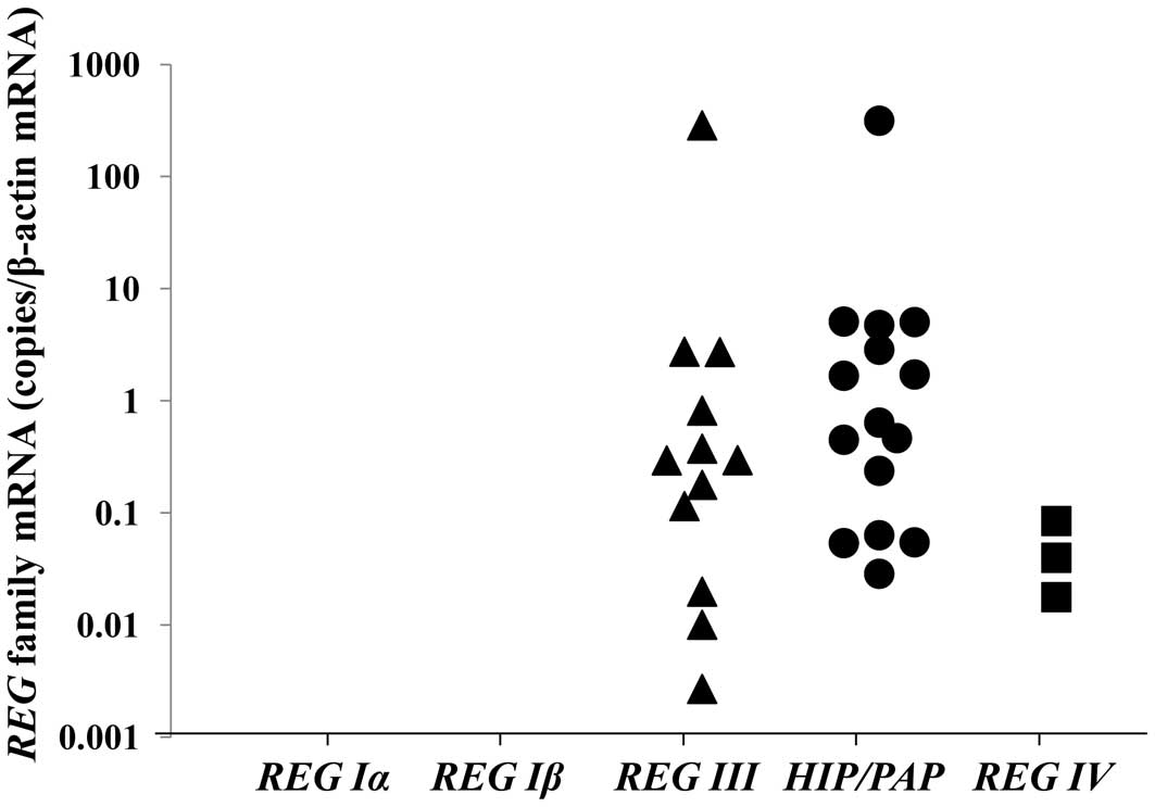
REG family gene expression in
hypopharyngeal squamous cell carcinomas
The mRNA expression of REG family genes in
each case was measured using real-time RT-PCR. No case with
positive expression of REG Iα and REG Iβ was noted,
and only 3 cases were positive for REG IV expression, while
there were 12 and 15 cases positive for REG III and
HIP/PAP expression, respectively (Fig. 1). No positive case among the normal
tissues in the hypopharyngeal area showed expression for any of the
REG family genes.
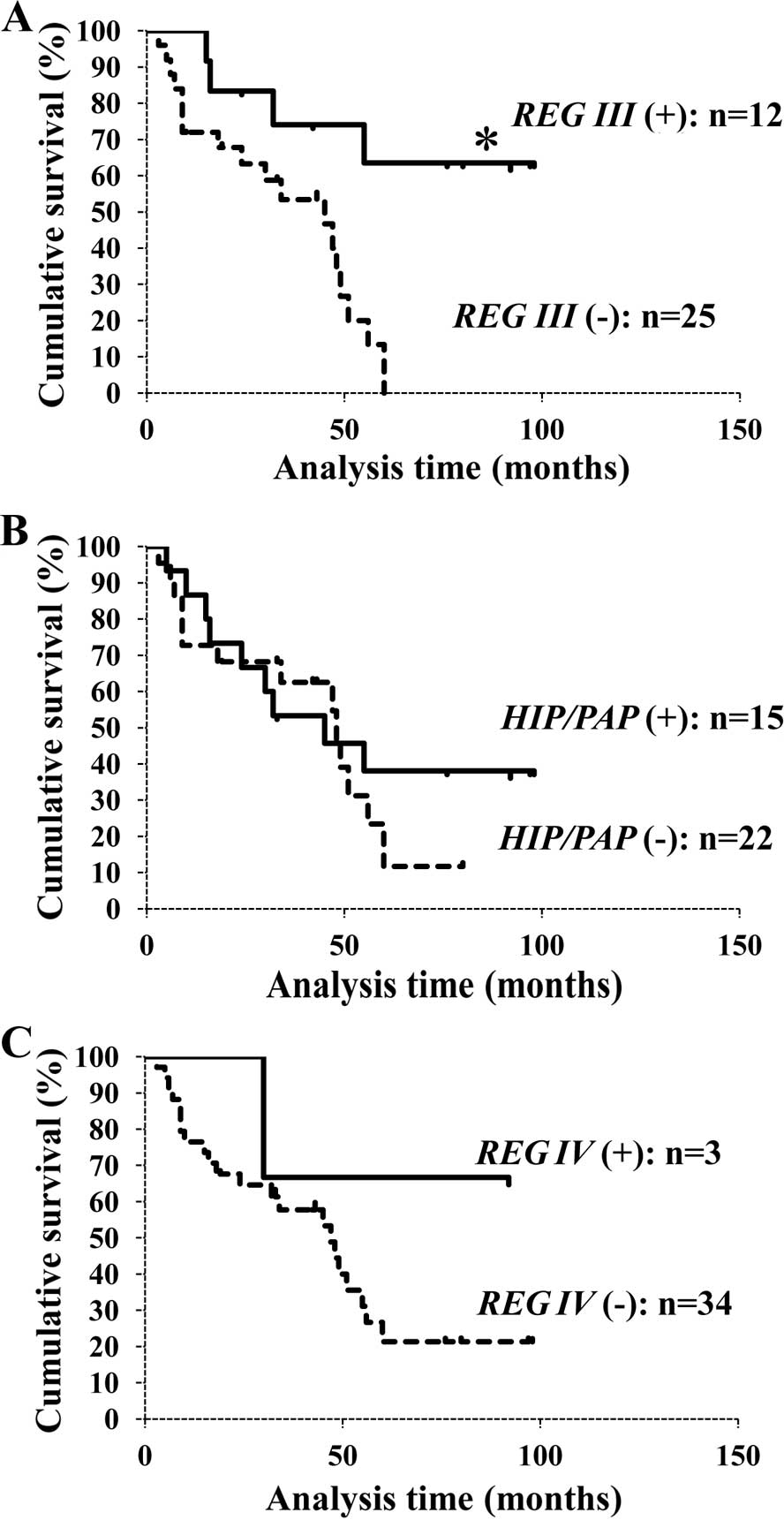
Differences in survival determined by the
clinical data
Each overall survival rate was calculated using the
Kaplan-Meier method. The REG III expression-positive group
showed long-term survival when compared to the negative group with
significant difference (Fig. 2A),
whereas there were no differences between groups in regards to
HIP/PAP and REG IV expression (Fig. 2B and C). These data suggest that
REG III expression is associated with a more favorable
prognosis of hypopharyngeal squamous cell carcinoma.
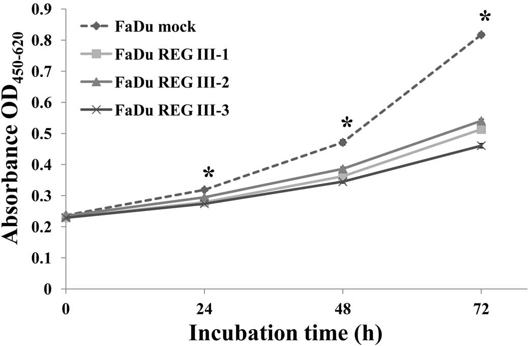
REG III suppresses the growth of FaDu
cells
To estimate the effect of REG III on
hypopharyngeal cancer cell growth, we stably transfected FaDu
cells, which originally express very low level of REG III
mRNA, with an expression plasmid for REG III, after which
the expression of REG III mRNA was assessed (data not
shown). FaDu cells transfected with the REG III expression
plasmid (FaDu REG III-1, -2 and -3 cells) showed higher expression
of REG III than the cells transfected with the
neomycin-resistance gene alone (FaDu mock).
In the cell proliferation assay using WST-8
cleavage, FaDu REG III-1, -2 and -3 cells showed a significant
decrease in growth rate when compared with the rate in the FaDu
mock cells (Fig. 3).
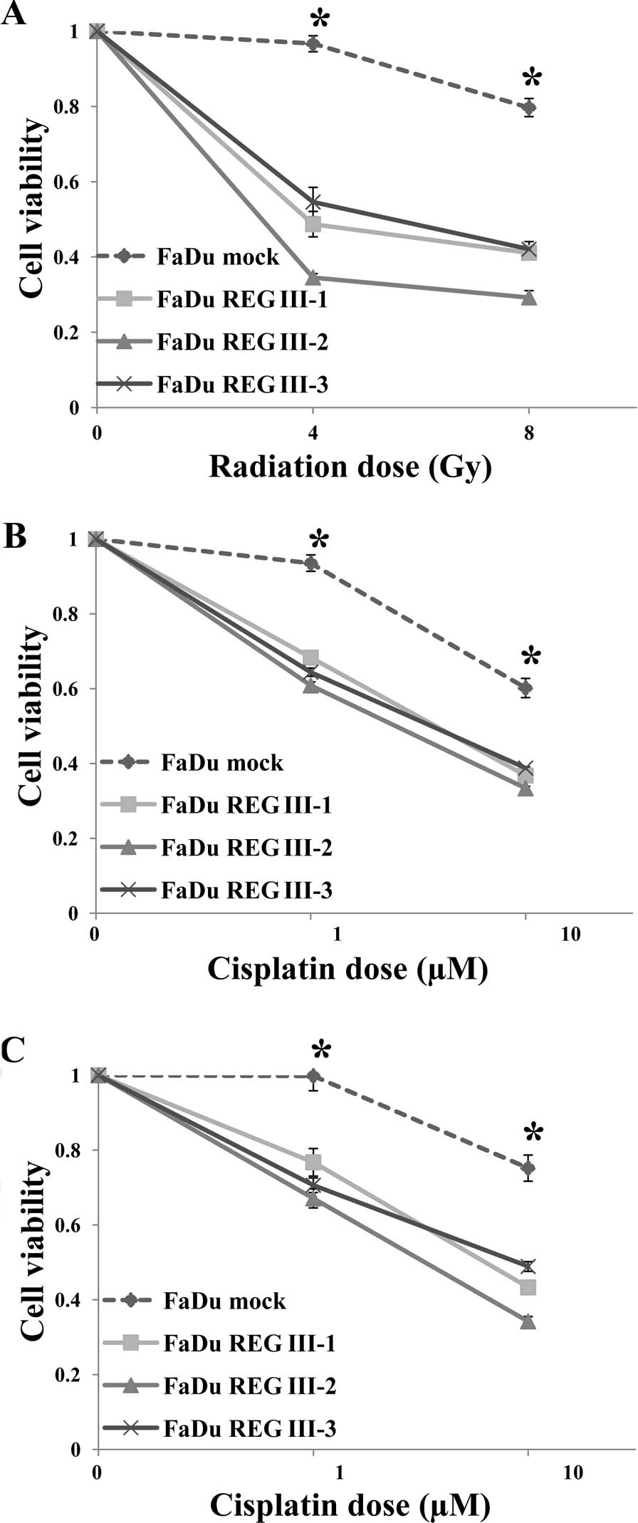
REG III enhances the chemosensitivity
and/or radiosensitivity of FaDu cells
FaDu REG III-1, -2 and -3 cells showed a significant
increase in radiosensitivity at 4 and 8 Gy and chemosensitivity at
1.0 and 10 μM cisplatin, as compared with the FaDu mock cells
(Fig. 4A and B). Furthermore,
chemoradiosensitivity was also significantly higher in the FaDu REG
III-1, -2 and -3 cells at 1.0 and 10 μM cisplatin (Fig. 4C). Thus, these results imply that
REG III enhances the chemosensitivity and/or
radiosensitivity of hypopharyngeal cancer cells.
Discussion
Members of the Reg family are grouped into
four subtypes: types I, II, III, and IV; the human REG
family is composed of five subclasses: REG Iα, REG
Iβ, REG III,
hepatocarcinoma-intestine-pancreas/pancreatitis-associated-protein
(HIP/PAP) and REG IV. REG III and HIP/PAP
belong to type III. The nucleotide sequence of REG III mRNA
is very similar to that of HIP/PAP mRNA (29). Although there are many reports
concerning REG Iα, HIP/PAP and REG IV, little
is known regarding REG III. REG III is strongly expressed in
the pancreas, moderately in the testis and weakly in the heart,
kidney and placenta, whereas HIP/PAP is strongly expressed
in the pancreas and small intestine and weakly in hepatoma,
stomach, brain and heart (29).
Type III Reg proteins as well as type I Reg proteins are suggested
to be induced in response to tissue inflammation such as
pancreatitis (29,34). However, the details of their
biological function have not been fully elucidated.
In the present study, we observed many cases of
hypopharyngeal cancer expressing REG III or HIP/PAP.
HIP/PAP expression was not associated with a significant
difference in survival, while the survival rate of patients with
REG III expression was significantly prolonged when compared
with that of the negative cases. In vitro, we also observe a
reduction in cell growth rates and the enhancement of
chemosensitivity and/or radiosensitivity in the FaDu cells
transfected with REG III when compared with the control.
These outcomes were compatible with the clinical data.
Type III Reg proteins have been suggested to be
involved in cellular proliferation of intestinal, hepatic and
neuronal cells (35,36). High expression of type III Reg
proteins has been observed in carcinomas in digestive organs and
inflammatory diseases such as pancreatitis, enterocolitis and UC
(29,31,37–39).
Furthermore, type III proteins are also present in response to
neuron damage and participate in the regeneration of neurons
(35,36,40).
However, as the details regarding the effects of type III Reg
proteins on intracellular signaling remain to be elucidated, it is
still unclear how REG III functioned to enhance the
chemosensitivity and/or radiosensitivity and improve the
survival of patients with hypopharyngeal squamous cell carcinoma in
the present study.
Several reports have shown that interleukin-6 (IL-6)
and dexamethasone activate the transcription of REG
I(23,34). and that type III is induced by
various cytokines, such as IL-6, INF-γ and TNF-α (41,42).
To investigate how REG III expression is regulated in HNSCC
cells, we determined the expression of IL-6, -8 and -11 using
real-time RT-PCR method using clinical samples in the present
study. The expression of REG III and IL-11 had no
correlation, while the expression of IL-6 and IL-8 had a positive
correlation with the expression of REG III (data not shown).
Although the details are still unknown, these results indicate the
possibility that IL-6 and IL-8 can become key factors to elucidate
the relationship between the expression of REG III and the
prognosis of hypopharyngeal cancer patients.
It has been demonstrated that Reg is highly
expressed in regenerating islets and tissues of pancreatitis,
whereas this expression declines when the function of the pancreas
is improved (17,37,43,44).
Moreover, in vivo, transfection with Reg into a
normal rat caused neither proliferation of β-cells nor hyperplasia
of islets (19). These results
suggest that there is an unknown suppressive function in
vivo in contrast with the proliferative activity by Reg
expression. In the present study, it was also expected that REG
III may act as a suppression factor with various functions in
hypopharyngeal cancer.
These data suggest that REG III, which can be
easily detected in formalin-fixed paraffin-embedded tissues with
RT-PCR analysis, may be a reliable biomarker of the
chemosensitivity and/or radiosensitivity and prognosis of
hypopharyngeal cancer. However, the biological function and cell
signaling pathway of REG III require further elucidation.
The critical mechanisms warrant further investigation. This is the
first report concerning the association between REG III
expression and the chemosensitivity and/or radiosensitivity and
prognosis of HNSCC including hypopharyngeal cancer.
Acknowledgements
We are grateful to Dr Kan-ichi Nakagawara (NGRL,
Sendai, Japan) for designing and providing the primers for RT-PCR.
The present study is partial academic fulfillment for Thesis by
T.M. of Medical Science at Nara Medical University.
Abbreviations:
|
REG
|
regenerating gene
|
|
HNSCC
|
head and neck squamous cell
carcinoma
|
|
CRT
|
chemoradiotherapy
|
|
UC
|
ulcerative colitis
|
|
HIP/PAP
|
hepatocarcinoma-intestine-pancreas/pancreatitis-associated-protein
|
|
RT-PCR
|
reverse transcriptase-polymerase chain
reaction
|
|
IL
|
interleukin
|
References
|
1
|
Ang KK, Trotti A, Brown BW, et al:
Randomized trial addressing risk features and time factors of
surgery plus radiotherapy in advanced head-and-neck cancer. Int J
Radiat Oncol Biol Phys. 51:571–578. 2001. View Article : Google Scholar : PubMed/NCBI
|
|
2
|
Ozer E, Grecula JC, Agrawal A, Rhoades CA,
Young DC and Schuller DE: Long-term results of a multimodal
intensification regimen for previously untreated advanced
resectable squamous cell cancer of the oral cavity, oropharynx, or
hypopharynx. Laryngoscope. 116:607–612. 2006. View Article : Google Scholar
|
|
3
|
Kumar B, Cordell KG, Lee JS, et al: EGFR,
p16, HPV titer, Bcl-xL and p53, sex, and smoking as indicators of
response to therapy and survival in oropharyngeal cancer. J Clin
Oncol. 26:3128–3137. 2008. View Article : Google Scholar : PubMed/NCBI
|
|
4
|
Worden FP, Kumar B, Lee JS, et al:
Chemoselection as a strategy for organ preservation in advanced
oropharynx cancer: response and survival positively associated with
HPV16 copy number. J Clin Oncol. 26:3138–3146. 2008. View Article : Google Scholar : PubMed/NCBI
|
|
5
|
Watanabe T, Yonekura H, Terazono K,
Yamamoto H and Okamoto H: Complete nucleotide sequence of human reg
gene and its expression in normal and tumoral tissues. The reg
protein, pancreatic stone protein, and pancreatic thread protein
are one and the same product of the gene. J Biol Chem.
265:7432–7439. 1990.PubMed/NCBI
|
|
6
|
Fukui H, Kinoshita Y, Maekawa T, et al:
Regenerating gene protein may mediate gastric mucosal proliferation
induced by hypergastrinemia in rats. Gastroenterology.
115:1483–1493. 1998. View Article : Google Scholar : PubMed/NCBI
|
|
7
|
Higham AD, Bishop LA, Dimaline R, et al:
Mutations of RegIalpha are associated with enterochromaffin-like
cell tumor development in patients with hypergastrinemia.
Gastroenterology. 116:1310–1318. 1999. View Article : Google Scholar : PubMed/NCBI
|
|
8
|
Shinozaki S, Nakamura T, Iimura M, et al:
Upregulation of Reg 1α and GW112 in the epithelium of inflamed
colonic mucosa. Gut. 48:623–629. 2001.
|
|
9
|
Ose T, Kadowaki Y, Fukuhara H, et al: Reg
I-knockout mice reveal its role in regulation of cell growth that
is required in generation and maintenance of the villous structure
of small intestine. Oncogene. 26:349–359. 2007. View Article : Google Scholar : PubMed/NCBI
|
|
10
|
Macadam RC, Sarela AI, Farmery SM,
Robinson PA, Markham AF and Guillou PJ: Death from early colorectal
cancer is predicted by the presence of transcripts of the REG gene
family. Br J Cancer. 83:188–195. 2000.PubMed/NCBI
|
|
11
|
Violette S, Festor E, Pandrea-Vasile I, et
al: Reg IV, a new member of the regenerating gene family, is
overexpressed in colorectal carcinomas. Int J Cancer. 103:185–193.
2003. View Article : Google Scholar
|
|
12
|
Yonemura Y, Sakurai S, Yamamoto H, et al:
REG gene expression is associated with the infiltrating growth of
gastric carcinoma. Cancer. 98:1394–1400. 2003. View Article : Google Scholar : PubMed/NCBI
|
|
13
|
Dhar DK, Udagawa J, Ishihara S, et al:
Expression of regenerating gene I in gastric adenocarcinomas:
correlation with tumor differentiation status and patient survival.
Cancer. 100:1130–1136. 2004. View Article : Google Scholar
|
|
14
|
Bishnupuri KS, Luo Q, Murmu N, Houchen CW,
Anant S and Dieckgraefe BK: Reg IV activates the epidermal growth
factor receptor/Akt/AP-1 signaling pathway in colon
adenocarcinomas. Gastroenterology. 130:137–149. 2006. View Article : Google Scholar : PubMed/NCBI
|
|
15
|
Hayashi K, Motoyama S, Koyota S, et al:
REG I enhances chemo- and radiosensitivity in squamous cell
esophageal cancer cells. Cancer Sci. 99:2491–2495. 2008. View Article : Google Scholar : PubMed/NCBI
|
|
16
|
Zhou L, Zhang R, Wang L, et al:
Upregulation of REG Iα accelerates tumor progression in pancreatic
cancer with diabetes. Int J Cancer. 127:1795–1803. 2010.
|
|
17
|
Terazono K, Yamamoto H, Takasawa S, et al:
A novel gene activated in regenerating islets. J Biol Chem.
263:2111–2114. 1988.PubMed/NCBI
|
|
18
|
Watanabe T, Yonemura Y, Yonekura H, et al:
Pancreatic beta-cell replication and amelioration of surgical
diabetes by Reg protein. Proc Natl Acad Sci USA. 91:3589–3592.
1994. View Article : Google Scholar : PubMed/NCBI
|
|
19
|
Unno M, Nata K, Noguchi N, et al:
Production and characterization of Reg knockout mice: reduced
proliferation of pancreatic beta-cells in Reg knockout mice.
Diabetes. 51(Suppl 3): S478–S483. 2002. View Article : Google Scholar : PubMed/NCBI
|
|
20
|
Takasawa S, Ikeda T, Akiyama T, et al:
Cyclin D1 activation through ATF-2 in Reg-induced pancreatic
beta-cell regeneration. FEBS Lett. 580:585–591. 2006. View Article : Google Scholar : PubMed/NCBI
|
|
21
|
Sanchez D, Figarella C, Marchand-Pinatel
S, Bruneau N and Guy-Crotte O: Preferential expression of reg Iβ
gene in human adult pancreas. Biochem Biophys Res Commun.
284:729–737. 2001.
|
|
22
|
Hervieu V, Christa L, Gouysse G, et al:
HIP/PAP, a member of the reg family, is expressed in
glucagon-producing enteropancreatic endocrine cells and tumors. Hum
Pathol. 37:1066–1075. 2006. View Article : Google Scholar : PubMed/NCBI
|
|
23
|
Okamoto H and Takasawa S: Recent advances
in the Okamoto model: the CD38-cyclic ADP-ribose signal system and
the regenerating gene protein (Reg)-Reg receptor system in
beta-cells. Diabetes. 51(Suppl 3): S462–S473. 2002. View Article : Google Scholar : PubMed/NCBI
|
|
24
|
Dusetti NJ, Frigerio JM, Fox MF, Swallow
DM, Dagorn JC and Iovanna JL: Molecular cloning, genomic
organization, and chromosomal localization of the human
pancreatitis-associated protein (PAP) gene. Genomics. 19:108–114.
1994. View Article : Google Scholar : PubMed/NCBI
|
|
25
|
Moriizumi S, Watanabe T, Unno M, et al:
Isolation, structural determination and expression of a novel reg
gene, human regI beta. Biochim Biophys Acta. 1217:199–202. 1994.
View Article : Google Scholar : PubMed/NCBI
|
|
26
|
Lasserre C, Simon MT, Ishikawa H, et al:
Structural organization and chromosomal localization of a human
gene (HIP/PAP) encoding a C-type lectin overexpressed in primary
liver cancer. Eur J Biochem. 224:29–38. 1994. View Article : Google Scholar : PubMed/NCBI
|
|
27
|
Miyashita H, Nakagawara K, Mori M, et al:
Human REG family genes are tandemly ordered in a 95-kilobase region
of chromosome 2p12. FEBS Lett. 377:429–433. 1995. View Article : Google Scholar : PubMed/NCBI
|
|
28
|
Hartupee JC, Zhang H, Bonaldo MF, Soares
MB and Dieckgraefe BK: Isolation and characterization of a cDNA
encoding a novel member of the human regenerating protein family:
Reg IV. Biochim Biophys Acta. 1518:287–293. 2001. View Article : Google Scholar : PubMed/NCBI
|
|
29
|
Nata K, Liu Y, Xu L, et al: Molecular
cloning, expression and chromosomal localization of a novel human
REG family gene, REG III. Gene. 340:161–170. 2004.
View Article : Google Scholar : PubMed/NCBI
|
|
30
|
Asahara M, Mushiake S, Shimada S, et al:
Reg gene expression is increased in rat gastric
enterochromaffin-like cells following water immersion stress.
Gastroenterology. 111:45–55. 1996. View Article : Google Scholar : PubMed/NCBI
|
|
31
|
Ogawa H, Fukushima K, Naito H, et al:
Increased expression of HIP/PAP and regenerating gene III in human
inflammatory bowel disease and a murine bacterial reconstitution
model. Inflamm Bowel Dis. 9:162–170. 2003. View Article : Google Scholar
|
|
32
|
Takasawa S, Kuroki M, Nata K, et al: A
novel ryanodine receptor expressed in pancreatic islets by
alternative splicing from type 2 ryanodine receptor gene. Biochem
Biophys Res Commun. 397:140–145. 2010. View Article : Google Scholar : PubMed/NCBI
|
|
33
|
Ota H, Tamaki S, Itaya-Hironaka A, et al:
Attenuation of glucose-induced insulin secretion by intermittent
hypoxia via down-regulation of CD38. Life Sci. 90:206–211. 2012.
View Article : Google Scholar : PubMed/NCBI
|
|
34
|
Akiyama T, Takasawa S, Nata K, et al:
Activation of Reg gene, a gene for insulin-producing beta-cell
regeneration: poly(ADP-ribose) polymerase binds Reg promoter and
regulates the transcription by autopoly(ADP-ribosyl)ation. Proc
Natl Acad Sci USA. 98:48–53. 2001.PubMed/NCBI
|
|
35
|
Livesey FJ, O’Brien JA, Li M, Smith AG,
Murphy LJ and Hunt SP: A Schwann cell mitogen accompanying
regeneration of motor neurons. Nature. 390:614–618. 1997.
View Article : Google Scholar : PubMed/NCBI
|
|
36
|
Namikawa K, Fukushima M, Murakami K, et
al: Expression of Reg/PAP family members during motor nerve
regeneration in rat. Biochem Biophys Res Commun. 332:126–134. 2005.
View Article : Google Scholar : PubMed/NCBI
|
|
37
|
Iovanna J, Orelle B, Keim V and Dagorn JC:
Messenger RNA sequence and expression of rat
pancreatitis-associated protein, a lectin-related protein
overexpressed during acute experimental pancreatitis. J Biol Chem.
266:24664–24669. 1991.
|
|
38
|
Dieckgraefe BK, Stenson WF, Korzenik JR,
Swanson PE and Harrington CA: Analysis of mucosal gene expression
in inflammatory bowel disease by parallel oligonucleotide arrays.
Physiol Genomics. 4:1–11. 2000.PubMed/NCBI
|
|
39
|
Lawrance IC, Fiocchi C and Chakravarti S:
Ulcerative colitis and Crohn’s disease: distinctive gene expression
profiles and novel susceptibility candidate genes. Hum Mol Genet.
10:445–456. 2001.
|
|
40
|
Nishimune H, Vasseur S, Wiese S, et al:
Reg-2 is a motoneuron neurotrophic factor and a signalling
intermediate in the CNTF survival pathway. Nat Cell Biol.
2:906–914. 2000. View Article : Google Scholar : PubMed/NCBI
|
|
41
|
Dusetti NJ, Ortiz EM, Mallo GV, Dagorn JC
and Iovanna JL: Pancreatitis-associated protein I (PAP I), an acute
phase protein induced by cytokines. Identification of two
functional interleukin-6 response elements in the rat PAP I
promoter region. J Biol Chem. 270:22417–22421. 1995. View Article : Google Scholar
|
|
42
|
Ortiz EM, Dusetti NJ, Vasseur S, et al:
The pancreatitis-associated protein is induced by free radicals in
AR4–2J cells and confers cell resistance to apoptosis.
Gastroenterology. 114:808–816. 1998.PubMed/NCBI
|
|
43
|
Rouquier S, Verdier JM, Iovanna J, Dagorn
JC and Giorgi D: Rat pancreatic stone protein messenger RNA.
Abundant expression in mature exocrine cells, regulation by food
content, and sequence identity with the endocrine reg transcript. J
Biol Chem. 266:786–791. 1991.
|
|
44
|
Bluth MH, Patel SA, Dieckgraefe BK,
Okamoto H and Zenilman ME: Pancreatic regenerating protein (reg I)
and reg I receptor mRNA are upregulated in rat pancreas after
induction of acute pancreatitis. World J Gastroenterol.
12:4511–4516. 2006.PubMed/NCBI
|