Introduction
In 1996, eligibility criteria such as the Milan
criteria (MC) of liver transplantation (LT) for hepatocellular
carcinoma (HCC) were reported by Mazzaferro et al(1). MC emphasized LT as a therapeutic
option for patients with HCC. Living donor liver transplantation
(LDLT) is virtually the only option for patients with HCC in the
east Asian countries such as Korea (2) or Japan (3–9), where
the number of deceased donors is limited, and for patients above MC
in western countries such as the United States and in Europe.
Therefore, understanding how far the criteria of LT for HCC can be
extended in LDLT from MC is key in improving the outcomes in
regions with limited organ donors. There have been several reports
of expanded criteria as indications of LT for HCC, such as the
Up-to-seven (Up-to-7) criteria (10), University California of San
Francisco (UCSF) criteria (11),
Asan criteria (2), Tokyo (5–5 rule)
criteria (3), Kyoto criteria
(4,5) and Kyushu criteria (6–8). In
addition, Kyoto criteria (4,5) and
Kyushu criteria (6–8) showed pre-operative tumor markers such
as the des-γ-carboxyprothrombin (DCP) level. In the present study,
we evaluated the predictive values of the previously proposed
selection criteria, including Up-to 7 criteria, UCSF criteria, Asan
criteria, Tokyo criteria, Kyoto criteria and Kyushu criteria, on
the overall survival (OS) and HCC disease-free survival (DFS) of
LDLT recipients with HCC. These criteria are categorized into
several types which are based only on pre-operative imaging
diagnosis, or on pathological diagnosis of the explant liver, which
consider microvascular invasion as above criteria, and take account
of tumor markers. According to Japanese national data (9), in addition to the MC, it is reported
that the values of tumor markers [α-fetoprotein (AFP) and DCP]
define prognosis, but as various factors are involved in tumor
markers, it is difficult to incorporate them into international
eligibility criteria of LT for HCC. Regardless of whether it is
deceased donor liver transplantation (DDLT) or LDLT, the criteria
of LT for HCC should be defined solely by simple factors such as
tumor diameter or number to guarantee their international
applicability. Furthermore, eligibility criteria of LT for HCC must
significantly define the prognosis for recipients in evaluations
which are based not only on pathological diagnosis of the explant
liver, but also on pre-operative imaging diagnosis. However, it is
important to perform pre-operative imaging diagnosis of HCC close
to post-operative pathological diagnosis. If the accuracy of
imaging diagnosis of HCC is low, the reliability of the criteria
decreases, therefore, pre-operative imaging diagnosis should be
performed accurately using some imaging diagnostic modalities. In
order to enhance imaging diagnostic accuracy for HCC, in addition
to dynamic multi-detectable-row computer tomography (dynamic MDCT)
and gadolinium ethoxybenzyl diethylenetriamine pentaacetic
acid-enhanced magnetic resonance imaging (Gd-EOB-DTPA-MRI)
(12,13), we also obtained images as far as
possible using CT under angiography [during arterial portography
(CTAP) and during hepatic arteriography (CTHA)] (14–17).
In view of the fact that a healthy living donor is exposed to major
risks by hepatectomy, recurrence of HCC after LT in the recipient
must be avoided. To receive LDLT under Japanese health insurance,
although no restrictions are imposed as to therapeutic history 3
months prior to LT, the recipient must satisfy the MC in the
pre-operative final imaging diagnosis. Eligibility criteria have
been reported by various high-volume centers in Japan (3–8) and
there are attempts to widen eligibility of LDLT for HCC under
health insurance. In this context, in order to expand the
eligibility criteria from within MC, we evaluated which criteria
were the most suitable from the two viewpoints of pre-operative
imaging diagnosis and pathological diagnosis with recipients who
had performed precise pre-operative diagnostic imaging and had been
observed for >5 years after LDLT for HCC. Furthermore, we
evaluated the appropriateness of the above criteria from the
viewpoint of proliferation of α-smooth muscle actin (SMA)-positive
cancer-associated fibroblasts (CAFs), which are strongly related to
cancer progression and invasion (18,19).
However, there are no reports which evaluate HCC recurrence after
LT from the viewpoint of α-SMA-positive CAFs. We therefore
evaluated the relationship between HCC recurrence after LDLT and
proliferation of α-SMA-positive CAFs, as well as the correlation
between eligibility criteria and α-SMA-positive CAFs.
Materials and methods
Patients
From July 2003 to December 2007, 22 consecutive
LDLTs for liver cirrhosis (LC) with HCC were performed at Kanazawa
University Hospital (Ishikawa, Japan) after receiving approval from
the Ethics and Indications Committee of Kanazawa University. Our
selection criteria for the patients with HCC were as follows: no
modality except LDLT available to cure patients with HCC and
end-stage liver disease, no extrahepatic metastasis and no
macrovascular invasion such as portal vein or hepatic vein
infiltrations. We limited adaptation of LDLT for HCC to within MC
under health insurance of Japan since January 2008, but performed
LDLT for above MC recipients by own expenses until December 2007.
Therefore targeted cases for the present study were limited to
recipients who had undergone LDLT by December 2007. Twenty-two
patients had HCC, proven histologically. The median age of the 22
patients was 55.5 years (range, 47–64 years). Written informed
consent for the present study was obtained from each patient. In
addition, the study was approved by the Kanazawa University Ethics
Committee. Tumor-specific evaluations, including abdominal and
thoracic dynamic MDCT, abdominal CTAP, abdominal CTHA, abdominal
Gd-EOB-DTPA-MRI, bone scintigraphy, and the determination of AFP
and DCP (Protein induced by Vitamin K, PIVKA-II), were performed
for all LDLT candidates. The diameter and number of HCCs were
determined by multiple radiologists, based on pre-operative imaging
studies within one month of LT. Thus, the variables used in the
criteria, including tumor diameter and number, were based on these
data. The explants were examined histologically. For pathological
examination, whole liver explants were fixed in 10% formalin and
cut into 5-mm slices to facilitate gross and histological
examinations. Following macroscopic examination, the nodular
lesions were embedded in paraffin, cut into 4-inch sections and
stained with hematoxylin and eosin. The incidence of microvascular
invasion and histological grades were subsequently estimated within
these criteria. Microvascular invasion was defined as microscopic
portal vein or hepatic vein invasion of cancer cells. The stage was
determined for each patient according to the AJCC/UICC (6th
edition) guidelines (20) and UNOS
TMN (21). Among the 22 patients,
10 (45.5%) met the MC according to pre-operative first imaging
diagnosis, while 12 did not. According to previous studies, Up-to
7, UCSF, Asan, Tokyo, Kyoto and Kyushu criteria were applied and
the predictive impacts of these criteria for HCC recurrence were
evaluated by univariate analyses. The previously proposed selection
criteria for HCC are briefly described below and are shown in
Table I. The Up-to-7 criteria are
defined as HCC with seven as the sum of the diameter of the largest
tumor (in cm) and the number of tumors. The UCSF criteria are
defined as HCC meeting the following criteria: solitary tumor of
≤6.5 cm, or ≤3 nodules with the largest lesion of ≤4.5 cm and a
total tumor diameter of ≤8 cm. The Asan criteria are defined as HCC
meeting the following criteria: tumor up to 6 nodules with a
maximum diameter of 5 cm without gross vascular invasion. The Tokyo
criteria are defined as HCC meeting the following criteria: tumor
of up to 5 nodules with a maximum diameter of 5 cm (5–5 rule) that
are evaluated with pre-operative imaging. The Kyoto criteria are
defined as HCC meeting the following criteria: ≤10 tumors that are
all ≤5 cm in diameter and DCP of ≤400 mAU/ml. The Kyushu University
criteria are defined as HCC with tumor diameter <5 cm or DCP
<300 mAU/ml. In the 7 above MC recipients who underwent pre-LDLT
therapy to downstage HCC, transarterial chemo-lipiodolisation
(TACL) was performed in all cases, and radiofrequency ablation
therapy (RFA) was also performed in 2 cases. The recipients who
underwent pre-LDLT therapy for HCC were observed for ≥3 months from
the end of the pre-operative therapy to LDLT. There were 4 out of 5
recipients who were downstaged from above MC (pre-operative first
imaging diagnosis) to within MC (pre-operative final imaging
diagnosis) by pre-LDLT therapy. In the 15 cases were LDLT was
performed without prior therapy, the pre-operative first imaging
diagnosis was considered the pre-operative final imaging diagnosis.
There were 13 cases in total that received therapy for HCC in the
past before LDLT; TACL had been performed in 11 cases and several
treatments in 10 cases. RFA had been performed in 8 cases,
percutaneous ethanol injection therapy (PEIT) in 5 cases and
hepatectomy in 2 cases. In addition, transarterial infusion
chemotherapy had been performed in only 1 case. The clinical
follow-up of patients after LDLT for HCC followed a strict
protocol, which did not change during the study period. The
patients were seen biweekly for the first 6 months and then
monthly. The patients underwent enhanced MDCT or Gd-EOB-DTPA-MRI at
4–6 month intervals. Liver biopsy, hepatic angiography with CT,
bone scintigraphy or 2-Fluoro 2-deoxyglucose positron emission
tomography (FDG-PET) CT was also performed if deterioration in the
graft function or a rise in the AFP or DCP levels was noted. The
mean follow-up period was 7 years.
 | Table ISummary of published outcomes of
liver transplantation for HCC between recipients satisfying
expanded/extended eligibility criteria. |
Table I
Summary of published outcomes of
liver transplantation for HCC between recipients satisfying
expanded/extended eligibility criteria.
| | | | | OS (%) | DFS (%) |
|---|
| | | | |
|
|
|---|
| Eligibility
criteria name and definition | Authors, year
(ref.) | Study design and
staging method | Tumor
characteristics | Cases (n) | 1-year | 3-year | 5-year | 1-year | 3-year | 5-year |
|---|
| UCSF criteria: no
extrahepatic spread or macrovascular invasion. Solitary tumor with
diameter ≤65 mm, or ≤3 nodules with maximum diameter ≤45 mm and
total tumor diameter ≤80 mm. | Yao et al,
2001 (11) | Retrospective
analysis. Staging: explant pathology | Within UCSF
criteria and above MC | 60 | 90 | | 75.2 | | | |
| Above UCSF | 10 | 50 | 20 | - | | | |
| Up-to-7 criteria:
no extrahepatic disease or microvascular invasion. Sum of number of
nodules and diameter of largest nodule (cm) ≤7 | Mazzoferro et
al, 2001 (10) | Retrospective
analysis. Staging: explant pathology | Within Up-to-7
criteria and above MC without microvasular invasion | 283 | | 77.7 | 71.2 | | | |
| Within Up-to-7
criteria and above MC with microvasular invasion | 116 | | 60.2 | 47.4 | | | |
| Within MC without
microvascular invasion | 361 | | 81.8 | 76.1 | | | |
| Within MC with
microvascular invasion | 44 | | 77.1 | 71.6 | | | |
| Above Up-to-7
criteria and without microvasular invasion | 333 | | 71.8 | 64 | | | |
| Above Up-to-7
criteria with microvasular invasion | 338 | | 41.7 | 33 | | | |
| Asan criteria: no
extrahepatic disease or macrovascular invasion. ≤6 nodules with
maximum diameter 50 mm. | Lee et al,
2008 (2) | Retrospective
analysis. Staging: explant pathology | Within Asan
criteria and above MC | 22 | 100 | 88.9 | 80 | | | |
| Within MC | 152 | 86.6 | 79.2 | 76 | | | |
| Beyond Asan
criteria | 32 | 65.7 | 34.1 | 18.9 | | | |
| Tokyo criteria
(5–5): No extrahepatic disease or macrovascular invasion. ≤5
nodules with maximum diameter 50 mm. | Sugawara et
al, 2007 (3) | Analysis against
predefined criteria. Staging: pre-LDLT radiology (imaging). | Within Tokyo
criteria | 72 | | | | 97 | 94 | |
| Above Tokyo
criteria | 6 | | | | 50 | 50 | |
| Kyoto criteria: no
extrahepatic disease or macrovascular invasion. ≤10 nodules with
maximum diameter 50 mm. PIVKA-II ≤400 mAU/ml. | Ito et al,
2007 (4) | Retrospective
analysis. Staging: explant pathology. | Within Kyoto
criteria | 78 | | | 86.7 | | | |
| Above Kyoto
criteria | 40 | | | 34.4 | | | |
| Takada et
al, 2007 (5) | Retrospective
analysis. Staging: pre-LDLT radiology (imaging). | Within Kyoto
criteria | 83 | | | 87 | | | |
| Above Kyoto
criteria | 44 | | | 37 | | | |
| Kyushu criteria: no
extrahepatic disease or macrovascular invasion. Any nodules with
maximum diameter 50 mm. PIVKA-II ≤300 mAU/ml. | Shirabe et
al, 2011 (8) | Retrospective
analysis. Staging: pre-LDLT radiology (imaging). | Within Kyushu
criteria and above MC | 48 | | | | 85 | 80 | 80 |
| Above Kyushu
criteria | 6 | | | | 16.7 | 0 | 0 |
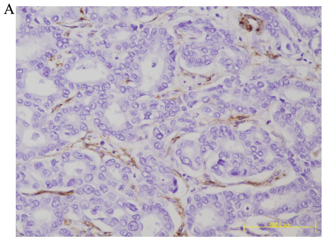
Immunohistochemistry
The proliferation of α-SMA-positive CAFs was
evaluated immunohistologically. When several tumors were present,
the tumor with microvascular invasion was evaluated. If no
microvascular invasion was found, tumors which had the poorer
histological degree of differentiation or differentiated into
biliary tract type (CK7-positive or CK19-positive), were evaluated.
Tumor specimens were fixed in 10% formalin and embedded in
paraffin. The expressions of α-SMA in HCC were examined
immunohistochemically using respective primary antibodies using
EnVision+ System (DAKO). De-waxed 4-μm sections were
incubated with 1:50 with protein blocking serum for 10 min to block
non-specific binding and immunostaining was performed using
EnVision+ System. Briefly, the slides were incubated
with each primary antibody (1:50) at 4°C overnight. After washing,
the EnVision+ polymer solution was applied for 1 h. The
reaction products were visualized via a diaminobenzidine (DAB)
reaction. The specimens were then lightly counterstained with
hematoxylin and examined under a fluorescence microscope. Primary
antibody used for immunostaining was Actin α2 Smooth Muscle rabbit
anti-human polyclonal antibody (Novus Biologicals, Littleton, CO,
USA).
Computer-assisted image analysis
(19)
We used computer-assisted image analysis to quantify
the value of α-SMA expression in HCC. After staining for α-SMA, the
histological sections were observed using a microscope equipped
with a charge coupled-device color camera (Olympus Co., Japan)
under constant electrical and optical conditions. A random
selection of 10 fields in most poorly differentiated and
α-SMA-positive CAF proliferating lesions of HCC were assessed for
α-SMA expression. Using an imaging processor (VH Analyzer; Keyence
Co., Japan) the percentage of α-SMA expression stromal area was
quantified as the relative percentage of the α-SMA-positive stromal
area to the selected fields of cancer.
Statistical analysis
All statistical analyses were performed using SPSS
Software v20 (IBM-Japan, Tokyo, Japan). The continuous variables
were compared using the Mann-Whitney U test. All variables are
expressed as means ± standard deviation (SD). The categorical data
were compared using χ2 tests. We compared Kaplan-Meier
distributions of time to mortality or HCC recurrence after LDLT
with the log-rank test or generalized Wilcoxon. Cox’s proportional
hazard model was used to identify independent variables for
post-operative recurrence of HCC. The comparative evaluation was
performed among the Milan, Up-to-7, Asan, Tokyo, Kyoto, Kyushu
criteria, degree of α-SMA-positive CAFs and clinicopathological
variables including pre-operative serum AFP levels and serum DPC
levels, presence of microvascular invasion, histological grade of
the tumor (poorly differentiated), the number of tumors and maximum
diameter of tumor on the resected specimen. The differences were
considered statistically significant when the P-value was
<0.05.
Results
Table II shows
background characteristics of 22 recipients who underwent LDLT for
HCC according to post-LDLT with or without HCC recurrence. The
average age of recipients was 56 years, 17 of whom were males, and
the average MELD score of recipients was 14 points. There were 12
recipients with hepatitis C viral (HCV) hepatitis and 10 recipients
with hepatitis B viral (HBV) hepatitis. Seventeen recipients were
given right hepatic graft and 5 recipients were given left hepatic
graft with caudate lobe. The average graft volume/standard liver
volume ratio (GV/SLV) (22) of
recipients was 46%. The average age of the donors was 36 years.
There were 5 cases with acute cellular rejection (ACR) after LDLT
(23%). No operation-related mortality of recipients occurred. All
donors returned to society promptly after the donor operation. For
immunosuppressive drugs, tacrolimus (FK) was used in 17 cases
(77.3%) and cyclosporine (CyA) was used in 5 cases. Administration
of steroids (prednisone) was limited to 1 week after LDLT in 11
cases (50%), while in the remaining 11 cases administration was
continued for a longer period of ≥6 months post-operatively.
Mycophenolate mofetil (MMF) was also used in 13 cases (59%). As to
UNOS TNM, 2 cases were stage I, 4 cases were stage II and 16 cases
(73%) were stage IV. Regarding UICC TNM, 2 cases were stage I, 18
cases (82%) were stage II and 2 cases were stage III. Concerning
histological differentiation of HCC, well-differentiated HCC was
only one case, moderately differentiated HCC were 15 cases (68%)
which accounted for the majority, poorly differentiated HCC were 3
cases and the combined type were 3 cases. In the statistical study,
well and moderately differentiated were both considered as
differentiated type, while poorly and combined were both considered
as poorly differentiated type. There were 13 recipients (59%) who
had received prior therapy for HCC and 7 recipients who had
received pre-LT therapy to downstage HCC. A total of 15 cases (68%)
had a history of prior therapy or had received pre-operative
therapy for HCC prior to LDLT. In the 7 cases where pre-LT therapy
was performed to downstage HCC, 4 cases were downstaged from above
MC to within MC. However, in 2 of these 4 cases where downstaging
of HCC was attempted, recurrence of HCC was found after LT. To
date, 8 recipients have died. The cause of mortality was HCC
recurrence in 6 cases, accounting for the majority, liver failure
due to recurrence of HCV hepatitis in 1 case and cancer in another
organ (oropharyngeal carcinoma) in 1 case, but in all 8 cases,
recurrence of HCC was found. HCC recurrence in post-LT recipients
was found most often in graft liver, but lung metastasis, bone
metastasis, adrenal metastasis and peritoneal dissemination or
lymph node metastasis were also observed concurrently. There were
no operation-related deaths in the recipients. As shown in Table II, a significant correlation of HCC
recurrence after LT was found only with UNOS TNM.
| Table IIBackground characteristics of
recipients who underwent LDLT for HCC according to post-LDLT with
or without HCC recurrence. |
Table II
Background characteristics of
recipients who underwent LDLT for HCC according to post-LDLT with
or without HCC recurrence.
| Factor | All recipients (22
cases) | Recipients without
post-LDLT HCC recurrence (13 cases) | Recipients with
post-LDLT HCC recurrence (9 cases) |
|---|
| Age, years (mean ±
SD) | 56±4 (range
47–64) | 56±4 | 55±3 |
| MELD score (mean ±
SD) | 14±8 (range
1–30) | 15±9 | 11±7 |
| GV/SLV (mean ±
SD) | 46.3±7.0(range
36–60) | 46.1±7.5 | 46.6±7.2 |
| Donor age (mean ±
SD) | 36±12 (range
20–61) | 38±13 | 35±12 |
| AFP (ng/ml) (mean ±
SD) | 148±264 | 169±323 | 118±182 |
| DCP (mAU/l) (mean ±
SD) | 183±388 | 85±179 | 323±573 |
| Gender
(female/male) | 5/17 | 5/8 | 0/9 |
| HCV/HBV | 12/10 | 7/6 | 5/4 |
| LDLT graft
(Left/Right) | 5/17 | 2/11 | 3/6 |
| Post LDLT
complication, n (%) |
| Bile duct
stenosis | 6 (27) | 4 (31) | 2 (22) |
| CMV infection | 9 (41) | 6 (46) | 3 (33) |
| ACR | 5 (23) | 2 (15) | 3 (33) |
|
Immunosuppressant |
| CNI (FK/CyA) | 17/5 | 10/3 | 7/2 |
| Prednisolone, n
(%) | 11 (50) | 8 (62) | 3 (33) |
| MMF, n (%) | 13 (59) | 8 (62) | 5 (56) |
| Child Pugh, n
(%) |
| A | 4 (18) | 1 (8) | 3 (33) |
| B | 12 (55) | 8 (61) | 12 (55) |
| C | 6 (27) | 4 (31) | 6 (27) |
| UNOS TNM, n
(%) |
| I,II | 6 (28) | 6 (46) | 0 |
| IV | 16 (72) | 7 (54) | 9 (100)a |
| UICC TNM, n
(%) |
| I | 2 (9) | 2 (15) | 0 |
| II | 18 (82) | 10 (77) | 8 (89) |
| III | 2 (9) | 1 (8) | 1 (11) |
| Histological grade
(poorly and combined), n (%) | 6 (27.2) | 4 (31) | 2 (22) |
| Microvascular
invasion, n (%) | 16 (73) | 9 (69) | 7 (78) |
| Bile duct invasion,
n (%) | 1 (5) | 1 (8) | 0 |
| Intrahepatic
metastasis, n (%) | 11 (50) | 5 (39) | 6 (67) |
| SVR, n (%) | 14 (64) | 7 (54) | 7 (78) |
| Pre LDLT treatment
for HCC, n (%) | 15 (68) | 9 (69) | 6 (67) |
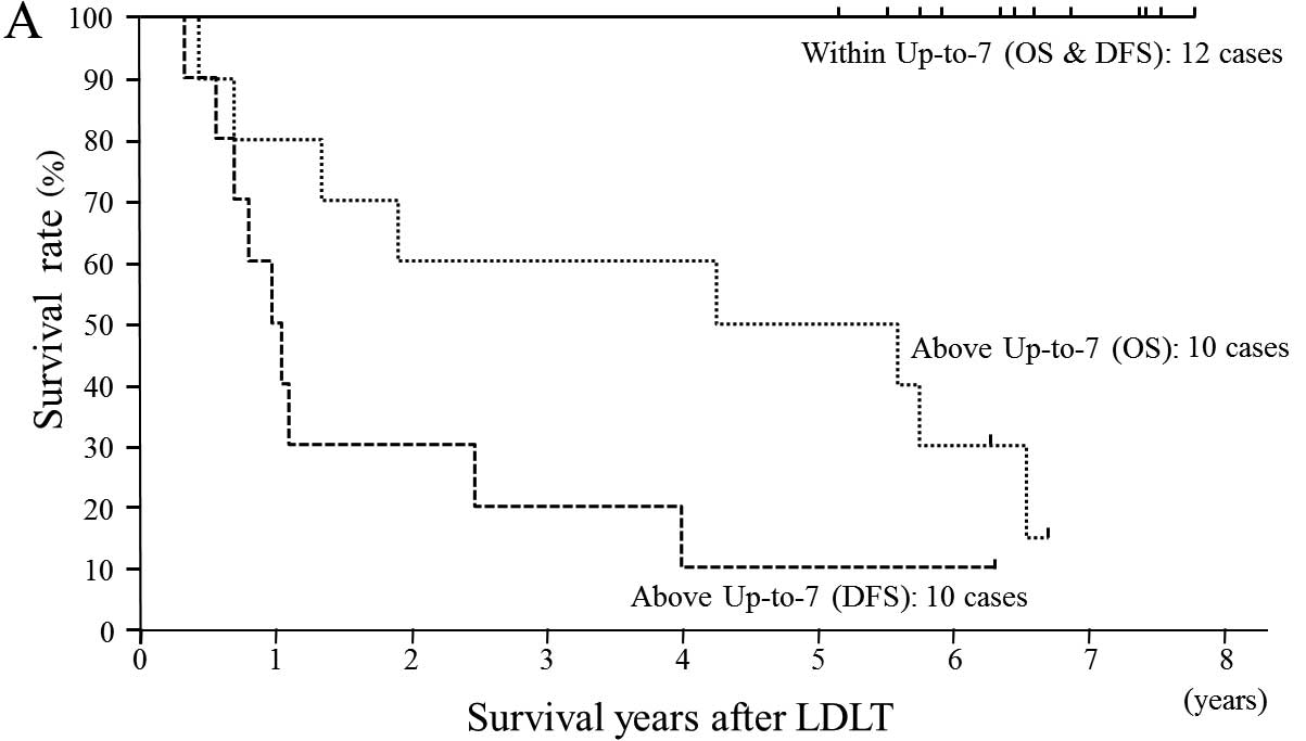
As shown in Fig. 1A and
C, all cases judged as within Up-to-7 criteria in the
pre-operative first imaging diagnosis or pathological diagnosis
survived without HCC recurrence. On the other hand, in cases judged
as above Up-to-7 criteria in the pre-operative first imaging
diagnosis and pathological diagnosis, the 5-year DFS was 10 and
21%, respectively, which is poor prognosis. Fig. 1B shows the OS and DFS of recipients
based on the Up-to-7 criteria evaluated by pre-operative final
imaging diagnosis. Comparing OS and DFS of within Up-to-7 criteria
with above criteria, a significant difference was found, and there
were 2 cases of recurrence in within Up-to-7 criteria.
Table III shows
the OS and DFS of recipients according to the MC, Up-to-7, Asan,
Tokyo, Kyoto and Kyushu criteria based on the pre-operative first
imaging diagnosis and pathological diagnosis. Recipients within
Asan criteria which permit wider eligibility than Up-to-7 or UCSF
criteria had a significantly better prognosis than above criteria,
but there were 2 cases of HCC recurrence among the within criteria
in pre-operative first imaging diagnosis. The Tokyo criteria gave
the same results as the Asan criteria. According to the Asan
criteria or Tokyo criteria, two deaths due to HCC recurrence were
confirmed based on the pre-operative first imaging diagnosis, thus
regarding eligibility for LDLT, the Up-to-7 criteria were deemed
the most appropriate criteria. On the other hand, according to the
Kyoto criteria, in an evaluation based on pre-operative final
imaging diagnosis, there was a significant correlation with the
prognosis of recipients, but in an evaluation based on the
pathological diagnosis, the prognosis was not reflected. According
to the Kyushu criteria, a significant difference was found in DFS
between within criteria and above criteria, but for OS, there was
no significant difference. These two criteria appear useful to
distinguish patients at very high risk of HCC recurrence in a
single high-volume center, but in the current situation where it is
not possible to prevent recurrence and no particularly effective
therapy after HCC recurrence, they cannot be considered universal
standard criteria. The most appropriate criteria which define the
prognosis of recipients after LDLT for HCC, for both pre-operative
imaging diagnosis and pathological diagnosis, are the Up-to-7
criteria, and in view of the burden of living donors, it should be
made the global standard of eligibility criteria for LT in HCC.
| Table IIIOutcome of recipients of LDLT for HCC
according to published eligibility criteria and α-SMA-positive CAF
in HCC. |
Table III
Outcome of recipients of LDLT for HCC
according to published eligibility criteria and α-SMA-positive CAF
in HCC.
| | | | OS (%) | | DFS (%) | |
|---|
| | | |
| |
| |
|---|
| Eligibility
criteria name | Staging method | Classification | Cases (n) | 1-year | 3-year | 5-year | 7-year | P-value | 1-year | 3-year | 5-year | 7-year | P-value |
|---|
| Milan criteria
(1) | Pre-operative
first | Within
criteria | 10 | 10 (100%) | 10 (100%) | 10 (100%) | 4 (100%) | <0.005 | 10 (100%) | 10 (100%) | 10 (100%) | 4 (100%) | <0.001 |
| imaging
diagnosis | Above criteria | 12 | 10 (83%) | 8 (67%) | 7 (58%) | - | | 7 (58%) | 4 (33%) | 3 (25%) | - | |
| Pre-operative
final | Within
criteria | 14 | 13 (93%) | 13 (93%) | 13 (93%) | 4 (77%) | <0.005 | 12 (86%) | 12 (86%) | 12 (86%) | 4 (86%) | <0.001 |
| imaging
diagnosis | Above criteria | 8 | 7 (88%) | 5 (63%) | 4 (50%) | - | | 5 (63%) | 2 (25%) | 1 (13%) | 0 | |
| Pathological
diagnosis | Within
criteria | 7 | 7 (100%) | 7 (100%) | 7 (100%) | 4 (100%) | <0.05 | 7 (100%) | 7 (100%) | 7 (100%) | 4 (100%) | <0.05 |
| Above criteria | 15 | 13 (87%) | 11 (73%) | 10 (67%) | 1 (40%) | | 10 (67%) | 7 (47%) | 6 (40%) | 1 (40%) | |
| Up-to-7 criteria
(10) | Pre-operative
first | Within
criteria | 12 | 12 (100%) | 12 (100%) | 12 (100%) | 4 (100%) | <0.0005 | 12 (100%) | 12 (100%) | 12 (100%) | 4 (100%) | <0.00001 |
| imaging
diagnosis | Above criteria | 10 | 8 (80%) | 6 (60%) | 5 (50%) | - | | 5 (50%) | 2 (20%) | 1 (10%) | - | |
| Pre-operative
final | Within
criteria | 15 | 14 (93%) | 14 (93%) | 14 (93%) | 4 (80%) | <0.0005 | 13 (87%) | 13 (87%) | 13 (87%) | 4 (87%) | <0.00005 |
| imaging
diagnosis | Above criteria | 7 | 6 (86%) | 4 (57%) | 3 (43%) | - | | 4 (57%) | 1 (14%) | 0 | 0 | |
| Pathological
diagnosis | Within
criteria | 9 | 9 (100%) | 9 (100%) | 9 (100%) | 3 (100%) | <0.01 | 9 (100%) | 9 (100%) | 9 (100%) | 3 (100%) | <0.005 |
| Above criteria | 13 | 11 (85%) | 9 (69%) | 8 (62%) | 1 (35%) | | 6 (55%) | 3 (27%) | 2 (21%) | - | |
| Asan criteria
(2) | Pre-operative
first | Within
criteria | 14 | 13 (93%) | 13 (93%) | 13 (93%) | 4 (80%) | <0.005 | 12 (86%) | 12 (86%) | 12 (86%) | 4 (86%) | <0.005 |
| imaging
diagnosis | Above criteria | 8 | 7 (88%) | 5 (63%) | 4 (50%) | - | | 5 (63%) | 2 (25%) | 1 (13%) | - | |
| Pre-operative
final | Within
criteria | 15 | 14 (93%) | 14 (93%) | 14 (93%) | 4 (80%) | <0.0005 | 13 (88%) | 13 (88%) | 13 (88%) | 4 (88%) | <0.00005 |
| imaging
diagnosis | Above criteria | 7 | 6 (71%) | 4 (57%) | 3 (43%) | - | | 4 (57%) | 1 (14%) | 0 | 0 | |
| Pathological
diagnosis | Within
criteria | 13 | 12 (92%) | 12 (92%) | 12 (92%) | 4 (79%) | <0.05 | 11 (85%) | 11 (85%) | 11 (85%) | 4 (85%) | <0.005 |
| Above criteria | 9 | 8 (89%) | 6 (67%) | 5 (56%) | - | | 6 (67%) | 3 (33%) | 2 (22%) | - | |
| Tokyo criteria (5-5
rule) (3) | Pre-operative
first | Within
criteria | 14 | 14 (100%) | 14 (100%) | 13 (93%) | 4 (79%) | <0.005 | 13 (93%) | 13 (93%) | 11 (86%) | 4 (86%) | <00001 |
| imaging
diagnosis | Above criteria | 8 | 6 (75%) | 4 (50%) | 3 (38%) | - | | 4 (50%) | 1 (13%) | 1 (13%) | - | |
| Pre-operative
final | Within
criteria | 16 | 15 (94%) | 15 (94%) | 15 (94%) | 4 (74%) | <0.005 | 14 (88%) | 14 (88%) | 13 (81%) | 4 (81%) | <0.00001 |
| imaging
diagnosis | Above criteria | 6 | 5 (83%) | 3 (50%) | 2 (33%) | - | | 3 (50%) | 0 | 0 | 0 | |
| Pathological
diagnosis | Within
criteria | 11 | 11 (100%) | 11 (100%) | 11 (100%) | 4 (100%) | <0.005 | 11 (100%) | 11 (100%) | 11 (100%) | 4 (100%) | <0.00001 |
| Above criteria | 11 | 9 (82%) | 7 (64%) | 6 (55%) | - | | 6 (55%) | 3 (27%) | 2 (21%) | - | |
| Kyoto criteria
(4,5) | Pre-operative
first | Within
criteria | 16 | 15 (94%) | 14 (88%) | 14 (88%) | 4 (69%) |
>0.05 | 13 (81%) | 13 (81%) | 12 (75%) | 4 (75%) | <0.01 |
| imaging
diagnosis | Above criteria | 6 | 5 (83%) | 4 (67%) | 3 (33%) | - | | 4 (67%) | 1 (17%) | 1 (17%) | - | |
| Pre-operative
final | Within
criteria | 17 | 16 (94%) | 15 (88%) | 15 (88%) | 4 (70%) | <0.05 | 14 (82%) | 14 (82%) | 13 (76%) | 4 (76%) | <0.005 |
| imaging
diagnosis | Above criteria | 5 | 4 (80%) | 3 (60%) | 2 (40%) | - | | 3 (60%) | 0 | 0 | 0 | |
| Pathological
diagnosis | Within
criteria | 16 | 15 (94%) | 14 (88%) | 14 (88%) | 4 (63%) | >0.1 | 13 (81%) | 12 (75%) | 11 (69%) | 4 (69%) | >0.1 |
| Above criteria | 6 | 5 (83%) | 4 (67%) | 3 (50%) | - | | 4 (67%) | 2 (33%) | 2 (33%) | - | |
| Kyushu criteria
(6–8) | Pre-operative
first | Within
criteria | 18 | 17 (94%) | 15 (83%) | 14 (78%) | 4 (61%) | >0.5 | 15 (83%) | 13 (72%) | 12 (67%) | 4 (67%) | <0.05 |
| imaging
diagnosis | Above criteria | 4 | 3 (75%) | 3 (75%) | 3 (75%) | - | | 2 (50%) | 1 (25%) | 1 (25%) | - | |
| Pre-operative
final | Within
criteria | 18 | 17 (94%) | 15 (83%) | 14 (78%) | 4 (61%) | >0.5 | 15 (83%) | 13 (72%) | 12 (67%) | 4 (67%) | <0.05 |
| imaging
diagnosis | Above criteria | 4 | 3 (75%) | 3 (75%) | 3 (75%) | - | | 2 (50%) | 1 (25%) | 1 (25%) | - | |
| Pathological
diagnosis | Within
criteria | 18 | 17 (89%) | 15 (83%) | 14 (78%) | 4 (61%) | >0.5 | 15 (83%) | 13 (72%) | 12 (67%) | 4 (67%) | <0.05 |
| Above criteria | 4 | 3 (25%) | 3 (25%) | 3 (25%) | - | | 2 (50%) | 1 (25%) | 1 (25%) | - | |
| α-SMA-positive
CAF | Pathological
diagnosis | Grade I | 10 | 10 (100%) | 10 (100%) | 10 (100%) | 3 (88%) | <0.0001 | 10 (100%) | 10 (100%) | 9 (90%) | 3 (90%) | <0.00001 |
| Grade II | 8 | 8 (100%) | 7 (88%) | 6 (75%) | 1 (60%) | | 7 (88%) | 4 (50%) | 4 (50%) | 1 (50%) | |
| Grade III | 4 | 2 (50%) | 1 (25%) | 1 (25%) | 0 | | 0 | 0 | 0 | 0 | |
As shown in Table
IV, the degree of histological differentiation of HCC, the
values of serum AFP and serum DCP, and the presence of
microvascular invasion were not significantly correlated with the
prognosis after LDLT. In other words, microvascular invasion should
be admitted as within criteria.
| Table IVCorrelation between α-SMA-positive CAF in HCC of LDLT
recipients with clinicopathological factors and published
eligibility criteria. |
Table IV
Correlation between α-SMA-positive CAF in HCC of LDLT
recipients with clinicopathological factors and published
eligibility criteria.
| | α-SMA-positive CAF |
|---|
| |
|
|---|
| Factor | All redipients (22
cases) | Grade I (10
cases) | Grade II, III (12
cases) |
|---|
| Age, years (mean ±
SD) | 56±4 | 56±3 | 55±4 |
| Gender
(female/male) | 5/17 | 5/5 | 0/12a |
| MELD score (mean ±
SD) | 14±8 (range
1–30) | 15±9 | 11±7 |
| HCV/HBV | 12/10 | 5/5 | 7/5 |
| Child-Pugh, n
(%) |
| A | 4 (18) | 0 | 4 (33)a |
| B, C | 12 (55) | 10 (100) | 8 (67)a |
| Pre-LDLT treatment
for HCC, n (%) | 15 (68) | 7 (70) | 8 (67) |
| AFP (ng/ml) (mean ±
SD) | 148±264 | 53±87 | 227±343 |
| DCP (mAU/l) (mean ±
SD) | 183±388 | 106±202 | 246±508 |
| CEA (ng/ml) (mean ±
SD) | 4.4±1.5 | 4.4±1.7 | 4.5±1.3 |
| CA19-9 (U/ml) (mean
± SD) | 73.4±82.6 | 101.7±94.7 | 45.1±62.7 |
| HCC numbers
(pre-LDLT first imaging diagnosis) (mean ± SD) | 5.3±5.8 | 2.2±2.3 | 7.8±6.7b |
| HCC numbers
(pathological diagnosis) (mean ± SD) | 6.6±6.0 | 4.0±3.3 | 8.8±7.0 |
| HCC maximum
diameter (pre-LDLT first imaging diagnosis) (mean ± SD) (cm) | 2.2±1.6 | 1.4±1.4 | 2.9±1.5 |
| HCC maximum
diameter (pathological diagnosis) (cm) | 2.9±1.2 | 2.5±1.2 | 3.2±1.1 |
| Sum of all HCC
diameters (pre-LDLT first imaging diagnosis) (mean ± SD) (cm) | 7.6±10.1 | 2.0±3.8 | 12.2±11.9b |
| Sum of all HCC
diameters (pathological diagnosis) (mean ± SD) (cm) | 10.3±9.4 | 6.6±5.9 | 13.4±10.8 |
| UNOS TNM, n
(%) |
| I, II | 6 (28) | 5 (50) | 1 (8)a |
| IV | 16 (72) | 5 (50) | 11 (92)a |
| Histological grade
(poorly and combined), n (%) | 6 (27.2) | 2 (20) | 4 (33) |
| Microvascular
invasion, n (%) | 16 (73) | 7 (70) | 9 (75) |
| Intrahepatic
metastasis, n (%) | 11 (50) | 5 (50) | 6 (50) |
| Post-LDLT HCC
recurrence, n (%) | 9 (41) | 1 (10) | 8 (67)a |
| Recipient
mortality, n (%) | 8 (36) | 1 (10) | 7 (53)a |
| Above Milan
criteria, n (%) |
| Imaging | 12 (55) | 3 (25) | 9 (75)a |
| Pathology | 15 (68) | 5 (33) | 10 (67) |
| Above Up-to-7
criteria, n (%) |
| Imaging | 10 (46) | 2 (20) | 8 (80)a |
| Pathology | 13 (59) | 4 (31) | 9 (69) |
| Above Asan
criteria, n (%) |
| Imaging | 8 (36) | 2 (25) | 6 (75) |
| Pathology | 9 (41) | 3 (33) | 6 (67) |
| Above Tokyo
criteria, n (%) | | | |
| Imaging | 8 (36) | 1 (13) | 7 (87)a |
| Pathology | 11 (50) | 3 (27) | 8 (73) |
| Above Kyoto
criteria, n (%) |
| Imaging | 6 (27) | 1 (17) | 5 (83) |
| Pathology | 6 (27) | 2 (33) | 4 (67) |
| Above Kyushu
criteria, n (%) |
| Imaging | 4 (18) | 1 (25) | 3 (75) |
| Pathology | 4 (18) | 1 (25) | 3 (75) |
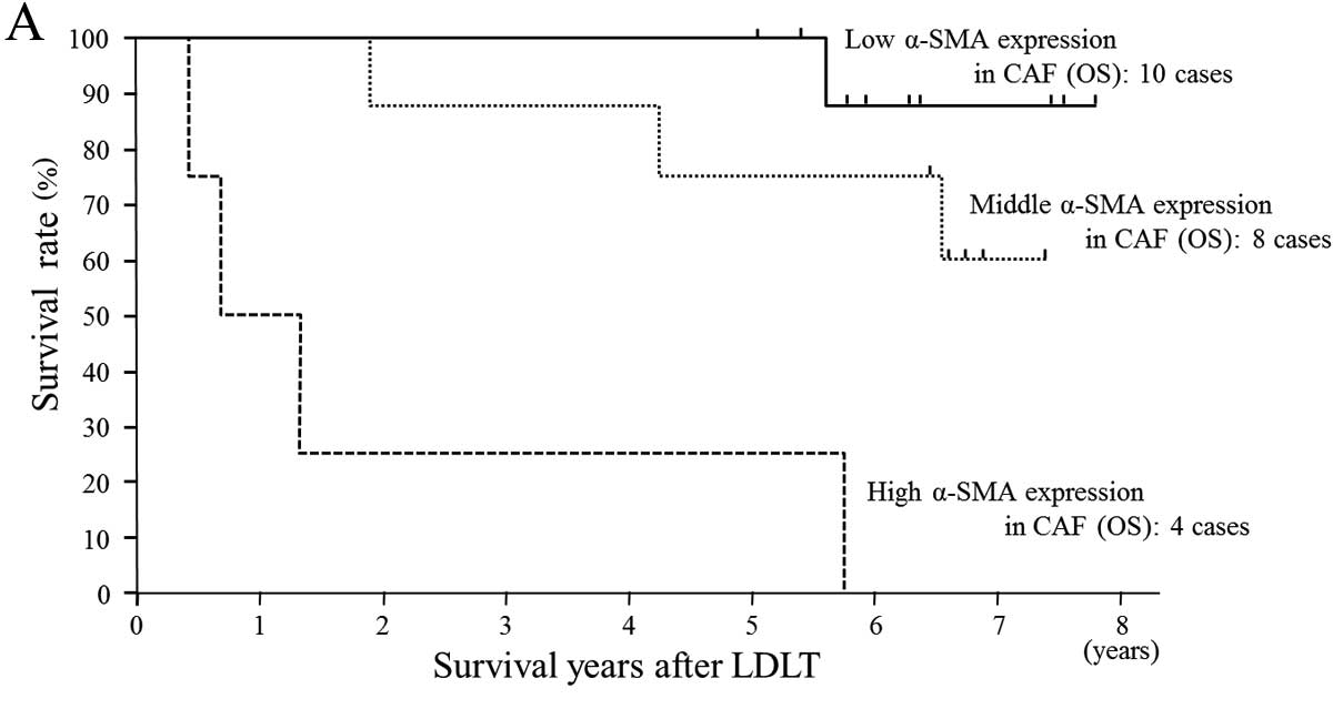
Proliferation of α-SMA-positive CAFs which is
thought to be strongly related to cancer progression and invasion,
clearly specifies the prognosis after LDLT in HCC. However, α-SMA
was not found to be expressed in HCC cancer cells. We categorized
the proliferation of α-SMA-positive CAF into the following 3
groups.
Group I (Fig. 2A),
10 cases: low grade proliferation of α-SMA-positive CAFs;
proliferation of cancer stroma not found, only slight proliferation
of α-SMA-positive CAFs and staining was <1% of ten fields under
high power view.
Group II (Fig. 2B),
8 cases: middle grade proliferation of α-SMA-positive CAFs; cancer
nests were bordered over their whole circumference by
α-SMA-positive CAFs, but the cancer stroma (CAFs) accounted for
<10% of ten fields under high power view.
Group III (Fig. 2C),
4 cases: high grade proliferation of α-SMA-positive CAFs group;
extensive proliferation of α-SMA-positive CAFs and cancer stroma
accounted for >10% of ten fields under high power view.
As shown in Fig. 3,
as the proliferation of α-SMA-positive CAFs increased, recurrence
of HCC increased significantly, and all 4 patients in group III
died soon after LDLT due to recurrence of HCC. In above Up-to-7
criteria recipients, significant proliferation of α-SMA-positive
CAFs was found. In the 2 of 3 recipients who underwent HCC
downstaging from above Up-to-7 to within MC by pre-LDLT therapy,
recurrence of HCC was found after LDLT, and in one of these 2
cases, proliferation of α-SMA-positive CAFs was observed. On the
other hand, in the one case without recurrence, proliferation of
α-SMA-positive CAFs was not observed. In above Up-to-7 criteria
recipients accompanied by proliferation of α-SMA-positive CAFs, the
risk of HCC recurrence is very high, therefore post-operative
adjuvant chemotherapy should be performed to improve survival. To
improve the prognosis of recipients with HCC recurrence,
appropriate anticancer therapy following recurrence is critical. We
applied RFA to HCC recurrence in graft liver, and in 2 cases of
recurrence with metastasis only to the lung, partial lung resection
was performed. We also performed surgical resection for lymph node
metastasis in the abdominal cavity. Moreover, we performed
irradiation and administered molecular target drugs, and confirmed
that in cases of HCC recurrence, the prognosis was improved by
these intensive therapies. The proliferation of α-SMA-positive CAFs
is closely related to the Up-to-7 criteria. The proliferation of
α-SMA-positive CAFs, as shown in Table
IV, is unrelated to pre-operative therapy or histological grade
of HCC, values of tumor markers, presence of microvascular
invasion, or hepatitis viruses (HBV or HCV), and appears to be a
major prognostic factor in the recurrence of HCC.
From the predictors which define HCC recurrence
after LT in univariate analysis, we performed a Cox-proportional
multivariate analysis using three key factors such as the Up-to-7
criteria, the Tokyo criteria and proliferation of α-SMA-positive
CAFs (Table V). From the results,
we determined that the Up-to-7 criteria and proliferation of
α-SMA-positive CAFs are both independent, most significant
lyprognostic factors of LDLT for HCC.
| Table VMultivariate analysis for recipient
OS using Cox’s proportional hazard model of statistically more
significant prognostic indicators such as Up-to-7 criteria, Tokyo
criteria and α-SMA-positive CAF in hepatocellular carcinoma in
univariate analysis. |
Table V
Multivariate analysis for recipient
OS using Cox’s proportional hazard model of statistically more
significant prognostic indicators such as Up-to-7 criteria, Tokyo
criteria and α-SMA-positive CAF in hepatocellular carcinoma in
univariate analysis.
| Predictors | Hazard ratio |
|---|
|
|---|
| 95% CI |
|---|
| Up-to-7
criteriaa | 61.62 | 2.24–1697.97 |
| α-SMA-positive
CAFb | 8.46 | 1.32–54.06 |
| Tokyo
criteriaa | 0.03 | 0.00–1.74 |
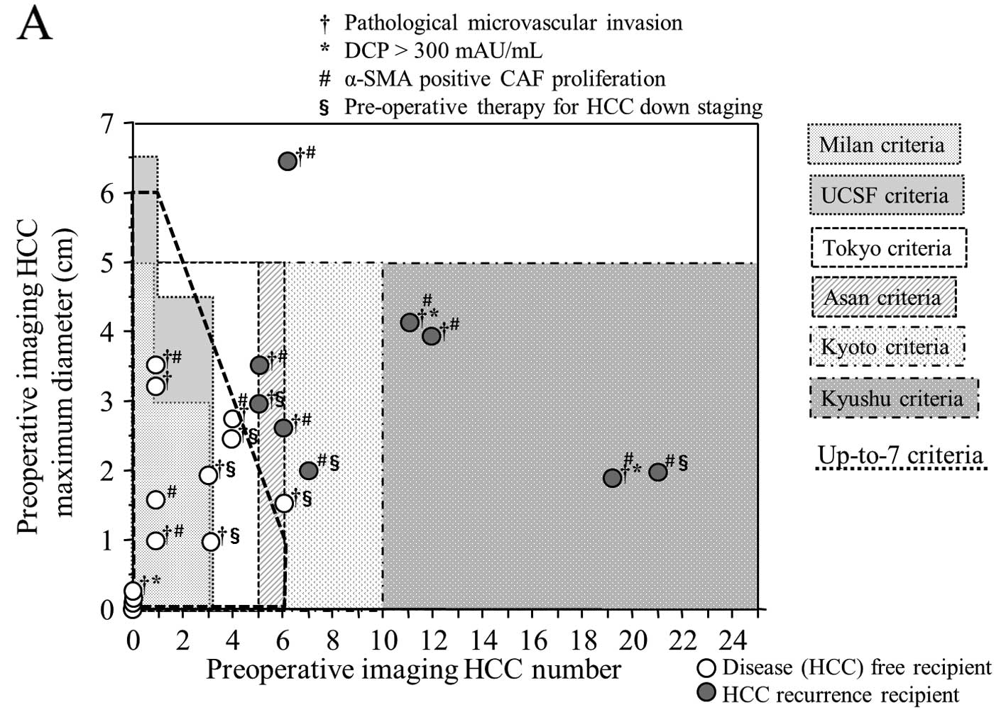
Fig. 4 shows the
outcome of the present study which is the relationship between
tumor number, maximum tumor diameter, microvascular invasion and
proliferation of α-SMA-positive CAFs respectively for the
pre-operative first imaging diagnosis prior to LDLT and
pathological diagnosis. As shown in Fig. 4A based on the pre-operative first
imaging diagnosis, all within Up-to-7 criteria cases survived
without HCC recurrence, accounting for the largest number of cases,
so these criteria appear to be the most suitable for LDLT for HCC.
There were 4 cases that were diagnosed with no viable cancer
lesions by pre-operative therapy for HCC such as TACL and RFA in
pre-operative first imaging diagnosis, but these 4 cases had viable
HCC cells. On the other hand, based on pathological diagnosis
(Fig. 4B), all within Tokyo
criteria recipients survived without HCC recurrence, accounting for
the largest number of cases, so this would appear to be the most
significant criteria from the viewpoint of recipient benefit. No
proliferation of α-SMA-positive CAFs in HCC was found in the 4
recipients without post-LDLT HCC recurrence, who were diagnosed
above Up-to-7 criteria by post-operative pathological
diagnosis.
Discussion
LT continues to be associated with significant
morbidity and mortality despite improvements in surgical techniques
and immunosuppressive regimens. Furthermore, unlike other forms of
oncological surgery, LT requires a donor organ. In view of this,
utility and fairness need to be considered in relation to
allocation for both donor and recipient. For this reason, the
application of strict eligibility criteria such as MC has evolved
as an important aspect of current clinical practice. In Japan,
whether or not pre-transplant therapy is performed for HCC at 3
months prior to LT, within MC, is an essential requirement for
receiving DDLT, and is also an essential requirement for receiving
LDLT under the health insurance. In LDLT, since a healthy donor
takes a major risk, HCC recurrence must be avoided after LT in
recipients. As there is no absolute curative treatment for HCC
recurrence, LDLT should be performed while adhering to strict
eligibility criteria so that HCC does not recur after LT. In the
present study, the OS and DFS after LDLT in patients who met
Up-to-7 criteria in both pre-operative evaluation and pathological
evaluation were 100% although including recurrence of HCV
hepatitis, we support that Up-to-7 criteria are well-established
tools for assessing the prognosis of HCC. In the pre-operative
first imaging diagnosis and pathological diagnosis, there were no
cases which were above MC and within UCSF criteria; however, there
were 2 cases which were above MC and within Up-to-7 criteria.
Therefore, we did not compare Up-to-7 criteria exceeding MC and
UCSF criteria but, as shown in Fig.
4, the Up-to-7 criteria broadly cover the UCSF criteria.
Moreover, in view of the fact that the only patient who survived
without HCC recurrence above Up-to-7 criteria was also close to
within criteria, it seems most appropriate to take the Up-to-7
criteria as suitable global standard criteria for LT in HCC.
Some of the published criteria of LT for HCC do not
affect the OS after LT and appear to underestimate the risk of HCC
recurrence. The reason for this may be that benign/malignant
borderline lesions, such as high-grade degenerative nodules
(17,23–25),
are counted as HCC. High-grade degenerative nodules must be clearly
distinguished from HCC and a consensus has already been reached
regarding this difference (17,25).
If high-grade degenerative nodules are included in HCC, it detracts
from the reliability of the criteria itself. Pathologically, the
ideal criteria are the Tokyo criteria, but 2 cases of recurrence
were found in pre-operative first imaging diagnosis, and it is
difficult to conclude that Tokyo criteria would have better
eligibility criteria than the Up-to-7 criteria.
Downstaging
The downstaging refers specifically to treatment
undertaken to convert a tumor with morphology beyond established LT
criteria (and therefore not a candidate for LT) to a size that is
within criteria and therefore enable a patient to become an LT
candidate. Any assessment of the efficacy of a downstaging protocol
needs not only a clear definition of which patients would be
considered for downstaging, but also a clear definition of
eligibility criteria that need to be met for the patient to qualify
for LT. Furthermore, some protocols require a period of stability
once LT criteria have been met prior to activation on waiting. Such
a restriction should ensure that patients with tumors that exhibit
unfavorable biology, which would be expected to translate into an
increased risk of recurrence, are excluded. However, comparable
post-LT outcomes in recipients who had been successfully downstaged
to recipients within MC (26–28)
have been demonstrated. In the present study, we performed pre-LT
therapy for downstaging in 7 recipients. Pre-LT therapy consisted
of 5 cases in which only TACL was performed, and 2 cases in which
RFA was performed in addition to TACL. In both situations, LDLT was
performed at 3 months or more after pre-LT therapy. Four of the 5
recipients above Up-to-7 criteria who underwent pre-LT therapy were
judged to be within Up-to-7 criteria from above Up-to-7 criteria in
the pre-operative final diagnosis, but in the pathological
diagnosis, all of these cases were judged to be above Up-to-7
criteria (allowing microvascular invasion). The reasons for the
discrepancy between the pre-operative final imaging diagnosis and
the pathological diagnosis are that minute, residual viable cancer
lesions of TACL therapy were not identified in the images, and
small HCC was judged as high-grade degenerative nodules in the
pre-operative diagnosis.
α-SMA-positive CAF (myofibroblastic
CAF)
Lysophostatidic acid (LPA) accelerates HCC
progression by recruiting peritumoral tissue fibroblasts (PTFs) and
promoting their transdifferentiation into myofibroblasts (18). Following transdifferentiation,
pretumoral tissue fibroblast expressed α-SMA and enhanced
proliferation, migration and invasion of HCC cells occur. In the
present study, proliferation of α-SMA-positive CAFs in HCC was
significantly correlated with metastasis of HCC and above Up-to-7
criteria, and was therefore significantly considered a poorer
prognosis factor equivalent to Up-to-7 criteria in post-LDLT
recipients with HCC. It is generally accepted that HCC originates
from hepatocytes but grows and advances while fully embedded in a
surrounding microenvironment with a rich content of myofibroblasts,
fibroblasts, and other cell types due to the underlying cirrhosis.
Liver myofibroblasts, derived from quiescent fibroblasts and
hepatic stellate cells activated by the chronic injury, can be
recognized by their expression of α-SMA (29,30).
Myofibroblasts have been detected at the advanced edge of several
different malignancies as the predominant phenotype in the CAF
population (31). Although the
origin of CAF remains controversial, their immunophenotypical
characterization, which primarily includes α-SMA and excludes
epithelial and endothelial common markers, is widely accepted
(29,32,33).
CAFs differ from PTFs not in terms of somatic mutations but,
rather, in terms of molecular and functional differences in
modulating neighboring cancer cells (34,35).
However, the paracrine crosstalk between HCC and stromal
fibroblasts such as CAF or pretumoral tissue fibroblast is poorly
understood. Stromal myofibroblasts in HCC and matching peritumoral
tissues is detected by staining with anti-α-SMA antibody (29). It was found that α-SMA-positive
cells were mainly expressed within the tumor stroma (18).
We also performed an immunohistochemical study for
biological markers of epithelial mesenchymal transitions (EMT) in
HCC which are thought to be related to cancer invasion and
metastasis (36,37) (data not shown). It is reported that
downregulation of E-cadherin (36–39),
weakened expression (39) or
overexpression of N-cadherin (40),
overexpression of β-catenin (36–38),
overexpression of vimentin (41,42),
overexpression of Snail (36,43),
overexpression of Slug (36) and
overexpression of TWIST (36,39)
are poor prognosis factors for HCC. We performed
immunohistochemical study of these markers. The recipients with
overexpression of vimentin or Snail had significantly higher risk
of HCC recurrence after LT, but it did not have as much of an
impact as expression of α-SMA-positive CAFs by α-SMA
immunostaining. Also, when we performed a multivariate analysis
using Cox’s proportional method with Up-to-7 criteria or
α-SMA-positive CAFs, and other histological factors in HCC, a clear
correlation was found for Up-to-7 criteria and proliferation of
α-SMA-positive CAFs. It was determined that these two factors alone
were independent factors that specified prognosis or DFS after LDLT
(Table V). In other words,
proliferation of α-SMA-positive CAFs leads to a high risk of HCC
metastasis and the prognosis is extremely poor even if LT is
performed.
Microvascular invasion of HCC
The investigated cases in the present study did not
include any cases of macrovascular invasion, and since it is
reported that macrovascular invasion is a significant risk factor
for recurrence of HCC (2,44–46),
there is no indication of LT. On the other hand, as regards
microvascular invasion, it has been reported to worsen prognosis
after LT for HCC (47) and there is
a conflicting report that it has no effect on the prognosis
(44). If we limit the discussion
to within Up-to-7 criteria, it has also been reported that the
presence of microvascular invasion does not define the prognosis
after LT for HCC (48). As regards
the MC and Up-to-7 criteria, microvascular invasion is regarded as
a factor of above criteria but, in this study, microvascular
invasion did not contribute to HCC recurrence after LDLT. It is
more difficult to determine the presence of microvascular invasion
from pre-operative imaging (49–51),
and we consider microvascular invasion should be included in the
within eligibility criteria of LT for HCC. The histological type
such as the combined HCC type or the poorly differentiated HCC
type, and the presence of intrahepatic metastasis did not
contribute to HCC recurrence after LDLT.
Pre-operative imaging diagnosis
One common methodological flaw in studies
identifying clinical predictors of favorable outcome is the use of
explant pathology to provide information on tumor maximum diameter
and number, with the derived criteria being subsequently applied to
radiological assessments of tumor burden. However, radiological
staging can be limited in accuracy; indeed, review of the
Eurotransplant Allocation System demonstrated a 34% accuracy of
radiology in comparison to explant pathology, with tumor absent in
8.3% of patients, overstaging of the tumor in 36.2% and
understaging in 10.4% (52). This
is clinically significant as radiological understaging translates
into inferior outcomes (53). If
the precision of HCC imaging diagnosis is low, the reliability of
the criteria decreases, so pre-operative imaging diagnosis must be
performed accurately using multiple modalities. In order to enhance
imaging diagnostic ability in HCC, the authors, in addition to
dynamic MDCT and Gd-EOB-DTPA-MRI (12,13),
also perform CT with angiography (CTAP and CTHA) as far as possible
(14–17). We believe that by combining these
tools, the ability to diagnose HCC can be enhanced to the maximum
level. By performing these three tools of pre-operative imaging,
the ability to diagnose benign/malignant borderline lesions and
local recurrence foci after RFA or TACL therapy is also enhanced,
which made it possible to obtain a pre-operative imaging diagnosis
close to a pathological diagnosis. In practice, comparing the
pre-operative first diagnosis and pathological diagnosis, the
sensitivity of the within Up-to-7 criteria was 100%, and the
specificity was 75% which is a satisfactory result. The Up-to-7
criteria which specify eligibility for LT in HCC were found to be
the criteria which clearly define prognosis in the diagnostic
results obtained using these three imaging diagnostic modalities.
At present, in Japan, in order to receive LDLT under health
insurance, the history of therapy for HCC 3 months prior to LT is
not a limitation, but satisfaction of the within MC in the
pre-operative final imaging diagnosis is a requirement. However, it
appears there is a sufficient scientific foundation for extending
this eligibility to within Up-to-7 criteria from MC.
LDLT vs. DDLT
In general, it is said that separate consideration
for LDLT for HCC is required, as the patient already has an
allocated liver graft and is therefore not dependent on the donor
pool. It can therefore be argued that the application of strict
eligibility criteria as required with cadaveric grafts for patients
with HCC is not necessary. However, in these circumstances, the
risk to the donor must be incorporated into any decision, since it
is clearly unethical to expose a donor to a significant risk of
morbidity or mortality. Therefore, we must consider that similar
criteria would apply to patients undergoing DDLT and LDLT. In fact,
similar outcomes are observed in patients receiving DDLT or LDLT
for HCC within Up-to-7 criteria (47). In several countries where DDLT is
mainly performed for LT, the Up-to-7 criteria are accepted as the
appropriate criteria (2,10,47,48,54)
and it is also clear from our present study that, similarly, there
must be appropriate criteria in the LDLT (48). In other words, eligibility for LT in
HCC should be within the Up-to-7 criteria regardless of whether it
is DDLT or LDLT.
In conclusion, the ideal eligibility criteria of
LDLT for HCC is the Up-to-7 criteria and although there were some
recipients in HCV hepatitis recurrence, all recipients within the
criteria survived without HCC recurrence. Also, in above Up-to-7
criteria, proliferation of α-SMA-positive CAFs was found more
frequently than within criteria, and this appeared to be a major
factor in recurrence of HCC after LT. On the other hand, no
significant correlation was found between pre-transplant treatment
for HCC, histological differentiation and tumor markers with
recurrence of HCC after LDLT. At present, while there is still no
effective treatment for recurrence of HCC after LT and also from
the viewpoint of proliferation of α-SMA-positive CAF, LT, in
particular LDLT, should be limited to recipients who are within
Up-to-7 criteria in pre-operative imaging diagnosis.
Abbreviations:
|
AFP
|
α-fetoprotein
|
|
CAFs
|
cancer-associated fibroblasts
|
|
CTAP
|
computer tomography under angiography
during arterial portography
|
|
CTHA
|
computer tomography under angiography
during hepatic arteriography
|
|
DCP
|
des-γ-carboxyprothrombin
|
|
DDLT
|
deceased donor liver
transplantation
|
|
HCC
|
hepatocellular carcinoma
|
|
DFS
|
disease-free survival
|
|
dynamic MDCT
|
dynamic multi-detectable-row computer
tomography
|
|
Gd-EOB-DTPA-MRI
|
gadolinium ethoxybenzyl
diethylenetriamine pentaacetic acid-enhanced magnetic resonance
imaging
|
|
HBV
|
hepatitis B virus
|
|
HCV
|
hepatitis C virus
|
|
LC
|
liver cirrhosis
|
|
LDLT
|
living donor liver transplantation
|
|
LT
|
liver transplantation
|
|
OS
|
overall survival
|
|
RFA
|
radiofrequency ablation therapy
|
|
TACL
|
transarterial
chemo-lipiodolisation
|
|
Up-to-7
|
Up-to-seven criteria
|
References
|
1
|
Mazzaferro V, Regalia E, Doci R, et al:
Liver transplantation for the treatment of small hepatocellular
carcinoma in patients with cirrhosis. N Engl J Med. 334:693–699.
1996. View Article : Google Scholar : PubMed/NCBI
|
|
2
|
Lee SG, Hwang S, Moon DB, et al: Expanded
indication criteria of living donor liver transplantation for
hepatocellular carcinoma at one large-volume center. Liver Tanspl.
14:935–945. 2008.PubMed/NCBI
|
|
3
|
Sugawara Y, Tamura S and Makuuchi M:
Living donor liver transplantation for hepatocellular carcinoma:
Tokyo University series. Dig Dis. 25:310–312. 2007. View Article : Google Scholar : PubMed/NCBI
|
|
4
|
Ito T, Takada Y, Ueda M, et al: Expansion
of selection criteria for patients with hepatocellular carcinoma in
living donor liver transplantation. Liver Transpl. 13:1637–1644.
2007. View
Article : Google Scholar : PubMed/NCBI
|
|
5
|
Takada Y, Ito T, Ueda M, et al: Living
donor liver transplantation for patients with HCC exceeding the
Milan criteria: a proposal of expanded criteria. Dig Dis.
25:299–302. 2007. View Article : Google Scholar : PubMed/NCBI
|
|
6
|
Soejima Y, Taketomi A, Yoshizumi T, et al:
Extended indication for living donor liver transplantation in
patients with hepatocellular carcinoma. Transplantation.
83:893–899. 2007. View Article : Google Scholar : PubMed/NCBI
|
|
7
|
Taketomi A, Sanefuji K, Soejima Y, et al:
Impact of des-gamma-carboxy prothrombin and tumor size on the
recurrence of hepatocellular carcinoma after living donor liver
transplantation. Transplantation. 87:531–537. 2009. View Article : Google Scholar
|
|
8
|
Shirabe K, Taketomi A, Morita K, et al:
Comparative evaluation of expanded criteria for patients with
hepatocellular carcinoma beyond the Milan criteria undergoing
living-related donor liver transplantation. Clin Transplant.
25:E491–E498. 2011. View Article : Google Scholar
|
|
9
|
Furukawa H, Shimamura T, Suzuki T, et al:
Liver transplantation for hepatocellular carcinoma: the Japanese
experience. J Hepatobiliary Pancreat Surg. 17:533–538. 2010.
View Article : Google Scholar : PubMed/NCBI
|
|
10
|
Mazzaferro V, Llovet JM, Miceli R, et al:
Predicting survival after liver transplantation in patients with
hepatocellular carcinoma beyond the Milan criteria: a
retrospective, exploratory analysis. Lancet Oncol. 10:35–43. 2009.
View Article : Google Scholar
|
|
11
|
Yao FY, Ferrell L, Bass NM, et al: Liver
transplantation for hepatocellular carcinoma: expansion of the
tumor size limits does not adversely impact survival. Hepatology.
33:1394–1403. 2001. View Article : Google Scholar : PubMed/NCBI
|
|
12
|
Kobayashi S, Matsui O, Gabata T, et al:
Gadolinium ethoxybenzyl diethylenetriamine pentaacetic
acid-enhanced magnetic resonance imaging findings of borderline
lesions at high risk for progression to hypervascular classic
hepatocellular carcinoma. J Comput Asist Tomogr. 35:181–186. 2011.
View Article : Google Scholar
|
|
13
|
Kobayashi S, Matsui O, Gabata T, et al:
Intranodular signal intensity analysis of hypovascular high-risk
borderline lesions of HCC that illustrate multi-step
hepatocarcinogenesis within the nodule on Gd-EOB-DTPA-enhanced MRI.
Eur J Radiol. 81:3839–3845. 2012. View Article : Google Scholar : PubMed/NCBI
|
|
14
|
Miyayama S, Matsui O, Yamashiro M, et al:
Detection of hepatocellular carcinoma by CT during arterial
portography using a cone-beam CT technology: comparison with
conventional CTAP. Abdom Imaging. 34:502–506. 2009. View Article : Google Scholar : PubMed/NCBI
|
|
15
|
Kitao A, Zen Y, Matsui O, Gabata T and
Nakanuma Y: Hepatocarcinogenesis: multistep changes of drainage
vessels at CT during arterial portography and hepatic arteriography
- radiologic-pathologic correlation. Radiology. 252:605–614. 2009.
View Article : Google Scholar
|
|
16
|
Miyayama S, Yamashiro M, Okuda M, et al:
Detection of corona enhancement of hypervascular hepatocellular
carcinoma by C-arm dual-phase cone-beam CT during hepatic
arteriography. Cardiovasc Intervent Radiol. 34:81–86. 2011.
View Article : Google Scholar : PubMed/NCBI
|
|
17
|
Matsui O, Kobayashi S, Sanada J, et al:
Hepatocelluar nodules in liver cirrhosis: hemodynamic evaluation
(angiography-assisted CT) with special reference to multi-step
hepatocarcinogenesis. Abdom Imaging. 36:264–272. 2011. View Article : Google Scholar
|
|
18
|
Mazzocca A, Dituri F, Lupo L, Quaranta M,
Antonaci S and Giannelli G: Tumor-secreted lysophostatidic acid
accelerates hepatocellular carcinoma progression by promoting
differentiation of peritumoral fibroblasts in myofibroblasts.
Hepatology. 54:920–930. 2011. View Article : Google Scholar
|
|
19
|
Okabe H, Beppu T, Hayashi H, et al:
Hepatic stellate cells may relate to progression of intrahepatic
cholangiocarcinoma. Ann Surg Oncol. 16:2555–2564. 2009. View Article : Google Scholar : PubMed/NCBI
|
|
20
|
Greene FL, Page DL, Fleming ID, et al:
AJCC Cancer Staging Manual. 6th edition. Springer-Verlag; New York:
pp. 4352002
|
|
21
|
United Network for Organ Sharing: Policy
3.6. www.unos.orgurisimplewww.unos.org. Accessed September
8, 2002
|
|
22
|
Urata K, Kawasaki S, Matsunami H, et al:
Calculation of child and adult standard liver volume for liver
transplantation. Hepatology. 21:1317–1321. 1995.PubMed/NCBI
|
|
23
|
Authors not listed. Terminology of nodular
hepatocellular lesions. International Working Party Hepatology.
22:101–105. 1995.
|
|
24
|
Hirohashi S, Ishak KG, Kojiro M, et al:
Hepatocellular carcinoma. Pathology and Genetics of Tumours of the
Digestive System. Hamilton SR and Aaltonen LA: IARC Press; Lyon:
pp. 159–172. 2000
|
|
25
|
Hayashi M, Matsui O, Ueda K, et al:
Correlation between the blood supply and grade of malignancy of
hepatocellular nodules associated with liver cirrhosis: evaluation
by CT during intraarterial injection of contrast medium. AJR Am J
Roentgenol. 172:969–976. 1999. View Article : Google Scholar
|
|
26
|
Chapman WC, Majella Doyle MB, Stuart JE,
et al: Outcomes of neoadjuvant transarterial chemoembolization to
downstage hepatocellular carcinoma before liver transplantation.
Ann Surg. 248:617–625. 2008.
|
|
27
|
Ravaioli M, Grazi GL, Piscaglia F, et al:
Liver transplantation for hepatocellular carcinoma: results of
down-staging in patients initially outside the Milan selection
criteria. Am J Transplant. 8:2547–2557. 2008. View Article : Google Scholar
|
|
28
|
Yao FY, Kerlan RK Jr, Hirose R, et al:
Excellent outcome following down-staging of hepatocellular
carcinoma prior to liver transplantation: an intention-to-treat
analysis. Hepatology. 48:819–827. 2008. View Article : Google Scholar : PubMed/NCBI
|
|
29
|
Serini G and Gabbiani G: Mechanisms of
myofibroblast activity and phenotypic modulation. Exp Cell Res.
250:273–283. 1999. View Article : Google Scholar : PubMed/NCBI
|
|
30
|
Tomasek JJ, Gabbiani G, Hinz B, Chaponnier
C and Brown RA: Myofibroblasts and mechano-regulation of connective
tissue remodelling. Nat Rev Mol Cell Biol. 3:349–363. 2002.
View Article : Google Scholar : PubMed/NCBI
|
|
31
|
Sappino AP, Skalli O, Jackson B, Schürch W
and Gabbiani G: Smooth muscle differentiation in stromal cells of
malignant and non-malignant breast tissues. Int J Cancer.
41:707–712. 1988. View Article : Google Scholar : PubMed/NCBI
|
|
32
|
Di Tommaso L, Pasquinelli G and Damiani S:
Smooth muscle cell differentiation in mammary stromo-epithelial
lesions with evidence of a dual origin: stromal myofibroblasts and
myoepithelial cells. Histopathology. 42:448–456. 2003.PubMed/NCBI
|
|
33
|
Orimo A, Gupta PB, Sgroi DC, et al:
Stromal fibroblasts present in invasive human breast carcinomas
promote tumor growth and angiogenesis through elevated SDF-1/CXCL12
secretion. Cell. 121:335–348. 2005. View Article : Google Scholar
|
|
34
|
Qiu W, Hu M, Sridhar A, et al: No evidence
of clonal somatic genetic alterations in cancer-associated
fibroblasts from human breast and ovarian carcinomas. Nat Genet.
40:650–655. 2008. View
Article : Google Scholar : PubMed/NCBI
|
|
35
|
Haviv I, Polyak K, Qiu W, Hu M and
Campbell I: Origin of carcinoma associated fibroblasts. Cell Cycle.
8:589–595. 2009. View Article : Google Scholar
|
|
36
|
Yang MH, Chen CL, Chau GY, Chiou SH, Su
CW, Chou TY, et al: Comprehensive analysis of the independent
effect of twist and snail in promoting metastasis of hepatocellular
carcinoma. Hepatology. 50:1464–1474. 2009. View Article : Google Scholar : PubMed/NCBI
|
|
37
|
Guo C, Liu QG, Yang W, Zhang ZL and Yao
YM: Relation among p130Cas, E-cadherin and beta-catenin expression,
clinicopathologic significance and prognosis in human
hepatocellular carcinoma. Hepatobiliary Pancreat Dis Int.
7:490–496. 2008.
|
|
38
|
Korita PV, Wakai T, Shirai Y, et al:
Overexpression of osteopontin independently correlates with
vascular invasion and poor prognosis in patients with
hepatocellular carcinoma. Hum Pathol. 39:1777–1783. 2008.
View Article : Google Scholar : PubMed/NCBI
|
|
39
|
Zhan DQ, Wei S, Liu C, et al: Reduced
N-cadherin expression is associated with metastatic potential and
poor surgical outcomes of hepatocellular carcinoma. J Gastroenterol
Hepatol. 27:173–180. 2012. View Article : Google Scholar : PubMed/NCBI
|
|
40
|
Seo DD, Lee HC, Kim HJ, et al: Neural
cadherin overexpression is a predictive marker for early
postoperative recurrence in hepatocellular carcinoma patients. J
Gastroenterol Hepatol. 23:1112–1118. 2008. View Article : Google Scholar : PubMed/NCBI
|
|
41
|
Hu L, Lau SH, Tzang CH, et al: Association
of Vimentin overexpression and hepatocellular carcinoma metastasis.
Oncogene. 23:298–302. 2004.PubMed/NCBI
|
|
42
|
Pan TL, Wang PW, Huang CC, Yeh CT, Hu TH
and Yu JS: Network analysis and proteomic identification of
vimentin as a key regulator associated with invasion and metastasis
in human hepatocellular carcinoma cells. J Proteomics.
75:4676–4692. 2012. View Article : Google Scholar : PubMed/NCBI
|
|
43
|
Miyoshi A, Kitajima Y, Kido S, et al:
Snail accelerates cancer invasion by upregulating MMP expression
and is associated with poor prognosis of hepatocellular carcinoma.
Br J Cancer. 92:252–258. 2005.PubMed/NCBI
|
|
44
|
Shah SA, Tan JC, McGilvray ID, et al: Does
microvascular invasion affect outcomes after liver transplantation
for HCC? A histopathological analysis of 155 consecutive explants.
J Gastrointest Surg. 11:464–471. 2007. View Article : Google Scholar : PubMed/NCBI
|
|
45
|
Zheng SS, Xu X, Wu J, et al: Liver
transplantation for hepatocellular carcinoma: Hangzhou experiences.
Transplantation. 85:1726–1732. 2008. View Article : Google Scholar : PubMed/NCBI
|
|
46
|
Mergental H, Adam R, Ericzon BG, et al:
Liver transplantation for unresectable hepatocellular carcinoma in
normal livers. J Hepatol. 57:297–305. 2012. View Article : Google Scholar : PubMed/NCBI
|
|
47
|
Sandhu L, Sandroussi C, Guba M, et al:
Living donor liver transplantation versus deceased donor liver
transplantation for hepatocellular carcinoma: comparable survival
and recurrence. Liver Transpl. 18:315–322. 2012. View Article : Google Scholar
|
|
48
|
Chan SC, Fan ST, Chok KS, et al: Survival
advantage of primary liver transplantation for hepatocellular
carcinoma within the up-to-7 criteria with microvascular invasion.
Hepatol Int. Oct 21–2011.(Epub ahead of print).
|
|
49
|
Chou CT, Chen RC, Lee CW, Ko CJ, Wu HK and
Chen YL: Prediction of microvascular invasion of hepatocellular
carcinoma by pre-operative CT imaging. Br J Radiol. 85:778–783.
2012. View Article : Google Scholar : PubMed/NCBI
|
|
50
|
Chandarana H, Robinson E, Hajdu CH,
Drozhinin L, Babb JS and Taouli B: Microvascular invasion in
hepatocellular carcinoma: is it predictable with pretransplant MRI?
AJR Am J Roentgenol. 196:1083–1089. 2011. View Article : Google Scholar : PubMed/NCBI
|
|
51
|
Kim KA, Kim MJ, Jeon HM, et al: Prediction
of microvascular invasion of hepatocellular carcinoma: usefulness
of peritumoral hypointensity seen on gadoxetate disodium-enhanced
hepatobiliary phase images. J Magn Reson Imaging. 35:629–634. 2012.
View Article : Google Scholar
|
|
52
|
Adler M, De Pauw F, Vereerstraeten P, et
al: Outcome of patients with hepatocellular carcinoma listed for
liver transplantation within the Eurotransplant allocation system.
Liver Transpl. 14:526–533. 2008. View Article : Google Scholar
|
|
53
|
Chen JW, Kow L, Verran DJ, et al: Poorer
survival in patients whose explanted hepatocellular carcinoma (HCC)
exceeds Milan or UCSF Criteria. An analysis of liver
transplantation in HCC in Australia and New Zealand. HPB (Oxford).
11:81–89. 2009. View Article : Google Scholar
|
|
54
|
D’Amico F, Schwartz M, Vitale A, et al:
Predicting recurrence after liver transplantation in patients with
hepatocellular carcinoma exceeding the up-to-seven criteria. Liver
Transpl. 15:1278–1287. 2009.
|