Introduction
Tumstatin has been found to be a target of
autoantibodies in patients with Goodpasture syndrome (1). This novel endogenous anti-angiogenic
molecule is the bioactive NCI domain (28 kDa) of colIVα3 chain,
liberated from the basement membrane through cleavage by matrix
metalloproteinase (2–4). Tumstatin can specially inhibit
proliferation of endothelial cells, cause G1 arrest of vascular
endothelial growth factor (VEGF)- and basic fibroblast growth
factor (bFGF)-stimulated endothelial cells, induce apoptosis of
proliferating endothelial cells and consequently inhibit
pathological angiogenesis (5–7). Tumor
growth in many mouse xenograft models treated with tumstatin is
suppressed because tumstatin can induce endothelial cell-specific
apoptosis (8–10). It is currently considered to be a
promising anti-angiogenic and antitumor agent for its unique
property of causing ‘tumor stasis’ (11).
Polyamine, comprised of putrescine, spermine and
spermidine, not only plays an important role in the maintenance of
normal cell function, but also is involved in the formation of
multiple malignant phenotypes including tumor angiogenesis
(12–14). Ornithine decarboxylase (ODC), the
first rate-limiting enzyme of polyamine biosynthesis, catalyzes the
decarboxylation of ornithine to produce putrescine (15,16).
ODC, associated with cell growth, proliferation, transformation and
angiogenesis, has been shown to be overexpressed in various cancers
(15,17–19).
Nude mice inoculated with ODC-overproducing NIH3T3 cells developed
well-vascularized tumors which were vascularized abundantly
(18). This tumor
neovascularization was elicited not by VEGF and bFGF, but by a
novel angiogenesis factor which promotes endothelial cell
proliferation and migration. Concomitant to this is the production
of thrombospondins, an inhibitor of angiogenesis, which appear to
be decreased in ODC-transformed cells. On the other hand,
DL-α-difluoromethylornithine (DFMO), an irreversible inhibitor of
ODC, can inhibit tumor angiogenesis and subsequent tumor growth
(20,21). The inhibitory effect of DFMO on B16
melanoma cells was much less than that on bovine pulmonary artery
endothelial cells. Therefore, Takahashi et al presumed that
the antitumor effect of DFMO is mostly attributed to tumor
angiogenesis inhibition by polyamine depletion (20).
Nemoto et al found that ODC overexpression
facilitates angiogenesis by suppressing the expression of
endostatin, which is also an endogenous angiogenesis inhibitor
(22). We presumed that
overexpression of ODC may downregulate the expression of tumstatin.
To overexpress ODC, we generated the plasmid pcDNA-ODC and
transfected it into HEK293 cells to establish ODC transfectants.
Subsequently, the effect of ODC overexpression on tumstatin
expression was examined in the following cell lines: PBS-treated
cells, mock transfectants, ODC transfectants, ODC transfectants
transfected with pcDNA-ODCr, and putrescine-treated cells. The data
presented here show that ODC overexpression downregulates the
tumstatin level.
Materials and methods
Sample collection
Thirty-eight cancerous samples paired with
noncancerous tissues adjacent to the cancer tissue of kidney were
obtained from Shandong Provincial Hospital, Shandong, China.
Informed consent was obtained from all patients before surgery.
Plasmid construction
Full-length human ODC gene sequence was amplified
using the following primers: 5′-CCGTCTAGA ATGAACAACTTTGGTAATGA-3′
and 5′-AGAAAGCTTCT
ACACATTAATACTAGCCG-3′ (enzyme recognition sites are underlined). An
ODC-overexpressing plasmid pcDNA-ODC was constructed by inserting
the ODC cDNA into an expression vector pcDNA3.1/Myc-His(−)A, and
the correct plasmid was identified by restriction enzyme digestion
and DNA sequencing. The antisense ODC expressing vector pcDNA-ODCr
was constructed as described previously (23).
Cells, stable transfection and
treatment
HEK293 cells (human embryonic kidney cell line),
ACHN cells (human renal carcinoma cell line), HELF6 cells (human
embryonic lung fibroblast cell line) and A549 cells (human lung
carcinoma cell line) were cultured in Dulbecco's modified Eagle's
medium (DMEM), DMEM, RPMI-1640 medium and Ham's/F-12 medium,
respectively, supplemented with 10% fetal bovine serum, 100 U/ml of
penicillin and 100 μg/ml streptomycin.
HEK293 cells transfected with pcDNA3.1/Myc-His(−)A
and pcDNA-ODC were selected using G418. At 3–4 weeks later, the
positive clone was picked, digested with trypsinase and further
cultured to establish mock and ODC transfectants.
Untreated HEK293 cells, mock transfectants and ODC
transfectants were plated in 6-well plates. At 24 h later,
untreated HEK293 cells were treated with PBS or 100 μM putrescine,
while the ODC transfectants were transfected with pcDNA-ODCr. All
five groups of cells (PBS-treated, mock transfectants, ODC
transfectants, pcDNA-ODC and pcDNA-ODCr transfectants, and
putrescine-treated group) were harvested 72 h post-treatment for
further analysis.
Semi-quantitative reverse
transcriptase-polymerase chain reaction (RT-PCR)
Total RNA was isolated from kidney tissues and cells
were treated as described above and then used for reverse
transcription according to the manufacturer's protocol (Fermentas).
PCR analysis was performed using the following primers: β-actin
(5′-CCACTGGCATCGTGATGGAC-3′ and 5′-GCGGATGTCCACGTCACACT-3′), ODC
(5′-CCGTCTAGAATGAACAACTTTGGTAATGA-3′ and
5′-AGAAAGCTTCTACACATTAATACTAGCCG-3′), tumstatin
(5′-GACGGTACCATGCCAGGTTTGAAAGGAA-3′ and
5′-GGCTCGAGTCAGTGTCTTTTCTTCATGC-3′), endostatin
(5′-ACGCATCTTCTCCTTTGACG-3′ and 5′-TGGCTACTTGGAGGCAGTCA-3′) and
VEGF (5′-CATCCCTGTGGGCCTTGCTC-3′ and 5′-GCTCACCGCCTCGGCTTGTC-3′).
Band intensities for ODC, tumstatin, endostatin and VEGF fragment
were quantified and normalized to the intensity of the β-actin
signal.
Western blot analysis
Western blot analysis was performed using cell
lysates. ODC was observed using the specific mouse anti-human
monoclonal antibody (Sigma). Tumstatin was detected with the
antibody prepared by our laboratory (24). Bands were visualized by the
electrochemiluminescence protein detection system (Millipore). An
immunoblot with antibody against β-actin was used as a control.
Dual luciferase reporter assay of
tumstatin gene promoter
The tumstatin gene promoter was amplified from total
DNA extracted from human peripheral blood, using the following
primers: 5′-GGTACCAGCAACATCTGCGATATGGTC-3′ and
5′-AAGCTTTCAGAGCCTGGGCGAGTC-3′. The amplified products were
digested, purified and inserted into the pGL3-basic null vector to
form the reporter construct pGL-tumstatin (2.2 kb). HEK293 cells
were plated in 24-well plates at a density of 1×10⁵
cells/well prior to transfection. Using Lipofectamine™ 2000, the
cells were co-transfected with pRL-TK and the following constructs:
pGL3-basic (blank control), pGL3-control (positive control) or
pGL-tumstatin (2.2 kb). The cells were collected 48 h later for the
promoter activity assay.
Using the same procedure, pGL-tumstatin (2.2 kb) and
pRL-TK were co-transfected into HEK293 cells treated as described
above (categorized into five groups). The cells were harvested 48 h
later for the promoter activity assay using the dual-luciferase
reporter system (Promega) according to the manufacturer's
protocol.
Statistical analysis
Data were expressed as the means ± SD. Correlation
analysis and ANOVA were performed by SPSS 13.0 statistical software
package and P<0.05 was considered statistically significant.
Results
The expression of ODC and tumstatin in
renal tissues and cells
We detected the expression of ODC and tumstatin in
various tumor cells. In the ACHN and A549 cells, ODC was
overexpressed, while the expression of tumstatin and endostatin
were remarkably suppressed in comparison with the corresponding
normal cells (HEK293 and HELF6, respectively), as determined by
RT-PCR and western blot analysis (Table
I).
 | Table IThe expression of ODC, tumstatin and
endostatin in cells. |
Table I
The expression of ODC, tumstatin and
endostatin in cells.
| | ODC | Tumstatin | |
|---|
| |
|
| |
|---|
| Sample | n | mRNA | Protein | mRNA | Protein | Endostatin mRNA |
|---|
| HEK293 | 3 | 0.90±0.05 | 0.27±0.09 | 0.91±0.66 | 0.69±0.23 | 1.29±0.05 |
| ACHN | 3 | 2.39±0.43a | 0.93±0.13a | 0.30±0.02a | 0.11±0.10a | 0.50±0.10a |
| HELF6 | 3 | 1.25±0.61 | 0.65±0.14 | 1.01±0.06 | 0.52±0.10 | 1.20±0.16 |
| A549 | 3 | 2.93±0.65a | 1.45±0.06a | 0.20±0.03a | 0.07±0.12a | 0.35±0.26a |
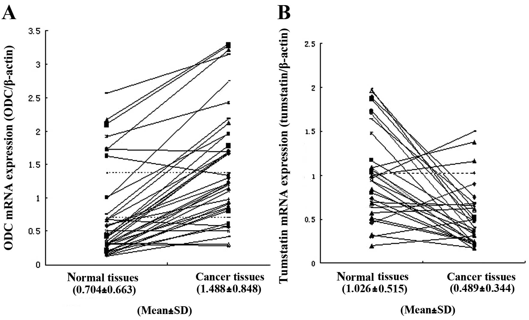
Subsequently, RT-PCR was performed to detect the
expression levels of ODC and tumstatin in human renal cancer and
adjacent normal tissues. ODC mRNA overexpression was detected in 32
of 38 cancerous tissues, and not in any of the corresponding normal
tissues (Fig. 1A). In these 32
ODC-overexpressing kidney cancerous samples, 24 had downregulated
tumstatin mRNA expression when compared with the adjacent normal
tissues (Fig. 1B). Statistical
analysis revealed a correlation between ODC gene expression and
tumstatin expression (P<0.05).
Establishment of mock transfectants and
ODC transfectants
Full-length human ODC cDNA (1,386 bp) was cloned
into pcDNA3.1/Myc-His(−)A expression vector to generate the
eukaryotic expression plasmid pcDNA-ODC, which was identified and
confirmed by restriction enzyme digestion and DNA sequencing.
Subsequently, RT-PCR and western blot analysis were performed to
detect whether the recombinant plasmid could be expressed in
eukaryotic cells (HEK293) (data not shown).
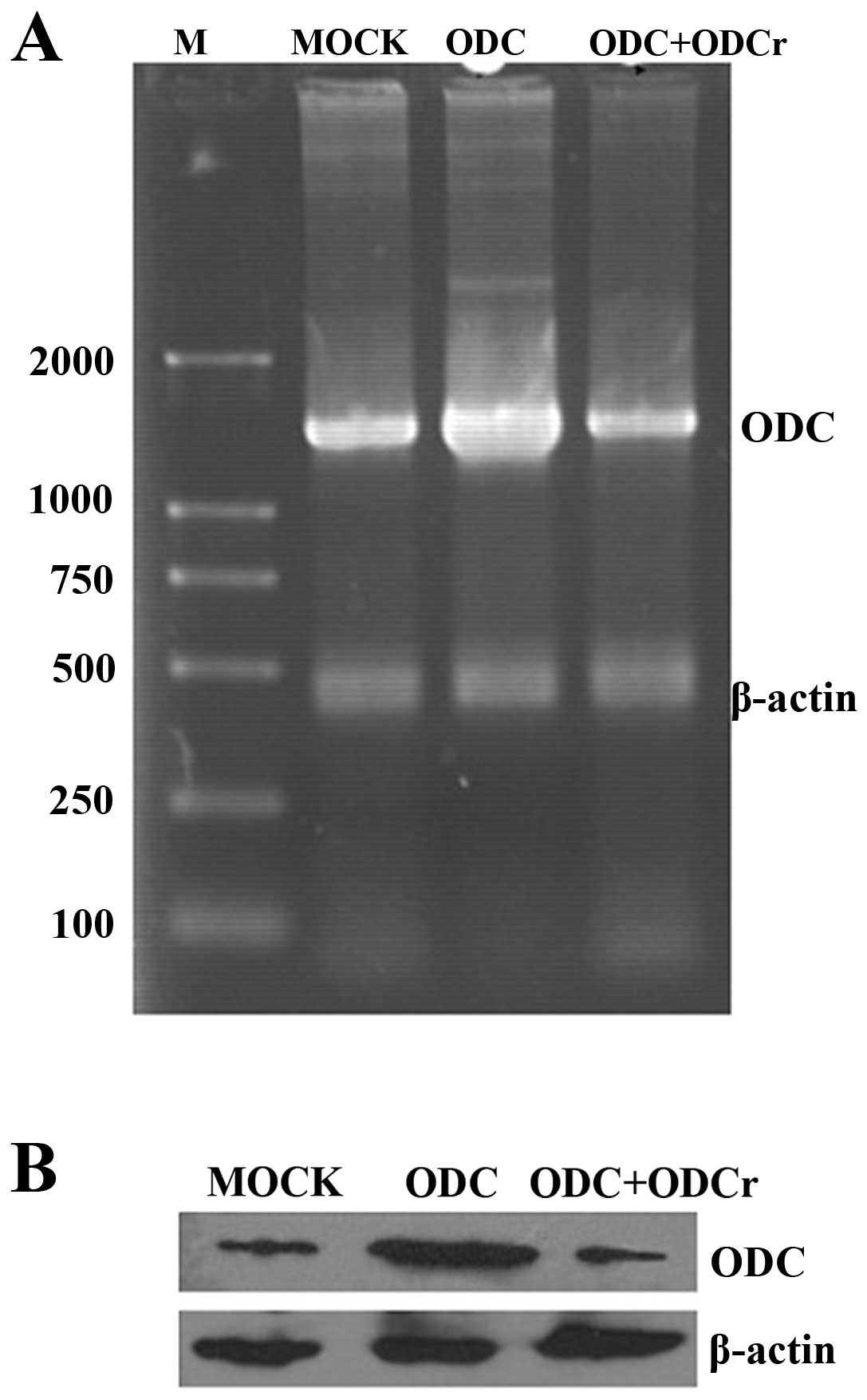
HEK293 cells transfected with pcDNA3.1/Myc-His(−)A
and pcDNA-ODC were selected in the presence of G418 for 3–4 weeks,
and the surviving cells were established as mock transfectants and
ODC transfectants, respectively. Compared with mock transfectants,
the expression level of ODC mRNA (Fig.
2A) and protein (Fig. 2B) in
ODC transfectants was increased by 200 and 196%, respectively.
However, the ODC mRNA and protein expression level in ODC
transfectants transfected with pcDNA-ODCr recovered to the same
level as that in mock transfectants (Fig. 2), which indicates that
pcDNA3.1-mediated antisense ODC could inhibit the expression of ODC
in ODC tranfectants.
ODC overexpression suppressed tumstatin
expression
In order to examine the effect of ODC overexpression
on tumstatin expression, we generated transfectants overexpressing
ODC and mock transfectants containing vector alone. Then HEK293
cells were subjected to different conditions: PBS treatment, mock
transfection, ODC transfection, pcDNA-ODC + pcDNA-ODCr (ODC
transfectants transfected with pcDNA-ODCr) and putrescine
treatment. The expression levels of ODC and tumstatin in HEK293
cells treated as described above were detected by semi-quantitative
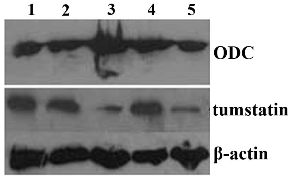
RT-PCR and western blot analysis. ODC mRNA (Fig. 3A) and protein (Fig. 4) were overexpressed in ODC
transfectants, and the elevated ODC expression level approached
that of mock transfectants in the pcDNA-ODC + pcDNA-ODCr group. No
change in ODC expression level was detected in the
putrescine-treated group, as compared with PBS-treated group
(Table II). However, the
expression of tumstatin at both the mRNA (Fig. 3B) and protein levels (Fig. 4) was significantly less in ODC
transfectants and putrescine-treated group than in PBS-treated
group. The suppression of tumstatin expression in ODC transfectants
was rescued after transfection of pcDNA-ODCr (Table II). We analyzed the expression of
endostatin and VEGF mRNA in treated HEK293 cells. The expression of
endostatin in ODC transfectants and putrescine-treated group was
significantly inhibited, but the downregulation of endostatin
expression in ODC transfectants was restored upon transfection of
pcDNA-ODCr (Fig. 3C), while VEGF
mRNA expression level remained unchanged (Fig. 3D) (Table II).
 | Figure 3mRNA expression of ODC, tumstatin,
endostatin and VEGF in HEK293 cells after treatment. (A) A 1,286 bp
band representing the ODC gene, (B) a 750 bp fragment representing
the tumstatin gene, (C) a 261 bp fragment representing endostatin
and (D) 156 bp fragment representing VEGF were amplified by RT-PCR
and visualized. β-actin was also amplified as an internal control.
Lane M, DL-2000 marker; lane 1, PBS-treated group; lane 2, mock
transfectants; lane 3, ODC transfectants; lane 4, ODC transfectants
transfected with pcDNA-ODCr; lane 5, putrescine-treated group. |
| Table IIThe expression of ODC, tumstatin,
endostatin and VEGF in treated HEK293 cells. |
Table II
The expression of ODC, tumstatin,
endostatin and VEGF in treated HEK293 cells.
| Treatment |
|---|
|
|
|---|
| Detection | PBS | Mock
transfectants | ODC
transfectants | ODC + ODCr | Putrescine |
|---|
| ODC mRNA | 1.89±0.02 | 1.91±0.43 | 3.31±0.29a | 1.90±0.21 | 1.60±0.41 |
| ODC protein | 0.237±0.04 | 0.245±0.03 | 0.598±0.14a | 0.308±0.04 | 0.239±0.01 |
| Tumstatin mRNA | 1.13±0.23 | 1.16±0.37 | 0.26±0.34a | 1.16±0.16 | 0.34±0.32a |
| Tumstatin
protein | 0.235±0.07 | 0.259±0.05 | 0.078±0.04a | 0.235±0.02 | 0.114±0.06a |
| Endostatin
mRNA | 0.97±0.25 | 0.96±0.09 | 0.31±0.07a | 1.08±0.10 | 0.40±0.24a |
| VEGF mRNA | 0.76±0.26 | 0.75±0.34 | 0.74±0.38 | 0.76±0.22 | 0.75±0.39 |
After demonstrating that ODC overexpression results
in the downregulation of tumstatin mRNA and protein levels, we then
examined the effect of ODC overexpression on the tumstatin gene
promoter. A luciferase reporter plasmid pGL-tumstatin 2.2 kb
containing the full-length promoter region (2,149 bp) was
constructed and identified by restriction enzyme digestion and DNA
sequencing. Subsequently, the tumstatin gene promoter luciferase
reporter plasmid was transfected into HEK293 cells and luciferase
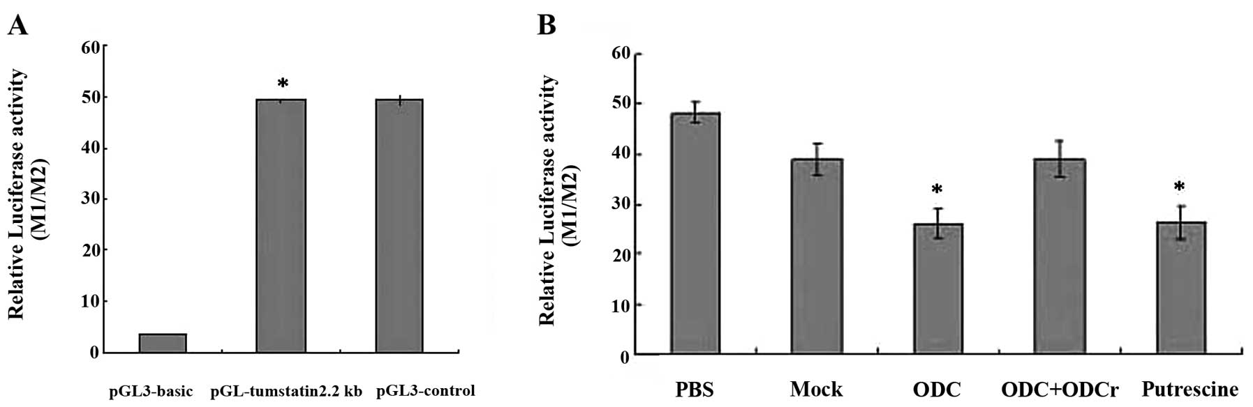
activity assay was performed. As shown in Fig. 5A, wherein M1/M2 represents the
relative luciferase activity, the 2,149 bp fragment exhibited
promoter activity. pGL-tumstatin (2.2 kb) exhibited pronounced
promoter activity (49.25±0.46) to the same degree as that of the
positive control pGL3-control (49.3±1.2). Cells transfected with
pGL3-basic (7.1±0.01) showed no promoter activity.
We next investigated whether pGL-tumstatin (2.2 kb)
could be regulated by ODC overexpression. Luciferase activity assay
results showed that the activity of pGL-tumstatin (2.2 kb)
decreased to 45.8% upon ODC overexpression (26.12±2.8) and to 45.2%
upon putrescine addition (26.36±3.42), compared with the
PBS-treated group (48.16±2.05). However, the luciferase activity of
pGL-tumstatin (2.2 kb) in ODC transfectants matched that in mock
transfectants (38.96±3.22) upon transfection with the antisense
plasmid pcDNA-ODCr (39.04±3.68) (Fig.
5B). These results indicate that ODC overexpression and
putrescine suppressed the expression of tumstatin by inhibiting
promoter activity.
Discussion
Angiogenesis, characterized by generation of new
capillaries, plays an important role in physiological processes
such as wound healing and in pathological disorders such as cancer
(25,26). Tumor angiogenesis is indispensable
for solid tumor growth and metastasis (27,28).
Neovascularization in tumors supplies tumor with oxygen and
nutrition, stimulates tumor progression by paracrine secretion and
facilitates the hematogenous metastasis of tumor cells (29). Targeting the blood vessels feeding
tumors could result in tumor starvation and tumor regression.
Therefore, tumor angiogenesis presents an essential target of
therapeutic intervention for cancer.
Tumor angiogenesis requires both upregulation of
angiogenic stimulators such as VEGF, bFGF, angiogenin and
downregulation of endogenous angiogenic inhibitors such as
tumstatin, endostatin and thrombospondin (30). Endogenous angiogenic inhibitors and
synthetic inhibitors or antibodies to angiogenic stimulators have
been investigated as potential therapeutic agents against tumors
because of their promising antitumor activity (27,28).
Tumstatin, a novel endogenous angiogenic inhibitor,
specifically suppresses proliferation of endothelial cells, induces
apoptosis of endothelial cells, and inhibits pathological
angiogenesis and tumor growth (31). It has distinct antitumor properties
with an N-terminal (amino acids 54–132) possessing anti-angiogenic
activity and a C-terminal (amino acids 185–203) having antitumor
cell activity. Tumstatin exerts an antitumor effect by binding to
αvβ3 integrin in endothelial cells and melanoma cells (5,7,10).
Endostatin, another endogenous inhibitor of angiogenesis,
originates from the α1 chain of type XVIII collagen (32). Tumstatin and endostatin share a 14%
amino acid homology and exhibit distinct anti-angiogenic activities
(6). Anti-angiogenesis effect of
tumstatin is ten times more potent than that of endostatin,
therefore tumstatin is regarded as a promising anticancer
therapeutic candidate (6,33). However, tumstatin alone failed to
achieve tumor regression. It might have an adjuvant role in tumor
treatment and is effective against tumors when administered in
combination with conventional therapy (33). Many studies have demonstrated that
the addition of tumstatin or endostatin increases the antitumor
efficacy of conventional therapies (34–36).
Thus the combined treatment of these agents could be used for
targeting cancer in the future.
The role of ODC and polyamine in cancer has been a
focus of many research studies. Elevated ODC activity has been
detected in many cancers and thought to be associated with cell
transformation, tumor invasion and angiogenesis (14,16–19).
The ODC gene is considered to be an oncogene because overexpression
of ODC results in malignant transformation of NIH3T3 cells
(18,37). B16 melanoma-induced angiogenesis,
rapid neovascularization and tumor growth are inhibited by DFMO by
irreversibly inactivating ODC, and these inhibitions are reversed
by exogenous putrescine and spermidine (20,38).
Due to this, DFMO has been used as chemotherapeutic agent in
clinical trials for cancers, although it exhibits dose-limiting
toxicity.
In the present study, we found that
ODC-overexpressing human cancer cells (ACHN and A549) and renal
cancer tissues have reduced expression of tumstatin. ODC
overexpression can downregulate the production of thrombospondin
and suppress the gene expression of type XVIII collagen and
endostatin (20,22). Therefore, we hypothesized that ODC
overexpression can inhibit the expression of tumstatin. To examine
the effect of ODC overexpression on tumstatin expression, we
generated an ODC overexpressing plasmid pcDNA-ODC and established
ODC-overexpressing HEK293 cells.
In this study, cells subjected to five different
conditions were examined: PBS treatment (control group), mock
transfection, ODC transfection, ODC transfectants transfected with
pcDNA-ODCr, and putrescine treatment. The overexpression of ODC in
ODC transfectants was suppressed by the antisense plasmid
pcDNA-ODCr. This experimental setup allowed us to examine the
expression level of tumstatin relative to the level of ODC. The
effect of the antisense plasmid pcDNA-ODCr on ODC was similar to
that of DFMO. RT-PCR and western blot results showed that ODC
overexpression and putrescine inhibited the expression of tumstatin
mRNA and protein, while the suppression of tumstatin expression in
ODC transfectants was rescued after transfection of pcDNA-ODCr. The
expression level of VEGF mRNA remained unchanged, demonstrating
that the effect of ODC overexpression and putrescine on promoting
angiogenesis was not associated with VEGF. This is consistent with
a previous report by Nemoto et al(22). In order to better understand the
negative effect of ODC on tumstatin expression, we examined the
effect of ODC on tumstatin gene promoter activity. The results from
the dual luciferase reporter assay indicate that ODC overexpression
and putrescine suppressed the expression of tumstatin by inhibiting
tumstatin promoter activity. Taken together, these results support
that ODC may promote tumor angiogenesis by suppressing tumstatin
expression in many cancers. This finding provides novel evidence
for the efficacy of combining anti-angiogenic therapy with
conventional therapy for cancer treatment.
Acknowledgements
This study was supported by the Foundation of the
Department of Science and Technology of Shandong Province (no.
2008GG30002030), the National Natural Science Foundation of China
(no. 81100323) and the Promotive Research Fund for Excellent Young
and Middle-aged Scientists of Shandong Province (BS2011YY033).
References
|
1
|
Saus J, Wieslander J, Langeveld JP,
Quinones S and Hudson BG: Identification of the Goodpasture antigen
as the alpha3(IV) chain of collagen IV. J Biol Chem.
263:13374–13380. 1988.
|
|
2
|
Hudson BG, Reeders ST and Tryggvason K:
Type IV collagen: structure, gene organization, and role in human
diseases. Molecular basis of Goodpasture and Alport syndromes and
diffuse leiomyomatosis. J Biol Chem. 268:26033–26036.
1993.PubMed/NCBI
|
|
3
|
Hamano Y and Kalluri R: Tumstatin, the NC1
domain of alpha3 chain of type IV collagen, is an endogenous
inhibitor of pathological angiogenesis and suppresses tumor growth.
Biochem Biophys Res Commun. 333:292–298. 2005. View Article : Google Scholar : PubMed/NCBI
|
|
4
|
Hamano Y, Zeisberg M, Sugimoto H, Lively
JC, Maeshima Y, Yang C, Hynes RO, Werb Z, Sudhakar A and Kalluri R:
Physiological levels of tumstatin, a fragment of collagen IV alpha3
chain, are generated by MMP-9 proteolysis and suppress angiogenesis
via alphaV beta3 integrin. Cancer Cell. 3:589–601. 2003. View Article : Google Scholar
|
|
5
|
Maeshima Y, Yerramalla UL, Dhanabal M,
Holthaus KA, Barbashov S, Kharbanda S, Reimer C, Manfredi M,
Dickerson WM and Kalluri R: Extracellular matrix-derived peptide
binds to αvβ3 integrin and inhibits angiogenesis. J Biol Chem.
276:31959–31968. 2001.
|
|
6
|
Sudhakar A, Sugimoto H, Yang C, Lively JC,
Zeisberg M and Kalluri R: Human tumstatin and human endostatin
exhibit distinct antiangiogenic activities mediated by αvβ3 and
α5β1 integrins. Proc Natl Acad Sci USA. 100:4766–4771.
2003.PubMed/NCBI
|
|
7
|
Esipov R, Beyrakhova K, Likhvantseva V, et
al: Antiangiogenic and antivascular effects of a recombinant
tumstatin-derived peptide in a corneal neovascularization model.
Biochimie. 94:1368–1375. 2012. View Article : Google Scholar : PubMed/NCBI
|
|
8
|
Maeshima Y, Colorado PC, Torre A, Holthaus
KA, Grunkemeyer JA, Ericksen MB, Hopfer H, Xiao Y, Stillman IE and
Kalluri R: Distinct antitumor properties of a type IV collagen
domain derived from basement membrane. J Biol Chem.
275:21340–21348. 2000. View Article : Google Scholar : PubMed/NCBI
|
|
9
|
Pasco S, Ramont L, Venteo L, Pluot M,
Maquart FX and Monboisse JC: In vivo overexpression of tumstatin
domains by tumor cells inhibits their invasive properties in a
mouse melanoma model. Exp Cell Res. 301:251–265. 2004. View Article : Google Scholar : PubMed/NCBI
|
|
10
|
Maeshima Y, Sudhakar A, Lively JC, Ueki K,
Kharbanda S, Kahn CR, Sonenberg N, Hynes RO and Kalluri R:
Tumstatin, an endothelial cell-specific inhibitor of protein
synthesis. Science. 295:140–143. 2002. View Article : Google Scholar : PubMed/NCBI
|
|
11
|
Maeshima Y, Colorado PC and Kalluri R: Two
RGD-independent alpha vbeta 3 integrin binding sites on tumstatin
regulate distinct anti-tumor properties. J Biol Chem.
275:23745–23750. 2000. View Article : Google Scholar : PubMed/NCBI
|
|
12
|
Joshi M: The importance of L-arginine
metabolism in melanoma: an hypothesis for the role of nitric oxide
and polyamines in tumor angiogenesis. Free Radic Biol Med.
22:573–578. 1997. View Article : Google Scholar : PubMed/NCBI
|
|
13
|
Kucharzewska P, Welch JE, Svensson KI and
Belting M: The polyamines regulate endothelial cell survival during
hypoxic stress through PI3K/AKT and MCL-1. Biochem Biophys Res
Commun. 380:413–418. 2009. View Article : Google Scholar : PubMed/NCBI
|
|
14
|
Auvinen M: Cell transformation, invasion,
and angiogenesis: a regulatory role for ornithine decarboxylase and
polyamines? J Natl Cancer Inst. 89:533–537. 1997. View Article : Google Scholar : PubMed/NCBI
|
|
15
|
Pegg AE: Polyamine metabolism and its
importance in neoplastic growth and a target for chemotherapy.
Cancer Res. 48:759–774. 1988.
|
|
16
|
Takigawa M, Ishida H, Takano T and Suzuki
F: Polyamine and differentiation: induction of ornithine
decarboxylase by parathyroid hormone is a good marker of
differentiated chondrocytes. Proc Natl Acad Sci USA. 77:1481–1485.
1980. View Article : Google Scholar : PubMed/NCBI
|
|
17
|
Kubota S, Kiyosawa H, Nomura Y, Yamada T
and Seyama Y: Ornithine decarboxylase overexpression in mouse
10T1/2 fibroblasts: cellular transformation and invasion. J Natl
Cancer Inst. 89:567–571. 1997. View Article : Google Scholar : PubMed/NCBI
|
|
18
|
Auvinen M, Laine A, Paasinen-Sohns A,
Kangas A, et al: Human ornithine decarboxylase-overproducing NIH3T3
cells induce rapidly growing, highly vascularized tumors in nude
mice. Cancer Res. 57:3016–3025. 1997.
|
|
19
|
Auvinen M, Paasinen A, Andersson LC and
Hölttä E: Ornithine decarboxylase activity is critical for cell
transformation. Nature. 360:355–358. 1992. View Article : Google Scholar : PubMed/NCBI
|
|
20
|
Takigawa M, Enomoto M, Nishida Y, Pan HO,
Kinoshita A and Suzuki F: Tumor angiogenesis and polyamines:
alpha-difluoromethylornithine, an irreversible inhibitor of
ornithine decarboxylase, inhibits B16 melanoma-induced angiogenesis
in ovo and the proliferation of vascular endothelial cells in
vitro. Cancer Res. 50:4131–4138. 1990.
|
|
21
|
Takahashi Y, Mai M and Nishioka K:
Alpha-difluoromethylornithine induces apoptosis as well as
anti-angiogenesis in the inhibition of tumor growth and metastasis
in a human gastric cancer model. Int J Cancer. 85:243–247. 2000.
View Article : Google Scholar
|
|
22
|
Nemoto T, Hori H, Yoshimoto M, Seyama Y
and Kubota S: Overexpression of ornithine decarboxylase enhances
endothelial proliferation by suppressing endostatin expression.
Blood. 99:1478–1481. 2002. View Article : Google Scholar : PubMed/NCBI
|
|
23
|
Zhang B, Liu XX, Zhang Y, Jiang CY, Hu HY
and Gong L: The screening of the sensitive antisense target in the
ODC mRNA. J Shandong Univ. 8:146–150. 2005.
|
|
24
|
Xu CX, Liu XX, Hou GS, Yan YF, Chen SM,
Wang W, Jiang GS, Liu B and Xin JX: The expression of tumstatin is
down-regulated in renal carcinoma. Mol Biol Rep. 37:2273–2277.
2010. View Article : Google Scholar : PubMed/NCBI
|
|
25
|
Folkman J: Angiogenesis in cancer,
vascular, rheumatoid and other disease. Nat Med. 1:27–31. 1995.
View Article : Google Scholar : PubMed/NCBI
|
|
26
|
Folkman J and Shing Y: Angiogenesis. J
Biol Chem. 267:10931–10934. 1992.
|
|
27
|
Liekens S, De Clercq E and Neyts J:
Angiogenesis: regulators and clinical applications. Biochem
Pharmacol. 61:253–270. 2001. View Article : Google Scholar : PubMed/NCBI
|
|
28
|
Harris AL: Angiogenesis as a new target
for cancer control. EJC (Suppl). 2:1–12. 2003. View Article : Google Scholar
|
|
29
|
Folkman J: Tumor angiogenesis: therapeutic
implications. N Engl J Med. 285:1182–1186. 1971. View Article : Google Scholar : PubMed/NCBI
|
|
30
|
Hanahan D and Folkman J: Patterns and
emerging mechanisms of the angiogenic switch during tumorigenesis.
Cell. 86:353–364. 1996. View Article : Google Scholar : PubMed/NCBI
|
|
31
|
Yang YP, Xu CX, Hou GS, Xin JX, Wang W and
Liu XX: Effects of eukaryotic expression plasmid encoding human
tumstatin gene on endothelial cells in vitro. Chin Med J (Engl).
123:2269–2273. 2010.PubMed/NCBI
|
|
32
|
O'Reilly MS, Boehm T, Shing Y, Fukai N,
Vasios G, Lane WS, Flynn E, Birkhead JR, Olsen BR and Folkman J:
Endostatin: an endogenous inhibitor of angiogenesis and tumor
growth. Cell. 88:277–285. 1997. View Article : Google Scholar
|
|
33
|
Chung IS, Son YI, Ko YJ, Baek CH, Cho JK
and Jeong HS: Peritumor injections of purified tumstatin delay
tumor growth and lymphatic metastasis in an orthotopic oral
squamous cell carcinoma model. Oral Oncol. 44:1118–1126. 2008.
View Article : Google Scholar : PubMed/NCBI
|
|
34
|
Bertolini F, Fusetti L, Mancuso P, Gobbi
A, Corsini C, Ferrucci PF, Martinelli G and Pruneri G: Endostatin,
an anti-angiogenic drug, induces tumor stabilization after
chemotherapy or anti-CD20 therapy in a NOD/SCID mouse model of
human high-grade non-Hodgkin lymphoma. Blood. 96:282–287.
2000.PubMed/NCBI
|
|
35
|
te Velde EA, Vogten JM, Gebbink MF, van
Gorp JM, et al: Enhanced antitumor efficacy by combining
conventional chemotherapy with angiostatin or endostatin in a liver
metastasis model. Br J Surg. 89:1302–1309. 2002.PubMed/NCBI
|
|
36
|
Yao B, He QM, Tian L, Xiao F, Jiang Y,
Zhang R, et al: Enhanced antitumor effect of the combination of
tumstatin gene therapy and gemcitabine in murine models. Hum Gene
Ther. 16:1075–1086. 2005. View Article : Google Scholar : PubMed/NCBI
|
|
37
|
Moshier JA, Dosescu J, Skunca M and Luk
GD: Transformation of NIH/3T3 cells by ornithine decarboxylase
overexpression. Cancer Res. 53:2618–2622. 1993.PubMed/NCBI
|
|
38
|
Meyskens FL Jr and Gerner EW: Development
of difluoromethyornithine (DFMO) as a chemoprevention agent. Clin
Cancer Res. 5:945–951. 1999.PubMed/NCBI
|