Introduction
The FUS/ERG fusion gene (FUS is also
named TLS) was first described in 1994 by two different
groups. Ichikawa et al(1)
found the FUS/ERG in four acute myeloid leukemia (AML)
patients whereas Panagopoulos et al(2) identified FUS/ERG in bone marrow
cells carrying a t(16;21)(p11;q22) in a 3-year-old boy diagnosed
with AML M1. Since then, the t(16;21)(p11;22) and/or its fusion
product FUS/ERG has been reported in 66 cases of AML and 3
cases of acute lymphoblastic leukemia (ALL; http://cgap.nci.nih.gov/Chromosomes/Mitelman,
database updated May 15, 2013). It is seen in all ages and appears
to be associated with a dismal prognosis (3,4).
Nevertheless, survivals longer than 36 months have been reported in
3 childhood cases one of which was an ALL (4–6). The
same fusion FUS/ERG has also been found in 3 Ewing tumors
(8,9); it is thus one of the relatively few
fusion genes that exert pathogenetic influence in widely disparate
neoplastic entities.
Pereira et al(7) showed that retroviral transduction of
FUS/ERG to human umbilical cord blood cells altered myeloid
and arrested erythroid differentiation and led to a dramatic
increase in the proliferative and self-renewal capacity of
transduced myeloid progenitors. They concluded that ‘TLS-ERG
expression alone induced a leukemogenic program that exhibited
similarities to the human disease associated with this
translocation’. Zou et al(10) showed that FUS/ERG activated
different sets of genes in mouse L-G myeloid progenitor cells and
NIH3T3 fibroblasts; the two cell types show little similarity. They
concluded that FUS/ERG transformed hematopoietic cells and
fibroblasts via different pathways.
In the two original articles, FUS/ERG was
identified based on the candidate gene approach (1,2). Prior
to those two studies, the breakpoints of the t(16;21)(p11;q22) had
already been shown to cluster in a specific intron of ERG in
chromosome band 21q22 (11).
ERG was furthermore known to be fused with EWSR1 (on
22q12) in a subset of Ewing sarcomas (12,13),
and EWSR1 was known to display a high degree of homology
with FUS, the 16p11 gene rearranged in the t(12;16)(q13;p11)
that characterizes myxoid liposarcomas (14,15).
It was therefore decided to investigate whether FUS was also
involved in the t(16;21) of AML. In both studies, Southern blot
analysis demonstrated that FUS was rearranged and PCR
examinations showed the formation of a FUS/ERG fusion gene
(1,2).
Recently, RNA-sequencing (RNA-Seq, also known as
whole transcriptome sequencing) was shown to be an efficient tool
in the detection of fusion genes in cancer (16). However, it suffers from the
shortcoming of identifying as ‘fusion genes’ also many technical,
biological and, perhaps in particular, clinical ‘false positives’,
thus making the assessment of which fusions are important and which
are noise extremely difficult. We and others have shown that a
combination of cytogenetics and RNA-Seq is an excellent approach to
detect the ‘primary’ fusion genes in neoplasms carrying only one or
a few chromosomal rearrangements. In solid tumors, this approach
was used to identify the WWTR1-CAMTA1 and
YWHAE-FAM22A/B chimeric genes in epithelioid
hemangioendothelioma and in high-grade endometrial stromal
sarcomas, respectively (17,18),
ZC3H7-BCOR in endometrial stromal sarcomas (19), IRF2BP2-CDX1 in a mesenchymal
chondrosarcoma (20), and
EWSR1-YY1 in a subset of mesotheliomas (21). Except the ZC3H7-BCOR, which
was detected in a tumor whose karyotype contained two chromosome
translocations (19), all the other
studies started with malignancies in which the karyotype had a
single chromosomal translocation. In hematologic malignancies,
likewise, an NFIA-CBFA2T3 (NFIA is located in 1p31)
chimeric transcript was found in an acute erythroid leukemia with
the translocation t(1;16)(p31;q24) and a FISH-detected split of
CBFA2T3 in 16q24 (22,23).
The same approach comparing karyotypic and sequencing data was also
used to identify the ZMYND8-RELA fusion in a congenital
acute erythroid leukemia carrying a t(11;20)(p11;q13) translocation
as a sole chromosome aberration (24). In the present study, we applied
RNA-Seq methodology to an acute myeloid leukemia with a rather
complex karyotype and identified a cryptic FUS/ERG fusion
gene.
Case report
Ethics statement
The study was approved by the Regional Ethics
Committee (Regional Komité for Medisinsk Forskningsetikk Sør-Øst,
Norge, http://helseforskning.etikkom.no), and written
informed consent was obtained from the patient’s parents to
publication of the case details.
Case report
A 2 years and 9 months old girl who had been treated
for pneumonia 3 months ago was referred to the local hospital with
fever, diarrhea, abdominal pain, reduced general condition, and
anemia. Upon admission, she had a hemoglobin value of 9.2 g/dl,
thrombocytes of 132×109/l, and signs of cholestasis and
pancreatitis (increased pancreas-amylase 418 U/l and lipase 1653
U/l, edematous pancreas on abdominal MRI, some peritoneal cavity
fluid). No infectious origin could be found, and investigations for
the presence of Epstein-Barr virus, parvovirus B19, hepatitis A, B
and C, chlamydia pneumoniae, cytomegalo-virus, and streptococci
were negative. After a short period of spontaneous clinical
improvement and normalization of liver and pancreatic values, her
condition again worsened. Due to recurrent fever and a sore throat,
she was started on penicillin. However, she deteriorated, and
hemoglobin fell to 7 g/dl. At this point, a bone marrow examination
was performed and an increased number of blast-like cells were
seen. The patient was then referred to the tertiary care pediatric
hematology unit.
Upon admission, the patient was in good clinical
shape. She had hypertrophic tonsils without signs of inflammation.
There was no pathological enlargement of lymph nodes and no
hepatosplenomegaly. Hemoglobin was 7.3 g/dl, the platelet count
222×109/l, and the white blood cell count
6.2×10⁹/l. Lactate dehydrogenase was elevated (914 U/l)
but liver parameters were normal. Examination of a bone marrow
aspirate revealed 50% myeloid blasts, consistent with acute myeloid
leukemia. The myeloid cell lineage of the blasts was confirmed by
flow cytometric immunophenotyping showing the expression of the
myeloid markers CD13, CD33, CD15, and myeloperoxidase in addition
to the aberrant expression of CD7 and CD56. The blasts were
positive for the progenitor cell markers CD34, CD117, and CD133,
but negative for HLA-DR antigens. Interestingly, a leukemic stem
cell population comprising 3% of the total number of cells was also
identified. These CD34 brightly positive/CD38 negative leukemic
stem cells displayed an abnormal phenotype being negative for
HLA-DR antigens but positive for CD7, CD56, and CD123. Molecular
genetic analysis did not show any Flt3 internal tandem duplication
(ITD) mutation nor were CEPBα mutations seen. As expected, markedly
increased levels of Wilms’ tumor (WT) 1 expression were
demonstrated.
The girl was started on AML chemotherapy according
to the NOPHO-AML 2004 protocol (25) (Nordic Society of Pediatric
Hematology and Oncology). She went into remission after the first
course (AIET: cytarabine, idarubicin, etoposide, thioguanin).
Minimal residual disease by flow cytometry was positive, but
<0.1%. This was also the case after the second course (AM:
cytarabine, mitoxantrone). However, WT1 levels were normalized.
This was also true after the second chemotherapy course (AM:
cytarabine, mitoxantrone). Following the third course (HA1M: high
dose cytarabine, mitoxantrone), MRD began slowly to increase. After
the fourth course (HA2E), the MRD level by flow cytometry was 0.8%
indicating relapse, and also the WT1 levels gradually increased.
The patient was taken off protocol and re-induced with two courses
of FLA (fludarabine, cytarabine). After an initial MRD decrease to
0.4%, it again rose to 3.5% after the second FLA course. Awaiting
completion of a bone marrow donor search, the patient is now
scheduled for a CloEC course (clofarabine, etoposide,
cyclophosphamide) after which the plan is to perform allogenic stem
cell transplantation.
G-banding analysis
Bone marrow cells were cytogenetically investigated
by standard methods. Chromosome preparations were made from
metaphase cells of a 24-h culture, G-banded using Leishman stain,
and karyotyped according to ISCN 2009 guidelines (26).
Fluorescence in situ hybridization
(FISH)
Fluorescence in situ hybridization (FISH) was
performed on metaphase spreads using the Vysis FUS Break Apart FISH
Probe Kit (Abbott Norge Molecular, Snaroya, Norway) and whole
painting probes for chromosomes 1, 2, 3, and 16 (Cytocell,
BioNordika, Lysaker, Norway). Fluorescent signals were captured and
analyzed using the CytoVision system (Leica Biosystems, Newcastle,
UK).
Whole transcriptome sequencing
Total RNA (3 μg) extracted from the patient’s bone
marrow at the time of diagnosis was sent for high-throughput
paired-end RNA-sequencing to the Norwegian Sequencing Centre at
Ullevål Hospital (http://www.sequencing.uio.no). The Illumina software
pipeline was used to process image data into raw sequencing data
and only sequence reads marked as ‘passed filtering’ were used in
the downstream data analysis. A total of 103 million reads were
obtained. The FASTQC software was used for quality control of the
raw sequence data (http://www.bioinformatics.babraham.ac.uk/projects/fastqc).
The software FusionMap was used for the discovery of fusion
transcripts (27) (release date
2012-04-16) together with the pre-built Human B37 and RefGene from
the FusionMap website (http://www.omicsoft.com/fusionmap).
PCR analyses
Total RNA (1 μg) was reverse-transcribed in a 20-μl
reaction volume using iScript Advanced cDNA Synthesis Kit for
RT-qPCR according to the manufacturer’s instructions (Bio-Rad,
Oslo, Norway). cDNA corresponding to 50 ng total RNA was used as
template in subsequent PCR assays. The 25 μl PCR volume contained
12.5 μl Premix Ex Taq™ DNA Polymerase Hot Start Version (Takara
Bio, AH Diagnostics, Oslo, Norway), 1 μl of diluted cDNA, and 0.2
μM of each of the forward and reverse primers. For the detection of
the FUS-ERG fusion transcript the forward FUS-358F (CAG AGC
TCC CAA TCG TCT TAC GG) and the reverse ERG-1163R (CAG GAG CTC CAG
GAG GAA CTG C) primers were used. The PCR was run on a C-1000
Thermal cycler (Bio-Rad) with an initial denaturation at 94°C for
30 sec, followed by 35 cycles of 7 sec at 98°C, 30 sec at 60°C, 1
min at 72°C, and a final extension for 5 min at 72°C. PCR products
(4 μl) were stained with GelRed (Biotium, VWR International, Oslo,
Norway), analyzed by electrophoresis through 1.0% agarose gel, and
photographed. The remaining PCR products were purified using the
NucleoSpin® Gel and PCR Clean-up kit (Macherey-Nagel,
VWR International) and sequenced at GATC Biotech (Germany,
http://www.gatc-biotech.com/en/home.html). The BLAST
software (http://blast.ncbi.nlm.nih.gov/Blast.cgi) was used for
computer analysis of sequence data.
Results
The G-banding analysis together with whole
chromosome paint FISH yielded the karyotype
46,XX,add(1)(p36),der(2)t(2;3)(q21;q21),del(3)(q21),der(10)t(1;10)(q32;q24),der(16)(2qter-->
2q21::16p11-->16q24::16p11-->16pter)[13]/46,XX[2] (Fig. 1A–C). FISH with a FUS break
apart probe showed that the FUS gene had been rearranged and
the 5′-end part of FUS (green probe in Fig. 1D) moved to the q arm of the
der(16), whereas the 3′-end part
of the gene (red probe in Fig. 1D)
remained on 16p11 of the der(16).
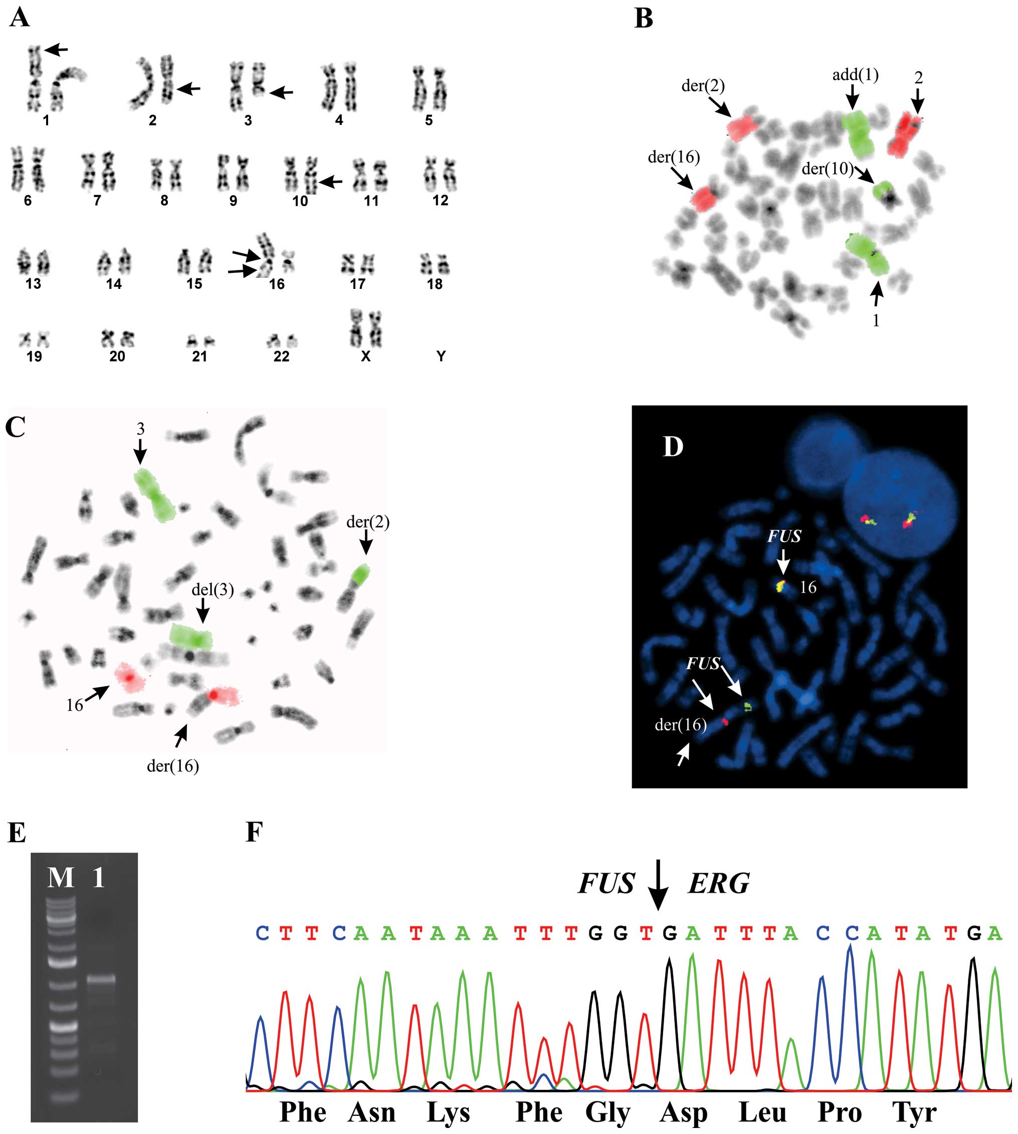 | Figure 1G-banding, FISH, and PCR analyses of
the AML. A, G-banded karyotype showing the chromosome aberrations
(arrows). B, FISH using WCP for chromosome 1 (green signal) and
chromosome 2 (red signal) shows (arrows) the add(1)(p36),
der(2)t(2;3)(q21;q21), and der(10)t(1;10)(q32;q24). C, FISH using
WCP for chromosomes 3 (green signal) and 16 (red signal) shows
(arrows) the der(2)t(2;3)(q21;q21), del(3), and der(16). D, FISH
using a FUS break apart probe shows (arrows) rearrangement
of FUS together with the normal chromosome 16. The 5′-part
of the FUS gene (green probe) has moved to the q arm of the
der(16), while the 3′-part of the
gene (red probe) remains on 16p11 of the der(16). E, Amplification of FUS-ERG
fusion cDNA fragments using the primers FUS-358F and ERG-1163R
(lane 1). M, 1 kb DNA ladder. F, Partial sequence chromatogram
showing the junctions of the FUS-ERG chimeric transcript and
part of the in-frame coding protein. |
Using FusionMap on the raw sequencing data obtained
by the Norwegian Sequencing Centre, 500 fusion genes were found.
The FUS-ERG fusion transcript was ranked fourth with 21 seed
counts. Because of this and because the chromosome band 16p11 was
involved in the complex karyotype (FUS maps to chromosome
bands 16p11), we decided to investigate further the FUS-ERG
fusion transcript. PCR and direct sequencing verified the presence
of a FUS-ERG chimeric transcript in which exon 7 of
FUS (nt 904 in sequence with accession number NM_004960
version 3) was fused in frame to exon 8 of ERG from sub-band
21q22.2 (nt 967 in NM_004449 version 4) (Fig. 1E and F).
Discussion
The present case of AML had a karyotype with five
structural chromosome aberrations, all of which could have
generated fusion genes. For example, the PRDM16 gene in
1p36, which codes for a transcription factor, and the RPN1
gene in 3q21, which codes for type I integral membrane protein
found in the rough endoplasmic reticulum, are rearranged in AML
with t(1;3)(p36;q21) (28). Both
chromosome bands, 1p36 and 3q21, were seen rearranged in the
abnormal karyotype of the present case. Likewise, on chromosome
band 16p11, the FUS gene was found to be rearranged and
fused to ERG in a subset of AML with t(16;21)(p11;q22)
(1,2), and on 16q24, the CBFA2T3 gene was found, also in
AML, to be a partner gene in the fusions RUNX1-CBFA2T3
[t(16;21)(q24;q22)], NFIA-CBFA2T3 [t(1;16)(p31;q24)], and
CBFA2T3-GLIS2 [inv(16)(p13q24)] (22,23,29,30).
The aberrations der(2)t(2;3)(q21;q21) and der(10)t(1;10)(q32;q24)
could also conceivably generate fusion genes. Screening with FISH
for all possibly rearranged genes associated with the present
abnormal karyotype would have been laborious and a very
time-consuming procedure. We therefore decided to perform RNA-Seq
to compare karyotyping and sequencing data and concentrate
exclusively on those suggested fusion genes that are found in
chromosomal breakpoints. From the 500 fusion genes which were
indicated by the RNA-Seq data using the FusionMap algorithm, only
FUS-ERG showed correspondence with karyotype features
(FUS maps to 16p11, a chromosome band which was rearranged
in the karyotype). Furthermore, the FUS-ERG fusion gene has
previously been described in a subset AML with t(16;21)(p11;q22)
(1,2).
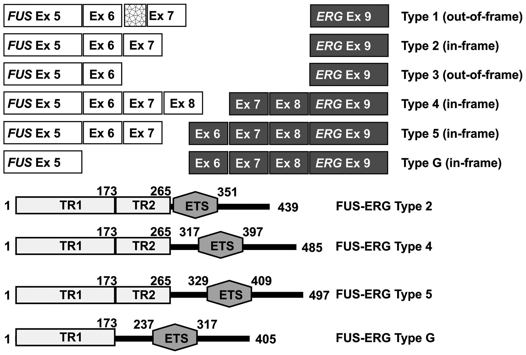
Five different FUS-ERG transcript types have
been reported in AML (Fig. 2), and
three additional types in Ewing tumors (types 4, 5, 8, 9, 31).
Types 1 and 3 (named A and C in 4, 8, 9) are out-of-frame
transcripts due to the presence of an extra 44 bp sequence of
intronic FUS material or the absence of 35 bp exonic
sequence of FUS(4). All
other FUS-ERG transcript types are in-frame with exons 5, 6,
7 or 8 of FUS (according to the reference sequence with
accession number NM_004960 version 3) fused with exons 8, 9, 10 or
11 of ERG (according to the reference sequence with
accession number NM_004449 version 3). Exons 8–11 of ERG
correspond to exons 6, 7, 8, and 9 of ERG in the study by
Kong et al(4) who used the
numeration of ERG exons presented by Zucman et
al(13). For the numbering of
ERG exons, Zucman et al(13) had assumed an identical genomic
organization for ERG and FLI1. All the FUS-ERG
isoforms contained the ETS domain which is encoded by exon 11 of
ERG and the N-terminal transactivation domain of FUS
(Fig. 2) (4,9). The
N-terminal transactivation domain of FUS can be further subdivided
into two independent functional domains, TR1 and TR2. TR1 consists
of amino acid residues 1–173 and comprises exons 1–5 of FUS,
whereas TR2 consists of amino acid residues 174–265 and comprises
exons 6 and 7 of FUS(32).
Deletion studies of the FUS/ERG fusion protein have shown that TR1
is necessary for transformation of mouse fibroblast NIH3T3 cells
while TR2 is essential for transformation of the mouse myeloid
precursor cell line L-G. This suggests that the TR2 domain is
critical for the leukemogenic potential of FUS-ERG fusion protein.
In fact, FUS exons 1–7 (representing the TR1 and TR2
domains) are always included in AML FUS/ERG transcripts,
whereas the TR2 domain is sometimes omitted from the fusion gene in
solid tumors (4,8).
The FUS-ERG transcript found here was of type
5 which has hitherto only been reported in two other cases of
childhood leukemia (in a 1-year-old boy and an 8-month-old boy)
both diagnosed with precursor B cell ALL (5,31).
This fusion transcript codes for a 497-aa FUS-ERG protein which,
similar to other AML-related FUS-ERG fusion proteins, would contain
both functional domains TR1 and TR2 of the transactivation domain
of FUS and the ETS domain of ERG (Fig.
2). It is still not clear whether any biological, let alone
clinical, significance can be attributed to the presence of exons
8, 9, and 10 of ERG in the FUS-ERG fusion proteins. Future
studies should be explicit as to whether exons 8, 9, and 10 of
ERG were present or not in the chimeric gene; however, given
the rarity of this type of transcript, statistically comparable
groups of cases with the various potentially important molecular
genetic characteristics may not be possible to establish.
Acknowledgements
The authors thank Hege Brandt Gehrken and Anne Mette
Eibak for their technical help. This work was supported by grants
from the Norwegian Cancer Society and The South-Eastern Norway
Regional Healthy Authority.
References
|
1
|
Ichikawa H, Shimizu K, Hayashi Y and Ohki
M: An RNA-binding protein gene, TLS/FUS, is fused to ERG in human
myeloid leukemia with t(16;21) chromosomal translocation. Cancer
Res. 54:2865–2868. 1994.PubMed/NCBI
|
|
2
|
Panagopoulos I, Aman P, Fioretos T, et al:
Fusion of the FUS gene with ERG in acute myeloid leukemia with
t(16;21)(p11;q22). Genes Chromosomes Cancer. 11:256–262. 1994.
View Article : Google Scholar : PubMed/NCBI
|
|
3
|
Fukushima Y, Fujii N, Tabata Y, et al:
AML(M7) associated with t(16;21)(p11;q22) showing relapse after
unrelated bone marrow transplantation and disappearance of
TLS/FUS-ERG mRNA. Rinsho Ketsueki. 42:502–506. 2001.PubMed/NCBI
|
|
4
|
Kong XT, Ida K, Ichikawa H, et al:
Consistent detection of TLS/FUS-ERG chimeric transcripts in acute
myeloid leukemia with t(16;21)(p11;q22) and identification of a
novel transcript. Blood. 90:1192–1199. 1997.PubMed/NCBI
|
|
5
|
Kanazawa T, Ogawa C, Taketani T, Taki T,
Hayashi Y and Morikawa A: TLS/FUS-ERG fusion gene in acute
lymphoblastic leukemia with t(16;21)(p11;q22) and monitoring of
minimal residual disease. Leuk Lymphoma. 46:1833–1835. 2005.
View Article : Google Scholar : PubMed/NCBI
|
|
6
|
Manola KN, Georgakakos VN, Stavropoulou C,
et al: Jumping translocations in hematological malignancies: a
cytogenetic study of five cases. Cancer Genet Cytogenet. 187:85–94.
2008. View Article : Google Scholar : PubMed/NCBI
|
|
7
|
Pereira DS, Dorrell C, Ito CY, et al:
Retroviral transduction of TLS-ERG initiates a leukemogenic program
in normal human hematopoietic cells. Proc Natl Acad Sci USA.
95:8239–8244. 1998. View Article : Google Scholar : PubMed/NCBI
|
|
8
|
Berg T, Kalsaas AH, Buechner J and Busund
LT: Ewing sarcoma-peripheral neuroectodermal tumor of the kidney
with a FUS-ERG fusion transcript. Cancer Genet Cytogenet.
194:53–57. 2009. View Article : Google Scholar : PubMed/NCBI
|
|
9
|
Shing DC, McMullan DJ, Roberts P, et al:
FUS/ERG gene fusions in Ewing’s tumors. Cancer Res. 63:4568–4576.
2003.PubMed/NCBI
|
|
10
|
Zou J, Ichikawa H, Blackburn ML, et al:
The oncogenic TLS-ERG fusion protein exerts different effects in
hematopoietic cells and fibroblasts. Mol Cell Biol. 25:6235–6246.
2005. View Article : Google Scholar : PubMed/NCBI
|
|
11
|
Shimizu K, Ichikawa H, Tojo A, et al: An
ets-related gene, ERG, is rearranged in human myeloid leukemia with
t(16;21) chromosomal translocation. Proc Natl Acad Sci USA.
90:10280–10284. 1993. View Article : Google Scholar : PubMed/NCBI
|
|
12
|
Sorensen PH, Lessnick SL, Lopez-Terrada D,
Liu XF, Triche TJ and Denny CT: A second Ewing’s sarcoma
translocation, t(21;22), fuses the EWS gene to another ETS-family
transcription factor, ERG. Nat Genet. 6:146–151. 1994.
|
|
13
|
Zucman J, Melot T, Desmaze C, et al:
Combinatorial generation of variable fusion proteins in the Ewing
family of tumours. EMBO J. 12:4481–4487. 1993.PubMed/NCBI
|
|
14
|
Crozat A, Aman P, Mandahl N and Ron D:
Fusion of CHOP to a novel RNA-binding protein in human myxoid
liposarcoma. Nature. 363:640–644. 1993. View Article : Google Scholar : PubMed/NCBI
|
|
15
|
Rabbitts TH, Forster A, Larson R and
Nathan P: Fusion of the dominant negative transcription regulator
CHOP with a novel gene FUS by translocation t(12;16) in malignant
liposarcoma. Nat Genet. 4:175–180. 1993. View Article : Google Scholar : PubMed/NCBI
|
|
16
|
Maher CA, Kumar-Sinha C, Cao X, et al:
Transcriptome sequencing to detect gene fusions in cancer. Nature.
458:97–101. 2009. View Article : Google Scholar : PubMed/NCBI
|
|
17
|
Tanas MR, Sboner A, Oliveira AM, et al:
Identification of a disease-defining gene fusion in epithelioid
hemangioendothelioma. Sci Transl Med. 3:98ra822011. View Article : Google Scholar : PubMed/NCBI
|
|
18
|
Lee CH, Ou WB, Marino-Enriquez A, et al:
14-3-3 fusion oncogenes in high-grade endometrial stromal sarcoma.
Proc Natl Acad Sci USA. 109:929–934. 2012. View Article : Google Scholar : PubMed/NCBI
|
|
19
|
Panagopoulos I, Thorsen J, Gorunova L, et
al: Fusion of the ZC3H7B and BCOR genes in endometrial stromal
sarcomas carrying an X;22-translocation. Genes Chromosomes Cancer.
52:610–618. 2013.PubMed/NCBI
|
|
20
|
Nyquist KB, Panagopoulos I, Thorsen J, et
al: Whole-transcriptome sequencing identifies novel IRF2BP2-CDX1
fusion gene brought about by translocation t(1;5)(q42;q32) in
mesenchymal chondrosarcoma. PLoS One. 7:e497052012. View Article : Google Scholar : PubMed/NCBI
|
|
21
|
Panagopoulos I, Thorsen J, Gorunova L, et
al: RNA sequencing identifies fusion of the EWSR1 and YY1 genes in
mesothelioma with t(14;22)(q32;q12). Genes Chromosomes Cancer.
52:733–740. 2013. View Article : Google Scholar : PubMed/NCBI
|
|
22
|
Micci F, Thorsen J, Haugom L, Zeller B,
Tierens A and Heim S: Translocation t(1;16)(p31;q24) rearranging
CBFA2T3 is specific for acute erythroid leukemia. Leukemia.
25:1510–1512. 2011. View Article : Google Scholar : PubMed/NCBI
|
|
23
|
Micci F, Thorsen J, Panagopoulos I, et al:
High-throughput sequencing identifies an NFIA/CBFA2T3 fusion gene
in acute erythroid leukemia with t(1;16)(p31;q24). Leukemia.
27:980–982. 2013. View Article : Google Scholar : PubMed/NCBI
|
|
24
|
Panagopoulos I, Micci F, Thorsen J, et al:
Fusion of ZMYND8 and RELA genes in acute erythroid leukemia. PLoS
One. 8:e636632013. View Article : Google Scholar : PubMed/NCBI
|
|
25
|
Abrahamsson J, Forestier E, Heldrup J, et
al: Response-guided induction therapy in pediatric acute myeloid
leukemia with excellent remission rate. J Clin Oncol. 29:310–315.
2011. View Article : Google Scholar : PubMed/NCBI
|
|
26
|
Schaffer LG, Slovak ML and Campbell LJ:
ISCN 2009: an International System for Human Cytogenetic
Nomenclature. Karger; Basel: 2009
|
|
27
|
Ge H, Liu K, Juan T, Fang F, Newman M and
Hoeck W: FusionMap: detecting fusion genes from next-generation
sequencing data at base-pair resolution. Bioinformatics.
27:1922–1928. 2011. View Article : Google Scholar : PubMed/NCBI
|
|
28
|
Duhoux FP, Ameye G, Montano-Almendras CP,
et al: PRDM16 (1p36) translocations define a distinct entity of
myeloid malignancies with poor prognosis but may also occur in
lymphoid malignancies. Br J Haematol. 156:76–88. 2012. View Article : Google Scholar : PubMed/NCBI
|
|
29
|
Gamou T, Kitamura E, Hosoda F, et al: The
partner gene of AML1 in t(16;21) myeloid malignancies is a novel
member of the MTG8(ETO) family. Blood. 91:4028–4037.
1998.PubMed/NCBI
|
|
30
|
Gruber TA, Larson Gedman A, Zhang J, et
al: An Inv(16)(p13.3q24.3)-encoded CBFA2T3-GLIS2 fusion protein
defines an aggressive subtype of pediatric acute megakaryoblastic
leukemia. Cancer Cell. 22:683–697. 2012. View Article : Google Scholar : PubMed/NCBI
|
|
31
|
Oh SH, Park TS, Choi JR, et al: Two
childhood cases of acute leukemia with t(16;21)(p11.2;q22): second
case report of infantile acute lymphoblastic leukemia with unusual
type of FUS-ERG chimeric transcript. Cancer Genet Cytogenet.
200:180–183. 2010. View Article : Google Scholar : PubMed/NCBI
|
|
32
|
Ichikawa H, Shimizu K, Katsu R and Ohki M:
Dual transforming activities of the FUS (TLS)-ERG leukemia fusion
protein conferred by two N-terminal domains of FUS (TLS). Mol Cell
Biol. 19:7639–7650. 1999.PubMed/NCBI
|