Introduction
Cancer is now believed to be a stem cell disease,
therefore targeting cancer stem cells is one of the major goals in
cancer treatment. Prodrug cancer gene therapy driven by mesenchymal
stem cells (MSCs) may be one of several treatment modalities
fulfilling these requirements. It represents an attractive tool for
activating the prodrug directly within the tumor mass, thus
avoiding systemic toxicity. In addition, MSCs lack major
histocompatibility complex MHC-II and show only minimal MHC-I
expression (1–3). The efficacy of suicide gene therapy
relies on efficient gene delivery to tumors and the localized
concentration of final gene products. MSCs possess tumor-tropic
properties and have been consequently utilized to deliver extrogene
for cancer treatment. These cells have a low immunogenic potential
that makes them a unique tool to convert the relatively nontoxic
prodrug into the highly toxic antitumor drug directly within the
tumor mass. The increased production of inflammatory mediators
found at the sites of a tumor is potentially responsible for
recruitment and engraftment of MSCs (4). Tumor secreted chemotactic protein
(MCP-1) has also been shown to be responsible for MSC migration to
the tumor site (5). MSC-targeted
gene therapy is a two-step process. In the first step, the gene for
a foreign enzyme (bacterial, yeast or viral) is delivered and
targeted to the tumor by transduced MSCs. In the second step, the
enzymatic activity of gene product is able to convert a far less
toxic prodrug to its cytotoxic substance at the tumor site. The
prodrug produced by an enzymatic process within transduced MSCs
kills neighboring tumor cells and also the more resistant cells in
which it is formed.
In this study, we investigated the feasibility and
efficacy of using hUCB-MSCs as gene delivery vectors for ovarian
carcinoma gene therapy. We tested if these cells could be
engineered to stably express the herpes simplex virus thymidine
kinase gene (HSV-tk) and cytosine deaminase (CD) genes by using
lentivirus gene transfer methods, whether they could exhibit tumor
tropism and whether they could efficiently kill tumor cells when
coupled with the HSV-tk/ganciclovir (GCV) and CD/5-fluorocytosine
(5-FC) system.
Materials and methods
Materials
hUCB-MSCs (Shandong Province, umbilical cord blood
stem cell bank) were sorted to obtain a homogenous
CD105+ and CD24− population, and tested for
purity by flow cytometry and for their ability to differentiate
into osteogenic, chondrogenic and adipogenic lineages.
pWZLneoCDglyTK (donated by Professor Daoxin Ma,
Central Laboratory, Qilu Hospital of Shandong University), pGC-FU
vector 9955 bp, pGC-FU vector age I enzyme cutting production,
SKOV3 cell line (Shanghai GeneChem Co., Ltd.).
Methods
Construction of double suicide gene
overexpression lentivector (pGC-FU-CD-TK)
To acquire HSV-tk/CD genes, we used the plasmid
pWZLneoCDglyTK as a molding board for polymerase chain reaction
(PCR) amplification. The following primers were used for the PCR:
forward primer, GAGGATCCCCGGGTACCGGTCG-CCACCATGTCGAA
TAACGCTTTACAAAC and reverse primer, TCACCATGG
TGGCG-ACCGGCCTTCCGGTATTGTCTCCTTC. Enzyme linearized the lentivirus
vector (pGC-FU vector 9955 bp) and then directly connected them
with objective genes and the production transformed into competent
bacteria. Then PCR was used to identify the positive clone. The
positive clone was picked and sequenced following which sequence
analysis was performed. The following primers were used for the PCR
and DNA sequencing: HSV-TK/CD fusion genes-SEQF, CATCT
ACACCACACAACACC, ubiquitin-F, GGGTCAATATGTA ATTTTCAGTG and
EGFP-N-R, CGTCGCCGTCCAGCTC GACCAG. The total PCR volume was 20 μl:
12.4 μl double-filtered nanopure water, 0.4 μl HSV-TK/CD fusion
gene-SEQF primer (10 μM), 0.4 μl EGFP-N-R primer (10 μM), 4 μl 5X
Taq buffer, 1.6 μl DNTPs (2.5 mM), 1 μl DNA template, 0.2 μl Taq
polymerase. Reactions were performed at 94°C for 2 min, followed by
30 cycles at 94°C for 30 sec, 60°C for 30 sec and 72°C for 1 min
and 72°C for 6 min. The water was replaced as template to eliminate
the possibility of exogenous nucleic acid contamination and false
positive results. Using the automatic connection control
transformant as template to eliminate non-specific amplification
induced false positive results; using the positive control
transformant as templates, amplification of part of the GAPDH gene,
to troubleshoot PCR reagents, PCR instrument, and PCR reaction
conditions caused false-negative results. The amplicons were
verified by 1% agarose gel electrophoresis and western blotting was
used to verify protein expression of objective gene after
lentivirus transduction. The following antibodies were used for the
western blotting: mouse anti-GFP and goat anti-mouse (Santa Cruz
Biotechnology, Inc.) at a dilution ratio of 1:2,000.
Overexpression of lentivector, packaging
and titration
Vectors were produced by standard transient
transfection of a three-plasmid system into producer cells.
Briefly, recombined expression plasmid pGC-LV, packaging plasmid
pHelper 1.0 and envelope plasmid pHelper 2.0 were transferred into
packaging cell line 293T using Lipofectamine 2000 transfection
reagent (Invitrogen Life Technologies), according to the
manufacturer’s instructions. The transfection efficiency was
observed through fluorescence microscopy and the lentivirus
particles were observed by means of electron microscopy. High titer
of lentivirus was harvested from the supernatant of virus producing
cell culture and concentrated by high speed centrifugation with
poly-l-lysine (PLL). Virus titer was determined by real-time
quantitative PCR method. RNA was extracted using TRIzol (Invitrogen
Life Technologies) according to the manufacturer’s instructions.
cDNA was synthesized according to the M-MLV (Promega Corporation)
instructions. All manipulations were RNase-free. The determined
recombinant plasmids were serially diluted 10-fold in TE buffer,
from 10−1 to 10−5. Each dilution was tested
and used as amplification templates to construct curves by plotting
the plasmid copy number logarithm against the Ct values.
All reactions were performed in triplicate and in at least 3
independent reactions, and the average relative content of
pGC-FU-CD-TK transcripts was calculated using the 2−ΔΔCt
method by iCycler IQ Multicolor Real-Time PCR Detection System
(Bio-Rad, USA) procedure. SYBR master mixture was from Takara Bio,
Inc. HSV-tk/CD fusion gene primer (forward primer,
TGCTTCAGCCGCTACCC and reverse primer, AGTTCACCTTGATGCCGTTC). ACTIN
primer (forward primer, GTGGACATCCGCAA-AGAC and reverse primer,
AAAGGGTGTAACGCAACTA). The total volume of PCR protocol was 20 μl:
10 μl SYBR Premix Ex Taq, 1.0 μl of each primer (5 μM each), 1 μl
DNA template, sterile water was added into the mixture to make 20
μl. Each run consisted of initial denaturation at 95°C for 15 sec
following by 40 consecutive cycles of denaturation at 95°C for 5
sec and annealing at 60°C for 30 sec. The absorption values were
read at the extended stage each time. Positive control (standard
plasmid without dilution) and negative control (NTC) were added to
each experiment, for quality control. Melting curve analysis was
performed to verify the specificity of primers. Following PCR,
denaturation at 95°C for 1 min, then cooling to 55°C was performed
to make DNA double chain to fully combine. From 55 to 95°C, the
temperature was increased by 0.5°C every step and the absorption
value was read.
Transfection of hUCB-MSCs with
lentivector co-expressing CD and HSV-tk
The hUCB-MSC cells have been shown to be easily
infected with lentivirus in conditions where enhanced infection
solution supplemented with 5 μg/ml polybrene were used. It has been
determined that to obtain an 80% infection rate, the best
multiplicity of infection (MOI) is 2.
The third generation hUCB-MSC cells were seeded in
12-well plates at 1×10⁶ cells/well and were randomly
divided into 3 groups: objective gene infected, empty lentiviral
vector and control group. MSCs were grown overnight and
consecutively transduced with lentivectors for three days in a
minimal volume of medium. Transgene expression was measured 72 h
post-transduction by immunofluorescent staining. MSCs were
harvested for analysis when fluorescence rate was >80%.
Real-time PCR confirmed the integration and expression of
extraneous gene. For RT-PCR assay, 106 cells were used.
GAPDH primer (forward primer, TGACTTCAACAGCGACACCCA and
reverse primer, CAC CCTGTTGCTGTAGCCAAA) and double suicide gene
primer (forward primer, AGCCTGGATGCCGAACAA and reverse primer,
GCCTTCAAACAGCGTGCC) were used.
In vitro cytotoxicity assay of
MSCs/tk+CD+ on SKOV3 cells
In order to observe if
MSCs/tk+CD+ had any effect on ovarian cancer
cell line SKOV3, they were co-cultured in a 24-well Transwell plate
with a filter size of 0.4 μm. The upper chamber was seeded with
MSCs/tk+CD+, and the lower chamber was seeded
with SKOV3 cells. MSCs/tk−CD− were used as
negative control and blank cells as positive controls. Twenty-four
hours later, prodrug GCV/5-FC were added at different
concentrations (10/80, 20/100, 100/160 μg/ml). MTT assay was
performed to measure SKOV3 cell viability everyday up to 5 days and
repeated three times. MSC viability was determined by standard MTT
assay as previously described.
Apoptosis levels were assessed by using Annexin
V-APC and PI dye kit (88–8007; eBioscience) according to the
manufacturer’s instructions. The data were acquired via flow
cytometry (FCM) using a FACSCalibur system and analyzed using
CellQuest acquisition software (BD Pharmingen).
Effect of prodrugs and
MSC/tk+CD+ on SKOV3 growth
Twenty-four-well Transwell plates with a diameter of
0.4 μm were used to co-culture MSCs/tk+CD+
and SKOV3 cells. MSCs/tk+CD+ cells were
plated in the upper compartment, while the lower compartment was
plated with SKOV3 cells. GCV (20 μg/ml), 5-FC (100 μg/ml) or
GCV/5-FC (20/100 μg/ml) were separately added to the co-culture
after 24 h. Cells were incubated for 5 days and growth inhibition
rate (GIR) of SKOV3 cells was measured every other day by MTT assay
and the experiment was repeated 3 times.
Statistical analysis
Statistical comparisons of groups were performed
using SPSS 16.0 software. All data are presented as means ± SE. The
statistical significance was determined by one-factor analysis of
variance (ANOVA) with replication followed by the Holm-Sidak
method. A P-value of <0.05 was considered to indicate
statistically significant differences.
Results
Identification of the recombinant
lentiviral plasmid pGC-FU-CD-TK vector
HSV-tk/CD genes were amplified by PCR using Taq DNA
polymerase and agarose gel electrophoresis displayed HSV-tk/CD
genes exist in the PCR product, as shown in Fig. 1. Age I Enzyme was used to cut the
plasmid pGC-FU and the linear plasmid vector was recovered for
agarose gel electrophoresis (Fig.
2). The linear lentivector was then fused with HSV-tk/CD gene
and transferred into competent bacteria. PCR was used to identify
the positive clone. The results showed that the 8th and 9th
transformed clone successfully produced lentivector which include
fusion gene of CD and HSV-TK (Fig.
3). DNA sequencing showed that the gene sequence of positive
clone was consistent with objective sequence, identities=898/898
(100%). Western blotting was used to detect the transfected 293T
cells and we observed that HSV-TK/CD fused GFP gene was expressed
in the objective plasmid. The size of inserted purpose fusion gene
fragment in express cloning is 2472 bp. The above-mentioned PCR,
DNA sequencing and western blotting demonstrated that we
successfully constructed overexpression lentivector-pGC-FU-CD-TK,
which included CD and HSV-TK fusion genes (Fig. 4).
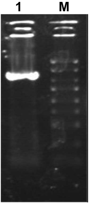
 | Figure 2Agarose gel electrophoresis displays
linear carrier pGC-FU vector existence. 1, 1 kb marker (10, 8, 6,
5, 4, 3.5, 3, 2.5, 2, 1.5, 1 kb, and 750, 500 and 250 bp); 2,
pGC-FU vector; 3, pGC-FU vector Age I Enzyme cutting product. |
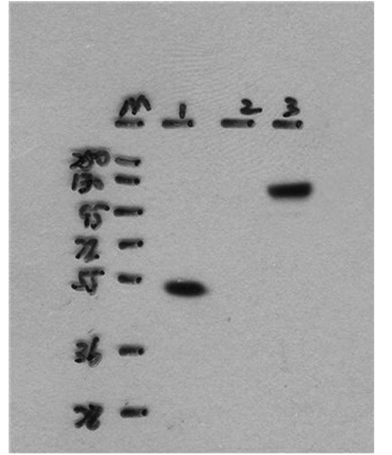
 | Figure 3Identification of positive clones by
PCR (941 bp). 1, Negative control (ddH2O); 2, negative
control (empty vector); 3, positive control (GAPDH); 4, marker (5,
3, 2, 1.5, 1 kb, and 750, 500, 250 and 100 bp); 5–12, PCR products
of recombinant plasmid pGC-FU-TK/CD DNA. |
Virus titer determination
Real-time quantitative reverse transcription
polymerase chain reaction assay (qRT-PCR) has become the benchmark
for detection and quantification of target gene expression level
and have been utilized increasingly in the detection of viral load
(Figs. 5 and 6). Melt curves are shown in Fig. 7, respectively. As a result, both
presented a single peak. The consequences of melting curve were
consistent with a single reaction product for each sample, that
supported the high specificity of primers in another aspect.
Pollution, primer dimer and nonspecific amplification were not
observed.
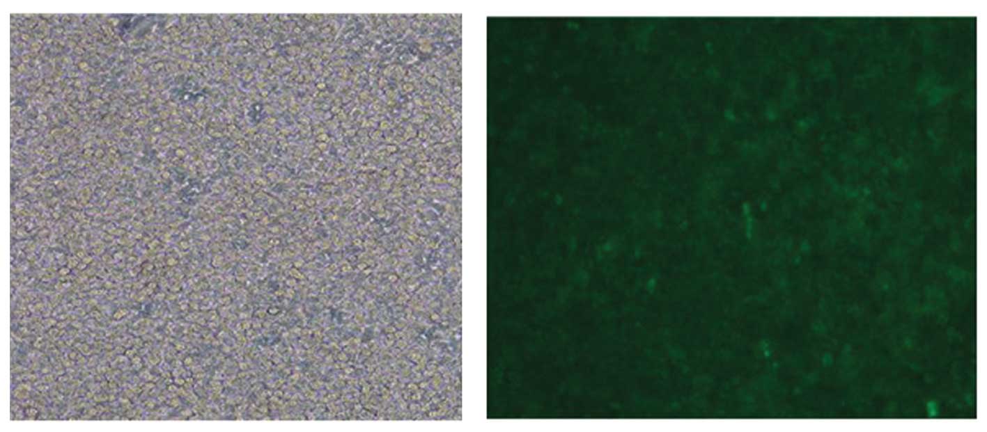
The efficiency of lentivector in
mediating gene transfer to MSCs
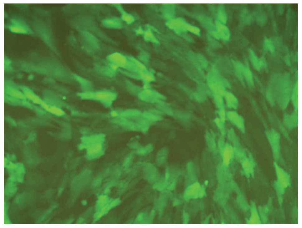
The GFP gene expression was observed by fluorescent
microscopy and, as shown in Fig. 6,
80–90% cells had green fluorescence. The Ct value showed
the expression level of the fusion gene in MSC was constant and
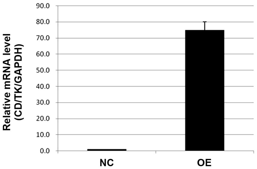
specific. RT-PCR demonstrated the expression level of the fusion
gene in the MSCs/tk+CD+ group was 75-fold
that in the negative control group (P<0.05) (Fig. 7). These results indicate that CD and
HSV-tk could be expressed efficiently and stably in hUCB-MSCs and
lentivector is an ideal tool for effective gene transfer into
hUCB-MSCs.
In vitro cytotoxicity assay of
MSCs/tk+CD+ on SKOV3 cells
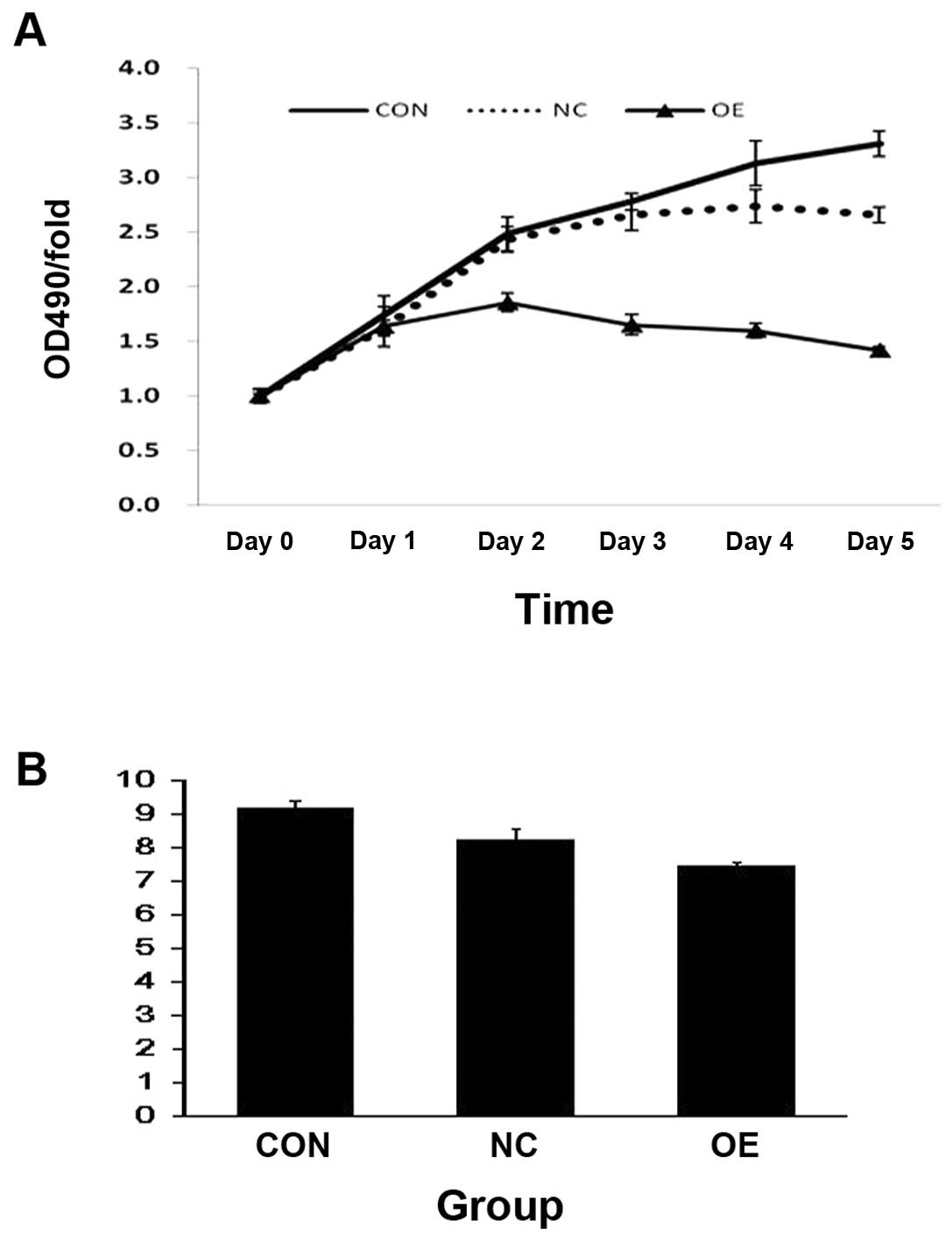
To study the effect of the
MSCs/tk+CD+ on SKOV3 cell survival and
proliferation, we co-cultured them and performed MTT and apoptosis
assays. Our results indicated that SKOV3 cells were not sensitive
to prodrugs in the negative control group and the blank control
group when the concentration of GCV and 5-FC was 20/100 μg/ml.
Notably, MTT assay revealed that cells co-cultured with
MSCs/tk+CD+ showed a significant inhibition
in proliferation (p<0.05) (Fig.
8A). Apoptosis assay also revealed that
MSCs/tk+CD+ induced significantly higher
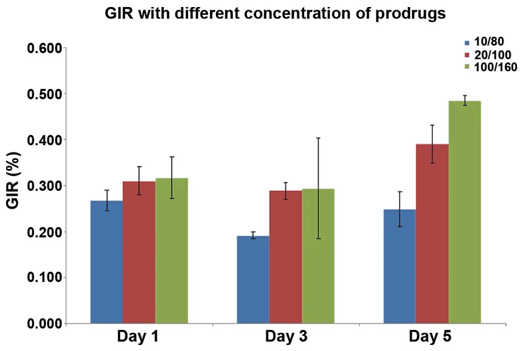
apoptosis in SKOV3 cells compared to controls (Fig. 8B). Furthermore, at day 5, the GIR
increased significantly in the presence of the prodrugs in a
dose-dependent manner (F=27.31, P<0.05) (Fig. 9). These results indicate that cell
viability of SKOV3 cells was significantly inhibited when
co-cultured with MSCs/tk+CD+.
Effect of the prodrugs in combination on
the growth rate of SKOV3 cells
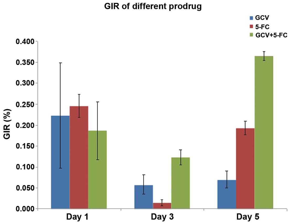
To understand the effect of the prodrugs, in
combination, on SKOV3 growth, we cultured the cells in the presence
of either GCV or 5-FC alone or in combination. We found that there
were no significant differences among the groups on day 1 and 3.
However, there were significant differences between the groups on
day 5. The GIRs were 7.00% in GCV group and 19.31% in the 5-FC
group, respectively (Fig. 10). The
GIR was 36.50%, when the cells were cultured with both drugs
together, and this was statistically significant (F=85.35,
P<0.05). This result suggested that the combined use of 2
prodrugs had stronger lethal effects than either one used
alone.
Discussion
Lentivectors are an attractive tool for gene
transfer. The features of lentiviral vector such as infecting both
dividing and non-dividing cells, holding larger segment size of
exogenous gene, lower immune response, better safety, producing
high titer of virus in host cells over long range, have made it
popular in the current gene therapy research (6,7). They
have the ability to incorporate the target gene into the host
chromosome, which forms the basis for stable gene expression for
long periods of time (8). Thus, the
efficiency of gene interference will not be lost with cell
division. Another characteristic, which could be a distinct
advantage, is that more rapid integration into the genome will
reduce the potential for genetic mosaicism (9). We successfully constructed double
suicide fusion gene overexpressing lentivector. It was determined
by enzyme digestion, DNA sequence and western blotting that the
fusion suicide gene was constructed with full function and
integrity and expression of the double suicide gene.
Suicide gene therapy is an important strategy in
cancer gene therapy. The optimization of vector system and the gene
targeting transfer expression can increase the specific expression
of suicide gene and enhance the effect of killing tumor cells. In
recent years, considerable evidence has accumulated to suggest that
MSCs have the potential to serve as vehicle to selectively deliver
anticancer molecules to tumors, as they possess inherent
tumor-tropic properties and can target tumor sites by migrating to
and infiltrating bulk tumors in vivo (10,11).
MSCs exhibit strong tropism toward tumors that express receptors
for chemokines and growth factors including SDF-1, MCP-1, HGF,
IL-8, NT3, TGF-b and VEGF (5,12–15).
In this study, MSCs were engineered to produce the fusion gene by
using lentiviral vector. Our results confirmed that hUCB-MSCs can
be easily infected by the lentivector at a high efficiency of over
80.0%. The hUCB-MSCs, express the target gene at a high efficiency
and RT-PCR demonstrated that the expression level of HSV-tk/cd
fusion gene in MSCs/tk+CD+ was 75-fold
greater than in negative control (P<0.05). It is in this context
that hUCB-MSCs have appeared to express exogenous gene efficiently,
and could be one of the new target cells for genetic engineering.
Furthermore, MSCs have immunosuppressive properties that may be
useful in targeted gene therapy for sustained tissue specific
engraftment. MSCs at the tumor site could continuously secrete
antitumor factors, thereby protecting against drug induced organ
specific (intestine, bladder and kidney) and hematological
toxicities (16) and reducing the
systemic drug adverse reactions. Lentiviral transduction does not
cause the transduced MSCs to lose their basic stem cell identity
(17). Our results suggest that
hUCB-MSCs could be important in target gene transfer.
In this study, we tested the feasibility and
efficacy of these therapeutic cells to function as cellular
vehicles of double suicide gene in ovarian cancer therapy. We
showed that hUCB-MSCs could be a potential tool for ex vivo
therapy in ovarian cancer. MSCs possess excellent migratory ability
and exerted inhibitory effects on the proliferation of ovarian
tumor cells. Gene-modification of MSCs with therapeutic genes
clearly augmented their antitumor effect. Gene therapy employing
MSCs as a targeting cytoreagent may be a promising approach.
Combined suicide gene therapies have recently
emerged as an attractive alternative therapy for the treatment of
various types of intractable cancer. CD/5-FC and HSV-TK/GCV system
employed in our study, are the two most studied suicide systems. CD
gene codes cytosine deaminase, an enzyme found in a variety of
bacteria and fungi, also able to deaminate the nontoxic prodrug
5-FC to its toxic metabolite 5-fluorouracil (5-FU), which inhibits
RNA and DNA synthesis during the S phase of the cell cycle. HSV-TK
gene codes thymidine kinase, which is able to initially
phosphorylate GCV, then subsequently incorporate into the host DNA
and cause tumor cell death. Both mechanisms are similar, but the
action segments are different (18,19).
Single suicide gene therapy has various limitations, drug
resistance being the most common. The high incidence of tumor gene
mutation is the foundation of drug resistance (20). Since different types of tumor cells
have differences in sensitivity to different enzyme/prodrug
systems, and tumor in vivo often exists in the form of a
variety of clonogenic tumor cells, making it difficult to achieve
radical tumor eradication by application of a single enzyme/prodrug
system. This makes fusion gene therapy a valuable option in cancer
treatment. It has been confirmed that HSV-TK has direct tumoricidal
effects and cytosine deaminase (CD) has attracted considerable
attention by virtue of its stronger bystander effects than other
suicide genes and using them in a complementary fashion can achieve
the best effect in killing tumor cells. Rogulski et al
(21) showed that, when combined,
these prodrug-converting enzymes were functionally superior to
either single agent alone. Therefore, our study adopted the CDglyTK
fusion gene, expecting to use these two systems with different
functions to overcome the tumor cell type dependence in suicide
gene therapy, and decrease the incidence of drug resistance. Our
analysis showed that MSCs/tk+CD+ cells have a
dual mode of action by both inhibiting cell growth and inducing
apoptosis. MTT test showed that the GIR of SKOV3 cells when using
GCV and 5-FC together, was significantly higher than that of either
drug when used alone. These results showed that using double
suicide gene therapy caused significant damage to tumor cells
compared to a single suicide gene.
We also showed that the prodrug concentration is
critical for the effectiveness of this therapy. When the
concentration of the prodrugs increased, there was a significant
inhibition of SKOV3 cells and this inhibition increased in a
dose-dependent manner in vitro.
Previous studies (22,23)
have shown that genetically modified MSCs can inhibit tumor growth
and prolong the lifespan of tumor-bearing animals. Transplantation
of MSCs has been shown to suppress tumor growth by inhibiting Akt
activation in a Kaposi’s sarcoma model and to reduce the size of
brain tumors and increase survival in a glioma model (16,24).
The bystander effect is believed to be mediated by gap junction
intercellular cell-to-cell contacts such as Cx43. The bystander
effect in suicide gene therapy is directly proportional to the
degree of gap junction intercellular communication. It has been
confirmed that MSCs and the human glioma cells can, through the gap
between connection communication, promote bystander effect and kill
tumor cells. On the other hand, due to the bystander effect of
suicide gene, while they destroy cancer cells they also kill the
MSCs, thereby reducing the long-term survival of MSCs (18,22).
The specific mechanism of interaction between MSCs
and tumor cells is not completely understood and this warrants
further studies, including a possible MSC contribution to tumor
stroma and vasculature, MSC-mediated antitumor immune suppression,
and the potential malignant transformation of cultured MSCs
(25–27). MSCs on tumor growth have two-way
regulating functions, which reflects that MSCs have heterogeneity.
Different subsets of the MSCs have differences in gene expression,
which in turn leads to the expression of different types of
proteins and chemokine receptors, which are not consistent.
Nonetheless, we highlight the novel prospects of MSC-based tumor
therapy, which appears to be a promising approach. While this
strategy remains to be tested in-depth, including various
orthotropic or metastatic tumor models, it may greatly improve the
suicide gene treatment results.
Acknowledgements
This study was supported by the Promotive Research
Fund for young and middle-aged scientists of Shandong Province (no.
2006BS03019).
References
|
1
|
Le Blanc K: Immunomodulatory effects of
fetal and adult mesenchymal stem cells. Cytotherapy. 5:485–489.
2003.PubMed/NCBI
|
|
2
|
Koppula PR, Chelluri LK, Polisetti N and
Vemuganti GK: Histocompatibility testing of cultivated human bone
marrow stromal cells - a promising step towards pre-clinical
screening for allogeneic stem cell therapy. Cell Immunol.
259:61–65. 2009. View Article : Google Scholar : PubMed/NCBI
|
|
3
|
Griffin MD, Ritter T and Mahon BP:
Immunological aspects of allogeneic mesenchymal stem cell
therapies. Hum Gene Ther. 21:1641–1655. 2010. View Article : Google Scholar : PubMed/NCBI
|
|
4
|
Kidd S, Caldwell L, Dietrich M, et al:
Mesenchymal stromal cells alone or expressing interferon-beta
suppress pancreatic tumors in vivo, an effect countered by
anti-inflammatory treatment. Cytotherapy. 12:615–625. 2010.
View Article : Google Scholar : PubMed/NCBI
|
|
5
|
Dwyer RM, Potter-Beirne SM, Harrington KA,
et al: Monocyte chemotactic protein-1 secreted in primary breast
tumors stimulates migration of mesenchymal stem cells. Clin Cancer
Res. 13:5020–5027. 2007. View Article : Google Scholar : PubMed/NCBI
|
|
6
|
Barraza RA and Poeschla EM: Human gene
therapy vectors derived from feline lentiviruses. Vet Immunol
Immunopathol. 123:23–31. 2008. View Article : Google Scholar : PubMed/NCBI
|
|
7
|
Barraza RA, Rasmussen CA, Loewen N, et al:
Prolonged transgene expression with lentivectors in the aqueous
humor outflow pathway of nonhuman primates. Hum Gene Ther.
20:191–200. 2009. View Article : Google Scholar : PubMed/NCBI
|
|
8
|
Pfeifer A and Hofmann A: Lentiviral
transgenesis. Methods Mol Biol. 530:391–450. 2009. View Article : Google Scholar : PubMed/NCBI
|
|
9
|
Park F: Lentivectors: are they the future
of animal transgenesis? Physiol Genomics. 31:159–173. 2007.
View Article : Google Scholar
|
|
10
|
Nakamizo A, Marini F, Amano T, et al:
Human bone marrow-derived mesenchymal stem cells in the treatment
of gliomas. Cancer Res. 65:3307–3318. 2005.PubMed/NCBI
|
|
11
|
Xiang J, Tang J, Song C, et al:
Mesenchymal stem cells as a gene therapy carrier for treatment of
fibrosarcoma. Cytotherapy. 11:516–526. 2009. View Article : Google Scholar : PubMed/NCBI
|
|
12
|
Khakoo AY, Pati S, Anderson SA, et al:
Human mesenchymal stem cells exert potent antitumorigenic effects
in a model of Kaposi’s sarcoma. J Exp Med. 203:1235–1247.
2006.PubMed/NCBI
|
|
13
|
Loebinger MR, Eddaoudi A, Davies D and
Janes SM: Mesenchymal stem cell delivery of TRAIL can eliminate
metastatic cancer. Cancer Res. 69:4134–4142. 2009. View Article : Google Scholar : PubMed/NCBI
|
|
14
|
Menon LG, Picinich S, Koneru R, et al:
Differential gene expression associated with migration of
mesenchymal stem cells to conditioned medium from tumor cells or
bone marrow cells. Stem Cells. 25:520–528. 2007. View Article : Google Scholar : PubMed/NCBI
|
|
15
|
Chang DY, Yoo SW, Hong Y, et al: The
growth of brain tumors can be suppressed by multiple
transplantation of mesenchymal stem cells expressing cytosine
deaminase. Int J Cancer. 127:1975–1983. 2010. View Article : Google Scholar : PubMed/NCBI
|
|
16
|
Rustum Y, Cao S, Durrani F and Fakih M:
Se-(methyl)selenocysteine (MSC) potentiates the antitumor activity
of irinotecan against human tumor xenografts and protects against
drug induced toxicity. J Clin Oncol. 22(Suppl 14): 20682004.
|
|
17
|
Wang F, Dennis JE, Awadallah A, et al:
Transcriptional profiling of human mesenchymal stem cells
transduced with reporter genes for imaging. Physiol Genomics.
37:23–34. 2009. View Article : Google Scholar : PubMed/NCBI
|
|
18
|
Kucerova L, Altanerova V, Matuskova M, et
al: Adipose tissue-derived human mesenchymal stem cells mediated
prodrug cancer gene therapy. Cancer Res. 67:6304–6313. 2007.
View Article : Google Scholar : PubMed/NCBI
|
|
19
|
Amano S, Li S, Gu C, et al: Use of
genetically engineered bone marrow-derived mesenchymal stem cells
for glioma gene therapy. Int J Oncol. 35:1265–1270. 2009.PubMed/NCBI
|
|
20
|
Huang SY, Zhang DS, Han JQ, et al:
Radiosensitization and anti-tumour effects of cytosine deaminase
and thymidine kinase fusion suicide gene in human adenoid cystic
carcinoma cells. J Int Med Res. 37:479–490. 2009. View Article : Google Scholar
|
|
21
|
Rogulski KR, Wing MS, Paielli DL, et al:
Double suicide gene therapy augments the antitumor activity of a
replication-competent lytic adenovirus through enhanced
cytotoxicity and radiosensitization. Hum Gene Ther. 11:67–76. 2000.
View Article : Google Scholar
|
|
22
|
Cavarretta IT, Altanerova V, Matuskova M,
et al: Adipose tissue-derived mesenchymal stem cells expressing
prodrug-converting enzyme inhibit human prostate tumor growth. Mol
Ther. 18:223–231. 2010. View Article : Google Scholar
|
|
23
|
Uchibori R, Okada T, Ito T, et al:
Retroviral vector-producing mesenchymal stem cells for targeted
suicide cancer gene therapy. J Gene Med. 11:373–381. 2009.
View Article : Google Scholar : PubMed/NCBI
|
|
24
|
Mori K, Iwata J, Miyazaki M, et al:
Bystander killing effect of tymidine kinase gene-transduced adult
bone marrow stromal cells with ganciclovir on malignant glioma
cells. Neurol Med Chir. 50:545–553. 2010. View Article : Google Scholar : PubMed/NCBI
|
|
25
|
Bagley RG, Weber W, Rouleau C, et al:
Human mesenchymal stem cells from bone marrow express tumor
endothelial and stromal markers. Int J Oncol. 34:619–627. 2009.
View Article : Google Scholar : PubMed/NCBI
|
|
26
|
Mishra PJ, Mishra PJ, Humeniuk R, et al:
Carcinoma-associated fibroblast-like differentiation of human
mesenchymal stem cells. Cancer Res. 68:4331–4339. 2008. View Article : Google Scholar : PubMed/NCBI
|
|
27
|
Yang SH, Park MJ, Yoon IH, et al: Soluble
mediators from mesenchymal stem cells suppress T cell proliferation
by inducing IL-10. Exp Mol Med. 41:315–324. 2009. View Article : Google Scholar : PubMed/NCBI
|