Introduction
Oral squamous cell carcinoma (SCC) is a major
neoplasm of the oral cavity with an increasing rate of incidence
(1–3). The optimal therapy for early oral SCC
is surgery, but the overall survival rate has exhibited only a
slight change (1–3). Therefore, more effective therapies for
oral SCC are needed.
Fatty acid binding proteins (FABPs) are a family of
small and highly conserved lipid chaperone molecules that bind
long-chain fatty acids and other hydrophobic ligands. Their
functions are wide ranging (4–6). Among
them, fatty acid binding protein 4 (FABP4, also known as aP2) is
highly expressed in adipocytes, macrophages and dendritic cells
(5,7). As a result of its distribution, FABP4
is the most extensively researched FABP in endocrinology and
metabolomics. FABP4 affects metabolic syndrome progression;
FABP4-deficient mice were found to have reduced
hyperinsulinemia and insulin resistance in obesity (7,8) and
showed protection from atherosclerosis (9).
However, little is known concerning the role of
FABP4 in cancer, including oral SCC. Recently, Nieman et al
(10) reported that adipocytes
promote ovarian cancer metastasis and tumor cell growth by
providing energy mediated by FABP4. Therefore, increased FABP4
expression may affect the growth of various tumor types. Our
research group also reported that molecules controlled by
peroxisome proliferator-activated receptor γ (PPARγ) play key roles
in SCC growth (11–15). As FABP4 is known to mediate
transcription with PPARγ (4,16), we
hypothesized that FABP4 may regulate SCC growth. Therefore, in the
present study, we investigated FABP4 expression and its effects on
SCC of the tongue.
Materials and methods
Tissue samples
All clinical studies were approved by the Ethics
Committee of Osaka University Dental Hospital, Osaka. Twenty-seven
SCC specimens from resected tongue tissue were obtained at the
Osaka University Dental Hospital during 1986–2008 after patient
informed consent (Table I).
Patients received no preoperative therapy, including chemotherapy
and irradiation therapy. The age range of the patients was 30–92
years (61.6±16.4 years, mean age ± SD); 17 patients were men and 10
were women.
 | Table ICharacteristics of the patients with
squamous cell carcinoma of the tongue, their histological diagnosis
and expression of FABP4 in the tissue specimens. |
Table I
Characteristics of the patients with
squamous cell carcinoma of the tongue, their histological diagnosis
and expression of FABP4 in the tissue specimens.
| | | | FABP4 expression |
|---|
| | | |
|
|---|
| Case no. | Age (years) | Gender | Differentiation | Tumor area | Non-tumor area |
|---|
| 1 | 34 | Male | Well-differentiated
SCC | 1 | 0 |
| 2 | 46 | Female | Well-differentiated
SCC | 2 | 0 |
| 3 | 57 | Male | Well-differentiated
SCC | 1 | 1 |
| 4 | 64 | Male | Well-differentiated
SCC | 0 | 0 |
| 5 | 69 | Male | Well-differentiated
SCC | 0 | 0 |
| 6 | 71 | Female | Well-differentiated
SCC | 1 | 0 |
| 7 | 71 | Male | Well-differentiated
SCC | 1 | 0 |
| 8 | 72 | Male | Well-differentiated
SCC | 2 | 0 |
| 9 | 78 | Female | Well-differentiated
SCC | 1 | 0 |
| 10 | 79 | Male | Well-differentiated
SCC | 1 | 1 |
| 11 | 47 | Male | Moderately
differentiated SCC | 1 | 0 |
| 12 | 61 | Male | Moderately
differentiated SCC | 1 | 0 |
| 13 | 61 | Female | Moderately
differentiated SCC | 1 | 0 |
| 14 | 72 | Male | Moderately
differentiated SCC | 1 | 0 |
| 15 | 77 | Male | Moderately
differentiated SCC | 1 | 2 |
| 16 | 83 | Female | Moderately
differentiated SCC | 3 | 2 |
| 17 | 92 | Female | Moderately
differentiated SCC | 1 | 0 |
| 18 | 30 | Male | Poorly
differentiated SCC | 2 | 1 |
| 19 | 33 | Male | Poorly
differentiated SCC | 0 | 0 |
| 20 | 38 | Male | Poorly
differentiated SCC | 0 | 0 |
| 21 | 51 | Female | Poorly
differentiated SCC | 0 | 0 |
| 22 | 52 | Male | Poorly
differentiated SCC | 2 | 0 |
| 23 | 55 | Female | Poorly
differentiated SCC | 2 | 0 |
| 24 | 65 | Male | Poorly
differentiated SCC | 1 | 3 |
| 25 | 67 | Male | Poorly
differentiated SCC | 3 | 2 |
| 26 | 41 | Female | Differentiation
unknown SCC | 0 | 0 |
| 27 | 68 | Female | Differentiation
unknown SCC | 2 | 0 |
Antibodies
The anti-FABP4 polyclonal antibody was obtained from
Bioss Inc. (Woburn, MA, USA). Antibodies against p44/42MAPK and the
phosphorylated p44/42MAPK antibody were from Cell Signaling
Technology (Beverly, MA, USA).
Immunohistochemical staining and
evaluation of FABP4 expression
FABP4 expression in tissues was detected by an
anti-FABP4 antibody using standard immunohistochemical techniques
(12–15). Formalin-fixed and paraffin-embedded
continuous sections were selected and sliced into 5-μm sections.
Briefly, incubation with an anti-FABP4 polyclonal antibody was
performed at 4°C for 16 h; sections were then washed. After
applying the secondary antibody, the Vectastain ABC kit (Vector
Laboratories, Burlingame, CA, USA) was used with a
3,3′-diaminobenzidine substrate kit, according to the
manufacturer’s instructions. The staining endpoint was determined
when the standard tissue sections were constantly stained at the
intensity as previously described (12,17).
The intensity of the immunohistochemical staining
with the anti-FABP4 antibody was evaluated by scoring according to
four groups: 0, <10%; 1, 10–20%; 2, >20–50%; and 3, >50%
of the cells exhibiting cytoplasmic staining (12,17).
To confirm the reproducibility, the anti-FABP4 immunohistochemical
staining was re-evaluated by a pathologist who was unaware of the
original assessment. Non-tumor areas were selected as comparatively
normal areas separated from the tumor areas by an appropriate
distance and confirmed by the pathologist (14,15).
Cell culture and cell growth assay
We used a human oral SCC cell line (SAS) that was
established from tongue SCC (13).
Cells were maintained in DMEM containing 10% fetal bovine serum
(FBS) at 37°C under 0.5% CO2. For the cell growth
experiment, cells were trypsinized and replated onto culture dishes
(11–15,17,18).
SCC cells were counted using a Countess Automated Cell Counter
(Invitrogen, Eugene, OR, USA). Inhibition of cell growth was
compared with the vehicle-treated controls.
RNA interference approach
The SAS cells were trypsinized and resuspended in
DMEM without FBS, and then separated placing ~2×10⁵
cells in each dish. The FABP4-specific siRNA (Stealth siRNA)
was purchased from Invitrogen Japan (Tokyo, Japan). We purchased
three sequences and performed preparatory experiments to determine
the most effective sequence. The sequences of the selected
FABP4-siRNA were: sense, 5′-CAC CAUUAAAUCUGAAAGUACCUUU-3′
and antisense, 5′-AAAGGUACUUUCAGAUUUAAUGGUG-3′. For transfection,
FABP4-siRNA or a negative control (Stealth RNAi negative
control duplex; Invitrogen Japan) solution was added to the DMEM
containing Lipofectamine RNAiMax (Invitrogen Japan) and incubated
for 20 min at room temperature to create the transfection mixture.
The transfection mixture was then added to the cells at the
indicated final siRNA concentrations. Following 24 h of
transfection, the medium was replaced by DMEM containing 10% FBS,
at which time viable cells were counted using a Countess Automated
Cell Counter. Cell growth was expressed as a percentage of the
vehicle-treated control growth.
Western blot analysis
Adherent or suspended cells were washed in PBS, and
the cell extracts were prepared by lysing the cells in lysis
buffer. Proteins were separated by electrophoresis using 10%
SDS-PAGE and transferred to a nitrocellulose membrane (Millipore,
Bedford, MA, USA). Detection of proteins was performed with each
polyclonal antibody and visualized using an ECL detection kit
(Amersham, London, UK) following the manufacturer’s recommended
procedure.
Statistical analysis
Results are expressed as the means ± SEM or ± SD.
Statistical comparisons were carried out using the Student’s t-test
or the Scheffé’s method after analysis of variance. P<0.05 was
considered to indicate a statistically significant result.
Results
Tongue SCC tissues express FABP4
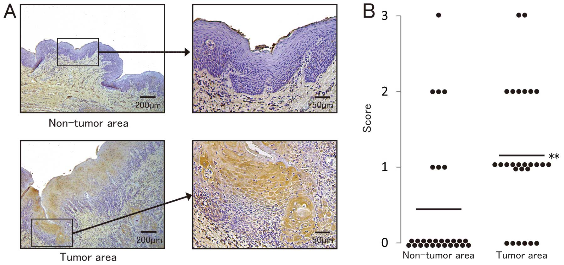
We stained tongue SCC tissues using the
FABP4-specific antibody. Within single tumor specimens, the
non-tumor areas were unstained (Fig.
1A, upper panel), whereas tumor areas showed positive FABP4
staining (Fig. 1A, lower panel).
According to the scoring as described above, FABP4 expression
between the non-tumor and the tumor area differed significantly
(Fig. 1B), and FABP4 was expressed
in the tumor areas, but not in the normal tissues.
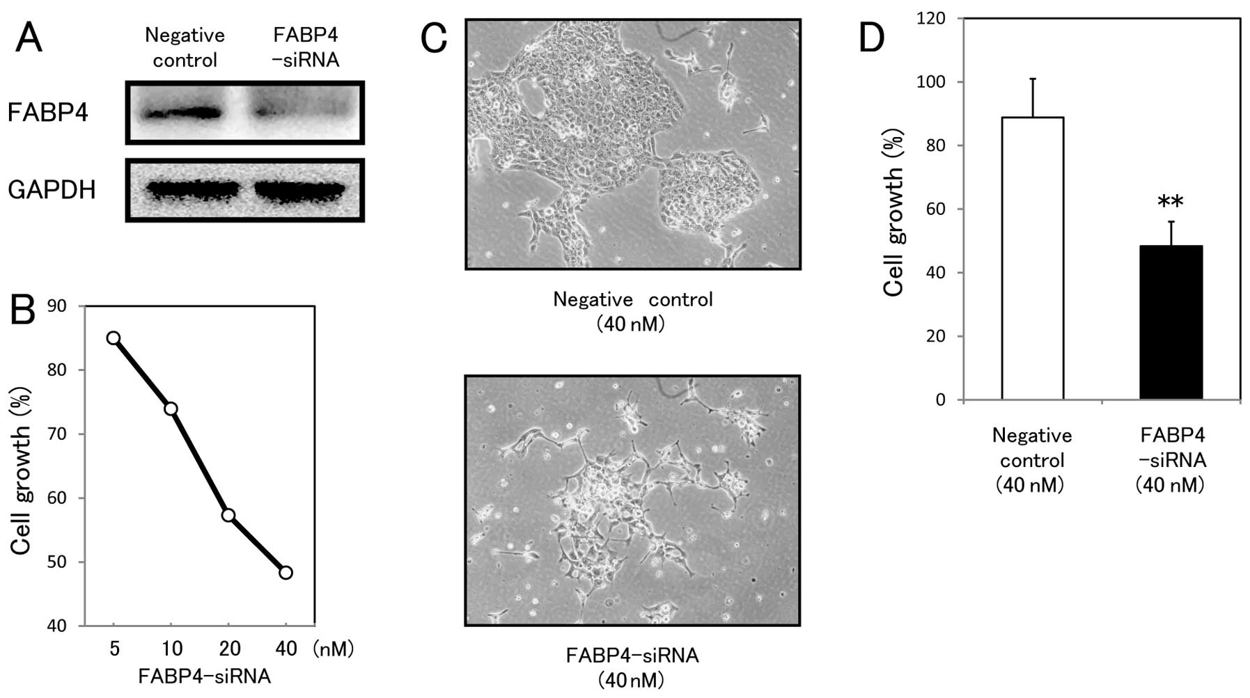
FABP4-specific siRNA suppresses the
growth of tongue SCC
Treatment with FABP4-siRNA markedly decreased
FABP4 protein levels in the SAS cells (Fig. 2A), and suppressed SAS cell growth in
a concentration-dependent manner (Fig.
2B). Inhibition of SCC growth was also visibly altered
(Fig. 2C), and significantly
differed between the SAS controls and the FABP4-knockdown
SAS cells (Fig. 2D).
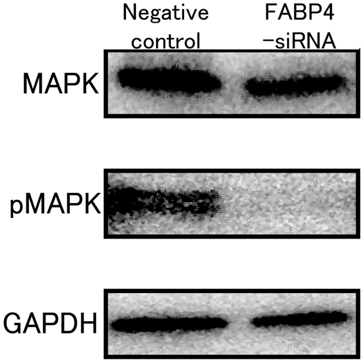
FABP4-specific siRNA inhibits expression
and phosphorylation of mitogen-activated protein kinase (MAPK)
To investigate the mechanisms involved in the growth
inhibition induced by suppression of FABP4, we analyzed a type of
MAPK, serine/threonine protein kinases. Since they affect cell
proliferation, survival and differentiation, aberrant MAPK cascades
contribute to cancer and other diseases (19–21).
Therefore, we studied the effects of FABP4 knockdown on MAPK
expression and phosphorylation. Western blot analysis showed
decreased phosphorylated MAPK (pMAPK; Fig. 3, middle panel). Notably, MAPK
expression itself was also regulated by FABP4 knockdown
(Fig. 3, upper panel).
Discussion
FABP4 expression has been reported in various types
of tumors such as ovarian and bladder cancers (10,22),
and FABP5 (E-FAPB) has been found in oral SCC (23,24).
However, the expression and exact role of FABP4 in oral SCC have
not been widely investigated
In the present study, using an immunohistochemical
approach, we consistently found significantly higher expression of
FABP4 protein in the tumor area of tongue SCC than in the non-tumor
area in the same tissue samples. Therefore, FABP4 expression in
tumors may affect SCC cell growth. In fact, we showed that
suppression of FABP4 protein by the FABP4-specific siRNA
clearly inhibited the growth of SCC cell lines. These results
clearly indicate the important role of FABP4 in SCC growth.
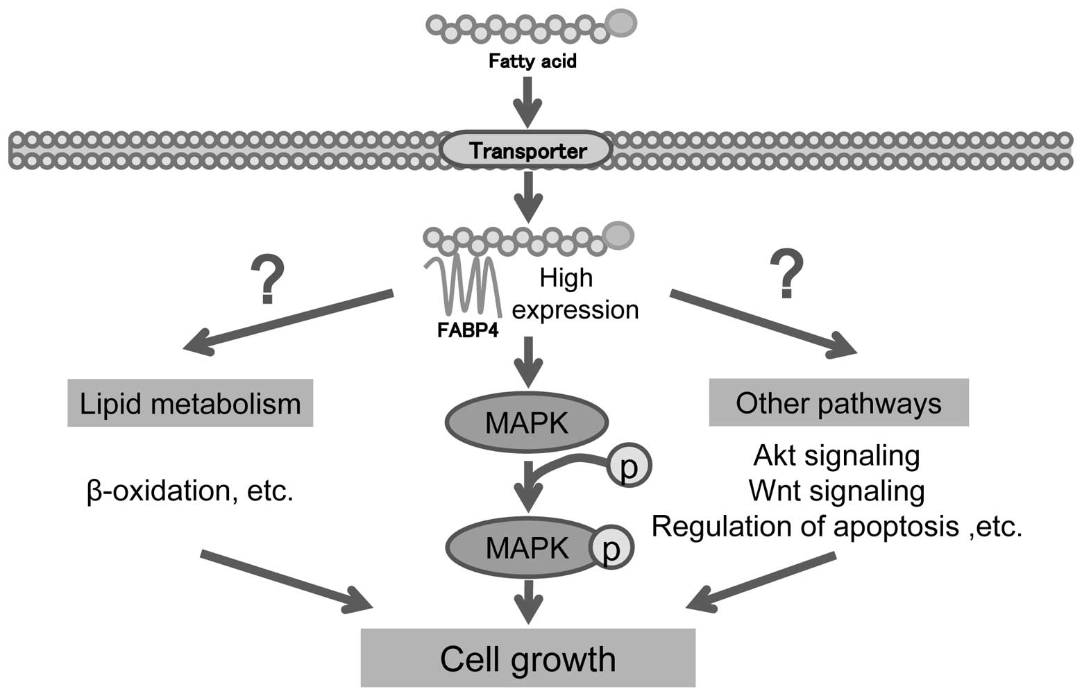
FABPs actively facilitate the transport of lipids to
specific cellular compartments, including lipid droplets for
storage; endoplasmic reticulum for signaling, trafficking and
membrane synthesis; mitochondria or peroxisome for oxidation;
cytosolic or other enzymes for activity regulation; nuclei for
lipid-mediated transcriptional regulation; or even outside the cell
for autocrine or paracrine signaling. Among the FABPs, FABP4 is
highly expressed in adipocytes, macrophages and dendritic cells and
affects these cells in various manners (5). In cancer cells, FABP4 transports
energy by carrying fatty acids, encouraging metastasis and tumor
cell growth (10). Yet, FABP4
performs other roles in tumor growth, through its various
functions. Therefore, we studied the role of FABP4 in the MAPK
pathway and the mechanisms of growth inhibition induced by FABP4
suppression.
As the MAPK pathway helps to mediate cell
proliferation and cancer growth, it has been widely studied as a
potential target for cancer therapy (19–21).
In fact, our present research showed decreased MAPK expression and
phosphorylation 12 h following treatment with FABP4-specific
siRNA, which indicates that FABP4 affects cell growth through the
MAPK pathway. Notably, expression of MAPK itself was also
suppressed by FABP4 knockdown; FABP4 may affect
transcription of MAPK, which implies a complex role for FABP4 in
tumor growth. Inhibition of the MAPK pathway may be one of the
several mechanisms through which FABP4 mediates tumor growth
(Fig. 4). Further investigation is
warranted.
In the present study, FABP4 expression in the tumor
tissues was not correlated with age, gender, histological tumor
differentiation or survival rate of the cases. Of our 27 cases, 12
had neck lymph node metastasis, all 12 of whom showed FABP4
expression in lymph nodes, similar to that in the primary tumors
(data not shown). Thus, FABP4 expression may affect metastasis to
neck lymph nodes; this role merits further investigation.
In summary, we demonstrated FABP4 expression in
human tongue SCC tissues and cultured SCC cells. Our results
suggest an important role for FABP4 in SCC growth and indicate that
FABP4 is a potential target for the therapy of oral SCC.
Acknowledgements
The present study was supported in part by grants
(T245928310 to Y.T.) from the Japanese Society for the Promotion of
Science.
References
|
1
|
Prince S and Bailey BM: Squamous carcinoma
of the tongue: review. Br J Oral Maxillofac Surg. 37:164–174. 1999.
View Article : Google Scholar : PubMed/NCBI
|
|
2
|
Okura M, Hiranuma T, Adachi T, et al:
Induction chemotherapy is associated with an increase in the
incidence of locoregional recurrence in patients with carcinoma of
the oral cavity: results from a single institution. Cancer.
82:804–815. 1998. View Article : Google Scholar : PubMed/NCBI
|
|
3
|
Goepfert H: Squamous cell carcinoma of the
head and neck: past progress and future promise. CA Cancer J Clin.
48:195–198. 1998. View Article : Google Scholar : PubMed/NCBI
|
|
4
|
Schroeder F, Petrescu AD, Huang H, et al:
Role of fatty acid binding proteins and long chain fatty acids in
modulating nuclear receptors and gene transcription. Lipids.
43:1–17. 2008. View Article : Google Scholar : PubMed/NCBI
|
|
5
|
Furuhashi M and Hotamisligil GS: Fatty
acid-binding proteins: role in metabolic diseases and potential as
drug targets. Nat Rev Drug Discov. 7:489–503. 2008. View Article : Google Scholar : PubMed/NCBI
|
|
6
|
Storch J and Thumser AE: The fatty acid
transport function of fatty acid-binding proteins. Biochim Biophys
Acta. 1486:28–44. 2000. View Article : Google Scholar : PubMed/NCBI
|
|
7
|
Hotamisligil GS, Johnson RS, Distel RJ,
Ellis R, Papaioannou VE and Spiegelman BM: Uncoupling of obesity
from insulin resistance through a targeted mutation in aP2, the
adipocyte fatty acid binding protein. Science. 274:1377–1379. 1996.
View Article : Google Scholar : PubMed/NCBI
|
|
8
|
Uysal KT, Scheja L, Wiesbrock SM,
Bonner-Weir S and Hotamisligil GS: Improved glucose and lipid
metabolism in genetically obese mice lacking aP2. Endocrinology.
141:3388–3396. 2000. View Article : Google Scholar : PubMed/NCBI
|
|
9
|
Makowski L, Boord JB, Maeda K, et al: Lack
of macrophage fatty-acid-binding protein aP2 protects mice
deficient in apolipoprotein E against atherosclerosis. Nat Med.
7:699–705. 2001. View
Article : Google Scholar : PubMed/NCBI
|
|
10
|
Nieman KM, Kenny HA, Penicka CV, et al:
Adipocytes promote ovarian cancer metastasis and provide energy for
rapid tumor growth. Nat Med. 17:1498–1503. 2011. View Article : Google Scholar : PubMed/NCBI
|
|
11
|
Masuda T, Wada K, Nakajima A, et al:
Critical role of peroxisome proliferator-activated receptor γ on
anoikis and invasion of squamous cell carcinoma. Clin Cancer Res.
11:4012–4021. 2005.
|
|
12
|
Nagata M, Wada K, Nakajima A, et al: Role
of myeloid cell leukemia-1 in cell growth of squamous cell
carcinoma. J Pharmacol Sci. 110:344–353. 2009. View Article : Google Scholar : PubMed/NCBI
|
|
13
|
Kusayama M, Wada K, Nagata M, et al:
Critical role of aquaporin 3 on growth of human esophageal and oral
squamous cell carcinoma. Cancer Sci. 102:1128–1136. 2011.
View Article : Google Scholar : PubMed/NCBI
|
|
14
|
Ishimoto S, Wada K, Tanaka N, et al: Role
of endothelin receptor signalling in squamous cell carcinoma. Int J
Oncol. 40:1011–1019. 2012.PubMed/NCBI
|
|
15
|
Ishimoto S, Wada K, Usami Y, et al:
Differential expression of aquaporin 5 and aquaporin 3 in squamous
cell carcinoma and adenoid cystic carcinoma. Int J Oncol. 41:67–75.
2012.PubMed/NCBI
|
|
16
|
Tan NS, Shaw NS, Vinckenbosch N, et al:
Selective cooperation between fatty acid binding proteins and
peroxisome proliferator-activated receptors in regulating
transcription. Mol Cell Biol. 22:5114–5127. 2002. View Article : Google Scholar
|
|
17
|
Ishida H, Wada K, Masuda T, et al:
Critical role of estrogen receptor on anoikis and invasion of
squamous cell carcinoma. Cancer Sci. 98:636–643. 2007. View Article : Google Scholar : PubMed/NCBI
|
|
18
|
Takahashi H, Fujita K, Fujisawa T, et al:
Inhibition of peroxisome proliferator-activated receptor gamma
activity in esophageal carcinoma cells results in a drastic
decrease of invasive properties. Cancer Sci. 97:854–860. 2006.
View Article : Google Scholar
|
|
19
|
Roux PP and Blenis J: ERK and p38
MAPK-activated protein kinases: a family of protein kinases with
diverse biological functions. Microbiol Mol Biol Rev. 68:320–344.
2004. View Article : Google Scholar : PubMed/NCBI
|
|
20
|
Roberts PJ and Der CJ: Targeting the
Raf-MEK-ERK mitogen-activated protein kinase cascade for the
treatment of cancer. Oncogene. 26:3291–3310. 2007. View Article : Google Scholar : PubMed/NCBI
|
|
21
|
Santarpia L, Lippman SM and El-Naggar AK:
Targeting the MAPK-RAS-RAF signaling pathway in cancer therapy.
Expert Opin Ther Targets. 16:103–119. 2012. View Article : Google Scholar : PubMed/NCBI
|
|
22
|
Boiteux G, Lascombe I, Roche E, et al:
A-FABP, a candidate progression marker of human transitional cell
carcinoma of the bladder, is differentially regulated by PPAR in
urothelial cancer cells. Int J Cancer. 124:1820–1828. 2009.
View Article : Google Scholar : PubMed/NCBI
|
|
23
|
Uma RS, Naresh KN, D’Cruz AK, Mulherkar R
and Borges AM: Metastasis of squamous cell carcinoma of the oral
tongue is associated with down-regulation of epidermal fatty acid
binding protein (E-FABP). Oral Oncol. 43:27–32. 2007. View Article : Google Scholar : PubMed/NCBI
|
|
24
|
Fang LY, Wong TY, Chiang WF and Chen YL:
Fatty-acid-binding protein 5 promotes cell proliferation and
invasion in oral squamous cell carcinoma. J Oral Pathol Med.
39:342–348. 2010. View Article : Google Scholar : PubMed/NCBI
|