Introduction
Hepatocellular carcinoma (HCC) is the second leading
cause of cancer-related mortality in men and the sixth leading
cause of cancer-related mortality in women and is the most
frequently diagnosed cancer worldwide (1). In addition, HCC is one of the
deadliest primary cancers, and its 5-year survival rate is 10% or
less (2). Limited treatment options
and late diagnosis can explain the low survival rate (3). The discovery of more satisfactory
molecular biomarkers could significantly improve the survival rate
of HCC as early diagnosis and prediction help to assess tumor
metastasis, tumor advancement and evaluate therapeutic strategies
(4).
HCC carcinogenesis involves aberrant changes in
multiple molecular pathways and genetic alterations, which
consequently result in malignant transformation and progression of
HCC (5). Each step of the signaling
pathways involved in the pathogenesis of HCC could be a target by
which to inhibit HCC progression. Several novel molecularly
targeted drugs are currently under preclinical and clinical trials
for the treatment of HCC. Yet, to date, only sorafenib has been
found to prolong the survival of patients with advanced HCC
(6). The effects of the majority of
targeted drug treatments remain unsatisfactory. In conclusion, it
is of great clinical value to further identify effective biomarkers
of HCC not only for diagnosis and prognosis but also for use as
novel therapeutic targets.
Suppressor with morphogenetic effect on genitalia
(SMG-1) is the most recently characterized member of the family of
phosphatidylinositol 3-kinase-related protein kinases (PIKKs). It
plays a central role in cell growth and stress response pathways,
and is conserved across species. SMG-1 is involved in a mechanism
responsible for the degradation of premature stop codon containing
mRNA, also called NMD (nonsense mediated mRNA decay) in C.
elegans and in mammalian cells (7–9). In
addition to NMD, SMG-1 has been shown to play other cellular roles,
such as regulation of the G1/S checkpoint during oxidative stress
(10), protection against
TNFα-dependent apoptosis (11),
response to hypoxia, IR and UV radiation (12,13),
as well as lifespan regulation in Caenorhabditis elegans
(14), and a novel regulator of
regeneration and animal growth with an antagonistic role to mTOR
signaling in planarians (15).
Furthermore, recent studies have demonstrated that
SMG-1 is also involved in tumorigenesis. Gubanova et al
(16) reported that SMG-1
suppresses tumor growth via regulation of both the p53 and Cdc25A
signaling pathways. Similar findings in another study using a
SMG-1-knockout mouse model which predisposed to a whole range of
tumor formation and inflammation suggested that SMG-1 is a tumor
suppressor (17).
It is well known that deregulation of cell cycle
control plays an important role in the multistep process of
hepatocarcinogenesis. The signaling pathways involved in regulation
of the cell cycle from the G1 phase to the S phase are frequent
targets of carcinogenic events. A key regulator of G1/S arrest is
SMG-1. SMG-1 has been shown to control G1/S cell cycle arrest
through both the p53-dependent and the p53-independent pathways
(16). Yet, no report exists
regarding the mRNA and protein expression levels of SMG-1 in human
HCC, and whether SMG-1 is an oncogene or a tumor-suppressor gene in
HCC has not yet been demonstrated.
To clarify the role of SMG-1 in HCC, we investigated
the mRNA and protein expression of SMG-1 in different pathological
subtypes of primary HCC using real-time PCR assay and
immunohistochemistry. The relationship between expression of SMG-1
and clinicopathological parameters, and patient survival in primary
HCC was analysed. We showed abnormal expression of SMG-1 in HCC
tissues compared to the corresponding distant normal tissues. SMG-1
may be a key biomarker of HCC tumors and patient prognosis. In
conclusion, low expression of SMG-1 is closely related to
progression of HCC, and may be regarded as a predictor of poor
prognosis of HCC.
Materials and methods
Liver cancer tissue specimens
Formalin-fixed, paraffin-embedded primary HCC
specimens obtained from 157 patients, who received surgical
resection without prior radiotherapy or chemotherapy between August
2007 and December 2009 were randomly selected from the archives of
The First Affiliated Hospital of Medical College of Xi’an Jiaotong
University. The fresh HCC tissue specimens, together with their
paired adjacent non-cancerous tissues (5-cm from the cancer region)
from each patient, were obtained during the surgical operation,
snap frozen in liquid nitrogen and subsequently stored at −80°C
until use for experimental purposes. For the use of these clinical
materials for research purposes, prior patient written informed
consent and approval from the Institutional Research Ethics
Committee of the Medical College of Xi’an Jiaotong University and
its First Affiliated Hospital were obtained. Histological diagnosis
was performed for all of the cases by three independent experienced
pathologists. The cohort of patients with HCC included 137 (87.3%)
men and 20 (12.7%) women. The mean age was 48.7 years, with ages
ranging from 19 to 79 years, and the patient clinicopathological
data are summarized in Table I.
Tumor stages were assigned according to the 2002 American Joint
Committee on Cancer/International Union against Cancer Tumor/Lymph
Node Metastasis/Distal Metastasis (TNM) classification system.
Tumor cellular differentiation was assessed using Edmondson’s
classification. Hepatitis B virus (HBV) infection was diagnosed
when HBV surface antigen (HBsAg) was detected by ELISA in the
serum. The follow-up for all of the cases was completed in August
of 2013. The cases lost to follow-up and those ending in death from
causes other than HCC were regarded as censored data during the
survival analysis.
 | Table ICorrelation between the
clinicopathological variables and SMG-1 expression in the HCC cases
(n=157). |
Table I
Correlation between the
clinicopathological variables and SMG-1 expression in the HCC cases
(n=157).
| | SMG-1 protein | | |
|---|
| |
| | |
|---|
| Variable | All cases, n
(%) | Low expression, n
(%) | High expression, n
(%) | χ2 |
P-valuea |
|---|
| Age
(years)b | | | | 0.149 | 0.700 |
| <48.7 | 65 (41.4) | 45 (69.2) | 20 (30.8) | | |
| ≥48.7 | 92 (58.6) | 61 (66.3) | 31 (33.7) | | |
| Gender | | | | 0.001 | 0.976 |
| Female | 31 (19.7) | 21 (67.7) | 10 (32.3) | | |
| Male | 126 (80.3) | 85 (67.5) | 41 (32.5) | | |
| HBsAg | | | | 0.064 | 0.800 |
| Positive | 137 (87.3) | 92 (67.2) | 45 (32.8) | | |
| Negative | 20 (12.7) | 14 (70.0) | 6 (30.0) | | |
| AFP (ng/ml) | | | | 12.073 | 0.001 |
| <400 | 99 (63.0) | 57 (57.6) | 42 (42.) | | |
| ≥400 | 58 (37.0) | 49 (84.5) | 9 (15.5) | | |
| Cirrhosis | | | | 0.005 | 0.946 |
| Yes | 101 (64.3) | 68 (67.3) | 33 (32.7) | | |
| No | 56 (35.9) | 38 (67.9) | 18 (32.1) | | |
| Tumor size
(cm) | | | | 0.432 | 0.511 |
| <5 | 68 (43.3) | 44 (64.7) | 24 (35.3) | | |
| ≥5 | 89 (56.7) | 62 (69.7) | 27 (30.3) | | |
| Tumor
multiplicity | | | | 1.239 | 0.266 |
| Single | 93 (59.2) | 66 (71.0) | 27 (29.0) | | |
| Multiple | 64 (40.7) | 40 (62.5) | 24 (37.5) | | |
|
Differentiation | | | | 6.777 | 0.009 |
| Well-moderate | 72 (45.9) | 41 (56.9) | 31 (43.1) | | |
|
Poor-undifferentiated | 85 (54.1) | 65 (76.5) | 20 (23.5) | | |
| Stage | | | | 35.266 | 0.000 |
| I–II | 67 (42.7) | 28 (41.8) | 39 (58.2) | | |
| III–IV | 90 (57.3) | 78 (86.7) | 12 (13.3) | | |
| Hepatic vein
invasion | | | | 1.478 | 0.224 |
| Yes | 47 (30.0) | 35 (74.5) | 12 (25.5) | | |
| No | 110 (70.0) | 71 (64.5) | 39 (35.5) | | |
Immunohistochemistry
Semi-quantitative immunohistochemistry (IHC) was
used to determine the SMG-1 protein levels. Briefly, after
deparaffinization and re-hydration of tissue sections, the sections
were first subjected to antigen retrieval in a pressure cooker in
citric buffer for 10 min. The sections were then incubated with 3%
H2O2 for 10 min at room temperature to block
potential endogenous peroxidase activity and then incubated with
20% normal serum and further with a primary antibody against SMG-1
(sc-135563, 1:100; Santa Cruz Biotechnology, Inc., Santa Cruz, CA,
USA) at 4°C overnight. The negative control sections were incubated
with phosphate-buffered saline (PBS) only to replace the primary
antibody. The next day the sections were washed thrice with PBS and
then incubated with a biotinylated secondary antibody for 30 min at
room temperature. Diaminobenzidine substrate was used to reveal
immunoreactive products in the sections. After counterstaining with
hematoxylin to reveal nuclei, the sections were mounted on slides
and coverslipped. To assess immunopositive cells, 3 pathologists
reviewed the immunostained sections under a light microscope and
scored the sections in 10 random ×20 power fields. The staining
intensity was graded as ‘0’ (negative staining), ‘1’ (weak
staining), ‘2’ (moderate staining) and ‘3’ (strong staining). The
percentage of positively stained cells was scored as: ‘0’ (0%), ‘1’
(1–25%), ‘2’ (26–50%), ‘3’ (51–75%) and ‘4’ (76–100%). These 2
scores were added together. Based on the sum of the scores, each
tissue sample was categorized into 4 groups: 0, ≤5% cells were
stained; 1–3, weak expression; 4–5, moderate expression; and 6–7,
strong expression. Finally, we compared statistically the numbers
of cells with low-to-weak expression with those with
moderate-to-strong expression. Furthermore, 10 representative
staining fields of each section were analyzed to verify the mean
optical density (MOD), which represented the strengths of staining
signals as measured per positive pixels. The MOD data were
statistically analyzed using t-test to compare the average MOD
difference between different groups of tissues, and P<0.05 was
considered to indicate a statistically significant result.
Real-time reverse
transcriptase-polymerase chain reaction (qRT-PCR)
Total mRNA was extracted from tissue samples using
TRIzol reagent (Invitrogen-Life Technologies, Carlsbad, CA, USA)
and reverse-transcribed into cDNA using an RT-PCR kit (Takara,
Dalian, China). Amplification of these cDNA samples was performed
using SYBR Premix Ex Taq™II (Takara) in an iQ5 Multicolor real-time
PCR detection system (Bio-Rad Laboratories, Hercules, CA, USA). The
primers for SMG-1 and glyceraldehyde-3-phosphate dehydrogenase
(GAPDH; internal control) were designed using Primer Express v2.0
software (Applied Biosystems) and are provided as follows: SMG-1
(forward: 5′-GGTGGCTCGATGTTAC CCTC-3′ and reverse,
5′-CTGCGTGGCGAAGGTTTC-3′); GAPDH (forward,
5′-ACCACAGTCCATGCCATCAC-3′ and reverse,
5′-TCCACCACCCTGTTGCTGTA-3′). Expression levels of SMG-1 were
evaluated using a relative quantification approach
(2−ΔΔCt method) against GAPDH levels.
Statistical analysis
Comparisons between groups for statistical
significance were performed with a two-tailed paired Student’s
t-test. The Chi-square (χ2) test was performed to
analyze the correlation between SMG-1 expression and
clinicopathological parameters. The Kaplan-Meier method (the
log-rank test) was used for survival analysis and univariate
analysis. The Cox proportional hazards regression model was used
for the multivariate analysis of survival duration. Statistical
Package for Social Science (SPSS) version 17.0 was used. All
reported P-values were two-sided. P-values were considered
statistically significant at the P<0.05 level, and a value of
P<0.01 was considered to indicate a highly significant
difference.
Results
Expression of SMG-1 mRNA and protein in
hepatocellular carcinoma tissue samples by real-time PCR and
immunohistochemistry
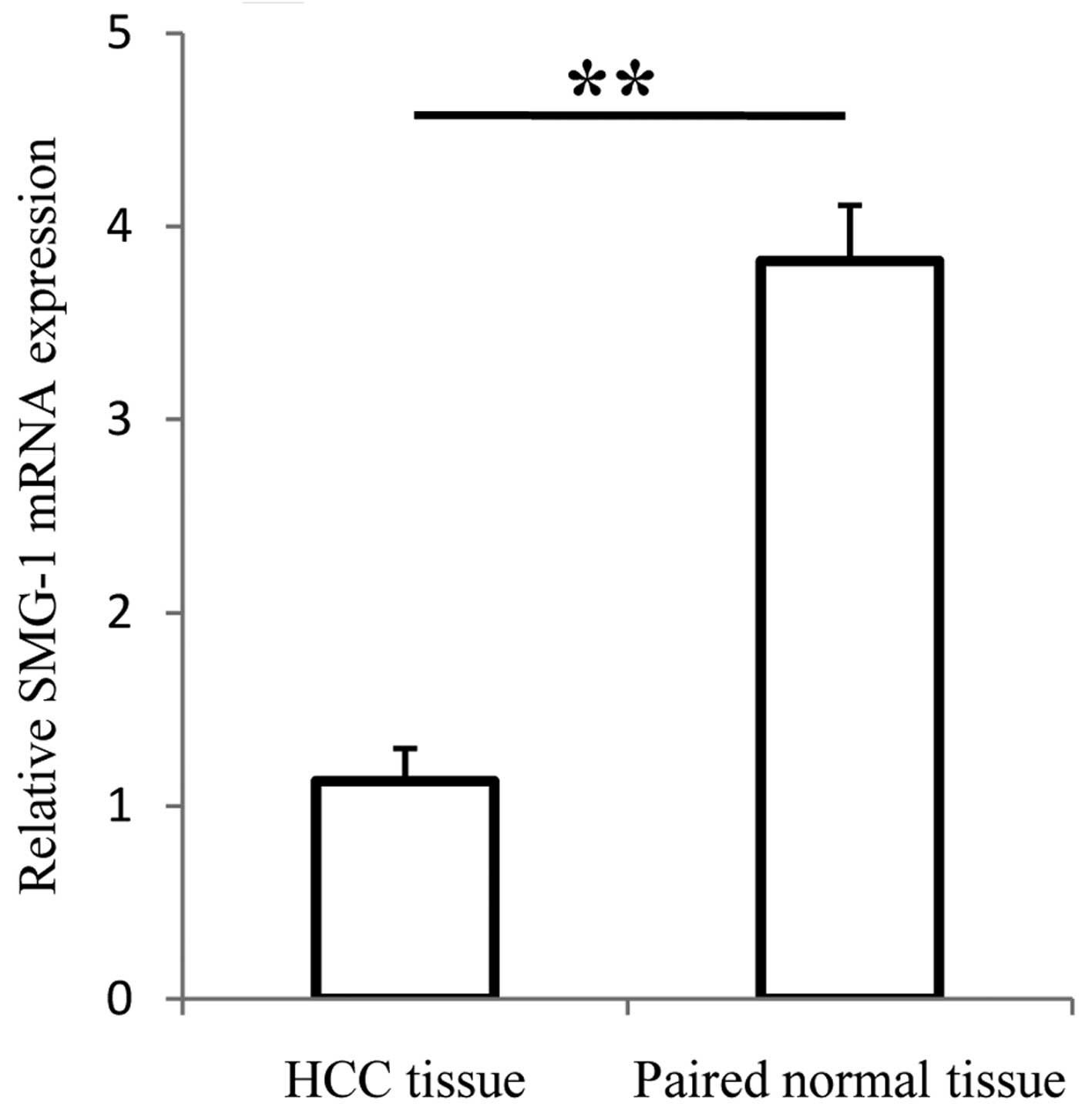
To examine the expression of SMG-1, 157 pairs of HCC
and adjacent non-cancerous tissues were subjected to qRT-PCR.
Results showed that the level of SMG-1 mRNA was significantly lower
in the HCC tissues than the level in the distant non-cancerous
tissues (1.13±0.17 compared with 3.82±0.29; t=23.32; P<0.01)
(Fig. 1). To further examine the
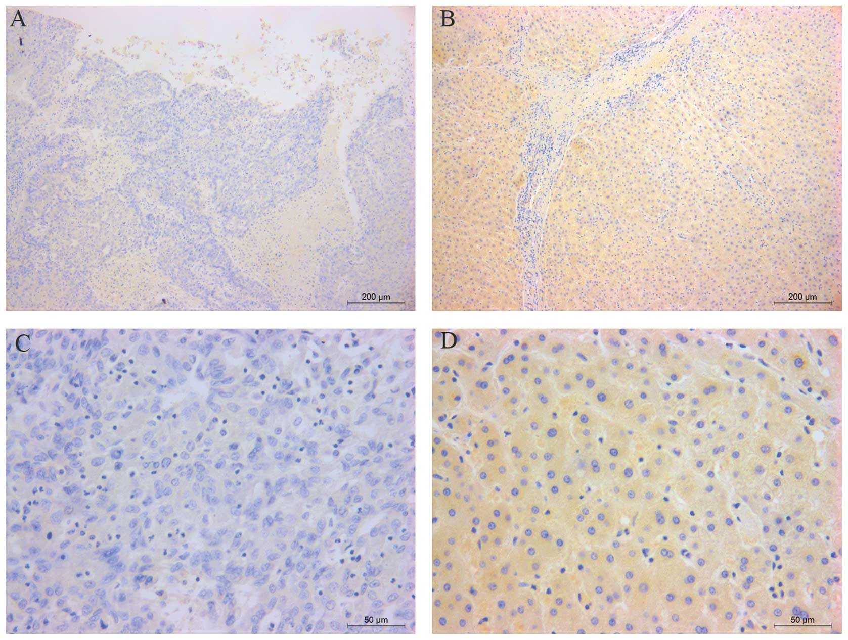
expression of SMG-1 in HCC, we analyzed the expression of SMG-1
protein using IHC. Based on the results of IHC, low SMG-1
expression in tumor tissues was identified in 67.5% (106/157) of
the cases. Moreover, in 71.3% (112/157) of the HCC patients, less
SMG-1 was expressed in the tumor tissues. IHC results confirmed the
qRT-PCR data and indicated that SMG-1 was significantly reduced in
the HCC tissues when compared with that in the distant
non-cancerous tissues (χ2=11.207, P=0.001) (Fig. 2). These results indicate that SMG-1
may represent a biological marker of the presence of HCC in
humans.
Association between SMG-1 expression and
clinical variables of the hepatocellular carcinoma cases
To determine the clinical significance of SMG-1 in
HCC, the correlation between expression of SMG-1 and
clinicopathological features was analyzed. Significant associations
were found with tumor differentiation (P=0.009), clinical stage
(P<0.001) and serum AFP level (P=0.001), indicating that HCC in
patients with low SMG-1 expression was frequently associated with a
high level of serum AFP, poor tumor differentiation and advanced
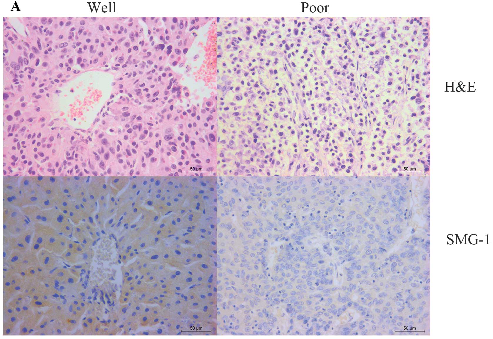
clinical stage (Table I). The
significant correlation between SMG-1 expression in HCC and tumor
differentiation was further confirmed. The percentage of cases with
low SMG-1 expression was markedly higher in poorly differentiated
HCC than that in well-differentiated HCC. Higher expression of
SMG-1 was observed in well-differentiated HCC. Conversely, low
SMG-1 expression was noticeably significant in poorly
differentiated HCC (Fig. 3).
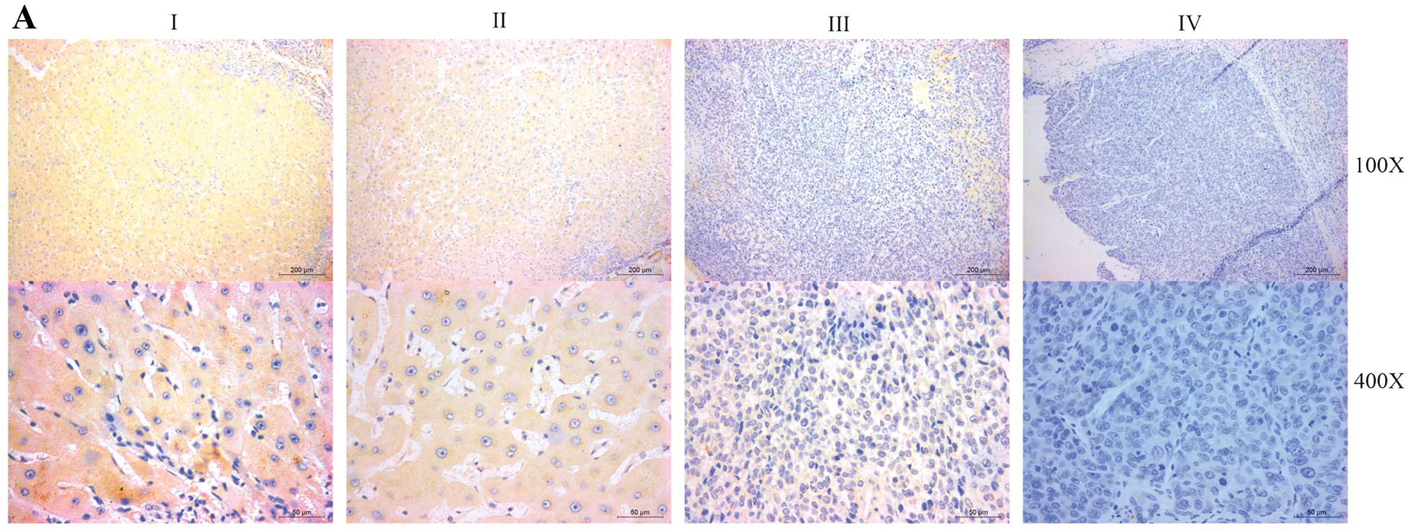
Furthermore, the IHC analysis showed that SMG-1 was
weak or negative in HCC lesions of patients with late stages of HCC
(TNM stages III–IV) as compared with that in early-stage HCC (TNM
stages I-II) tissues (P<0.05; Fig.
4). Quantitative analysis of the IHC staining indicated that
SMG-1 expression in clinical stage III–IV primary tumors was
statistically lower than that in clinical stage I–II primary tumors
(P<0.05; Fig. 4).
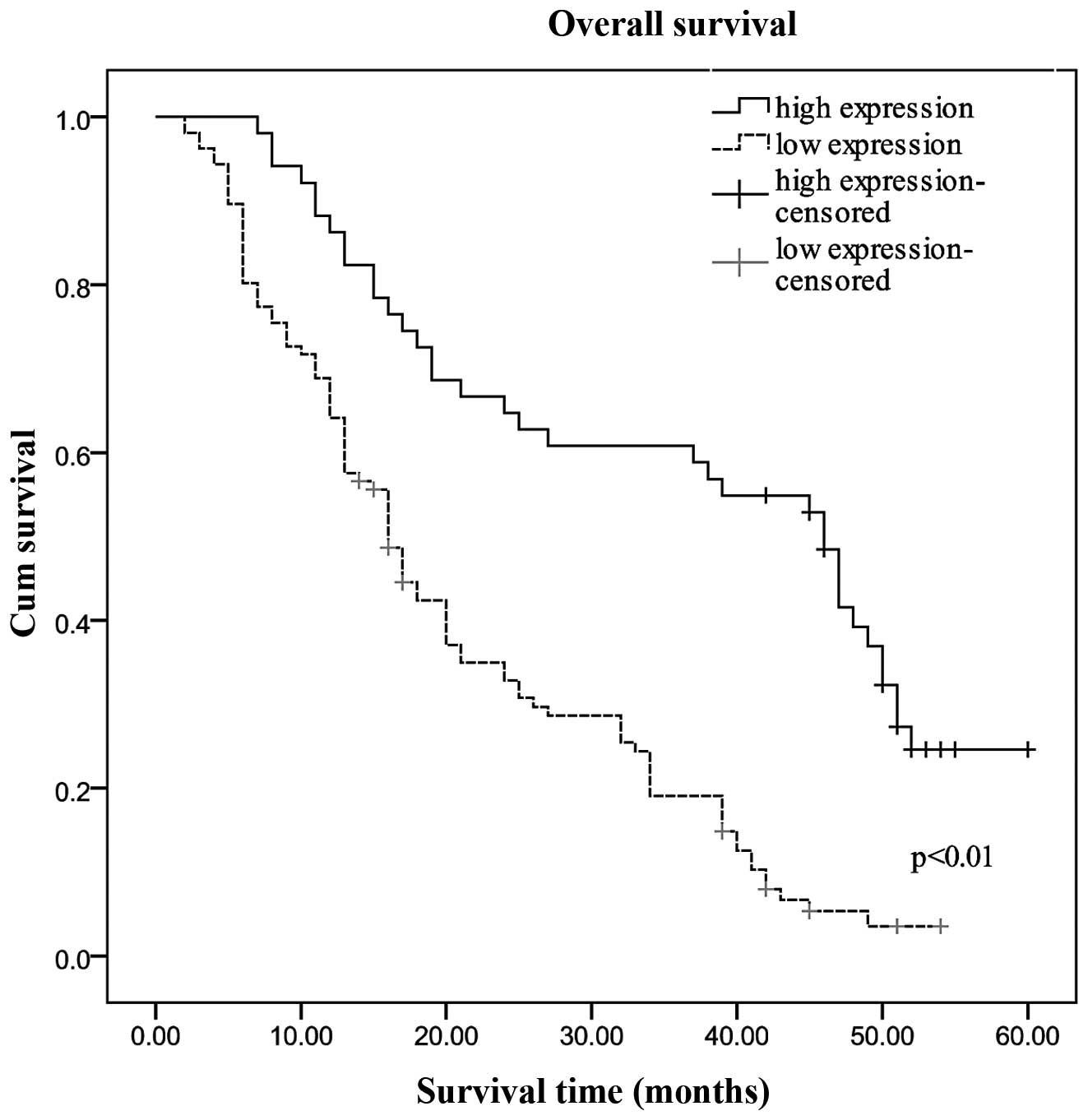
Correlation of SMG-1 expression with
overall survival of hepatocellular carcinoma patients
To determine the prognostic impact of SMG-1 on
survival of postsurgical HCC patients, Kaplan-Meier survival
analysis was performed. All of the 157 patients were followed up
for survival until August 2013, and their survival data were
stratified according to SMG-1 expression. Of the 157 patients, the
mean survival period was 20.583 months for the patients with low
SMG-1 expression, whereas it was 37.755 months for patients with
high levels of SMG-1 expression (P<0.01). As shown in Fig. 5, Kaplan-Meier and log-rank survival
tests indicated that low and high SMG-1 expression in HCC patients
were indicative of different survival times; the OS of patients
expressing high SMG-1 in their HCC lesions was much longer that the
OS of the cases with low SMG-1 expression (P<0.01).
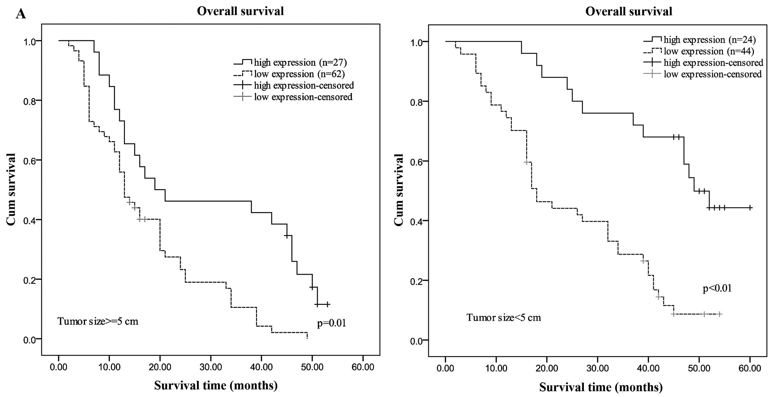
The prognostic effect of SMG-1 was further confirmed
by stratified survival analysis. Results showed that patients with
low SMG-1 expression had a significantly reduced survival following
surgical resection in 5 subgroups of HCC patients (Fig. 6). Expression of SMG-1 was correlated
with OS in subgroups of HCC patients. Kaplan-Meier survival
analyses were performed in subgroups according to factors that are
attributed to the outcome of HCC patients (log-rank test). This
supports the usefulness of SMG-1 protein as a new prognostic marker
for HCC patients.
Univariate and multivariate analyses of
prognostic variables in the hepatocellular carcinoma patients
To reveal variables with potential prognostic
significance in HCC patients, univariate analyses (Kaplan-Meier
survival analyses) for each variable were performed. SMG-1, as well
as tumor size, serum AFP level, tumor multiplicity, clinical stage
and tumor differentiation were shown to be responsible for the
outcome of HCC patients (Table
II). SMG-1, as well as tumor size, serum AFP level, tumor
multiplicity, clinical stage and tumor differentiation were all
significant prognostic indicators for OS of HCC patients.
| Table IIUnivariate analysis of survival
data. |
Table II
Univariate analysis of survival
data.
| Clinicopathological
characteristics | P-value |
|---|
| Age, <48.7 vs.
≥48.7 years | 0.343 |
| Gender, male vs.
female | 0.498 |
| AFP, <400 vs.
≥400 ng/ml | 0.001 |
| Hepatitis B virus
infection, positive vs. negative | 0.539 |
| Tumor size, <5
vs. ≥5 cm | 0.006 |
| Histological grade,
well/moderate vs. poor | 0.017 |
| TNM stage, I + II
vs. III + IV | 0.000 |
| SMG-1, high
expression vs. low expression | 0.001 |
According to the results of the multivariate
analysis of these factors, the predictive ability of SMG-1
expression, tumor size and tumor multiplicity was confirmed
(Table III). However, there were
no significant associations between prognosis and the other
clinicopathological features.
| Table IIICox multivariate analyses of
prognostic factors on overall survival. |
Table III
Cox multivariate analyses of
prognostic factors on overall survival.
| Variable | β | SE | Hazard ratio (95%
CI) | P-value |
|---|
| AFP | 0.512 | 0.185 | 1.699
(1.161–2.400) | 0.060 |
| Tumor size | 0.696 | 0.198 | 2.005
(1.361–2.953) | 0.000 |
| Tumor
multiplicity | 0.915 | 0.179 | 2.497
(1.757–3.548) | 0.000 |
|
Differentiation | 0.057 | 0.180 | 1.059
(0.744–1.507) | 0.752 |
| Stage | 0.126 | 0.216 | 1.134
(0.743–1.731) | 0.560 |
| SMG-1 | 1.494 | 0.254 | 4.457
(2.701–7.325) | 0.000 |
Discussion
SMG-1 participates in the regulation of normal cell
growth and TNFα-induced apoptosis (18), similar to other members of the PIKK
family such as ATM (ataxia telangiectagia mutated) and ATR (ATM-
and Rad3-related) (19), and has
been shown to control G1/S cell cycle arrest (16). The signal pathways involved in
regulation of the cell cycle from the G1-phase to the S phase are
frequent targets of carcinogenic events. Thus, loss of SMG-1 may
result in a general alteration in the homeostasis of normal cells
due to increased survival potential, which may subsequently lead to
tumorigenesis. A recent study demonstrated that SMG-1 is a
tumor-suppressor gene which suppresses tumor growth via the
regulation of both the p53 and Cdc25A signaling pathways (16). However, the role of SMG-1 in the
development of primary hepatocellular carcinoma has not been
reported.
In the present study, we analyzed the expression of
SMG-1 mRNA and protein in HCC patients using real-time PCR assay
and immunohistochemistry. The results derived from the different
assays strongly suggest a correlation between the SMG-1 level and
the clinical outcome of HCC. We found that the combination of SMG-1
mRNA and protein in HCC tissue specimens was significantly lower
than that in the distant normal tissues. The abnormal expression of
SMG-1 associated with HCC was also correlated with higher AFP
levels, poor differentiation and advanced TNM stages of HCC.
Multivariate analysis confirmed that tumor size, tumor multiplicity
and low expression of SMG-1 were risk factors for poor survival of
HCC patients. The present study suggests that SMG-1, as a
prognostic biomarker, is abnormally expressed in primary HCC
tissues and is related to tumorigenesis, and detection of SMG-1 may
provide new opportunities for prediction of HCC patient
survival.
It is known that SMG-1 inhibits the mTOR
(mechanistic target of rapamycin) signaling pathway which
contributes to the regulatation of cell growth, metabolism and
aging in response to nutrients, cellular energy stage and growth
factors, thus, inhibiting the development of cancer and other
diseases of aging (15). Numerous
studies have demonstrated the importance of the mTOR pathway in
patients with HCC (20–22). More importantly, it has also been
suggested that the mTOR pathway is associated with the poor
prognosis of HCC (23,24). Most of the genes which inhibit the
mTOR pathway have been confirmed as tumor-suppressor genes in HCC.
Furthermore, the mTOR inhibitor has been used as a potential target
for the prevention and treatment of HCC (25). This implies that SMG-1, which can
inhibit the mTOR signaling pathway, may be a new tumor-suppressor
gene in primary HCC. The present study found that SMG-1 was deleted
or weakly expressed in primary HCC tissues. Although the process by
which SMG-1 functions to suppress HCC development or progression is
still unclear, we believe that SMG-1 may contribute to inhibition
of the tumorigenesis and development of HCC by negatively
regulating the mTOR pathway. The mTOR pathway is more significantly
altered in poorly differentiated and high-TNM stage tumors, and
tumors with poor prognostic features (23,24).
In the present study, low SMG-1 expression was more frequently
observed in poorly differentiated and high-TNM stage tumors. These
findings can be explained by the fact that decreased or deleted
expression of SMG-1 results in the loss of the negative regulation
of the mTOR signaling pathway, resulting in the exacerbation of
HCC. Yet, further research is needed to fully understand the
underlying molecular mechanisms.
Due to their critical role in the regulation of the
cell cycle and cell growth, DNA damage restructuring and
maintaining telomere length, PIKK family proteins, including PI3K,
PI4K, TEL1, RAD3, ATM and ATR, have been attracting increased
attention in regards to their significance in human cancers.
Notably, altered expression of PIKKs commonly have a significant
prognostic value in cancer. Lee et al (26) reported that aberrant expression of
ATM plays a critical role in the development and progression of
gastric cancer. Expression of PI3K was significantly associated
with aggressive behavior and shorter DSS in non-gastrointestinal
stromal soft tissue sarcomas (27).
Bhattacharya et al (28)
showed that inactivation of the ATM pathway may have an important
role in the development of breast carcinoma with diagnostic,
prognostic and therapeutic implications. Similar findings in other
studies have found that expression of ATM may be a useful marker
with which to identify patients presenting with pancreatic
neuroendocrine tumors with poor prognosis who may benefit from
close follow-up and aggressive therapy (29), and may be a possible prognostic
factor in Chinese patients with CLL (30). Our findings suggest that SMG-1 also
has similar functions, and can be used as an independent prognostic
factor for the OS of HCC patients. In the present study, a decrease
in SMG-1 was associated with poor prognosis of the HCC patients.
Numerous independent studies have shown that the high potential for
vascular invasion and metastasis is often the main biological basis
for the poor prognosis of HCC (31,32).
p53, one of the key players in the processes of cell dissociation,
angiogenesis and cell migration in HCC (33,34),
has been shown to be regulated by SMG-1 (16). This suggests that expression of
SMG-1 may be associated with the prognosis of HCC via regulation of
the expression of P53, which is consistent with our results.
It is widely recognized that early diagnosis and
treatment are crucially needed for a more favorable prognosis of
HCC patients. Using ideal biomarkers to identify patients with a
higher risk of a worse prognosis may reduce the mortality rate of
HCC patients. In our study, the patients with early-stage HCC (TNM
stages I–II) also displayed a significantly lower level of SMG-1 in
HCC lesions than that in the normal liver tissues. In addition, as
HCC progressed to later stages, the SMG-1 level further decreased.
Thus, these characteristics of SMG-1 further confirm that SMG-1 may
be a significant biomarker for identifying patients with poor
prognosis after surgical intervention in early disease stages.
In summary, the present study demonstrated that
SMG-1 was frequently deleted or downregulated in HCC. Abnormal
expression of SMG-1 was significantly correlated with
differentiation, clinical stage and serum AFP level, suggesting
that SMG-1 may be involved in the pathogenesis and development of
HCC. Low SMG-1 expression adversely impacted the survival of HCC
patients. Consequently, our data revealed that low SMG-1 expression
plays a crucial role in predicting the postsurgical overall
survival of HCC patients.
Acknowledgements
The authors thank Professor Chen Huang of Xi’an
Jiaotong University (Xi’an, China) for providing the experimental
platform and expert opinions.
References
|
1
|
Jemal A, Bray F, Center MM, Ferlay J, Ward
E and Forman D: Global cancer statistics. CA Cancer J Clin.
61:69–90. 2011. View Article : Google Scholar
|
|
2
|
He G and Karin M: NF-κB and STAT3 - key
players in liver inflammation and cancer. Cell Res. 21:159–168.
2011.
|
|
3
|
Chen L, Ho DW, Lee NP, Sun S, Lam B, Wong
KF, Yi X, Lau GK, Ng EW, Poon TC, Lai PB, Cai Z, Peng J, Leng X,
Poon RT and Luk JM: Enhanced detection of early hepatocellular
carcinoma by serum SELDI-TOF proteomic signature combined with
alpha-fetoprotein marker. Ann Surg Oncol. 17:2518–2525. 2010.
View Article : Google Scholar : PubMed/NCBI
|
|
4
|
Sidransky D: Emerging molecular markers of
cancer. Nat Rev Cancer. 3:210–219. 2002. View Article : Google Scholar
|
|
5
|
El-Serag HB and Rudolph KL: Hepatocellular
carcinoma: Epidemiology and molecular carcinogenesis.
Gastroenterology. 132:2557–2576. 2007. View Article : Google Scholar : PubMed/NCBI
|
|
6
|
Zhao Y, Wang WJ, Guan S, Li HL, Xu RC, Wu
JB, Liu JS, Li HP, Bai W, Yin ZX, Fan DM, Zhangand ZL and Han GH:
Sorafenib combined with transurethral chemoembolization for the
treatment of advanced hepatocellular carcinoma: a large-scale
multicenter study of 222 patients. Ann Oncol. 7:1786–1792. 2013.
View Article : Google Scholar
|
|
7
|
Denning G, Jamieson L, Maquat L, Thompson
E and Fields A: Cloning of a novel phosphatidylinositol
kinase-related kinase: characterization of the human SMG-1 RNA
surveillance protein. J Biol Chem. 276:22709–22714. 2001.
View Article : Google Scholar : PubMed/NCBI
|
|
8
|
Yamashita A, Ohnishi T, Kashima I, Taya Y
and Ohno S: Human SMG-1, a novel phosphatidylinositol
3-kinase-related protein kinase, associates with components of the
mRNA surveillance complex and is involved in the regulation of
nonsense-mediated mRNA decay. Genes Dev. 15:2215–2228. 2001.
View Article : Google Scholar : PubMed/NCBI
|
|
9
|
Grimson A, O’Connor S, Newman CL and
Anderson P: SMG-1 is phosphatidylinositol kinase-related protein
kinase required for nonsense-mediated mRNA decay in
Caenorhabditis elegans. Mol Cell Biol. 24:7483–7490. 2004.
View Article : Google Scholar : PubMed/NCBI
|
|
10
|
Gehen S, Staversky R, Bambara R, Keng P
and O’Reilly M: SMG-1 and ATM sequentially and independently
regulate the G1 checkpoint during oxidative stress. Oncogene.
27:4065–4074. 2008. View Article : Google Scholar : PubMed/NCBI
|
|
11
|
Cheung H, St Jean M, Beug S, Lejmi-Mrad R,
LaCasse E, Baird S, Stojdl D, Screaton R and Korneluk R: SMG1 and
NIK regulate apoptosis induced by Smac mimetic compounds. Cell
Death Dis. 2:e1462011. View Article : Google Scholar : PubMed/NCBI
|
|
12
|
Brumbaugh K, Otterness D, Geisen C,
Oliveira V, Brognard J, Li X, Lejeune F, Tibbetts R, Maquat L and
Abraham R: The mRNA surveillance protein hSMG-1 functions in
genotoxic stress response pathways in mammalian cells. Mol Cell.
14:585–598. 2004. View Article : Google Scholar : PubMed/NCBI
|
|
13
|
Chen R, Yang Q, Chen Y, Oliveira V, Dalton
W, Fearns C and Lee J: Kinome siRNA screen identifies SMG-1 as a
negative regulator of hypoxia-inducible factor-1α in hypoxia. J
Biol Chem. 284:16752–16758. 2009.PubMed/NCBI
|
|
14
|
Masse I, Molin L, Mouchiroud L, Vanhems P,
Palladino F, Billaud M and Solari F: A novel role for the SMG-1
kinase in lifespan and oxidative stress resistance in
Caenorhabditis elegans. PLoS One. 3:e33542008. View Article : Google Scholar : PubMed/NCBI
|
|
15
|
González-Estévez C, Felix DA, Smith MD,
Paps J, Morley SJ, James V, Sharp TV and Aboobaker AA: SMG-1 and
mTORC1 act antagonistically to regulate response to injury and
growth in planarians. PLoS Genet. 3:e10026192012.PubMed/NCBI
|
|
16
|
Gubanova E, Issaeva N, Gokturk C,
Djureinovic T and Helleday T: SMG-1 suppresses CDK2 and tumor
growth by regulating both the p53 and Cdc25A signaling pathways.
Cell Cycle. 12:3770–3780. 2013. View
Article : Google Scholar : PubMed/NCBI
|
|
17
|
Roberts T, Ho U, Luff J, Lee C, Apte S,
MacDonald K, Raggat L, Pettit A, Morrow C and Waters M: Smg1
haploinsufficiency predisposes to tumor formation and inflammation.
Proc Natl Acad Sci USA. 110:E285–E294. 2013. View Article : Google Scholar : PubMed/NCBI
|
|
18
|
Oliveira V, Romanow WJ, Geisen C,
Otterness DM, Mercurio F, Wang HG, Dalton WS and Abraham RT: A
protective role for the human SMG-1 kinase against tumor necrosis
factor-α-induced apoptosis. J Biol Chem. 283:13174–13184.
2008.PubMed/NCBI
|
|
19
|
Shiloh Y: ATM and related protein kinases:
Safeguarding genome integrity. Nat Rev Cancer. 3:155–168. 2003.
View Article : Google Scholar : PubMed/NCBI
|
|
20
|
Matter MS, Decaens T, Andersen JB and
Thorgeirsson SS: Targeting the mTOR pathway in hepatocellular
carcinoma: current state and future trends. J Hepatol. 60:855–865.
2014. View Article : Google Scholar : PubMed/NCBI
|
|
21
|
Menon S, Yecies JL and Zhang HH: Chronic
activation of mTOR complex 1 is sufficient to cause hepatocellular
carcinoma in mice. Sci Signal. 5:ra242012. View Article : Google Scholar : PubMed/NCBI
|
|
22
|
Bassullu N, Turkmen I, Dayangac M, Yagiz
Korkmaz P, Yasar R, Akyildiz M, Yaprak O, Tokat Y, Yuzer Y and
Bulbul Dogusoy G: The predictive and prognostic significance of
c-erb-B2, EGFR, PTEN, mTOR, PI3K, p27, and ERCC1 expression in
hepatocellular carcinoma. Hepat Mon. 12:e74922012. View Article : Google Scholar : PubMed/NCBI
|
|
23
|
Villanueva A, Chiang DY, Newell P, Peix J,
Thung S, Alsinet C, Tovar V, Roayaie S, Minguez B, Sole M,
Battiston C, Van Laarhoven S, Fiel MI, Di Feo A, Hoshida Y, Yea S,
Toffanin S, Ramos A, Martignetti JA, Mazzaferro V, Bruix J, Waxman
S, Schwartz M, Meyerson M, Friedman SL and Llovet JM: Pivotal role
of mTOR signaling in hepatocellular carcinoma. Gastroenterology.
135:1972–1983. e1–11. 2008. View Article : Google Scholar : PubMed/NCBI
|
|
24
|
Zhou L, Huang Y, Li J and Wang Z: The mTOR
pathway is associated with the poor prognosis of human
hepatocellular carcinoma. Med Oncol. 27:255–261. 2010. View Article : Google Scholar : PubMed/NCBI
|
|
25
|
Buitrago-Molina LE and Vogel A: mTor as a
potential target for the prevention and treatment of hepatocellular
carcinoma. Curr Cancer Drug Targets. 9:1045–1061. 2012.PubMed/NCBI
|
|
26
|
Lee HE, Han N, Kim MA, Lee HS, Yang HK,
Lee BL and Kim WH: DNA damage response-related proteins in gastric
cancer: ATM, Chk2 and p53 expression and their prognostic value.
Pathobiology. 1:25–35. 2014. View Article : Google Scholar : PubMed/NCBI
|
|
27
|
Valkov A, Kilvaer TK, Sorbye SW, Donnem T,
Smeland E, Bremnes RM and Busund LT: The prognostic impact of Akt
isoforms, PI3K and PTEN related to female steroid hormone receptors
in soft tissue sarcomas. J Transl Med. 9:2002011. View Article : Google Scholar : PubMed/NCBI
|
|
28
|
Bhattacharya N, Mukherjee N, Singh RK,
Sinha S, Alam N, Roy A, Roychoudhury S and Panda CK: Frequent
alterations of MCPH1 and ATM are associated with primary breast
carcinoma: clinical and prognostic implications. Ann Surg Oncol.
3:424–432. 2013. View Article : Google Scholar : PubMed/NCBI
|
|
29
|
Shin JU, Lee CH, Lee KT, Lee JK, Lee KH,
Kim KM, Kim KM, Park SM and Rhee JC: Prognostic significance of ATM
and cyclin B1 in pancreatic neuroendocrine tumor. Tumour Biol.
5:1645–1651. 2012. View Article : Google Scholar : PubMed/NCBI
|
|
30
|
Xu W, Li JY, Wu YJ, Yu H, Shen QD, Li L,
Fan L and Qiu HX: Prognostic significance of ATM and TP53 deletions
in Chinese patients with chronic lymphocytic leukemia. Leuk Res.
7:1071–1077. 2008. View Article : Google Scholar : PubMed/NCBI
|
|
31
|
Llovet JM and Bruix J: Novel advancements
in the management of hepatocellular carcinoma in 2008. J Hepatol.
48(Suppl 1): S20–S37. 2008. View Article : Google Scholar : PubMed/NCBI
|
|
32
|
Bruix J, Sherman M, Llovet JM, Beaugrand M
and Lencioni R: Clinical management of hepatocellular carcinoma.
Conclusions of the Barcelona-2000 EASL conference European
Association for the Study of the Liver. J Hepatol. 35:421–430.
2001. View Article : Google Scholar : PubMed/NCBI
|
|
33
|
Tseng PL, Tai MH, Huang CC, Wang CC, Lin
JW, Hung CH, Chen CH, Wang JH, Lu SN, Lee CM, Changchien CS and Hu
TH: Overexpression of VEGF is associated with positive p53
immunostaining in hepatocellular carcinoma (HCC) and adverse
outcome of HCC patients. J Surg Oncol. 5:349–357. 2008. View Article : Google Scholar : PubMed/NCBI
|
|
34
|
Hsu H, Peng S, Lai P, Chu J and Lee P:
Mutations of p53 gene in hepatocellular carcinoma correlate with
tumor progression and patient prognosis: a study of 138 patients
with unifocal HCC. Int J Oncol. 6:1341–1347. 1994.PubMed/NCBI
|