Introduction
Retinoblastoma is a hypervascular tumor, the
proliferation of which is dependent on the vascular supply of the
tumor (1). It is also known that
the vascular density of human retinoblastoma is closely related to
the local invasion and distant metastasis of the disease (2). Although local therapy and systemic
chemotherapy are widely adopted for ocular salvage, enucleation is
still regarded as the treatment of choice in advanced cases of
retinoblastoma. Since the inhibitory effects of several
anti-angiogenic agents such as anecortave acetate (3), bevacizumab (4) and PEDF (5) on tumor growth have been proven in
transgenic and orthotopic murine models of retinoblastoma, vascular
targeted therapy is considered as one of the future adjuvant
treatment options for retinoblastoma.
Since differences in gene expression between tumor
vascular endothelium and normal endothelial cells have been noted,
the concept of tumor vascularization by sprouting has changed. In
human glioblastoma and neuroblastoma, chromosomal analysis using
fluorescence in situ hybridization (FISH) revealed that some
tumor vascular endothelium shows cytogenetic abnormality similar to
that of tumor cells (6,7). Moreover, direct evidence of tumor
vascularization by the endothelial differentiation of cancer
stem-like cells was found in glioblastoma (7,8).
The p53 protein is well known as a tumor suppressor,
which inhibits tumorigenesis through the activation of apoptosis
and induction of cell cycle arrest in various types of human
cancers including retinoblastoma (9). When stained with the DO-1 monoclonal
antibody, 91.4% of specimens were found to exhibit positive
staining, and the immunoreactivity to the p53 antibody was
prominent in poorly or moderately differentiated areas (9). According to another study which
explored the distribution of wild-type p53 in cultivated
retinoblastoma cell lines and human retinoblastoma tissue using the
p1801 monoclonal antibody (10),
nuclear staining of the p53 protein was noted in 89% of human
retinoblastoma. In contrast, retinoblastoma cell lines showed
cytoplasmic or mixed nuclear and cytoplasmic distribution of the
p53 protein (10).
Since tumor vascularization is also an important
issue in the prognosis and treatment of retinoblastoma and there is
increasing evidence of tumor stem-like cells in human and mouse
retinoblastoma (11,12), it is necessary to identify the
presence of genetically unstable tumor vascular endothelium in
retinoblastoma. In the present study, to investigate cytogenetic
abnormalities in tumor endothelium of retinoblastoma, we examined
the expression and distribution of the p53 protein in mature tumor
vascular endothelium in an orthotopic retinoblastoma model and in
human retinoblastoma.
Materials and methods
Retinoblastoma cells
Two types of human retinoblastoma cell lines were
used in this study. SNUOT-Rb1 (13), which was established by our group,
and Y79 (14) (American Type
Culture Collection, Manassas, VA, USA) were cultured in RPMI-1640
media (Welgene Inc., Korea) supplemented with 10% fetal bovine
serum (Gibco Invitrogen Corp., Carlsbad, CA, USA) and 1%
penicillin-streptomycin solution (Invitrogen, Carlsbad, CA, USA).
SNUOT-Rb1 cells maintained by our group were used at passage
3–7.
Orthotopic transplantation model
For induction of the orthotopic retinoblastoma mouse
model, cultivated SNUOT-Rb1 and Y79 cells were injected into the
vitreal cavity of BALB/c-nude mice (Samtako, Osan, Korea) as
previously described by our group (13). Three mice were assigned to each
treatment group. The right eyes of the mice were injected with
cultivated retinoblastoma cells (1×10⁷) suspended in
phosphate-buffered saline (4°C), using a 30-gauge needle. For the
control, phosphate-buffered saline (4°C) was injected into the left
eyes of the mice in the same manner. Intraocular tumorigenesis was
evaluated every week by indirect ophthalmoscope, and mice with
adequate tumorigenesis were sacrificed and enucleated at the 4th
week after injection. Mice were maintained and treated in
accordance with the ARVO Statement for the Use of Animals in
Ophthalmic and Vision Research.
Human retinoblastoma tissue
Formalin-fixed, paraffin-embedded human eyeball
sections (4 μm) of three patients primarily enucleated for
retinoblastoma were obtained from the Department of Pathology,
Seoul National University Hospital. The Institutional Review Board
of Seoul National University Hospital approved the study.
Double immunofluorescence staining
Formalin-fixed, paraffin-embedded blocks of tumor
sections (4-μm) (from both the orthotopic model and human
retinoblastoma specimens) were deparaffinized by serial xylene and
ethyl alcohol immersion. For antigen retrieval, sections were
treated with proteinase K (20 μg/ml) at 37°C for 20 min. After
being permeabilized with Triton X-100 (0.2%), sections were blocked
with blocking solution (BioGenex Laboratories, San Ramon, CA, USA).
Prepared sections were incubated overnight with primary antibodies
at 4°C and then with fluorescein tagged secondary antibody for 2 h
at room temperature after rinsing. The anti-p53 polyclonal antibody
(FL-393; sc-6243-G) and anti-von Willebrand factor (vWF) antibody
(sc-14014) (both from Santa Cruz Biotechnology, Santa Cruz, CA,
USA), for detecting mature vascular endothelium, were used as
primary antibodies at the concentration of 1:100. For the secondary
antibody, Alexa Fluor 594-conjugated donkey anti-goat IgG and Alexa
Fluor 488-conjugated donkey anti-rabbit IgG (both from Molecular
Probes, Carlsbad, CA, USA) were used at the concentration of 1:200.
Nuclear counterstaining with 4,6-diamidino-2-phenylindole
dihydrochloride (DAPI) was performed for the discrimination of
nuclear or cytoplasmic distribution of the p53 protein. After
aqueous mounting, the slides were examined under a fluorescence
microscope (Axio Observer; Carl Zeiss). After confirming the number
of total vWF-positive endothelium, the vWF-positive endothelium
with p53 nuclear accumulation was independently evaluated by two
investigators in 10 randomly selected high power fields (x400).
Then, the overall ratio of endothelium with p53 nuclear
accumulation among all vWF-positive tumor vascular endothelium was
calculated.
Statistical analysis
Statistical analysis was performed using SPSS
version 12.0 for Windows. The ratio of vWF-positive endothelium
with p53 nuclear accumulation among all tumor vascular endothelial
cells in the SNUOT-Rb1 and Y79 cell groups was compared using the
Mann-Whitney U test. The cut-off value of statistical significance
was p<0.05.
Results
Expression and distribution of the p53
protein in the orthotopic retinoblastoma model
Four weeks after intravitreal inoculation of
retinoblastoma cells, we assessed intraocular tumor formation with
indirect ophthalmoscopic examination and H&E staining. Adequate
tumorigenesis was achieved in both Y79 and SNUOT-Rb1 cell induced
mouse models. Although not quantitatively analyzed, the orthotopic
retinoblastoma model induced by SNUOT-Rb1 cells showed a more
aggressive growth pattern such as relatively rapid growth and
occasional extraocular extension compared to the Y79 cell induced
model. We did not note any other significant differences in
phenotype between the tumors induced by Y79 and SNUOT-Rb1 cells. In
both of the orthotopic retinoblastoma models induced by Y79 and
SNUOT-Rb1 cells, strong and uniform immunopositivity against the
anti-p53 antibody was observed in the area filled with
retinoblastoma cells (Fig. 1). In
contrast, the retinas of the contralateral eyes did not exhibit
significant immunoreactivity to the anti-p53 antibody.
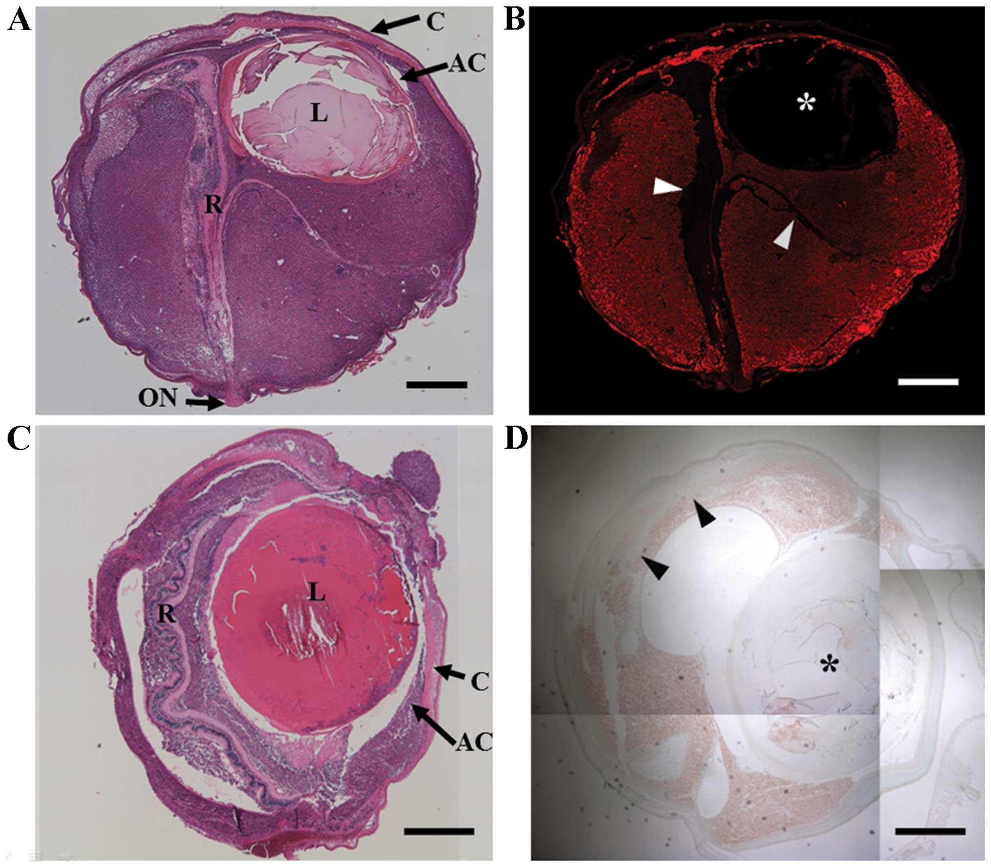 | Figure 1Diffuse p53 expression in an
orthotopic transplantation mouse model of retinoblastoma. (A) A
tumor mass filling the subretinal area, intravitreal cavity and
anterior chamber can be observed 4 weeks after the intravitreal
inoculation of SNUOT-Rb1 cells (H&E staining). (B) In a serial
section stained with fluorescein tagged anti-p53 antibody (red),
diffuse p53 immunoreactivity of the tumor mass is noted apart from
the detached retina (arrowheads) and crystalline lens (*). (C)
After inoculation of Y79 cells, intraocular tumor formation similar
to the SNUOT-Rb1 cell model was observed, but the tumor volume was
relatively smaller in the Y79 model. (D) Immunohistochemical
staining of the Y79 cell induced model also shows diffuse p53
immunopositivity of the tumor mass apart from the relatively
preserved retina (arrow heads) and crystalline lens (*). AC,
anterior chamber; C, cornea; L, lens; ON, optic nerve; R, detached
retina. Scale bars, A-D, 500 μm. |
Most of the tumor cells demonstrated p53
immunoreactivity and when merged with DAPI nuclear counterstaining,
we noted the nuclear location of the p53 protein in both Y79 and
SNUOT-Rb1 cell models.
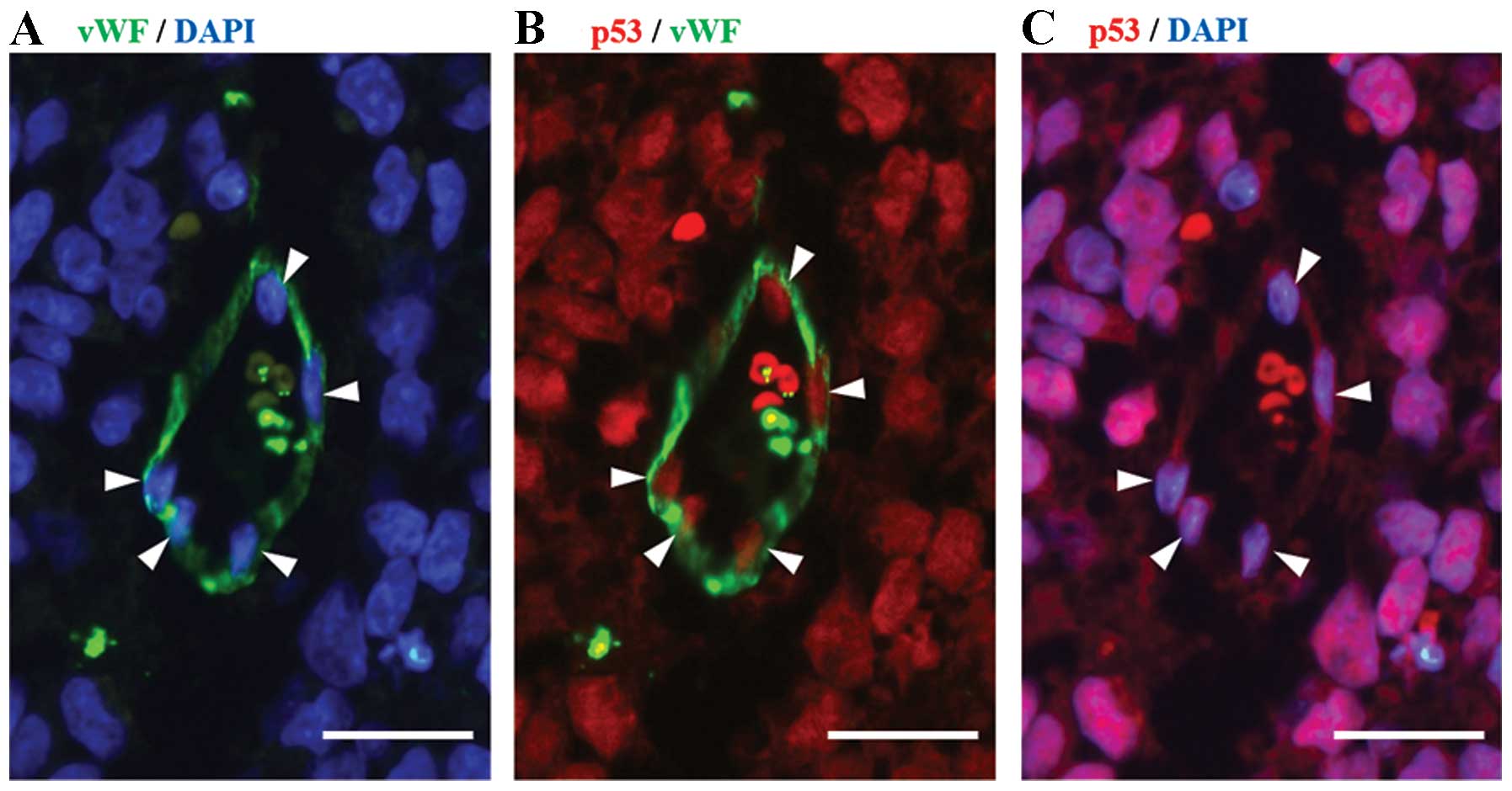
Mature tumor vascular endothelium with
p53 protein immunoreactivity in the orthotopic retinoblastoma
model
Some of the vWF-immunopositive tumor vascular
endothelium was also stained with anti-p53 antisera. In both Y79
and SNUOT-Rb1 models, the nuclear localization of the p53 protein
in the vWF-immunopositive tumor vascular endothelium was confirmed
by DAPI nuclear counterstaining. The distribution of the p53
protein in endothelial cells resembled that in the tumor cells
(Fig. 2). In contrast to the tumor
vascular endothelium in the retinoblastoma model, retinal vascular
endothelium of the contralateral normal eyes did not react with p53
antisera.
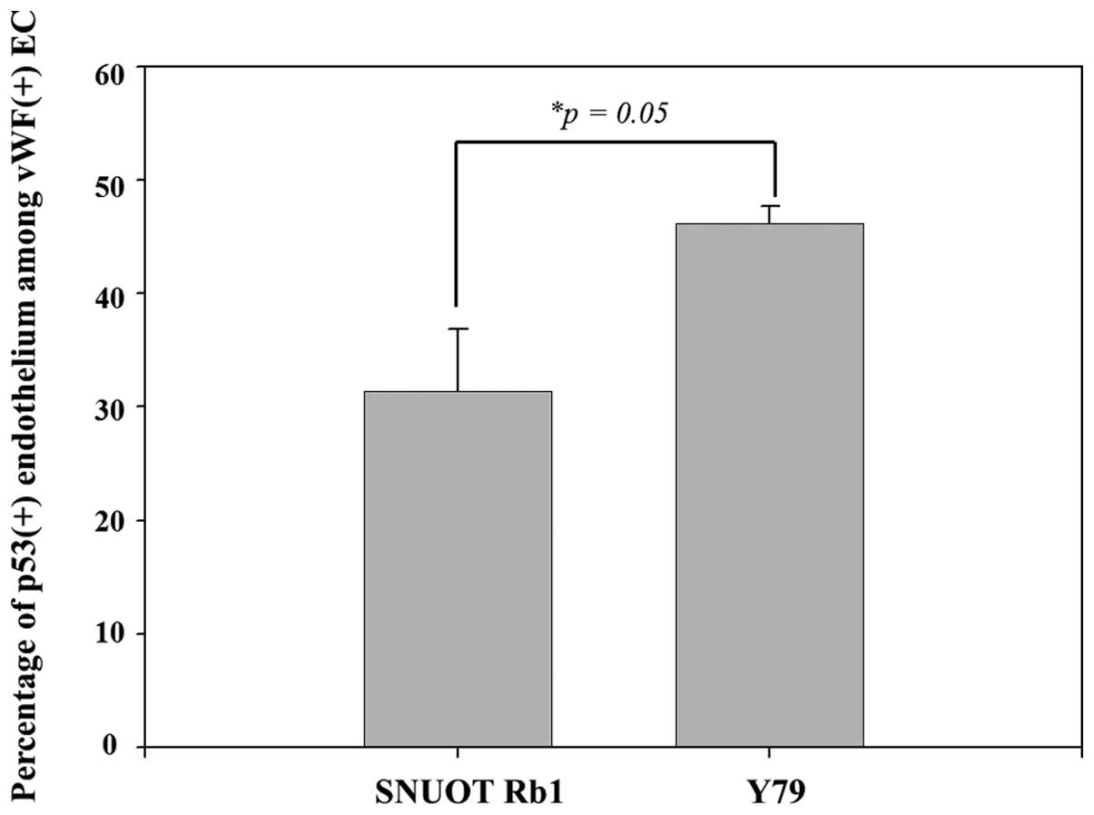
The ratio of p53 immunopositivity was different in
each orthotopic model induced by Y79 and SNUOT-Rb1 cells. The mean
percentage of p53-positive endothelium was 31.4±5.4% in 3 eyes from
the orthotopic transplantation model with SNUOT-Rb1 cells, and was
46.1±1.6% in the other 3 eyes from the Y79 cell model (Fig. 3). The ratio of tumor vascular
endothelium with p53 nuclear accumulation was higher in the Y79
cell induced orthotopic retinoblastoma model with a marginal
statistical significance (p=0.05).
Immunoreactivity of the human
retinoblastoma specimens against the anti-p53 antibody: tumor cells
and vascular endothelium
All three patients involved in this study were
primarily enucleated for extensive retinoblastoma which was
classified as group D according to the International Intraocular
Retinoblastoma Classification. Extraocular extension was not
detected in any patients at the time point of enucleation. The
clinicopathological features of the patients were similar (Table I).
 | Table IClinicopathological characteristics of
the human retinoblastoma cases. |
Table I
Clinicopathological characteristics of
the human retinoblastoma cases.
| | | | | | Gross pathology | Percentage of
p53-positive endothelium |
|---|
| | | | | |
|
|
|---|
| Patient no. | Age
(months)/gender | Laterality | International
classification | Growth pattern | MTD (mm) | AC involvement | Choroidal
involvement | Optic nerve
involvement | Observer 1 (%) | Observer 2 (%) |
|---|
| 1 | 8/M | Bilateral | Group D | Exophytic | 15 | No | No | No | 29.3 | 33.3 |
| 2 | 12/M | Unilateral | Group D | Exophytic | 16 | No | Yes | No | 31.0 | 36.2 |
| 3 | 9/M | Unilateral | Group D | Exophytic | 13 | No | No | No | 32.4 | 35.1 |
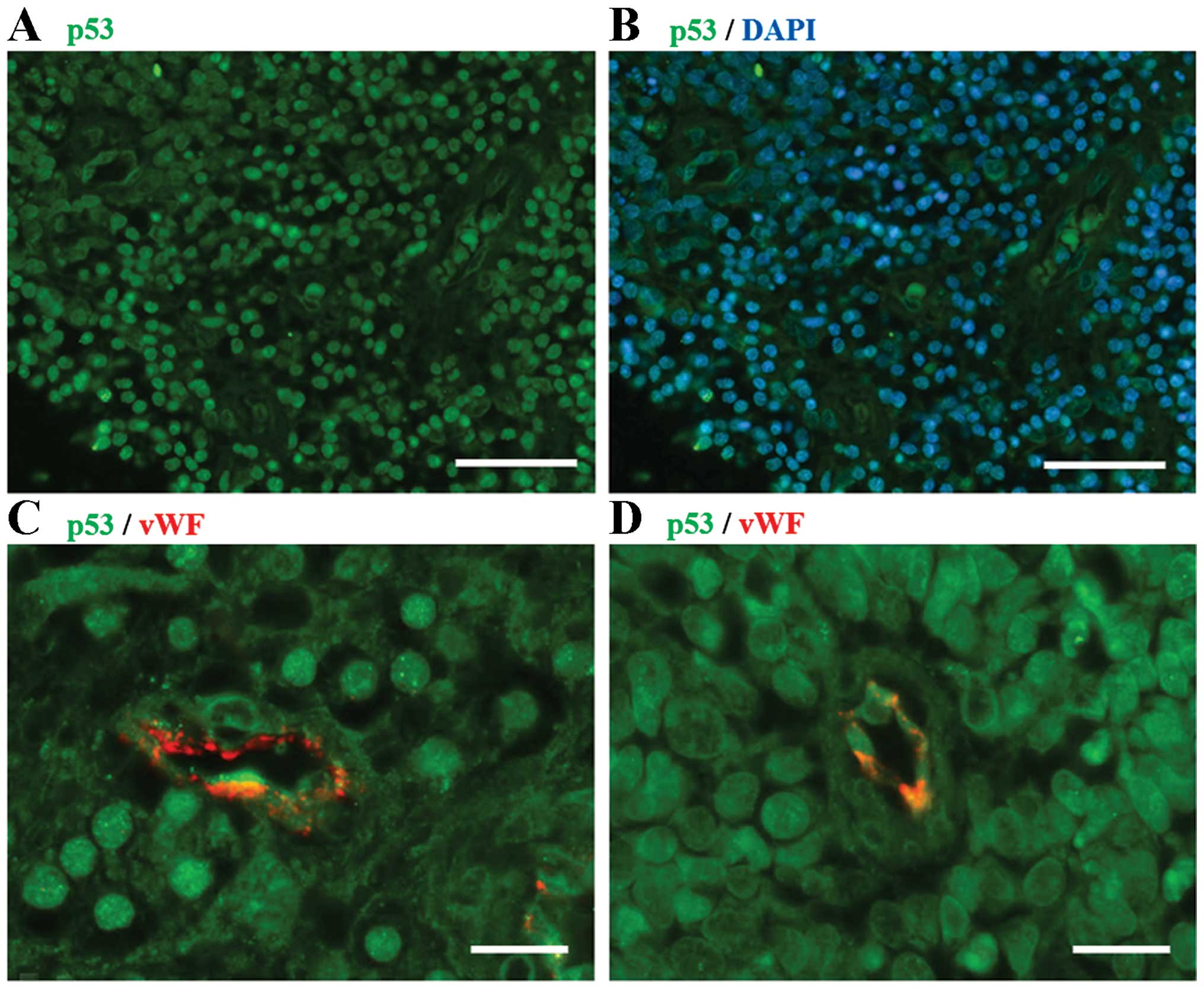
In the present study, human retinoblastoma cells
demonstrated a variable immunoreactivity against the anti-p53
antibody (FL-393), showing a similar trend with the results of a
previous study using the DO-1 monoclonal antibody (9). When comparing the distribution of p53
immunopositivity with DAPI nuclear counterstaining, the p53 protein
in retinoblastoma cells was mainly nuclear, with some cytoplasmic
localization. In cases of cytoplasmic p53 distribution, the
immunoreactivity was typically weak. In every specimen, a fraction
of vWF-positive tumor vascular endothelium presented nuclear
accumulation of the p53 protein (Fig.
4).
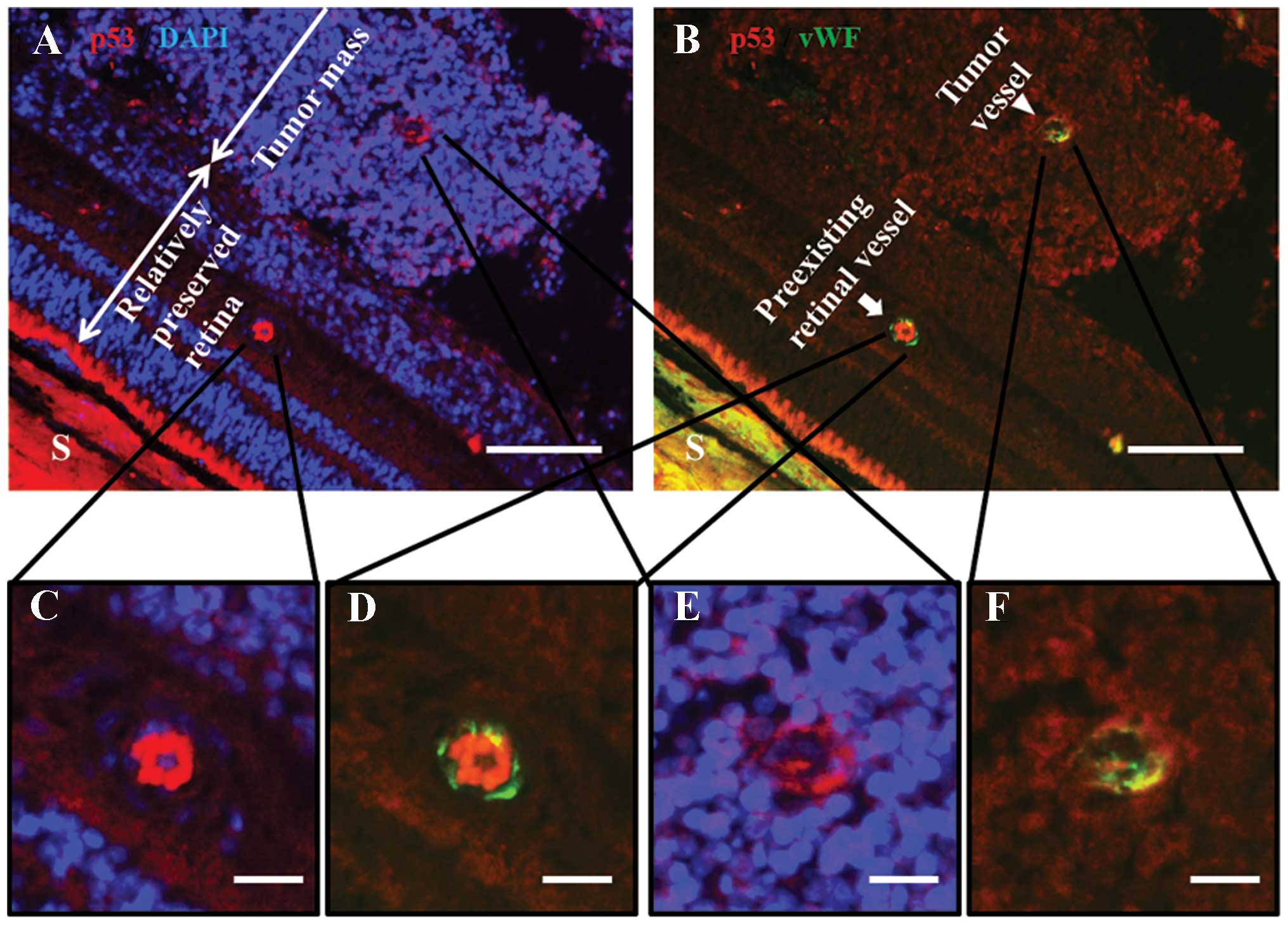
In a specimen which had a portion of relatively
preserved retinal layers, we found retinal vessels, presumed to be
pre-existing and not of neoplastic origin. To verify whether the
endothelial p53 immunopositivity exclusively occurs in tumor
vessels, we carefully reviewed the retinal vessels in the preserved
retina. The vWF-positive endothelial cells of those vessels did not
exhibit any p53 immunopositivity without exception (Fig. 5). The percentage of p53-positive
endothelium among the total vWF-positive tumor vascular endothelium
counted by two independent observers is presented in Table I. The average of the percentages
reported by each observer ranged from 31.3 to 33.8%.
Discussion
In human retinoblastoma, no genetic mutation of p53
was previously noted in the primary tumors (15). Yet, following a report by Laurie
et al (16), it is generally
accepted that in some human retinoblastoma cases, the p53 protein
remains wild-type but is functionally inactivated by abnormally
amplified MDM2 and MDMX. According to their results,
the amplification of MDMX which does not have ubiquitin
ligase activity is found in 65% of human retinoblastoma cases
(17), whereas that of MDM2
which results in ubiquitin-dependent p53 degradation is noted in
only 10% of human retinoblastomas (16,18).
Since several previous reports indicate that p53
nuclear staining of tumor cells is observed in most cases of human
retinoblastoma (9,10), the authors considered p53 nuclear
staining as a putative marker of cytogenetic abnormality in the
subjects with retinoblastoma (9,10). A
previous study concerning the p53 expression of normal mouse retina
revealed that the entire layer of retina did not show any
significant immunoreactivity against all three types of anti-p53
antibodies used for the immunofluorescence assay (including FL-393,
which was used in this study) (19). In another study that evaluated the
p53 expression of the normal mouse eye using anti-p53 driven CAT
staining, only the photoreceptor layer showed significant p53
expression throughout the entire retinal layers (20). Since we also evaluated the
contralateral eyes of the orthotopic model to rule out the
possibility of detecting physiological p53, nuclear staining of p53
could be considered as a putative marker of cytogenetic
abnormality.
Cultured retinoblastoma cell lines are known to show
cytoplasmic or mixed nuclear and cytoplasmic distribution of the
p53 protein in vitro. When stained with the p1801 monoclonal
antibody, the p53 protein exhibits a cytoplasmic localization in
Y79 cells (10). However, in the
present study, both Y79 and SNUOT-Rb1 cell induced orthotopic
retinoblastoma models consistently demonstrated nuclear
accumulation of p53 in the tumor cells. It has been reported that
the gene expression pattern of xenografted human glioma cells is
different from that of cells grown in vitro (21). A previous study which compared the
gene expression profile of human pancreatic cancer cells cultured
in ectopic and orthotopic conditions revealed that the organ
microenvironment is an important determinant of the gene expression
profile (22). The authors
postulated that the tumor microenvironment of the orthotopic
retinoblastoma model may have induced the molecular change which
affects the nuclear localization signal of the p53 protein.
Tumor-associated endothelial cells which share the
same genetic aberration of the original tumor have been noted in
several tumor types such as lymphoma and renal carcinoma (23,24).
More recently, direct evidence for the presence of tumor
cell-derived tumor-associated microvasculature has been noted in
human malignancies such as glioblastoma and neuroblastoma (6–8). To
the best of our knowledge, this is the first report concerning a
cytogenetic abnormality of the tumor vascular endothelium in human
and orthotopic models of retinoblastoma. Moreover, endothelial
immunoreactivity against anti-p53 antisera found in the orthotopic
retinoblastoma model could be a strong evidence for the direct
contribution of retinoblastoma cells to tumor vascularization.
The ratio of tumor-derived microvessels has been
reported to be as high as 78% in human neuroblastoma (6). According to a report which analyzed
tumor-specific chromosomal aberration by FISH assay in tumor
vascular endothelium of glioblastoma, 60.7% (ranging from 20 to
90%) of tumor endothelial cells showed chromosomal aberration
(7). In the present study, the
ratio of p53 nuclear accumulation among mature tumor vascular
endothelium was 32.9% (ranging from 31.3 to 33.8%). Although this
is a pilot study involving only a small number of cases, our
results showed a relatively low mean value and small inter-subject
variability compared to that of previous studies on glioblastoma.
In our opinion, the lower mean value in this study could have been
influenced by a low sensitivity of p53 immunostaining for the
detection of cytogenetic abnormalities. The small inter-subject
variability may be related to the homogeneous clinicopathological
features of our participants.
Briefly, p53 nuclear accumulation in some of the
mature tumor endothelium as well as in the tumor cells was
consistently observed in both the human and orthotopic models of
retinoblastoma. Our results imply a role of retinoblastoma cells in
tumor angiogenesis and this feature should be considered in future
vascular targeted therapy of retinoblastoma. A discrepancy in the
ratio of p53 immunopositivity between the SNUOT-Rb1 and Y79 cell
induced orthotopic model observed in this study may reflect the
different biological properties of each cell line. Previously our
group demonstrated that the doubling time of SNUOT-Rb1 cells was
shorter than Y79 cells (13), and
the tumor growth pattern was relatively more aggressive in the
SNUOT-Rb1 cell model. During tumor vascularization, the tumor
vascular endothelium is known to originate from neighboring
capillary (25) and bone
marrow-derived precursor cells (26). More recent studies revealed the role
of endothelial differentiation of tumor stem-like cells in tumor
angiogenesis. A more rapidly growing tumor needs an abundant
vascular supply, and if we assume that the endothelial
differentiation capacity of tumor stem-like cells is limited to a
certain degree, the proportion of tumor endothelium which is
derived from tumor stem-like cells could be lower in rapidly
growing tumors. Further study comparing the microvascular density
in both orthotopic retinoblastoma models could provide a better
understanding in regards to the role of p53-positive endothelium in
tumor vascularization.
Acknowledgements
This study was supported by the Bio-Signal Analysis
Technology Innovation Program (2009-0090895), the Pioneer Research
Program (2012-0009544), the Global Core Research Center (GCRC)
grant from NRF/MEST, Korea (2012-0001187), the Seoul National
University Research Grant (800-20130338), and the Seoul National
University Hospital Research Grant (04-2013-0520). We thank Ms.
Esther Yang for the technical assistance.
References
|
1
|
Burnier MN, McLean IW, Zimmerman LE and
Rosenberg SH: Retinoblastoma. The relationship of proliferating
cells to blood vessels. Invest Ophthalmol Vis Sci. 31:2037–2040.
1990.PubMed/NCBI
|
|
2
|
Rössler J, Dietrich T, Pavlakovic H, et
al: Higher vessel densities in retinoblastoma with local invasive
growth and metastasis. Am J Pathol. 164:391–394. 2004.PubMed/NCBI
|
|
3
|
Jockovich ME, Murray TG, Escalona-Benz E,
Hernandez E and Feuer W: Anecortave acetate as single and adjuvant
therapy in the treatment of retinal tumors of
LHBETATAG mice. Invest Ophthalmol Vis Sci.
47:1264–1268. 2006. View Article : Google Scholar : PubMed/NCBI
|
|
4
|
Lee SY, Kim DK, Cho JH, Koh JY and Yoon
YH: Inhibitory effect of bevacizumab on the angiogenesis and growth
of retinoblastoma. Arch Ophthalmol. 126:953–958. 2008. View Article : Google Scholar : PubMed/NCBI
|
|
5
|
Yang H, Cheng R, Liu G, et al: PEDF
inhibits growth of retinoblastoma by anti-angiogenic activity.
Cancer Sci. 100:2419–2425. 2009. View Article : Google Scholar : PubMed/NCBI
|
|
6
|
Pezzolo A, Parodi F, Corrias MV, Cinti R,
Gambini C and Pistoia V: Tumor origin of endothelial cells in human
neuroblastoma. J Clin Oncol. 25:376–383. 2007. View Article : Google Scholar : PubMed/NCBI
|
|
7
|
Ricci-Vitiani L, Pallini R, Biffoni M, et
al: Tumour vascularization via endothelial differentiation of
glioblastoma stem-like cells. Nature. 468:824–828. 2010. View Article : Google Scholar : PubMed/NCBI
|
|
8
|
Wang R, Chadalavada K, Wilshire J, et al:
Glioblastoma stem-like cells give rise to tumour endothelium.
Nature. 468:829–833. 2010. View Article : Google Scholar : PubMed/NCBI
|
|
9
|
Nork TM, Poulsen GL, Millecchia LL, Jantz
RG and Nickells RW: p53 regulates apoptosis in human
retinoblastoma. Arch Ophthalmol. 115:213–219. 1997. View Article : Google Scholar : PubMed/NCBI
|
|
10
|
Schlamp CL, Poulsen GL, Nork TM and
Nickells RW: Nuclear exclusion of wild-type p53 in immortalized
human retinoblastoma cells. J Natl Cancer Inst. 89:1530–1536. 1997.
View Article : Google Scholar : PubMed/NCBI
|
|
11
|
Seigel GM, Hackam AS, Ganguly A, Mandell
LM and Gonzalez-Fernandez F: Human embryonic and neuronal stem cell
markers in retinoblastoma. Mol Vis. 13:823–832. 2007.PubMed/NCBI
|
|
12
|
Zhong X, Li Y, Peng F, et al:
Identification of tumorigenic retinal stem-like cells in human
solid retinoblastomas. Int J Cancer. 121:2125–2131. 2007.
View Article : Google Scholar : PubMed/NCBI
|
|
13
|
Kim JH, Kim JH, Yu YS, Kim DH, Kim CJ and
Kim KW: Establishment and characterization of a novel,
spontaneously immortalized retinoblastoma cell line with adherent
growth. Int J Oncol. 31:585–592. 2007.PubMed/NCBI
|
|
14
|
Reid TW, Albert DM, Rabson AS, et al:
Characteristics of an established cell line of retinoblastoma. J
Natl Cancer Inst. 53:347–360. 1974.PubMed/NCBI
|
|
15
|
Kato MV, Shimizu T, Ishizaki K, et al:
Loss of heterozygosity on chromosome 17 and mutation of the p53
gene in retinoblastoma. Cancer Lett. 106:75–82. 1996. View Article : Google Scholar : PubMed/NCBI
|
|
16
|
Laurie NA, Donovan SL, Shih CS, et al:
Inactivation of the p53 pathway in retinoblastoma. Nature.
444:61–66. 2006.
|
|
17
|
Danovi D, Meulmeester E, Pasini D, et al:
Amplification of Mdmx (or Mdm4) directly contributes
to tumor formation by inhibiting p53 tumor suppressor activity. Mol
Cell Biol. 24:5835–5843. 2004.
|
|
18
|
Lu F, Chi SW, Kim DH, Han KH, Kuntz ID and
Guy RK: Proteomimetic libraries: design, synthesis, and evaluation
of p53-MDM2 interaction inhibitors. J Comb Chem. 8:315–325. 2006.
View Article : Google Scholar : PubMed/NCBI
|
|
19
|
Pokroy R, Tendler Y, Pollack A, Zinder O
and Weisinger G: p53 expression in the normal murine eye. Invest
Ophthalmol Vis Sci. 43:1736–1741. 2002.PubMed/NCBI
|
|
20
|
Weisinger G, Tendler Y and Zinder O:
Quantification of p53 expression in the nervous system. Brain Res
Brain Res Protoc. 6:71–79. 2000. View Article : Google Scholar : PubMed/NCBI
|
|
21
|
Camphausen K, Purow B, Sproull M, et al:
Influence of in vivo growth on human glioma cell line gene
expression: convergent profiles under orthotopic conditions. Proc
Natl Acad Sci USA. 102:8287–8292. 2005. View Article : Google Scholar : PubMed/NCBI
|
|
22
|
Nakamura T, Fidler IJ and Coombes KR: Gene
expression profile of metastatic human pancreatic cancer cells
depends on the organ microenvironment. Cancer Res. 67:139–148.
2007. View Article : Google Scholar : PubMed/NCBI
|
|
23
|
Fonsato V, Buttiglieri S, Deregibus MC,
Puntorieri V, Bussolati B and Camussi G: Expression of Pax2 in
human renal tumor-derived endothelial cells sustains apoptosis
resistance and angiogenesis. Am J Pathol. 168:706–713. 2006.
View Article : Google Scholar : PubMed/NCBI
|
|
24
|
Streubel B, Chott A, Huber D, et al:
Lymphoma-specific genetic aberrations in microvascular endothelial
cells in B-cell lymphomas. N Engl J Med. 351:250–259. 2004.
View Article : Google Scholar : PubMed/NCBI
|
|
25
|
Holash J, Maisonpierre PC, Compton D, et
al: Vessel cooption, regression, and growth in tumors mediated by
angiopoietins and VEGF. Science. 284:1994–1998. 1999. View Article : Google Scholar : PubMed/NCBI
|
|
26
|
Lyden D, Hattori K, Dias S, et al:
Impaired recruitment of bone-marrow-derived endothelial and
hematopoietic precursor cells blocks tumor angiogenesis and growth.
Nat Med. 7:1194–1201. 2001. View Article : Google Scholar : PubMed/NCBI
|