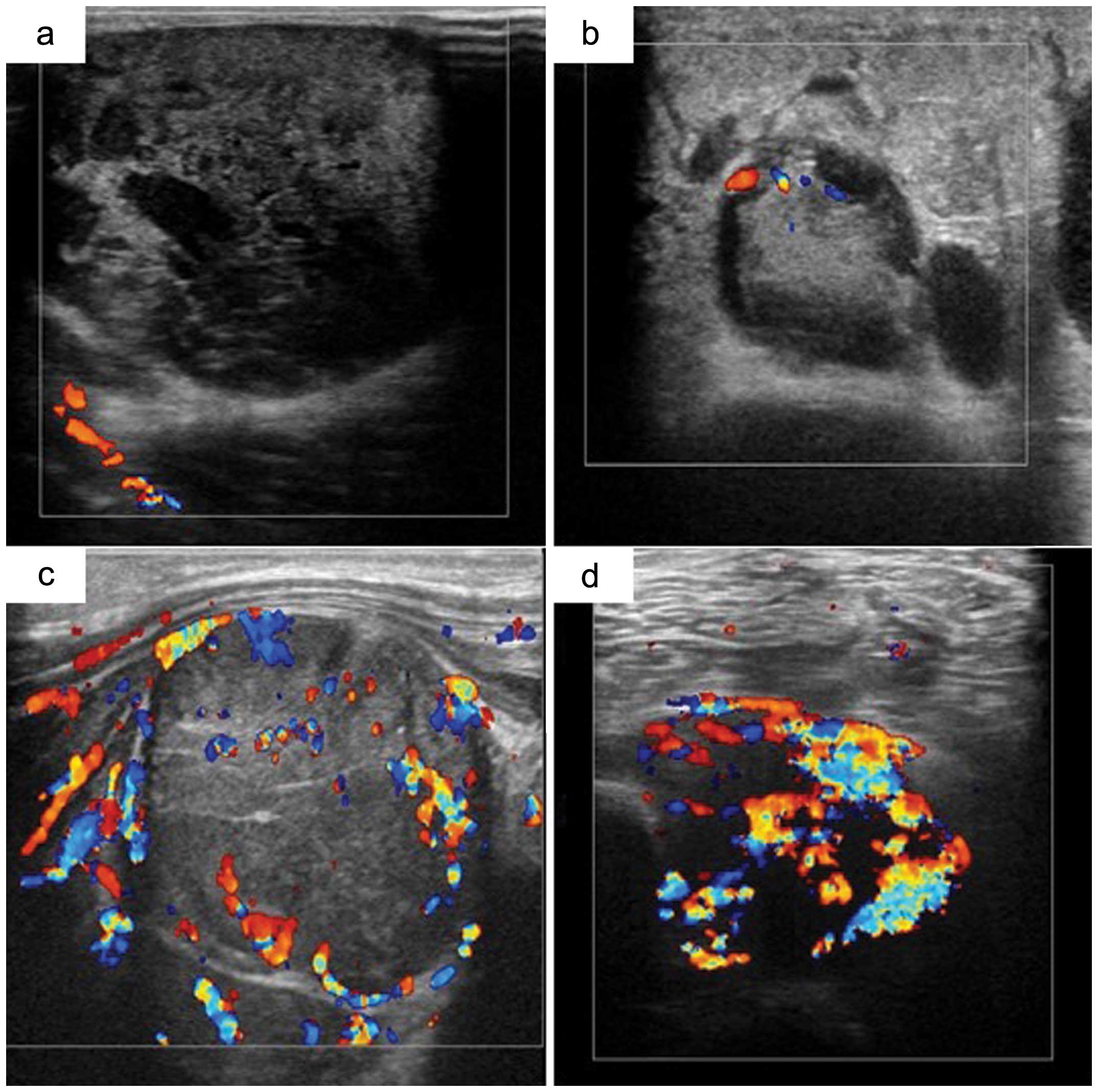
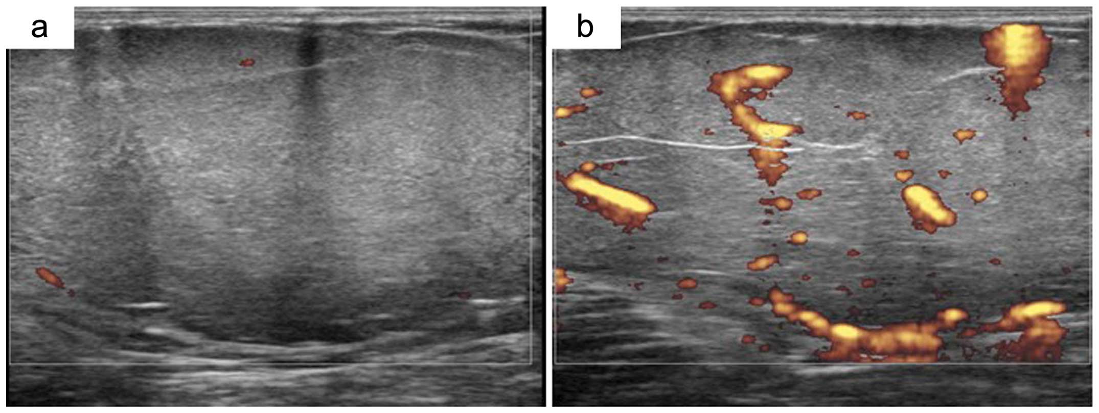
|
1
|
Kransdorf MJ and Murphey MD: Radiologic
evaluation of soft-tissue masses: a current perspective. AJR Am J
Roentgenol. 175:575–587. 2000. View Article : Google Scholar : PubMed/NCBI
|
|
2
|
Lagalla R, Iovane A, Caruso G, Lo Bello M
and Derchi LE: Color Doppler ultrasonography of soft-tissue masses.
Acta Radiol. 39:421–426. 1998. View Article : Google Scholar : PubMed/NCBI
|
|
3
|
Giovagnorio F, Andreoli C and De Cicco ML:
Color Doppler sonography of focal lesions of the skin and
subcutaneous tissue. J Ultrasound Med. 18:89–93. 1999.PubMed/NCBI
|
|
4
|
Widmann G, Riedl A, Schoepf D, Glodny B,
Peer S and Gruber H: State-of-the-art HR-US imaging findings of the
most frequent musculoskeletal soft-tissue tumors. Skeletal Radiol.
38:637–649. 2009. View Article : Google Scholar : PubMed/NCBI
|
|
5
|
Jo VY and Fletcher CD: WHO classification
of soft tissue tumours: an update based on the 2013 (4th) edition.
Pathology. 46:95–104. 2014. View Article : Google Scholar : PubMed/NCBI
|
|
6
|
Simon MA and Finn HA: Diagnostic strategy
for bone and soft-tissue tumors. J Bone Joint Surg Am. 75:622–631.
1999.PubMed/NCBI
|
|
7
|
Siegel R, Naishadham D and Jemal A: Cancer
statistics, 2013. CA Cancer J Clin. 63:11–30. 2013. View Article : Google Scholar
|
|
8
|
Drapé JL: Advances in magnetic resonance
imaging of musculoskeletal tumours. Orthop Traumatol Surg Res.
99(Suppl 1): S115–S123. 2013.
|
|
9
|
Landa J and Schwartz LH: Contemporary
imaging in sarcoma. Oncologist. 14:1021–1038. 2009. View Article : Google Scholar : PubMed/NCBI
|
|
10
|
Iwamoto Y: Diagnosis and treatment of soft
tissue tumors. J Orthop Sci. 4:54–65. 1999. View Article : Google Scholar
|
|
11
|
Simon MA and Biermann JS: Biopsy of bone
and soft-tissue lesions. J Bone Joint Surg Am. 75:616–621.
1993.PubMed/NCBI
|
|
12
|
Stacy GS and Dixon LB: Pitfalls in MR
image interpretation prompting referrals to an orthopedic oncology
clinic. Radio-graphics. 27:805–828. 2007.PubMed/NCBI
|
|
13
|
Kawaguchi N, Matsumoto S and Manabe J:
Clinical diagnosis of soft tissue tumors. J Orthop Sci. 3:225–238.
1998. View Article : Google Scholar : PubMed/NCBI
|
|
14
|
ESMO/European Sarcoma Network Working
Group. Soft tissue and visceral sarcomas: ESMO Clinical Practice
Guidelines for diagnosis, treatment and follow-up. Ann Oncol.
23(Suppl 7): vii92–vii99. 2012.PubMed/NCBI
|
|
15
|
Noria S, Davis A, Kandel R, Levesque J,
O’Sullivan B, Wunder J and Bell R: Residual disease following
unplanned excision of soft-tissue sarcoma of an extremity. J Bone
Joint Surg Am. 78:650–655. 1996.PubMed/NCBI
|
|
16
|
Hoshi M, Ieguchi M, Takami M, Aono M,
Taguchi S, Kuroda T and Takaoka K: Clinical problems after initial
unplanned resection of sarcoma. Jpn J Clin Oncol. 38:701–709. 2008.
View Article : Google Scholar : PubMed/NCBI
|
|
17
|
Jin W, Kim GY, Park SY, Chun YS, Rhyu KH,
Park JS and Ryu KN: The spectrum of vascularized superficial
soft-tissue tumors on sonography with a histopathologic
correlation: part 2, malignant tumors and their look-alikes. AJR Am
J Roentgenol. 195:446–453. 2010. View Article : Google Scholar : PubMed/NCBI
|
|
18
|
Braunstein EM, Silver TM, Martel W and
Jaffe M: Ultrasonographic diagnosis of extremity masses. Skeletal
Radiol. 6:157–163. 1981. View Article : Google Scholar : PubMed/NCBI
|
|
19
|
Adler RS, Bell DS, Bamber JC, Moskovic E
and Thomas JM: Evaluation of soft-tissue masses using segmented
color Doppler velocity images: preliminary observations. AJR Am J
Roentgenol. 172:781–788. 1999. View Article : Google Scholar : PubMed/NCBI
|
|
20
|
Lange TA, Austin CW, Seibert JJ, Angtuaco
TL and Yandow DR: Ultrasound imaging as a screening study for
malignant soft-tissue tumors. J Bone Joint Surg Am. 69:100–105.
1987.PubMed/NCBI
|
|
21
|
Chiou HJ, Chou YH, Chiu SY, Wang HK, Chen
WM, Chen TH and Chang CY: Differentiation of benign and malignant
superficial soft-tissue masses using grayscale and color doppler
ultrasonography. J Chin Med Assoc. 72:307–315. 2009. View Article : Google Scholar : PubMed/NCBI
|
|
22
|
Jin W, Kim GY, Park SY, Chun YS, Nam DH,
Park JS and Ryu KN: The spectrum of vascularized superficial
soft-tissue tumors on sonography with a histopathologic
correlation: part 1, benign tumors. AJR Am J Roentgenol.
195:439–445. 2010. View Article : Google Scholar
|
|
23
|
Verstraete KL, Van der Woude HJ,
Hogendoorn PC, De-Deene Y, Kunnen M and Bloem JL: Dynamic
contrast-enhanced MR imaging of musculoskeletal tumors: basic
principles and clinical applications. J Magn Reson Imaging.
6:311–321. 1996. View Article : Google Scholar : PubMed/NCBI
|
|
24
|
Ma LD, Frassica FJ, McCarthy EF, Bluemke
DA and Zerhouni EA: Benign and malignant musculoskeletal masses: MR
imaging differentiation with rim-to-center differential enhancement
ratios. Radiology. 202:739–744. 1997. View Article : Google Scholar : PubMed/NCBI
|
|
25
|
Belli P, Costantini M, Mirk P, Maresca G,
Priolo F and Marano P: Role of color Doppler sonography in the
assessment of musculoskeletal soft tissue masses. J Ultrasound Med.
19:823–830. 2000.PubMed/NCBI
|
|
26
|
Omoto K, Matsunaga H, Take N, et al:
Sentinel node detection method using contrast-enhanced
ultrasonography with sonazoid in breast cancer: preliminary
clinical study. Ultrasound Med Biol. 35:1249–1256. 2009. View Article : Google Scholar : PubMed/NCBI
|
|
27
|
Uemura H, Sano F, Nomiya A, et al:
Usefulness of perflubutane microbubble-enhanced ultrasound in
imaging and detection of prostate cancer: phase II multicenter
clinical trial. World J Urol. 31:1123–1128. 2013. View Article : Google Scholar : PubMed/NCBI
|
|
28
|
Goto E, Masuzaki R, Tateishi R, et al:
Value of post-vascular phase (Kupffer imaging) by contrast-enhanced
ultrasonography using Sonazoid in the detection of hepatocellular
carcinoma. J Gastroenterol. 47:477–485. 2012. View Article : Google Scholar : PubMed/NCBI
|
|
29
|
Chiou HJ, Chou YH, Chen WM, Chen W, Wang
HK and Chang CY: Soft-tissue tumor differentiation using 3D power
Doppler ultrasonography with echo-contrast medium injection. J Chin
Med Assoc. 73:628–633. 2010. View Article : Google Scholar : PubMed/NCBI
|
|
30
|
Metheny-Barlow LJ and Li LY: The enigmatic
role of angiopoietin- 1 in tumor angiogenesis. Cell Res.
13:309–317. 2003. View Article : Google Scholar : PubMed/NCBI
|
|
31
|
Bodner G, Schocke MF, Rachbauer F, Seppi
K, Peer S, Fierlinger A, Sununu T and Jaschke WR: Differentiation
of malignant and benign musculoskeletal tumors: combined color and
power Doppler US and spectral wave analysis. Radiology.
223:410–416. 2002. View Article : Google Scholar : PubMed/NCBI
|


