Introduction
Extrahepatic bile duct carcinoma is an epithelial
cancer originating from the bile ducts with features of
cholangiocytic differentiation. There is no significant
geographical variation in the incidence of extrahepatic bile duct
carcinoma. In the USA, extrahepatic bile duct carcinoma accounts
for 0.16% of all invasive cancers in males and 0.15% in females in
the general population (1).
Surgical treatment is the only curative therapy for extrahepatic
bile duct carcinoma and is therefore the treatment of choice if
feasible. The spreading cancer cells via the lymphatic to regional
lymph nodes are an important factor for tumor progression. In a
recent study, the median disease-specific survival rate after
surgery in patients with lymph node metastasis was lower than that
of patients without lymph node metastasis (19.3 vs. 53.5 months,
P<0.0001) (2).
Recent studies have demonstrated that an invasive
micropapillary carcinoma (IMPC) frequently shows aggressive tumor
behaviors with marked lymph-vascular invasion, resulting in poor
prognosis in several organs including the breast (3–5),
urinary bladder (6–8), lung (9–13),
parotid gland (14,15), pancreas (16), gallbladder (17), colorectum (18) and stomach (19–22).
To the best of our knowledge, however, there has been only one case
report of extrahepatic bile duct carcinoma which contains IMPC
(23). Therefore, the
clinicopathological significance of IMPC has not yet been
elucidated in extrahepatic bile duct. To clarify the significance
of IMPC in extrahepatic bile duct, the present study investigated
the clinicopathological features of 13 cases of extrahepatic bile
duct carcinoma containing IMPC, compared with 80 cases of
conventional extrahepatic bile duct carcinoma.
Materials and methods
Patients
We investigated consecutive bile duct carcinoma
surgical cases treated between January 2007 and December 2012,
after obtaining each patient’s informed consent for use of their
clinical records and pathological specimens at Hirosaki University
Hospital. The series consisted of 69 men and 24 women with a median
age of 70 years (range, 31–83 years). The carcinomas were located
in the perihilar (34 cases) and distal bile duct (59 cases),
according to the anatomic location (24). Curative resection and regional lymph
node dissection were dependent on the location of primary tumors:
pancreaticoduodenectomy or pylorus-preserving
pancreaticoduodenectomy was performed in 56 patients, bile duct
resection in 1 patient and combined hepatectomy with bile duct
resection in 29 cases and combined hepatectomy and
pancreaticoduodenectomy in 7 patients. Survival data were obtained
from hospital medical charts and the median observation period was
25.6 months (79 cases).
Pathological analysis
All surgically resected specimens were routinely
fixed with 10% formalin, then embedded in paraffin and stained with
hematoxylin and eosin (H&E) for pathological evaluation. The
following histological features were assessed: depth of invasion
(T-grade), histological type, lymphovascular invasion, perineural
invasion, the mode of infiltration pattern, lymph nodal metastasis
and IMPC component. We defined IMPC as tumor clusters of tumor
cells lying within clear spaces. The clear spaces have no
endothelial lining. Cases with tumor clusters only in the
lymphovascular channels or mucinous lesions, which resemble the
IMPC pattern, were excluded. We also evaluated the component ratio
of IMPC in the entire tumor tissues. In cases where the distinction
of lymphovascular invasion from the IMPC was difficult based on
H&E stained alone, we evaluated the cases with
immunohistochemical podoplanin (D2-40) staining. Degrees of
lymphatic, vessel and perineural invasion were classified as: 0, no
invasion; 1, mild invasion; 2, moderate invasion; 3, severe
invasion. Modes of infiltration pattern were classified into three
groups, i.e., INF-α, cancer nests showing expansive growth and
presenting the clear borderline between the tumor tissue and
stroma; INF-β, intermediate patterns of growth and invasive
structure of INF-α and INF-γ; and INF-γ, scirrhous growth with
unclear borderline of the invasive front. These data were evaluated
according to our previous study (25) and the General Rules for Surgical and
Pathological Studies on Cancer of the Biliary Tract (26) with reference to the World Health
Organization classification and staged according to the TMN
classification of the International Union Against Cancer (UICC)
(24). We also investigated
phenotypes of IMPC components using the immunohistochemical
procedure, as described below.
Immunohistochemistry
For histological examination, extrahepatic bile duct
carcinoma specimens were routinely fixed with formalin, embedded in
paraffin and thin-sectioned. Sections 4-μm-thick were mounted on
saline-coated glass slides. Immunohistochemical examination was
performed on deparaffinized sections using the standard
avidin-biotin-peroxidase complex method with automated
immunostainer (Benchmark XT; Ventana Medical System, Tucson, AZ,
USA). The characteristic ‘inside-out’ pattern of IMPC was confirmed
with the immunohistochemical MUC1 antibody. Furthermore, we
investigated the phenotypes of IMPC using MUC1, MUC2, MUC5AC and
MUC6 antibodies. Podoplanin (D2-40) was used for clarifying
lymphatic invasion. The antibodies we used were: MUC1 (1:50, clone
Ma696), MUC2 (1:50, clone Ccp), MUC5AC (1:100, clone CLH2), MUC6
(1:100, clone CLH5; all from Novocastra Laboratories), D2-40
(1:100, clone D2-40; Dako, Glostrup, Denmark).
Evaluation of immunohistochemistry
Luminal membranous immunoreactivities of the tumor
were judged as positive for MUC1, and cytoplasmic
immunoreactivities as positive for MUC2, MUC5AC and MUC6. The
results were classified into two groups based on the percentage of
positive cells with each staining as follows: negative group in
which <5% of cancer cells were stained, and positive group in
which ≥5% was stained.
Statistical analysis
Statistical comparisons between two groups were
analyzed using the Pearson’s Chi-square test for categorical data
and the Mann-Whitney test for continuous data. Survival curves were
constructed using the Kaplan-Meier method and differences in
survival were evaluated using the log-rank test. The relative
prognostic factors were analyzed with the Cox proportional hazards
regression model. Differences were considered to be statistically
significant if the P-value was <0.05. All statistical
evaluations were performed using R (http://www.r-project.org) and PASW statistics software
(version 18.0; SPSS, Inc., Chicago, IL, USA).
Results
Histological and immunohistochemical
findings of IMPC
We reviewed 93 cases of the extrahepatic bile duct
cancer and found 13 cases (14.0%) with IMPC component. The
clinicopathological findings are summarized in Table I. The 13 patients (11 men and two
women) with IMPC ranged in age from 49 to 80 years (mean, 68
years). Follow-up information was available for 10 patients, with a
median follow-up time of 17 months (range, 2–54 months). Eight
patients succumbed to the disease, one was alive with disease and
one was alive without recurrent disease. Histologically, IMPC
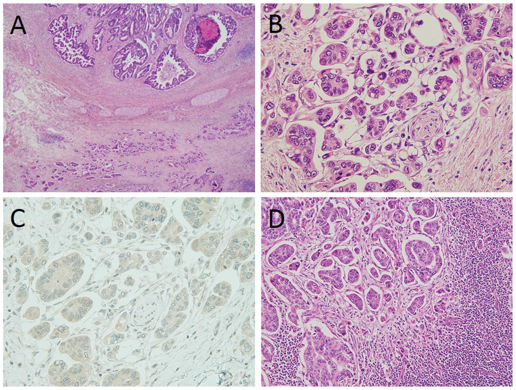
components ranging from 5 to 60% (mean ± SD, 17.7±15.4, data not
shown) of the entire tumor were mainly found at the front of the
tumor (Fig. 1A). The tumor was
characterized by small round to ovoid micropapillary tumor cell
clusters with no fibrovascular cores, lying within clear stromal
spaces (Fig. 1B). The clear stromal
spaces resembled lymphatic vessels, but were immunohistochemically
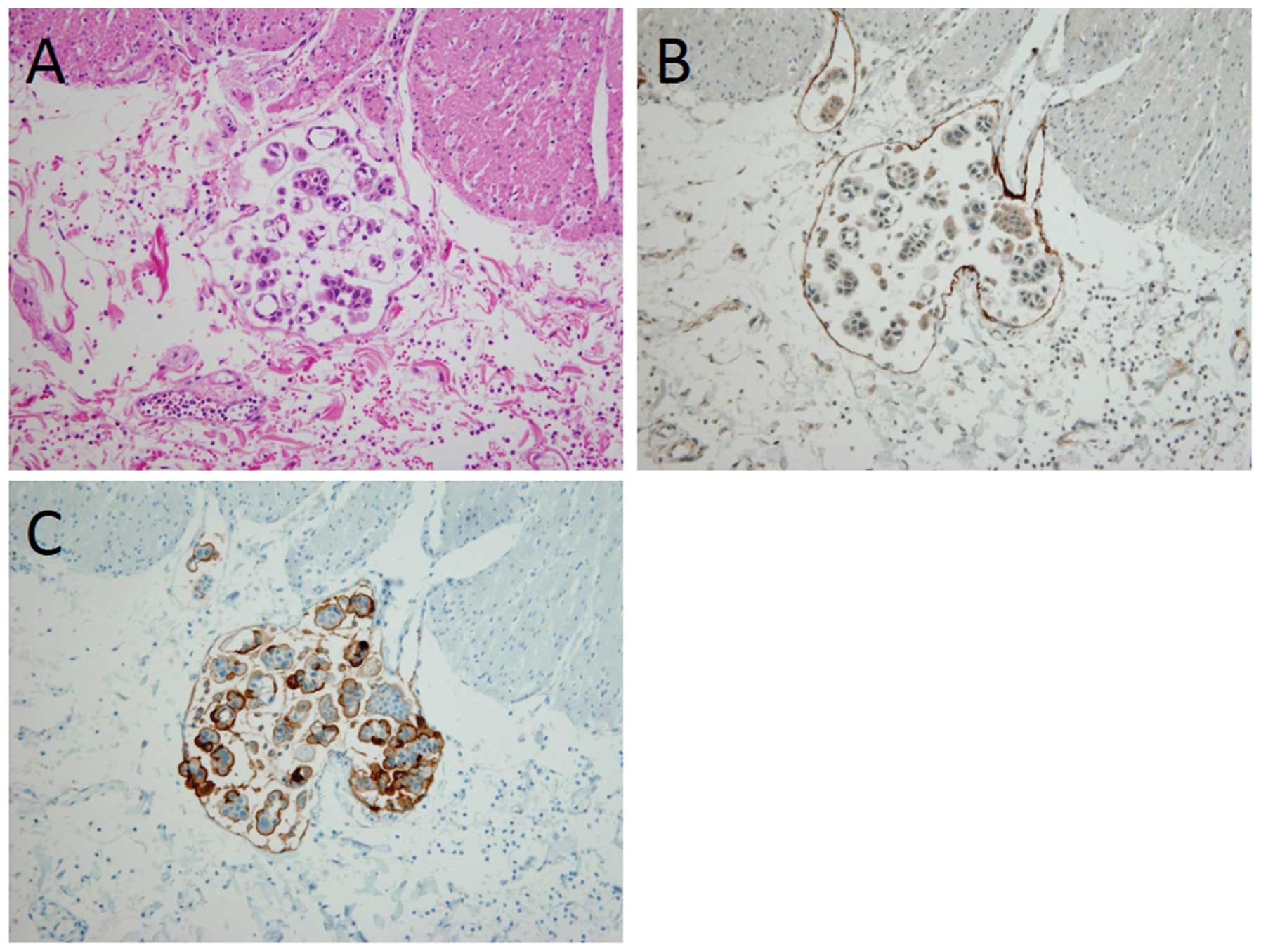
negative for D2-40, a marker of lymphatic vessel (Fig. 1C). Metastatic carcinomas of lymph
nodes also had IMPC component (Fig.
1D). The carcinoma cells characteristically displayed a reverse
polarity, known as an ‘inside-out’ growth pattern, mimicking
extensive lymphatic invasion (Fig. 2A
and B).
 | Table IExtrahepatic bile duct cancer with
IMPC component (13 cases). |
Table I
Extrahepatic bile duct cancer with
IMPC component (13 cases).
| Case No. | Age/gender | Percentage of IMPC
component | Lymph node
metastasis | Survival
(months) | Status |
|---|
| 1 | 74/M | 50–60 | 9/17 | 2 | Deceased |
| 2 | 49/F | 40–50 | 6/29 | 54 | Deceased |
| 3 | 71/M | 20–30 | 2/6 | N/A | N/A |
| 4 | 70/M | 10–20 | 2/6 | 24 | Deceased |
| 5 | 65/M | 10–20 | 1/8 | 7 | Deceased |
| 6 | 70/M | 10–20 | 1/10 | 18 | Alive |
| 7 | 72/M | 10–20 | 3/23 | N/A | N/A |
| 8 | 72/F | 5–10 | 2/21 | 30 | Deceased |
| 9 | 73/M | 5–10 | 3/39 | 8 | Deceased |
| 10 | 73/M | 5–10 | 0/19 | 27 | Alive |
| 11 | 43/M | 5–10 | 1/10 | 16 | Deceased |
| 12 | 74/M | 5–10 | 4/34 | 9 | Deceased |
| 13 | 80/M | 5–10 | 1/14 | N/A | N/A |
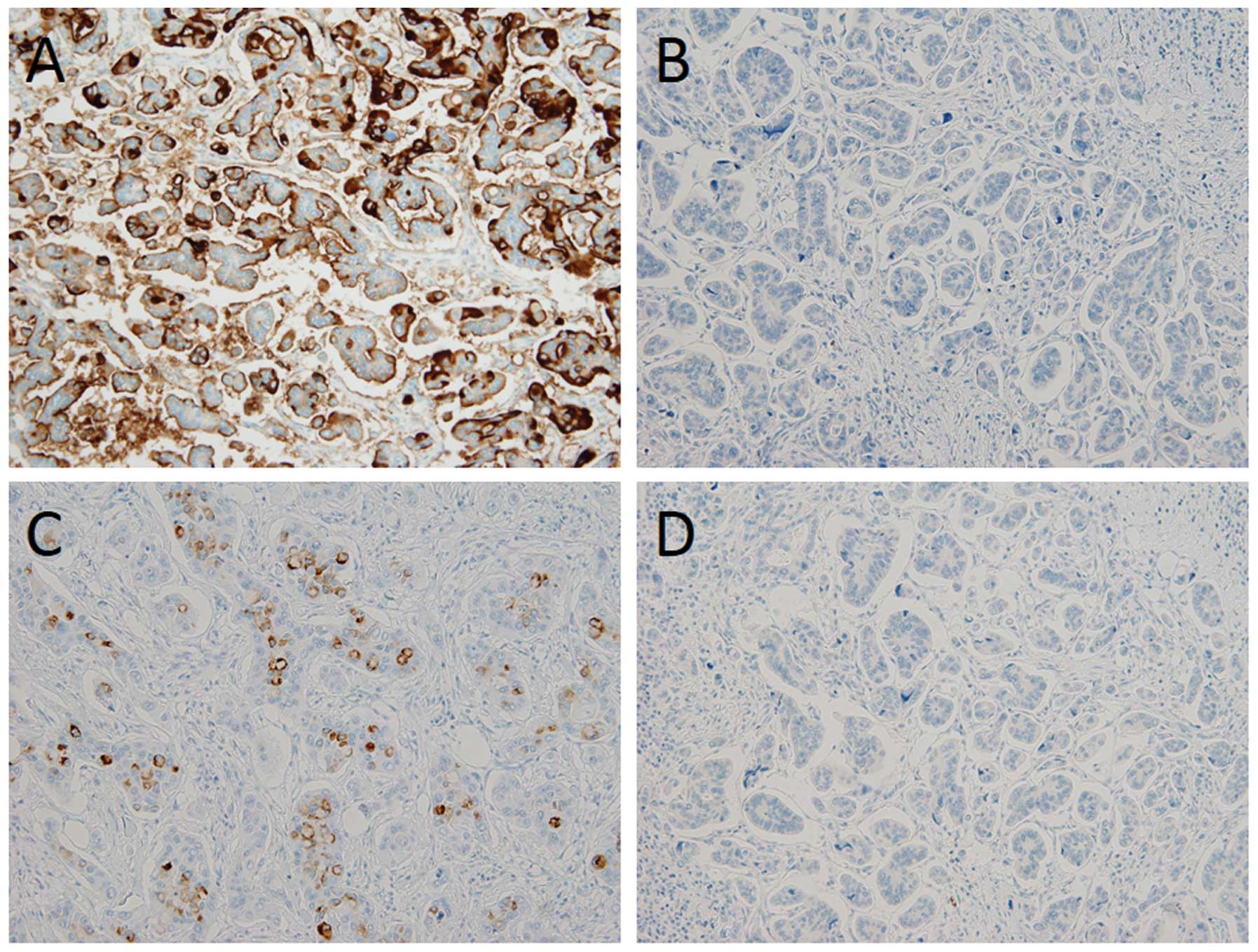
The results of immunohistochemistry are summarized
in Table II. Nine (69.2%) of the
13 cases of IMPC were positive for MUC1. MUC1 immunoreactivity was
predominantly detected at the surface of the cell cluster and
clearly exhibited the ‘inside-out’ growth pattern (Figs. 2C and 3A). MUC5AC was focally found in the
cytoplasm, as well as at the cell surface, in 4 of the 13 cases
(Fig. 3C). MUC2 and MUC6 staining
was negative in all cases of IMPC (Fig.
3B and D).
| Table IIImmunohistochemical characteristics
of IMPC component of extrahepatic bile duct cancer (13 cases). |
Table II
Immunohistochemical characteristics
of IMPC component of extrahepatic bile duct cancer (13 cases).
| Case No. | Age/ gender | Mucin
phenotype |
|---|
|
|---|
| MUC1 | MUC2 | MUC5AC | MUC6 |
|---|
| 1 | 74/M | + | − | + | − |
| 2 | 49/F | − | − | − | − |
| 3 | 71/M | − | − | − | − |
| 4 | 70/M | + | − | − | − |
| 5 | 65/M | + | − | + | − |
| 6 | 70/M | + | − | − | − |
| 7 | 72/M | + | − | − | − |
| 8 | 72/F | + | − | + | − |
| 9 | 73/M | − | − | − | − |
| 10 | 73/M | + | − | − | − |
| 11 | 43/M | − | − | − | − |
| 12 | 74/M | + | − | + | − |
| 13 | 80/M | + | − | − | − |
Clinicopathological findings of IMPC
The clinicopathological findings of extrahepatic
bile duct carcinoma with and without IMPC component are summarized
in Table III. The presence of
IMPC component was significantly correlated with lymph node
metastasis, lymphatic invasion and the mode of infiltration pattern
(P<0.001, P=0.016 and P=0.027, respectively). In addition, the
extrahepatic bile duct cancer with IMPC component frequently showed
lymph node metastasis with IMPC component (Table IV, P<0.001).
| Table IIIHistopathological characteristics of
extrahepatic bile duct cancer with or without IMPC component. |
Table III
Histopathological characteristics of
extrahepatic bile duct cancer with or without IMPC component.
| Variables | With IMPC component
(n=13) | Without IMPC
component (n=80) | P-value |
|---|
| Age | | | 0.956 |
| >65 | 3 | 58 | |
| ≤65 | 10 | 22 | |
| Gender | | | 0.503 |
| Male | 11 | 58 | |
| Female | 2 | 22 | |
| Location | | | 0.361 |
| Perihilara | 3 | 31 | |
| Distala | 10 | 49 | |
| Depth of
invasion | | | 0.071 |
| T1 or T2b | 3 | 40 | |
| T3 or T4b | 10 | 40 | |
| Histological
type | | | 0.332 |
|
Well-differentiated adenocarcinoma | 2 | 27 | |
| Other histological
type | 11 | 53 | |
| Lymph node
metastasis | | | <0.001 |
| pN (+) | 12 | 31 | |
| Lymphatic
invasion | | | 0.016 |
| ly0 or ly1 | 2 | 41 | |
| ly2 or ly3 | 11 | 39 | |
| Venous
invasion | | | 0.063 |
| v0 or v1 | 2 | 34 | |
| v2 or v3 | 11 | 46 | |
| Perineural
invasion | | | 0.332 |
| n0 or n1 | 2 | 27 | |
| n2 or n3 | 11 | 53 | |
| INFc | | | 0.027 |
| α or β | 1 | 34 | |
| γ | 12 | 46 | |
| Table IVHistological relationship between
primary tumor and lymph node metastasis with or without IMPC
component. |
Table IV
Histological relationship between
primary tumor and lymph node metastasis with or without IMPC
component.
| With IMPC component
(n=13) | Without IMPC
component (n=80) | P-value |
|---|
| Lymph node
status |
| pN (+) | 12 (92.3%) | 31 (38.8%) | <0.001 |
|
| Primary with IMPC
component (%) | Primary without
IMPC component (%) | P-value |
|
| Lymph node with
IMPC component | 11 (25.6) | 1 (2.3) | <0.001 |
| Lymph node without
IMPC component | 1 (2.3) | 30 (69.8) | |
Univariate and multivariate analysis
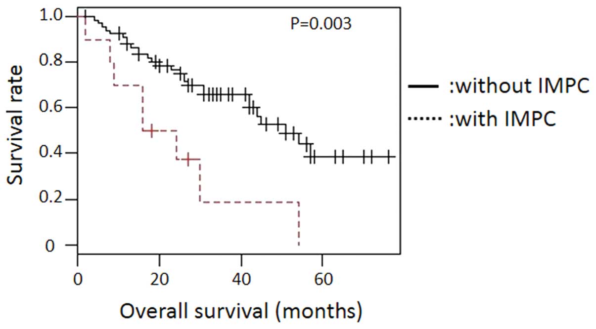
Survival curves based on univariate survival
analysis demonstrated that the patients with IMPC were associated
with poor prognosis (Fig. 4,
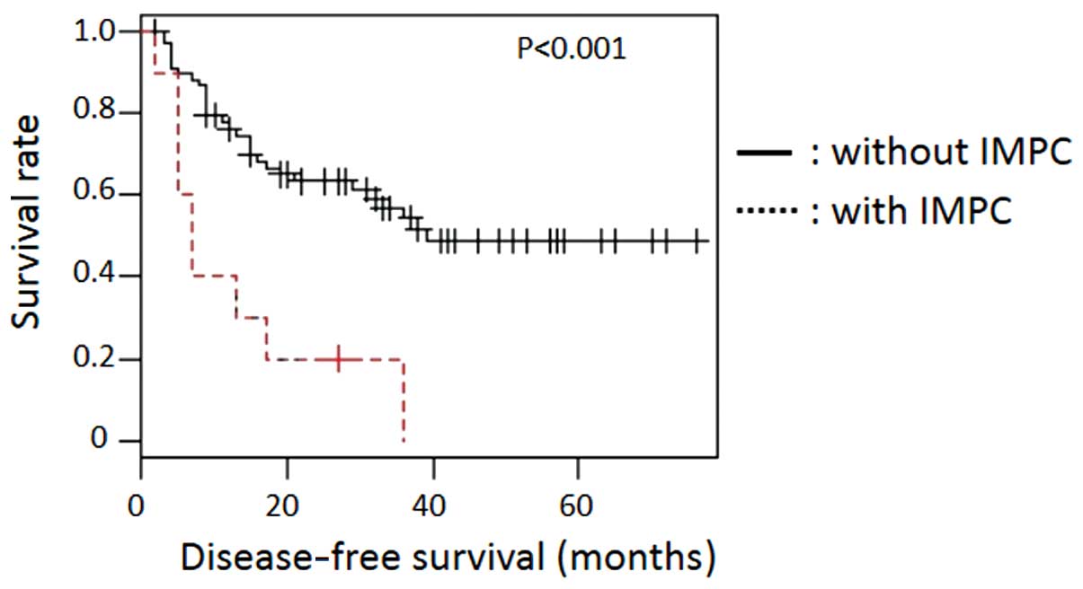
P=0.003) and poor disease-free survival (Fig. 5, P<0.001). To clarify potential
prognostic indicators, we analyzed various pathological factors
investigated (Table V). Univariate
analysis revealed that the following factors were correlated with
poor prognosis: IMPC component [relative risk (RR) 3.195, 95%
confidence interval (CI) 1.437–7.107, P=0.004], depth of invasion
(RR 3.261, 95% CI 1.583–6.719, P=0.001), histological type (RR
6.787, 95% CI 2.072–22.23, P=0.002), lymphatic invasion (RR 4.028,
95% CI 1.973–8.224, P<0.001), venous invasion (RR 2.714, 95% CI
1.327–5.551, P=0.006) and lymph node metastasis (RR 3.868, 95% CI
1.946–7.684, P<0.001).
| Table VUnivariate and multivariate analysis
of prognostic factors of survival. |
Table V
Univariate and multivariate analysis
of prognostic factors of survival.
| Variables | Values (%) | Univariate analysis
P-value | Multivariate
analysis P-value |
|---|
| IMPC component | | 0.004 | 0.643 |
| With IMPC
component | 10 (12.7) | | |
| Without IMPC
component | 69 (87.3) | | |
| Depth of
invasion | | 0.001 | 0.234 |
| T1 or T2a | 40 (50.6) | | |
| T3 or T4a | 39(49.6) | | |
| Histological
type | | 0.002 | 0.062 |
|
Well-differentiated adenocarcinoma | 24 (30.1) | | |
| Other histological
type | 55 (69.6) | | |
| Lymphatic
invasion | | <0.001 | 0.223 |
| ly0 or ly1 | 43 (54.4) | | |
| ly2 or ly3 | 36 (45.6) | | |
| Venous
invasion | | 0.006 | 0.76 |
| v0 or v1 | 36 (45.6) | | |
| v2 or v3 | 43 (54.4) | | |
| Perineural
invasion | | 0.071 | - |
| n0 or n1 | 29 (36.7) | | |
| n2 or n3 | 50 (63.3) | | |
| INFb | | 0.064 | - |
| α or β | 30 (38.0) | | |
| γ | 49 (62.0) | | |
| Lymph node
metastasis | | <0.001 | 0.116 |
| pN (−) | 50 (63.3) | | |
| pN (+) | 29 (36.7) | | |
Discussion
In the present study, we clarified
clinicopathological characteristics of IMPC of the extrahepatic
bile duct. We clarified that IMPC frequently showed aggressive
tumor growth with lymphatic invasion and lymph node metastasis,
resulting in short overall/disease-free survival of the patients.
This is the first report describing clinicopathological malignant
potential of IMPC of the extrahepatic bile duct.
IMPC component has been reported in several organs,
such as breast, urinary bladder, lung, parotid gland, pancreas,
gallbladder, colorectum and stomach (3–22). The
previous reports revealed that IMPC exhibited a tendency for
lymphatic invasion and lymph node metastasis. In our study, IMPC
component of extrahepatic bile duct ranged from 5 to 60% of the
entire tumor, while with IMPC components were found in the other
organs: 10–90% in salivary duct carcinoma (15), 10–90% in gastric carcinoma (21), 5–10% in gallbladder carcinoma
(17), 5–95% in breast cancer
(27). IMPC component, regardless
of its tumor volume, has been shown to have malignant potential for
lymphatic invasion and lymph node metastasis.
The molecular mechanisms of the malignant potential
of IMPC have not yet been fully elucidated, while IMPC is
histologically characterized by the ‘inside-out’ pattern. A
previous study proposed that extracellular matrix (ECM) contributed
to the IMPC structure (28).
Madin-Darby canine kidney (MDCK) cells exhibited ‘inside-out’
structure without type I collagen, but were able to reorient their
cell polarity under the presence of type I collagen in ECM. Here,
the reorientation of cell polarity was shown to be related to RAC1,
PI3-kinase and a PKC (28). Cancer
cells of IMPC may have abnormalities of RAC1 suppression cascade
and show the characteristic ‘inside-out’ structures (29). We also revealed that MUC1 expression
was predominantly at the surface of tumor clusters. The MUC1
expression was similar to the other organs (30). Human MUC1 is a high molecular weight
transmembrane glycoprotein, which is apically expressed in the
majority of glandular epithelia (31). Increased MUC1 expression has been
shown to inhibit integrin-mediated cell adhesion between cancer
cells and ECM (32) and to decrease
adhesion to type I collagen (33).
Based on this evidence, IMPC expressing MUC1 may reduce the cell
adhesion from ECM and result in forming the characteristic
‘inside-out’ structures.
Our study revealed that IMPC structures were found
not only in the primary carcinoma lesions, but also in the foci of
lymphatic vessels and metastatic lymph nodes. The ‘inside-out’ IMPC
structures were thought to play an important role in the lymph node
metastasis. Of note, IMPC exhibited stromal desmoplastic reactions
around the ‘inside-out’ cancer cell clusters. The desmoplastic
changes consisted of proliferation of fibroblasts and collagen
fibers and were found not only in the primary lesion, but also in
the parts of lymph node metastasis. The desmoplastic changes are
thought to be associated with epithelial-mesenchymal transition,
which may contribute to an aggressive growth of the invasive
cancer.
The results of univariate analysis revealed that
IMPC was significantly correlated with poor patient prognosis, but
the multivariate analysis using Cox proportional hazards model
showed that IMPC was not an independent prognostic factor for
overall survival. We suspected the reason why it was not an
independent factor was that IMPC may be strongly associated with
lymphatic invasion and lymph node metastasis. An important clinical
issue is that the presence of IMPC indicates malignant potential,
even if the component is small. Therefore, pathologists should
describe presence of IMPC component in the diagnostic report, even
if the component is a small part of the extrahepatic bile duct
carcinoma.
Acknowledgements
This study was supported by Grants-in Aid for
Science from the Ministry of Education, Culture, Sports, Science
and Technology in Japan, and a Grant for Hirosaki University
Institutional Research.
References
|
1
|
Bosman Fred T and Carnerio Fatima: World
health Organization Classification of Tumours of the Digestive
System. IARC Press; Lyon: pp. 272–273. 2012
|
|
2
|
Ito K, Ito H, Allen PJ, et al: Adequate
lymph node assessment for extrahepatic bile duct adenocarcinoma.
Ann Surg. 251:675–681. 2010. View Article : Google Scholar : PubMed/NCBI
|
|
3
|
Tavassoli FA and Devilee P: World Health
Organization Classification of Tumours of the Breast and Female
Genital Organs. IARC Press; Lyon: pp. 35–36. 2003
|
|
4
|
Siriaunkgul S and Tavassoli FA: Invasive
micropapillary carcinoma of the breast. Mod Pathol. 6:660–662.
1993.PubMed/NCBI
|
|
5
|
Nassar H, Wallis T, Andea A, Dey J, Adsay
V and Visscher D: Clinicopathologic analysis of invasive
micropapillary differentiation in breast carcinoma. Mod Pathol.
14:836–841. 2001. View Article : Google Scholar : PubMed/NCBI
|
|
6
|
Lopez-Beltran A, Montironi R, Blanca A and
Cheng L: Invasive micropapillary urothelial carcinoma of the
bladder. Hum Pathol. 41:1159–1164. 2010. View Article : Google Scholar : PubMed/NCBI
|
|
7
|
Perepletchikov AM and Parwani AV:
Micropapillary urothelial carcinoma: clinico-pathologic review.
Pathol Res Pract. 205:807–810. 2009. View Article : Google Scholar : PubMed/NCBI
|
|
8
|
Samaratunga H and Khoo K: Micropapillary
variant of urothelial carcinoma of the urinary bladder; a
clinicopathological and immunohistochemical study. Histopathology.
45:55–64. 2004. View Article : Google Scholar : PubMed/NCBI
|
|
9
|
Kuroda N, Hamaguchi N, Takeuchi E, Ohara
M, Hirouchi T and Mizuno K: Lung adenocarcinoma with a
micropapillary pattern: a clinicopathological study of 25 cases.
APMIS. 11:381–385. 2006. View Article : Google Scholar : PubMed/NCBI
|
|
10
|
Kuroda N, Hamaguchi N, Ohara M, Hirouchi
T, Miyzaki E and Mizuno K: Intracytoplasmic lumina in invasive
micropapillary carcinoma of the lung. Diagn Cytopathol. 34:224–226.
2006. View
Article : Google Scholar : PubMed/NCBI
|
|
11
|
Kuroda N, Hamauzu T, Toi M, et al:
Pulmonary adenocarcinoma with micropapillary component: an
immunohistochemical study. Case report. APMIS. 113:550–554. 2005.
View Article : Google Scholar : PubMed/NCBI
|
|
12
|
Makimoto Y, Nabeshima K, Iwasaki H, et al:
Micropapillary pattern: a distinct pathological marker to
subclassify tumours with a significantly poor prognosis within
small peripheral lung adenocarcinoma (</=20 mm) with mixed
bronchioloalveolar and invasive subtypes (Noguchi’s type C
tumours). Histopathology. 46:677–684. 2005.PubMed/NCBI
|
|
13
|
Amin MB, Tamboli P, Merchant SH, et al:
Micropapillary component in lung adenocarcinoma: a distinctive
histologic feature with possible prognostic significance. Am J Surg
Pathol. 26:358–364. 2002. View Article : Google Scholar : PubMed/NCBI
|
|
14
|
Yamamoto H, Uryu H, Segawa Y and
Tsuneyoshi M: Aggressive invasive micropapillary salivary duct
carcinoma of the parotid gland. Pathol Int. 58:322–326. 2008.
View Article : Google Scholar : PubMed/NCBI
|
|
15
|
Nagao T, Gaffey TA, Visscher DW, et al:
Invasive micropapillary salivary duct carcinoma: a distinct
histologic variant with biologic significance. Am J Surg Pathol.
28:319–326. 2004. View Article : Google Scholar : PubMed/NCBI
|
|
16
|
Khayyata S, Basturk O and Adsay NV:
Invasive micropapillary carcinomas of the ampullo-pancreatobiliary
region and their association with tumor-infiltrating neutrophils.
Mod pathol. 18:1504–1511. 2005. View Article : Google Scholar
|
|
17
|
Hara S, Kijima H, Okada K and Igarashi Y:
Invasive micropapillary variant of the gallbladder adenocarcinoma
and its aggressive potential for lymph node metastasis. Biomed Res.
31:89–95. 2010. View Article : Google Scholar : PubMed/NCBI
|
|
18
|
Kim MJ, Hong SM, Jang SJ, et al: Invasive
colorectal micropapillary carcinoma: an aggressive variant of
adenocarcinoma. Hum Pathol. 37:809–815. 2006. View Article : Google Scholar : PubMed/NCBI
|
|
19
|
Fujita T, Gotohda N, Kato Y, et al:
Clinicopathological features of stomach cancer with invasive
micropapillary component. Gastric cancer. 15:179–187. 2012.
View Article : Google Scholar : PubMed/NCBI
|
|
20
|
Ohtsuki Y, Kuroda N, Yunoki S, et al:
Immunohistochemical analysis of invasive micropapillary carcinoma
pattern in four cases of gastric cancer. Med Mol Morphol.
46:114–121. 2013. View Article : Google Scholar : PubMed/NCBI
|
|
21
|
Ushiku T, Matsusaka K, Iwasaki Y, et al:
Gastric carcinoma with invasive micropapillary pattern and its
association with lymph node metastasis. Histopathology.
59:1081–1089. 2011. View Article : Google Scholar : PubMed/NCBI
|
|
22
|
Shimoda M, Okada Y, Hayashi Y, et al:
Primary invasive micropapillary carcinoma of the stomach. Pathol
Int. 58:513–517. 2008. View Article : Google Scholar : PubMed/NCBI
|
|
23
|
Kondo T: Bile duct adenocarcinoma with
minor micropapillary component: a case report. Cases J. 2:512009.
View Article : Google Scholar : PubMed/NCBI
|
|
24
|
Sobin LH, Gospodarowicz MK and Wittekind
CH: TMN Classification of Malignant Tumors. 7th edition.
Wiley-Liss; New York: 2009
|
|
25
|
Okada K, Kijima H, Imaizumi T, et al:
Clinical significance of wall invasion pattern of
subserosa-invasive gallbladder carcinoma. Oncol Rep. 28:1531–1536.
2012.PubMed/NCBI
|
|
26
|
Japanese Society of Biliary Surgery.
General Rules for the Surgical and Pathological Studies on Cancer
of the Biliary Tract. 5th edition. Kanehara Shuppan; Tokyo:
2003
|
|
27
|
Walsh MM and Bleiweiss IJ: Invasive
micropapillary carcinoma of the breast: eighty cases of an
underrecognized entity. Hum Pathol. 32:583–589. 2001. View Article : Google Scholar : PubMed/NCBI
|
|
28
|
Liu KD, Datta A, Yu W, et al: Rac1 is
required for reorientation of polarity and lumen formation through
a PI 3-kinase-dependent pathway. Am J Physiol Renal Physiol.
293:F1633–F1640. 2007. View Article : Google Scholar : PubMed/NCBI
|
|
29
|
Sahai E and Marshall CJ: RHO-GTPases and
cancer. Nat Rev Cancer. 2:133–142. 2002. View Article : Google Scholar
|
|
30
|
Nassar H, Pansare V, Zhang H, et al:
Pathogenesis of invasive micropapillary carcinoma: role of MUC1
glycoprotein. Mod Pathol. 17:1045–1050. 2004. View Article : Google Scholar : PubMed/NCBI
|
|
31
|
Winterford CM, Walsh MD, Leggett BA and
Jass JR: Ultrastructural localization of epithelial mucin core
proteins in colorectal tissues. J Histochem Cytochem. 47:1063–1074.
1999. View Article : Google Scholar : PubMed/NCBI
|
|
32
|
Wesseling J, van der Valk SW, Vos HL,
Sonnenberg A and Hilkens J: Episialin (MUC1) overexpression
inhibits integrin-mediated cell adhesion to extracellular matrix
components. J Cell Biol. 129:255–265. 1995. View Article : Google Scholar : PubMed/NCBI
|
|
33
|
Hudson MJ, Stamp GW, Chaudhary KS, et al:
Human MUC1 mucin: a potent glandular morphogen. J Pathol.
194:373–383. 2001. View Article : Google Scholar : PubMed/NCBI
|