Introduction
Hepatocellular carcinoma (HCC) is one of the most
deadly and prevalent malignant diseases, not only in developing
countries but also in the US and Europe, and is therefore a serious
disease burden for public health (1). In recent years, the morbidity and
mortality rates of HCC have been continuously rising, placing HCC
as the second leading cause of deaths among nine most common
cancers in China (2). Due to the
lack of sensitive and specific biomarkers in the clinic, the
existing early-stage screening strategies have limited benefits to
the prevention of HCC. Even with advanced treatments, such as
surgery, chemotherapy and radiotherapy, HCC still presents a dismal
prognosis with an overall survival rate as low as 25–39% (3). Therefore, there is an urgent need to
identify new tumor markers and novel targeted agents for the early
diagnosis of HCC and develop efficacious regimens for treating HCC
in order to improve patient outcome.
In the past few decades, many cellular signaling
pathways and genes have been implicated in the pathogenesis of HCC,
including the Ras/Raf/MEK/ERK signaling pathway (4–7).
Progestin and adipoQ receptor family member III(PAQR3), also named
RKTG, negatively regulates the Ras/Raf/MEK/ERK signaling cascade
and the G protein-coupled receptor (GPCR) Gβγ subunit signaling
pathway (8,9). PAQR3 is a member of the PAQR family of
proteins comprising an intriguing group of recently discovered
receptors and a seven transmembrane protein localized at the Golgi
apparatus (8,10). PAQR3 expression inhibits
EGF-stimulated ERK and RSK phosphorylation, blocks NGF-mediated
cell differentiation, and antagonizes Ras and Raf-1-stimulated
Elk-1 transactivation (8). Through
interaction with Raf-1, PAQR3 alters the localization of Raf-1 from
the cytoplasm to the Golgi apparatus, blocks EGF-stimulated Raf-1
membrane translocation, and reduces the interaction of Raf-1 with
Ras and MEK1 (11). PAQR3 has a
negative effect on cell proliferation, migration, sprouting and
angiogenesis of endothelial cells by suppressing mitogen-activated
protein kinase signaling (12). In
addition, PAQR3 negatively regulates the transactivation activity
of hypoxia-inducible factor 1α (HIF-1α) by inhibiting formation of
the HIF-1α/p300 complex and suppressing VEGF transcription, thereby
reducing hypoxia-induced VEGF production (12). Furthermore, recent studies have
found that PAQR3 is implicated in a set of human cancers, including
clear-cell renal cell carcinoma and colorectal cancer (12,13).
However, the significance of altered expression of PAQR3 in HCC has
not been fully elucidated due to the lack of studies using fresh
human tumor samples.
In the present study, we investigated the expression
of PAQR3 in primary HCC using real-time quantitative RT-PCR,
western blotting and immunohistochemistry. We also identified the
relationship between PAQR3 expression and clinicopathological
features of the HCC cases and evaluated the potential prognostic
value of PAQR3 expression for the survival of HCC patients. In
addition, we proved that PAQR3 can inhibit tumor cell growth and
colony formation in vitro. To the best of our knowledge, the
data generated in the present study represent the first report
correlating the presence of PAQR3 with clinicopathological
characteristics and the survival of HCC patients.
Materials and methods
Ethics statement
This study was approved by the Institutional Ethics
Review Board (IERB) of Union Hospital, and written informed consent
was obtained from all patients studied.
Cell lines and culture conditions
The hepatic cancer cell lines, Hep-3B, Hep-G2 and
Huh-7, and the normal liver cell line LO2 were obtained from the
Committee of Type Culture Collection of the Chinese Academy of
Sciences (Shanghai, China). Cell lines were cultured in RPMI-1640
media supplemented with 10% heat-inactive fetal bovine serum (FBS).
The cells were incubated at 37°C in a humidified chamber containing
5% CO2.
Patients and tissue samples
To investigate PAQR3 expression in HCC, 62 HCC
tissues and paired adjacent non-cancerous tissues, not less than 2
cm away from the HCC tissues, were collected respectively from the
HCC patients undergoing hepatectomy at Union Hospital between March
and June 2012. The diagnosis of HCC was confirmed by
histopathology. Surgically removed fresh tissues were immediately
frozen in liquid nitrogen and stored at −70°C until use. In
addition, another 132 paraffin-embedded HCC tissues were collected
between 2004 and 2007. These patients did not receive preoperative
chemotherapy or radiotherapy.
The follow-up data of the HCC patients included in
this study were obtained as a routine practice. Our outpatient
department performed postoperative follow-up on the HCC patients at
intervals of every 3 months for the first 2 years, every 6 months
from the 3rd to the 5th year, and annually thereafter for an
additional 5 years, or until patient death, whichever occurred
first. Overall survival was defined as the time from surgery to
patient death or the last follow-up and was used as a measure of
prognosis, while disease-free survival was defined as the time from
surgery to disease recurrence or metastasis. The histological types
of HCC were defined according to the classification criteria of
WHO.
RNA extract preparation and quantitative
real-time PCR (qRT-PCR)
Total RNA extracts were prepared using TRIzol
reagent (Invitrogen; 15596018) according to the manufacturer’s
protocol. Next, RNA (2 μg) was reversely transcribed into first
strand cDNA by M-MLV reverse transcriptase (Promega; M1705)
according to the manufacturer’s instructions. PAQR3 and GAPDH were
then amplified by quantitative real-time PCR using the following
primers: PAQR3 forward, 5′-ATGGCAAAGGCTCCGTTCT-3′ and reverse,
5′-AGTTTAGTTGTCCTGGAAAGTACCG-3′; GAPDH forward,
5′-CTCCTCCTGTTCGACAGTCAGC-3′ and reverse,
5′-CCCAATACGACCAAATCCGTT-3′. Gene specific amplification was
performed in an ABI 7900HT real-time PCR system (Life Technologies,
USA) with a 15-μl PCR mix containing 0.5 μl of cDNA, 7.5 μl of 2X
SYBR-Green Master Mix (Invitrogen; 11760500) and 200 nM of the
appropriate primers. The mixture was preheated for 10 min at 95°C
followed by 45 cycles of amplification (30 sec at 95°C and 1 min at
60°C, respectively). The resolution curve was measured at 95°C for
15 sec, 60°C for 15 sec and 95°C for 15 sec. The Ct (threshold
cycle) value of each sample was calculated, and the relative
expression of PAQR3 mRNA was normalized to the GAPDH value
(2−ΔCt method).
Preparation of total protein extracts and
western blot analysis
The hepatic cancer cells and frozen HCC samples
including tumor or non-tumor tissue control were homogenized in
RIPA lysis buffer. After centrifugation at 12,000 rpm, at 4°C for
20 min, ~25 μg of the protein samples was run on a 12% SDS-PAGE gel
and transferred to polyvinylidene difluoride membranes (PVDF,
Millipore). After blocking non-specific binding sites for 45 min
with 5% non-fat milk, the membranes were incubated with goat
polyclonal antibody against PAQR3 (1:300) and GAPDH (1:1,000; both
from Santa Cruz, CA, USA) at 4°C overnight, respectively. The
membranes were then washed with TBST for three times, (15 min each
time) and then incubated with HRP-conjugated anti-goat secondary
antibody (1:10,000) for 45 min at room temperature. The membranes
were developed by an enhanced chemiluminescence system (ECL; Cell
Signaling) after being washed with TBST for three times. The
intensity of the protein bands was determined by densitometry using
ImageJ software (Image J; National Institutes of Health).
Immunohistochemistry and PAQR3
immunostaining scoring principle
Formalin-fixed and paraffin-embedded tissues were
sectioned (4-μm) for immunohistochemical analysis. The sections
were deparaffinized and rehydrated through a graded series of
aqueous ethanol solutions. For the antigen retrieval, the slides
were immersed in EDTA [1 mmol/l, (pH 8.0)] and boiled for 20 min in
a microwave oven. After rinsing with PBS, endogenous peroxidase was
blocked with 0.3% hydrogen peroxide for 15 min at room temperature.
The slides were incubated with the primary antibody (1:100; Santa
Cruz), in a humidified chamber at 4°C overnight. Following
additional washing with PBS for three times, the sections were
sequentially incubated with horseradish peroxidase-conjugated
secondary antibody (Envision™ detection kit; GK500705; Gene Tech)
at 37°C for 30 min, and then washed three times with PBS. Finally,
3,3′-diaminobenzidine tetrahydrochloride (DAB) was used for the
signal development and then the sections were lightly
counterstained with 20% hematoxylin. The slides were dehydrated and
mounted on coverslips. For the negative controls, PBS was used in
place of the primary antibody.
The total PAQR3 immunostaining score was calculated
as the sum of the percent positivity of the stained tumor cells and
the staining intensity. The percent positivity was scored as ‘0’
(<5%, negative), ‘1’ (5–25%, sporadic), ‘2’ (>25–50%, focal),
‘3’ (>50%, diffuse). The staining intensity was scored as ‘0’
(no staining), ‘1’ (weakly stained), ‘2’ (moderately stained), and
‘3’ (strongly stained). Both the percent positivity of cells and
the staining intensity were determined under double-blind
conditions. The PAQR3 immunostaining score was expressed as the
value of the percent positivity score multiply the staining
intensity score, which ranged from 0 to 9. The PAQR3 expression
levels were defined as follows: − (score 0–1), + (score 2–3), ++
(score 4–6) and +++ (score >6). Based on the PAQR3 expression
levels, the HCC patients were divided into two groups: low PAQR3
expression group (PAQR3− or PAQR3+) and high PAQR3 expression group
(PAQR3++ or PAQR3+++).
Expression plasmid and transient
transfections
A eukaryotic expression plasmid pcDNA3.1(+)
containing full-length human PAQR3 cDNA was obtained from the
GenePharma Company (Shanghai, China). Empty vector was used as the
negative control. Hep-3B cells were cultured in 6-well plates until
they reached 85–90% confluency, and then transient transfections
were performed using Lipofectamine 2000 (Invitrogen) according to
the manufacturer’s instructions. Forty-eight hours after
transfection, gene expression was examined by western blot
analysis, and then cell proliferation and colony formation analyses
were performed.
RNA oligonucleotides and cell
transfections
To silence PAQR3 expression, siRNAs were synthesized
by GenePharma Company (Shanghai, China). The siRNA sequences were
as follows: siRNA-PAQR3 sense, 5′-CGCAGAUUCAUCCCAA UUATT-3′ and
antisense, 5′-AUAUGCCAUAUUUGGUGG CTT-3′; the negative control (NC)
sense, 5′-UUCUCCGAACG UGUCACGUTT-3′ and antisense,
5′-ACGUGACACGUUCG GAGAATT-3′. Four hundred picomoles siRNA-PAQR3 or
NC was transfected into 2×10⁵ LO2 cells using
Lipofectamine RNAi MAX reagent (Invitrogen, USA) according to the
manufacturer’s protocol. After that, cell proliferation and colony
formation assays were performed.
Proliferation assay
The cell growth rate of Hep-G2, Huh-7, Hep-3B and
LO2 cells was detected by MTS cell proliferation assay. Cells were
seeded in a 96-well plate at a density of 5×10²
cells/well. The cell growth rate was detected using cell
proliferation MTS kit according to the manufacturer’s instructions
(Promega, USA). Each experiment was performed three times
independently.
Colony formation assay
For the colony formation assay, PAQR3-expressing
Hep-3B cells or control Hep-3B cells were plated in a 6-well plate
at a density of 1×10³ cells/well. After 10 days of
culture, surviving colonies (>50 cells/colony) were counted with
crystal violet (0.5%) staining. Colony-forming efficiency (CFE %)
was defined as the ratio of the number of colonies formed in the
culture to the number of cells inoculated. The experiment was
conducted three times independently.
Statistical analysis
Statistical analyses were performed using programs
including the Statistical Package for the Social Sciences, ver.
17.0 (SPSS Inc, Chicago, IL, USA). Paired-samples t-test was used
to compare protein expression of PAQR3 in HCC tissues with paired
adjacent non-tumor tissue samples. Comparisons of PAQR3 tumor
expression levels with clinicopathological features were evaluated
by χ2 tests. Follow-up time was censored if the patient
was lost to follow-up. Survival curves were calculated using the
Kaplan-Meier method and compared by the log-rank test. Cox
proportional-hazard analysis was used for the univariate and
multivariate analyses to explore the effect of PAQR3 expression on
HCC clinicopathological variables on survival. The two-tailed
unpaired Student’s t-test was used to assess differences in cell
growth rate and colony formation. Differences were considered
significant at P<0.05.
Results
PAQR3 mRNA expression in the primary HCC
tissues
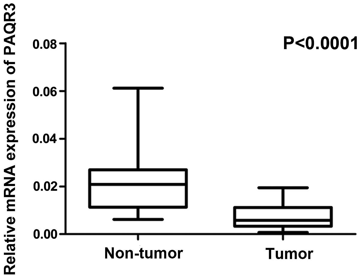
Real-time quantitative PCR was conducted on 62
paired surgical samples consisting of HCC (cancerous tissues and
the corresponding adjacent non-cancerous tissues from the same
patients) to explore expression of PAQR3 at the mRNA level. As
shown in Fig. 1, the median level
of PAQR3 mRNA expression was significantly lower in the cancer
tissues than that in the corresponding adjacent non-tumor tissues
(P<0.0001), and 80.64% of the subjects (50/62) displayed lower
mRNA expression of PAQR3 in cancer tissues. All of the 62 paired
tissues were independently tested twice.
PAQR3 protein expression in the primary
HCC tissue samples and cell lines
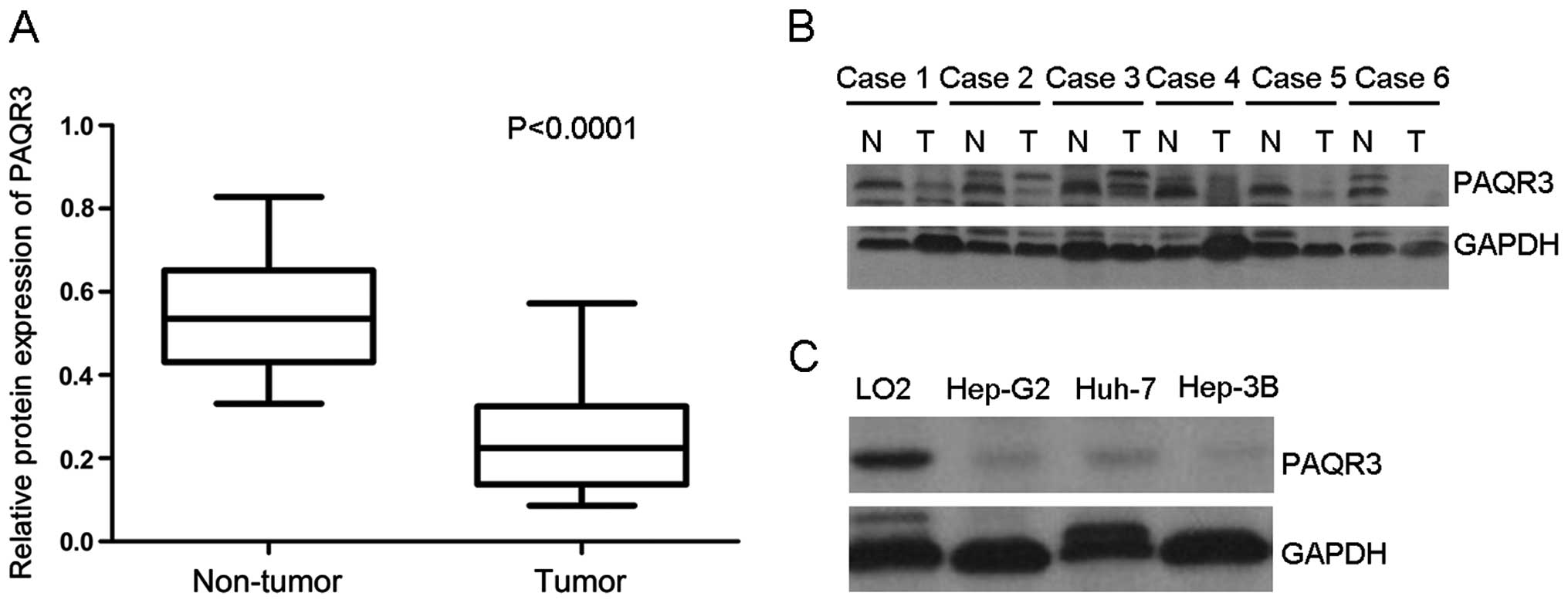
To investigate whether the expression of PAQR3 was
decreased at the protein level, western blotting was performed on
the 26 paired HCC cancer tissues and their corresponding adjacent
non-cancer tissues. Consistent with the qRT-PCR results, western
blot analyses showed that PAQR3 expression was decreased in 79.92%
(20/26) of cancerous tissues compared with their corresponding
non-cancerous tissues (P<0.001, Fig.
2A). As shown in Fig. 2B, 6 HCC
cancer tissues exhibited lower PAQR3 expression to some extent, and
all of the 6 paired tissues were tested three times independently.
Likewise, the PAQR3 protein expression was markedly decreased in
the hepatic cancer cell lines, Hep-G2, Huh-7, particularly in
Hep-3B, compared with normal hepatic cell line LO2 (Fig. 2C).
PAQR3 expression in HCC and its
relationship with clinicopathological features
We further examined PAQR3 expression in a total of
132 HCC surgical specimens using immunohistochemical staining.
Among them, 79/132 (59.85%) cases showed low expression of PAQR3
(PAQR3− or PAQR3+) and 53/132 (40.15%) cases exhibited high PAQR3
expression (PAQR3++ or PAQR3+++) (Table
I). In the positive cases, PAQR3 was detected as being
localized in the cytoplasm of the cells (Fig. 3B, D and G). PAQR3 expression was
found positive in normal liver tissues (Fig. 3G and H). In cases with adjacent
hyperplastic tissue, we often observed a sharp contrast between
infiltrative tumor areas of negative staining and the adjacent
non-tumor tissue of positive staining (Fig. 3H). Analysis of the relationship
between expression of PAQR3 and various clinicopathological
parameters is listed in Table I.
The expression of PAQR3 was significantly correlated with serum AFP
(P=0.034), mitotic count (P=0.012), tumor size (P=0.028),
histologic grade (P=0.005) and recurrence (P=0.004).
Well-differentiated cases showed strongly positive expression
(Fig. 3A and B), moderately
differentiated cases showed poorly positive expression of PAQR3
(Fig. 3C and D), and in most poorly
differentiated cases, we often did not detect PAQR3 expression
(Fig. 3E and F). There was no
statistically significant difference between PAQR3 expression and
age, HBsAg status, liver cirrhosis and metastasis.
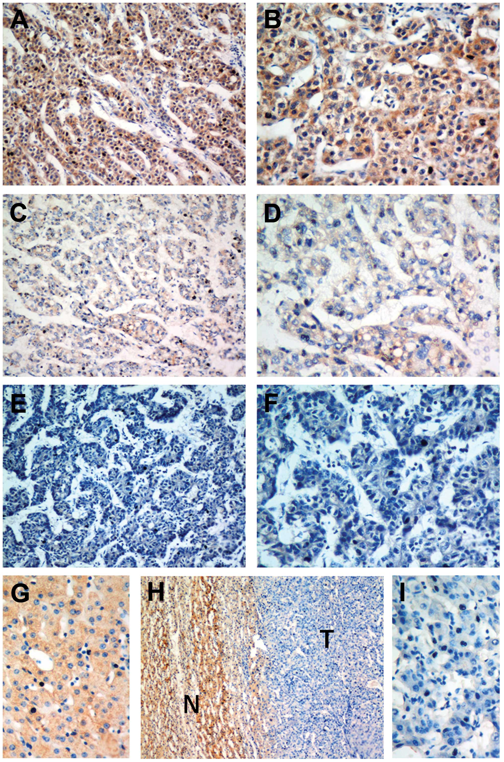 | Figure 3Immunohistochemical analysis of PAQR3
protein expression in primary HCC surgical specimens. (A and B)
Well-differentiated HCC scored as PAQR3(++). (C and D) Moderately
differentiated HCC scored as PAQR3(++). (E and F) Poorly
differentiated HCC scored as PAQR3(−). (H) Normal liver tissue
scored as PAQR3(+++). (G–I) Immunostaining of HCC tumor area and
the adjacent non-tumor area. N, non-tumor tissue; T, tumor tissue.
Magnifications: H, ×100; A, C, E, ×200; B, D, F, G, I, ×400. |
 | Table ICorrelation of PAQR3 expression and
clinicopathological data in the 132 hapatocarcinoma cases. |
Table I
Correlation of PAQR3 expression and
clinicopathological data in the 132 hapatocarcinoma cases.
| Clinicopathological
parameters | N | PAQR3 expression | χ2 | P-value |
|---|
|
|---|
| Low | High |
|---|
| All cases | 132 | 79 | 53 | | |
| Age (years) | | | | 0.807 | 0.369 |
| <50 | 67 | 42 | 25 | | |
| ≥50 | 65 | 37 | 28 | | |
| HBsAg status | | | | 2.504 | 0.114 |
| Negative | 19 | 10 | 9 | | |
| Positive | 113 | 69 | 44 | | |
| Serum AFP (μg/l) | | | | 4.515 | 0.034a |
| ≤400 | 41 | 19 | 22 | | |
| >400 | 91 | 60 | 31 | | |
| Mitotic count | | | | 6.356 | 0.012a |
| ≤20 | 67 | 33 | 34 | | |
| >20 | 65 | 46 | 19 | | |
| Liver
cirrhosis | | | | 0.556 | 0.456 |
| No | 60 | 38 | 22 | | |
| Yes | 72 | 41 | 31 | | |
| Tumor size
(cm) | | | | 4.803 | 0.028a |
| <5 | 57 | 28 | 29 | | |
| ≥5 | 75 | 51 | 24 | | |
| Histologic
grade | | | | 7.748 |
0.005b |
| Well | 23 | 9 | 14 | | |
| Moderate | 63 | 35 | 28 | | |
| Poor | 46 | 35 | 11 | | |
| Recurrence | | | | 8.100 |
0.004b |
| No | 94 | 49 | 45 | | |
| Yes | 38 | 30 | 8 | | |
| Metastasis | | | | 0.044 | 0.834 |
| No | 111 | 66 | 45 | | |
| Yes | 21 | 13 | 8 | | |
Relationship between PAQR3 expression and
patient survival
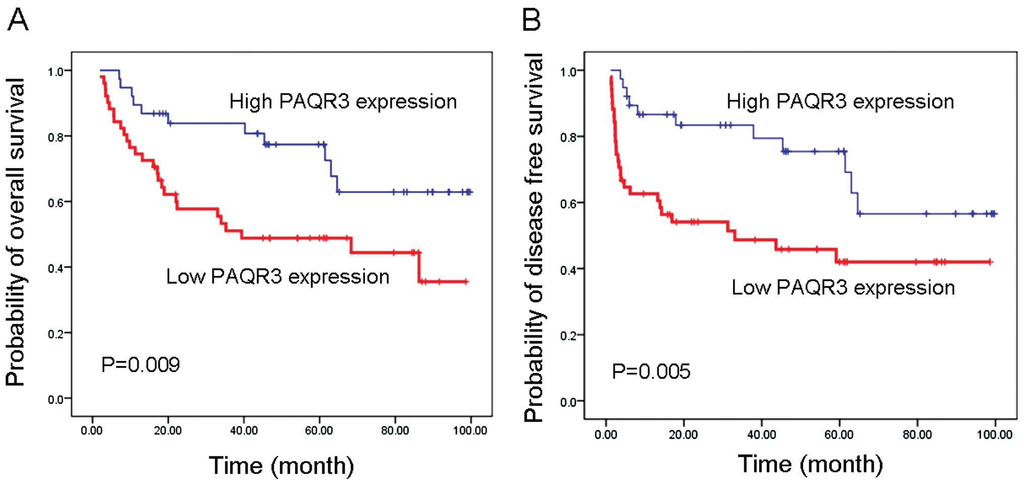
Having demonstrated the correlation of PAQR3
expression with clinicopathological features, we proceeded to
examine the relationship between PAQR3 expression and patient
survival. The prognostic value of PAQR3 for overall and
disease-free survival of HCC patients was evaluated between
patients with high and low PAQR3 expression phenotypes.
Kaplan-Meier curves showed that, in primary HCC category, patients
carrying high PAQR3 expression phenotypes had prolonged overall
survival time (77.98±5.84 months) and disease-free survival
(74.58±6.49 months), whereas patients expressing lower levels of
PAQR3 showed a much shorter overall survival time (53.07±5.85
months; log-rank test, P=0.009; Fig.
4A) and disease-free survival (47.98±6.37 months; log-rank
test, P=0.005; Fig. 4B). The
cumulative 5-year overall survival rate was 71.97% for patients
with high levels of PAQR3. Surprisingly, the survival rate
dramatically declined to 40.91% for patients with low levels of
PAQR3.
Univariate and multivariate analyses of
the prognostic variables in HCC patients
To determine whether PAQR3 could serve as a risk
factor with clinical usefulness, univariate and multivariate
analyses were carried out using Cox proportional hazards model to
compare the impact of PAQR3 expression on other clinical
pathological features of the HCC patients. Cox regression analyses
revealed that a low level of PAQR3 was associated with a
significantly increased risk of cancer-related death (P<0.01) in
HCC patients. The HRs indicated that serum AFP, histological grade
and recurrence were unfavorable predictors based on univariate
analysis (Tables II and III). After adjusting for potential
confounding factors, low expression of PAQR3 in HCC was found to
predict poorer survival in an independent manner (Tables II and III). Analyses using multivariate Cox
regression model for clinicopathological diagnoses showed that
PAQR3 and recurrence were independent prognostic parameters and
predicted poor overall survival and disease-free survival. Thus,
PAQR3 expression may play a role in predicting overall survival and
disease-free survival of HCC patients (Tables II and III).
| Table IIUnivariate and multivariate analyses
of the overall survival in 132 HCC patients. |
Table II
Univariate and multivariate analyses
of the overall survival in 132 HCC patients.
| Univariate
analysis | Multivariate
analysis |
|---|
|
|
|
|---|
| Parameters | HR | 95% CI | P-value | HR | 95% CI | P-value |
|---|
| PAQR3 | 0.445 | 0.182–0.793 |
0.006b | 0.608 | 0.375–0.984 | 0.010a |
| Age | 0.846 | 0.602–1.103 | 0.583 | | | |
| HBsAg status | 1.501 | 0.928–2.355 | 0.206 | | | |
| Serum AFP | 2.271 | 1.329–3.881 | 0.030a | 1.611 | 0.898–2.890 | 0.110 |
| Mitotic count | 1.072 | 0.894–2.460 | 0.225 | | | |
| Liver
cirrhosis | 0.812 | 0.654–1.081 | 0.108 | | | |
| Tumor size | 1.413 | 0.906–2.204 | 0.127 | | | |
| Histologic
grade | 1.840 | 0.223–3.211 | 0.027a | 0.945 | 0.495–1.808 | 0.869 |
| Recurrence | 9.173 | 3.052–18.655 |
<0.001c | 6.492 | 2.153–13.906 |
<0.001c |
| Metastasis | 1.845 | 0.858–2.638 | 0.154 | | | |
| Table IIIUnivariate and multivariate analyses
of the disease-free survival in 132 HCC patients. |
Table III
Univariate and multivariate analyses
of the disease-free survival in 132 HCC patients.
| Univariate
analysis | Multivariate
analysis |
|---|
|
|
|
|---|
| Parameters | HR | 95% CI | P-value | HR | 95% CI | P-value |
|---|
| PAQR3 | 0.395 | 0.155–0.623 |
0.004b | 0.425 | 0.307–0.722 |
0.006b |
| Age | 0.805 | 0.594–1.235 | 0.571 | | | |
| HBsAg status | 1.959 | 0.779–4.981 | 0.158 | | | |
| Serum AFP | 3.199 | 1.624–4.302 | 0.021a | 1.416 | 0.539–2.312 | 0.267 |
| Mitotic count | 1.035 | 0.923–2.211 | 0.232 | | | |
| Liver
cirrhosis | 0.922 | 0.637–1.334 | 0.136 | | | |
| Tumor size | 1.381 | 0.308–4.316 | 0.096 | | | |
| Histologic
grade | 1.552 | 0.969–2.986 | 0.043a | 1.032 | 0.532–2.485 | 0.553 |
| Recurrence | 7.786 | 4.052–15.304 |
<0.001c | 6.655 | 3.863–10.574 |
<0.001c |
| Metastasis | 2.035 | 1.326–3.085 | 0.095 | | | |
Role of PAQR3 in cell proliferation and
colony formation in the LO2 and Hep-3B cell lines
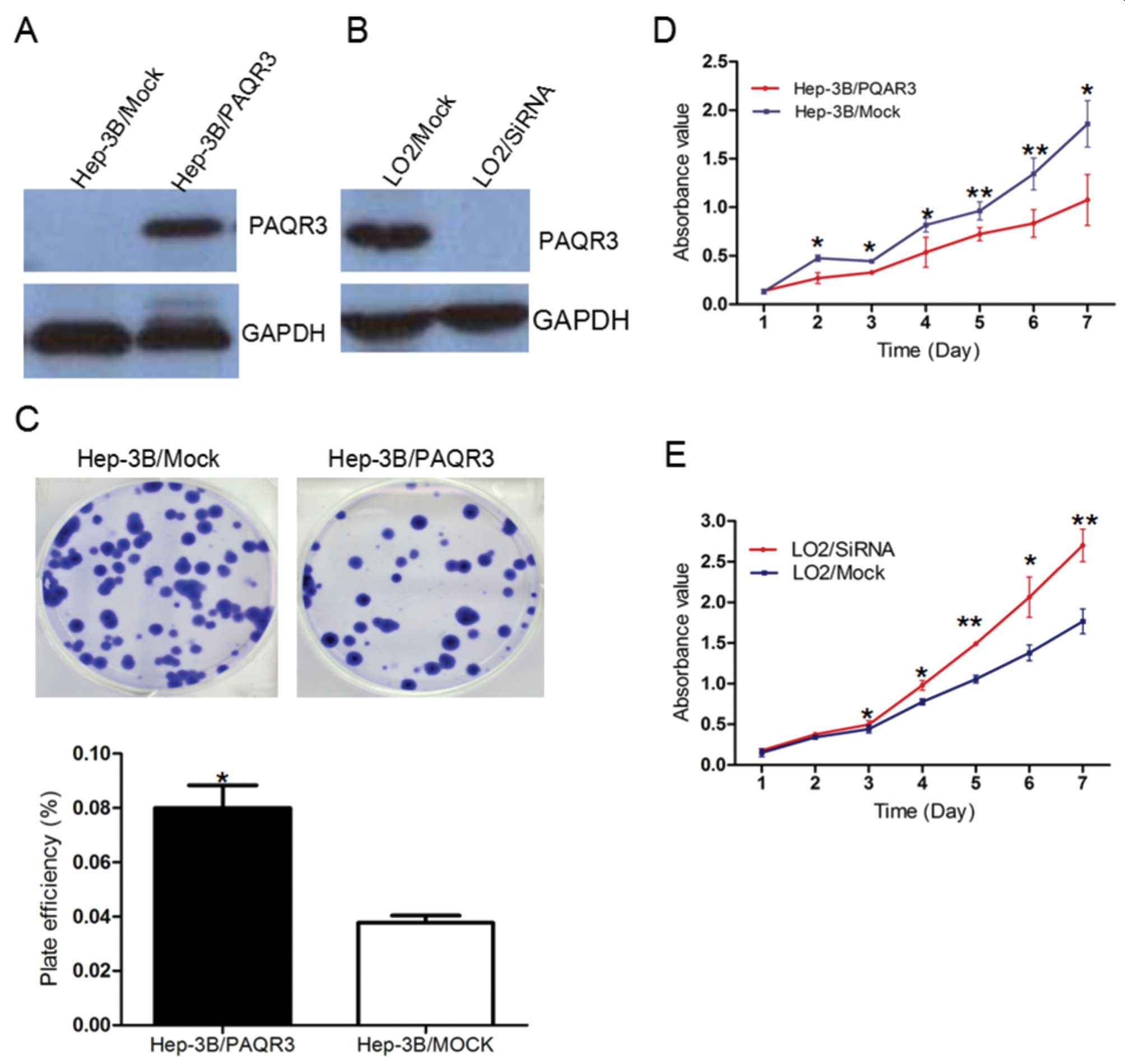
To evaluate the effects of PAQR3 on cell
proliferation, the PAQR3 expression vector and the control vector
were respectively transfected into Hep-3B cells. PAQR3 expression
in the transfected cells was detected by western blotting (Fig. 5A). The cell growth assay revealed
that the cell growth rate of the PAQR3-transfected Hep-3B cells was
significantly lower than that of the control vector-transfected
Hep-3B cells (Fig. 5D). Meanwhile,
the efficiency of colony formation was significantly (P=0.0379)
inhibited in the PAQR3-transfected hepatic cancer cells compared
with control vector-transfected hepatic cancer cells (Fig. 5C). To further confirm the
proliferation suppression function of PAQR3, we silenced PAQR3
expression in the LO2 cell line with siRNA. PAQR3 expression in the
transfected cells was detected by western blotting (Fig. 5B). We found that silencing of the
expression of PAQR3 in LO2 cells significantly enhanced cell
proliferation when compared with rates of cell proliferation in the
mock siRNA-treated cells (Fig.
5E).
Discussion
PAQR3 has been identified as a tumor-suppressor gene
in many cancer cell lines in vitro, as overexpression of
PAQR3 results in the inhibition of cell proliferation, migration,
sprouting and angiogenesis, and knockdown of PAQR3 promotes cell
proliferation, migration, sprouting and angiogenesis (11,12,14).
In a study by Xie et al, the number and size of papillomas
were increased in PAQR3−/− mice, accompanied by
shortened tumor latency and enhanced keratinocyte proliferation
(12). The possible mechanism is
associated with its regulation of the ERK signaling pathway, and
suppressing mitogen-activated protein kinase signaling, negatively
regulates the transactivation activity of hypoxia-inducible factor
1α (HIF-1α) by inhibiting formation of the HIF-1α/p300 complex and
suppressing VEGF transcription, thereby reducing hypoxia-induced
VEGF production (11,12,14).
Our observations, presented here for the first time, indicate that
PAQR3 acts as a tumor-suppressor gene in human HCC.
In the present study, we examined the expression of
PAQR3 at the mRNA and protein levels in paired primary HCC samples
using qRT-PCR and western blotting. The results demonstrated that
PAQR3 was downregulated in the majority of the primary HCC cancer
tissues at both the transcriptional and translational levels.
Consistently, immunohistochemical analyses also showed that PAQR3
expression was decreased in most HCC cancer tissues compared with
their corresponding non-cancer tissues. We favor the hypothesis
that a serious alteration, insertion and/or deletion in the gene
may possibly result in diminished or abolished expression of PAQR3
in HCC, which will require further investigation in future
research. These results indicated that downregulated expression of
PAQR3 may play a role in HCC carcinogenesis.
In the immunohistochemical analysis, downregulation
of PAQR3 expression in HCC was significantly associated with AFP,
mitotic count, tumor size, histologic grade and recurrence. Mitotic
count is used in HCC to quantify proliferation rates, and low PAQR3
expression was related with a high mitotic count. Fan et al
demonstrated that knockdown of PAQR3 enhanced malignant melanoma
cell proliferation and motility via Ras/Raf/MEK/ERK signaling
cascade phosphorylation (7). The
relationship between low PAQR3 expression and large tumor size in
the present study suggested that the lack of PAQR3 may facilitate
rapid expansion of the tumor. Additionally, most
well-differentiated HCC tissues showed high PAQR3 expression, in
contrast with a profoundly lower PAQR3 expression in the moderately
and poorly differentiated tumor samples. Recently, Jiang et
al demonstrated that there exists a functional interplay
between PAQR3 and p53 in EMT and tumorigenesis in HepG2 epithelial
cells (11). These findings
indicate that loss of PAQR3 expression may suppress the
differentiation of HCC cells and further promote the recurrence of
HCC. The low expression of PAQR3 was associated with a high serum
AFP level, which, to the best of our knowledge, has not been
reported, yet the exact mechanism still remains unclear, thus
requiring further investigation. Wang et al found PAQR3
downregulation in colorectal cancers, resulting in increased tumor
grade in vivo (10). These
observations are consistent with our clinicopathological findings
that low expression of PAQR3 in HCC is associated with larger tumor
size. Our findings, together with the findings of other authors,
strongly suggest that PAQR3 may be a novel, yet important,
prognostic marker for HCC patients.
Kaplan-Meier survival analysis revealed that low
PAQR3 expression was significantly correlated with a poor prognosis
of HCC patients after surgical resection. Furthermore, PAQR3
expression was found to be an independent prognostic factor
relative to overall and disease-free survival using multivariate
analysis. Consistent with our observations, downregulation of PAQR3
expression in colorectal cancer was found to be significantly
associated with a poor survival status of colorectal cancer
patients (10). These findings
suggest that PAQR3 may serve as a new and independent predictor of
prognosis for HCC patients after surgical resection.
We further investigated the functional role of PAQR3
in hepatic cell lines. Restoring PAQR3 expression in Hep-3B cells
significantly inhibited cell proliferation and colony formation.
Silencing the expression of PAQR3 in LO2 cells significantly
enhanced the cell growth rate. These results indicated that PAQR3
may play an important role in inhibiting tumor cell growth.
Recently, functional assays of PAQR3 in colorectal cancer cell
lines by Wang et al suggest that PAQR3 exerts
tumor-suppressor activity (10).
Fan et al demonstrated that restoring the expression of
wild-type PAQR3 inhibits ERK activation, cell proliferation and
transformation of A375 cells in vitro, and is sufficient to
suppress tumorigenicity of xenografts with human malignant melanoma
cell line A375 harboring B-Raf mutation V600E (7), while RNA interference-mediated PAQR3
silencing enhances cellular proliferation migration, sprouting and
angiogenesis of endothelial cells (12). Notably, knockdown of PAQR3 led to
enhancement of LPA-stimulated AKT and GSK3β phosphorylation,
together with increased accumulation of β-catenin and the
appearance of EMT features that were antagonized by p53
overexpression in HepG2 epithelial cells (15). Aberrant mTOR signaling has been
detected in up to 48% of HCCs and represents a suitable therapeutic
target in HCC (16), indicating a
link between PAQR3 and the AKT/MTOR signaling pathway in HCC
carcinogenesis and further investigation is warranted. These data,
together with ours, suggest that loss of PAQR3 may play an
important role in the process of carcinogenesis.
Collectively, our results demonstrate that PAQR3
expression is downregulated in the majority of HCC tissues at both
the mRNA and protein levels, and low PAQR3 expression may indicate
a dismal prognosis of HCC patients. In addition, we confirmed that
PAQR3 inhibits tumor cell growth and colony formation in
vitro. To date, the data generated in the present study
represent the first findings to correlate the presence of PAQR3
with clinicopathological characteristics and the survival of HCC
patients. Our findings provide evidence that PAQR3 may be a novel
prognostic biomarker for HCC and may serve as a potential
therapeutic candidate for HCC.
Acknowledgements
This study was supported by the Union Hospital Key
Laboratory Foundation of Biological Target Therapy (no.
02.03.2013-80 to P.D.L.) and the Independent Innovation Research
Foundation of Huazhong University of Science and Technology (no.
01-08-530059 to P.D.L.).
References
|
1
|
Parkin DM, Bray F, Ferlay J, et al:
Estimating the world cancer burden: Globocan 2000. Int J Cancer.
94:153–156. 2001. View
Article : Google Scholar : PubMed/NCBI
|
|
2
|
He J, Gu D, Wu X, et al: Major causes of
death among men and women in China. N Engl J Med. 353:1124–1134.
2005. View Article : Google Scholar : PubMed/NCBI
|
|
3
|
Thomas MB and Zhu AX: Hepatocellular
carcinoma: The need for progress. J Clin Oncol. 23:2892–2899. 2005.
View Article : Google Scholar : PubMed/NCBI
|
|
4
|
Zender L, Villanueva A, Tovar V, et al:
Cancer gene discovery in hepatocellular carcinoma. J Hepatol.
52:921–929. 2010. View Article : Google Scholar : PubMed/NCBI
|
|
5
|
Feng L, Xie X, Ding Q, et al: Spatial
regulation of Raf kinase signaling by RKTG. Proc Natl Acad Sci USA.
104:14348–14353. 2007. View Article : Google Scholar : PubMed/NCBI
|
|
6
|
Luo X, Feng L, Jiang X, et al:
Characterization of the topology and functional domains of RKTG.
Biochem J. 414:399–406. 2008. View Article : Google Scholar : PubMed/NCBI
|
|
7
|
Fan F, Feng L, He J, et al: RKTG
sequesters B-Raf to the Golgi apparatus and inhibits the
proliferation and tumorigenicity of human malignant melanoma cells.
Carcinogenesis. 29:1157–1163. 2008. View Article : Google Scholar : PubMed/NCBI
|
|
8
|
Zhang Y, Jiang X, Qin X, et al: RKTG
inhibits angiogenesis by suppressing MAPK-mediated autocrine VEGF
signaling and is downregulated in clear-cell renal cell carcinoma.
Oncogene. 29:5404–5415. 2010. View Article : Google Scholar : PubMed/NCBI
|
|
9
|
Jiang Y, Xie X, Zhang Y, et al: Regulation
of G-Protein signaling by RKTG via sequestration of the G betagamma
subunit to the Golgi apparatus. Mol Cell Biol. 30:78–90. 2010.
View Article : Google Scholar : PubMed/NCBI
|
|
10
|
Wang X, Li X, Fan F, et al: PAQR3 plays a
suppressive role in the tumorigenesis of colorectal cancers.
Carcinogenesis. 33:2228–2235. 2012. View Article : Google Scholar : PubMed/NCBI
|
|
11
|
Jiang Y, Xie X, Li Z, et al: Functional
cooperation of RKTG with p53 in tumorigenesis and
epithelial-mesenchymal transition. Cancer Res. 71:2959–2968. 2011.
View Article : Google Scholar : PubMed/NCBI
|
|
12
|
Xie X, Zhang Y, Jiang Y, et al:
Suppressive function of RKTG on chemical carcinogen-induced skin
carcinogenesis in mouse. Carcinogenesis. 29:1632–1638. 2008.
View Article : Google Scholar : PubMed/NCBI
|
|
13
|
Grabinski N, Ewald F, Hofmann BT, et al:
Combined targeting of AKT and mTOR synergistically inhibits
proliferation of hepatocellular carcinoma cells. Mol Cancer.
20:112012.PubMed/NCBI
|
|
14
|
Psyrri A, Arkadopoulos N, Vassilakopoulou
M, et al: Pathways and targets in hepatocellular carcinoma. Expert
Rev Anticancer Ther. 12:1347–1357. 2012. View Article : Google Scholar : PubMed/NCBI
|
|
15
|
Jain S, Singhal S, Lee P, et al: Molecular
genetics of hepatocellular neoplasia. Am J Transl Res. 2:105–118.
2010.
|
|
16
|
Guichard C, Amaddeo G, Imbeaud S, et al:
Integrated analysis of somatic mutations and focal copy-number
changes identifies key genes and pathways in hepatocellular
carcinoma. Nat Genet. 44:694–698. 2012. View Article : Google Scholar : PubMed/NCBI
|