Introduction
Pancreatic cancer is almost the deadliest of all
malignancies (1). In Japan,
pancreatic cancer is currently the fifth leading cause of
cancer-related death among individuals of both genders (2,3).
Resection surgery is still the only potentially curative treatment
for pancreatic cancer, and recent improvements in operative
technique have been reported (4).
Although advances in adjuvant treatment have been observed
(5), in general, the prognosis of
patients with pancreatic cancer is still poor. Further studies of
the mechanisms of pancreatic carcinogenesis and cancer development
are needed, and new therapeutic options are highly desirable.
Endoplasmic reticulum (ER) stress response in tumor
cells is critical for tumor cell growth and cancer progression
(6). The ER stress response is
mediated by at least three sensor molecules: inositol-requiring
enzyme 1α (IRE1α), PKR-like ER kinase (PERK), and activating
transcription factor 6 (ATF6), which are usually associated with
glucose-regulated protein 78 (GRP78/Bip) (7). ER stress, which is associated with the
accumulation of unfolded proteins, induces unfolded protein
response (UPR), yet if ER stress is overloaded, cells could face
death such as by apoptosis and autophagy. Downstream of IRE1α and
PERK, the effector molecules, X-box-binding protein 1 (XBP1) and
C/EBP homologous protein (CHOP), and growth arrest and DNA damage
gene 34 (GADD34) all exist, and they are activated by ER stress. ER
stress also leads to the phosphorylation of eukaryotic translation
initiation factor 2α (eIF2α) (8).
For example, p90ATF6 is converted to the activated form p50ATF6,
and p50ATF6 translocates to the nucleus (9). Basic leucine-zipper family factors
p50ATF6 and XBP1 could induce expression of a subset of UPR-related
genes, which include ER stress elements, and are involved in
efficient protein folding, maturation and degradation in the ER
(6).
The association between ER stress response and tumor
growth and progression has been reported (10). We and others have reported that
GRP78 is involved in cancer development and innate immune response
in the liver (11–14). Liver and pancreas progenitors
commonly develop from endoderm cells in the embryonic foregut
(15). Pancreatic epithelial cells
have a highly developed ER due to a strong engagement in digestive
enzyme secretion (16). GRP78 is
the main target of UPR signaling that promotes pancreatic cancer
cell survival (17). GRP78 is
involved in cancer progression as well as drug resistance (18,19).
Hence, to decrease the ability of pancreatic cancer cells to
survive and proliferate, it may be necessary to block GRP78
expression (17).
We previously demonstrated that blocking of the
induction of UPR, as well as inhibition of GRP78 expression is
associated with the cleavage of poly(ADP-ribose) polymerase (PARP)
(13). In the present study, we
examined the expression of ER stress-related molecules in human
pancreatic cancer cell lines in the presence or absence of
thapsigargin, one of the ER stress-inducers. We also investigated
whether knockdown of GRP78 by small interfering RNA (siRNA)
enhances the PARP cleavage in human pancreatic cancer cell lines
exposed to ER stress.
Materials and methods
Cell culture
Human pancreatic cancer cell lines (KP-2, MIAPaCa-2,
Panc-1 and SUIT-2) were grown in RPMI-1640 medium (Sigma, St.
Louis, MO, USA) supplemented with 10% fetal bovine serum, 100 U/ml
penicillin and 100 μg/ml streptomycin at 37°C in a humidified
atmosphere with 5% CO2. Inhibitor of
sarcoplasmic/endoplasmic reticulum (ER) Ca2+ ATPases
(SERCA), thapsigargin, control siRNA (si-control) and siRNA for
GRP78 (si-GRP78) were purchased from BioVision (Milpitas, CA, USA)
and Santa Cruz Biotechnology (Santa Cruz, CA, USA),
respectively.
Western blotting
Twenty-four hours after thapsigargin (1 μM)
treatment, cells were lysed in sodium dodecyl sulfate sample
buffer, and after sonication, lysates were processed for western
blot analysis (11). Briefly,
protein samples were subjected to electrophoresis on 5–20%
polyacrylamide gels and transferred onto polyvinylidene difluoride
membranes (ATTO, Tokyo, Japan). Membranes were probed with
antibodies specific for ATF4, ATF6 and tubulin (Abcam, Cambridge,
UK); GADD34, gyceraldehyde-3-phosphate dehydrogenase (GAPDH) and
XBP1 (Santa Cruz); eIF2α, phospho-eIF2α (Ser51), GRP78/Bip and PARP
(Cell Signaling Technology, Tokyo, Japan). After washing with
PBS-T, the membranes were incubated with secondary horseradish
peroxidase-conjugated antibodies. Signals were detected by means of
enhanced chemiluminescence (GE Healthcare, Tokyo, Japan) and
scanned by image analyzer LAS-4000 and Image Gauge (version 3.1)
(Fuji Film, Tokyo, Japan) and ImageJ software (NIH, Bethesda, MD,
USA).
Transfection of siRNA
To confirm the effects of GRP78 knockdown on
apoptosis, we examined GRP78 knockdown by small-interfering RNA
(siRNA). Cells were transfected with 50 nM si-GRP78 or si-control,
using Effectene transfection reagent (Qiagen, Hilden, Germany)
according to the manufacturer’s protocol (20). After 24 h of transfection, cells
were treated with 1 μM thapsigargin for 24 h.
Statistical analysis
Results are expressed as means ± standard deviation
(SD). Statistical analysis was performed using the Student’s
t-test. A P-value <0.05 was considered to indicate a
statistically significant result.
Results
Human pancreatic cancer cell lines
express GRP78
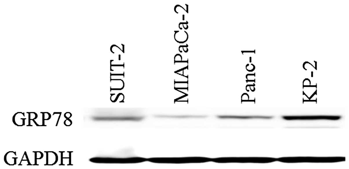
First, we examined the GRP78 expression in the human
pancreatic cancer cell lines SUIT-2, MIAPaCa-2, Panc-1 and KP-2
(3). Protein samples were collected
from the four pancreatic cancer cell lines, and protein levels of
GRP78 were investigated by western blotting with a specific
antibody for GRP78 (Fig. 1). We
confirmed that all four pancreatic cancer cell lines variably
expressed GRP78.
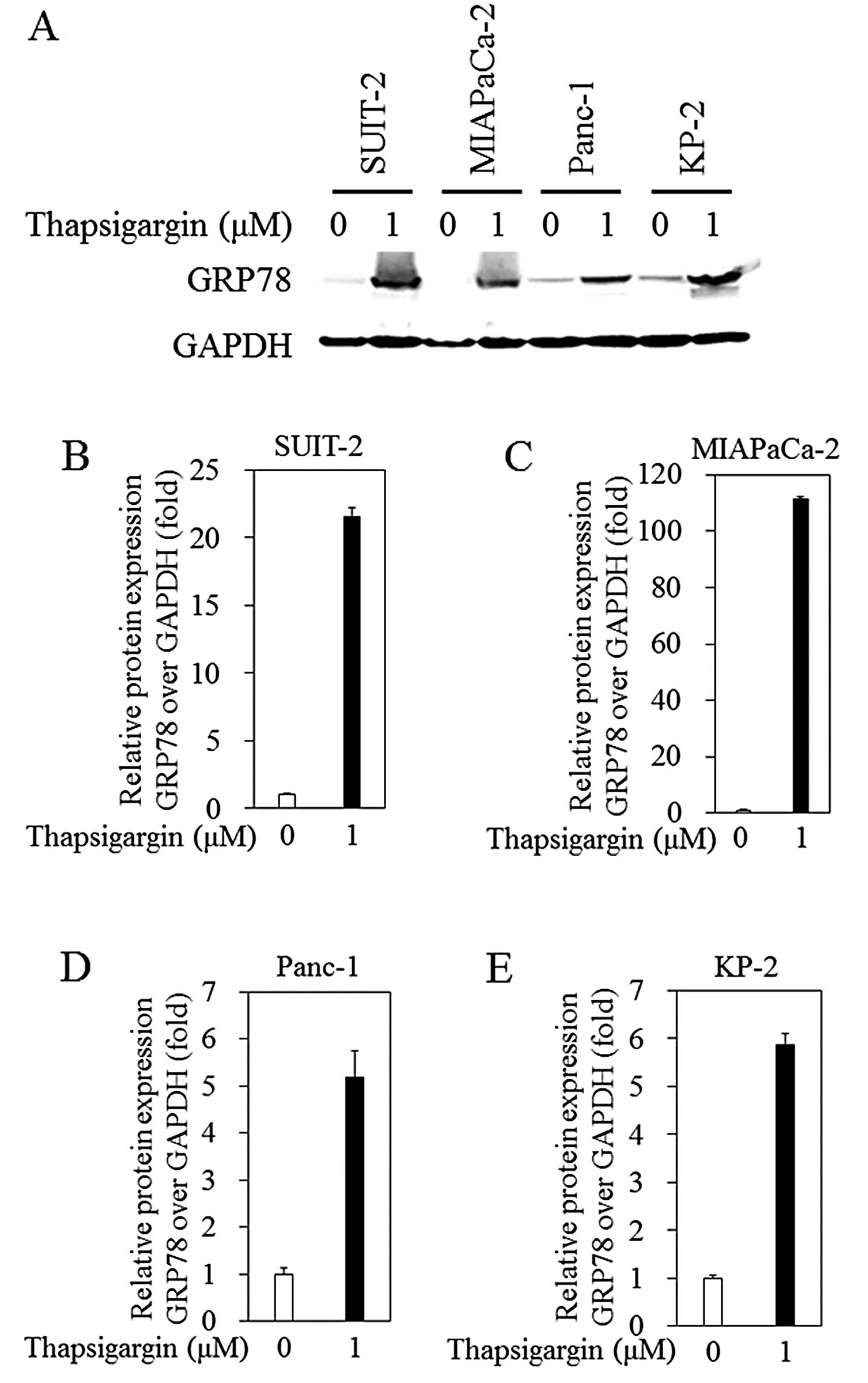
Thapsigargin upregulates the protein
levels of GRP78 in the human pancreatic cancer cell lines
Next, we examined the effect of thapsigargin, one of
the ER stress-inducers, on GRP78 expression in the human pancreatic
cancer cell lines (Fig. 2).
Treatment of 1 μM thapsigargin for 24 h led to the upregulation of
GRP78 expression at the protein level [21.5±0.7 vs. 1±0.1 (in
untreated control), n=3, p=0.00015; 111.5±1.0 vs. 1±0.12, n=3,
p=0.000010; 5.2±0.57 vs. 1±0.1, n=3, p=0.0023; and 5.9±0.2 vs.
1±0.1, n=3, p=0.00013, respectively, in the SUIT-2, MIAPaCa-2,
Panc-1 and KP-2 cells]. In the MIAPaCa-2, cells GRP78 expression
was more strongly induced than in the other three cell lines.
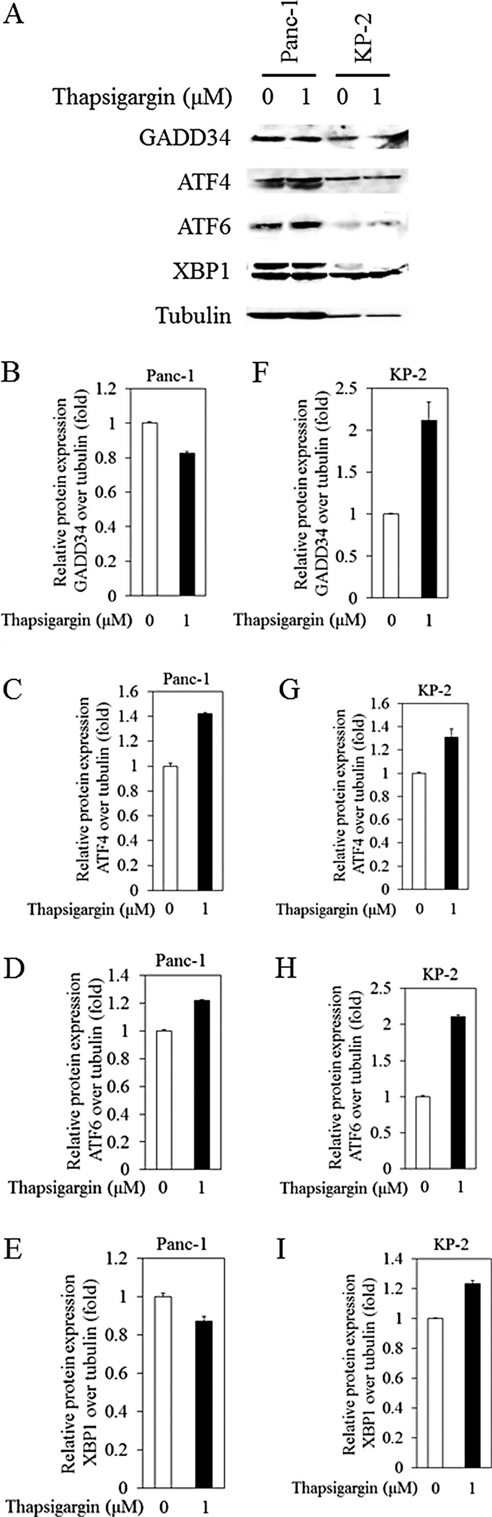
Effects of thapsigargin on GADD34, ATF4,
ATF6 and XBP1 protein expression levels in the human pancreatic
cancer cell lines
We examined the protein expression of ER stress
signaling-associated molecules in the human pancreatic cell lines
treated with or without thapsigargin. The results for the Panc-1
and KP-2 cells are shown in Fig. 3.
In the Panc-1 cells, ATF4 and ATF6 expression was upregulated in
the presence of 1 μM thapsigargin [1.4±0.010 vs. 1±0.023 (in
untreated control), n=3, p=0.000089; and 1.2±0.0027 vs. 1±0.010,
n=3, p=0.00019, respectively] (Fig. 3A,
C and D. In the Panc-1 cells, GADD34 and XBP1 expression at the
protein level was down-regulated in the presence of 1 μM
thapsigargin [0.82±0.012 vs. 1±0.0076 (in untreated control), n=3,
p=0.0000414; and 0.87±0.024 vs. 1±0.019, n=3, p=0.0012,
respectively] (Fig. 3A, B and
E).
On the other hand, in KP-2 cells, the protein
expression levels of GADD34, ATF4, ATF6 and XBP1 were upregulated
in the presence of 1 μM thapsigargin [2.1±0.22 vs. 1±0.012 (in
untreated control), n=3, p=0.0063; 1.3±0.073 vs. 1±0.0062, n=3,
p=0.0088; 2.1±0.022 vs. 1±0.014, n=3, p=0.0000008; and 1.2±0.019
vs. 1±0.0063, n=3, p=0.00043, respectively] (Fig. 3A and F–I).
XBP1 was also upregulated in the presence of 1 μM
thapsigargin in both SUIT-2 and MIAPaCa-2 cells, yet we did not
observe any enhancement of GADD34, ATF4 or ATF6 by thapsigargin
(data not shown).
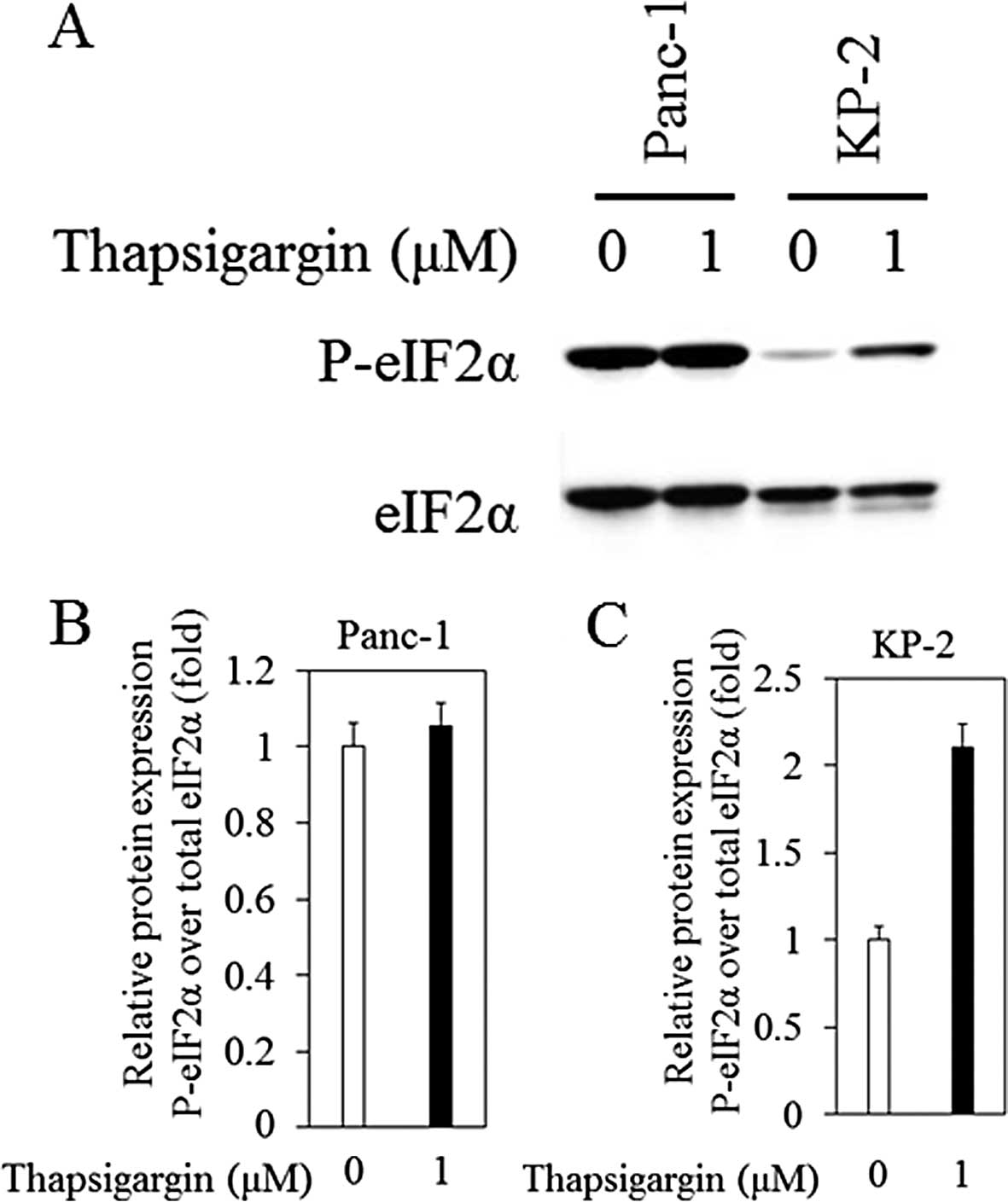
Effects of thapsigargin on the
phosphorylation of eIF2α in the human pancreatic cancer cell
lines
We also examined the phosphorylation status of eIF2α
to understand how thapsigargin affects ER stress signaling in
Panc-1 and KP-2 cells (Fig. 4A). In
Panc-1 cells, phosphorylation of Ser51-eIF2α in the presence of
thapsigargin tended to increase, compared with that in the absence
of thapsigargin (Fig. 4B; 1.1±0.059
vs. 1±0.064, n=3, p=0.17). In the KP-2 cells, significant
phosphorylation of Ser51-eIF2α in the presence of thapsigargin was
observed when compared with that in the absence of thapsigargin
(Fig. 4C; 2.1±0.14 vs. 1±0.075,
n=3, p=0.00050).
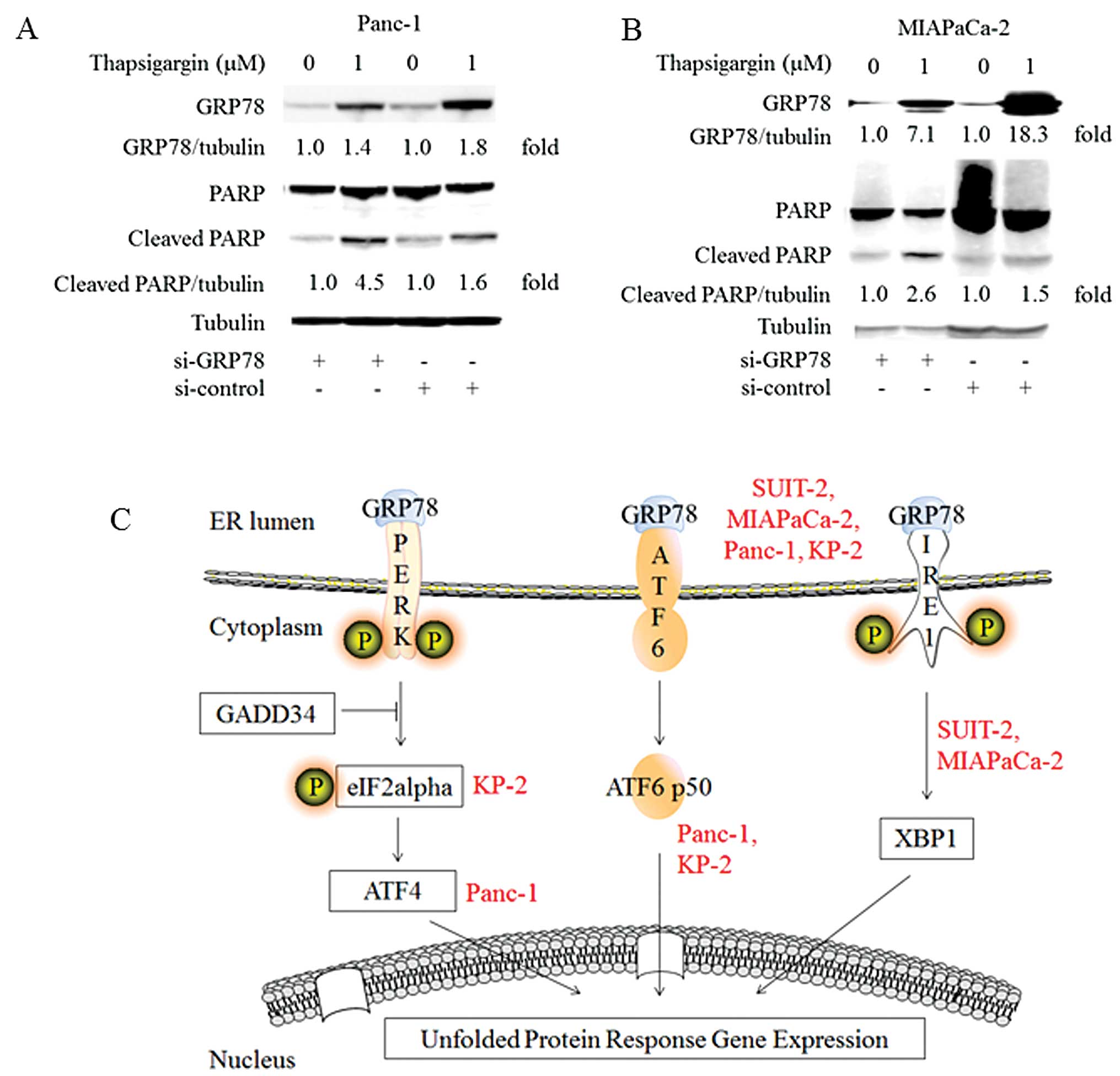
Knockdown of endogenous GRP78 enhances
PARP cleavage in the pancreatic cancer cells
We confirmed that the expression of GRP78 at the
protein level was upregulated in all four human pancreatic cancer
cell lines tested, yet other molecules downstream of GRP78 reported
to be involved in ER stress were expressed at variable levels
depending on the individual cell line. Thus, we focused our
examination on GRP78. Our previous study (13) demonstrated that blocking of GRP78
induction led to PARP cleavage in hepatocyte apoptosis. We
investigated the effect of knockdown of GRP78 by siRNA on PARP
cleavage in pancreatic cancer cells treated with thapsigargin
(Fig. 5A and B).
GRP78 expression was significantly inhibited by
transfection with si-GRP78 in the presence of thapsigargin,
compared with that with si-control [1.4±0.040 vs. 1.8±0.040, n=3,
p=0.00014; and 7.1±0.24 vs. 18.3±0.37, n=3, p=0.0000038,
respectively, in Panc-1 (Fig. 5A)
and MIAPaCa-2 cells (Fig. 5B)].
PARP cleavage was significantly enhanced by
transfection with si-GRP78 in the presence of thapsigargin,
compared with that with si-control [4.5±0.045 vs. 1.6±0.085, n=3,
p=0.00000080; and 2.6±0.13 vs. 1.5±0.047, n=3, p=0.00016,
respectively, in Panc-1 (Fig. 5A)
and MIAPaCa-2 cells (Fig. 5B)].
Discussion
In the present study, we demonstrated that i) human
pancreatic cancer cell lines expressed GRP78; ii) ER stress induced
by thapsigargin upregulated protein levels of GRP78 in human
pancreatic cancer cell lines; iii) ER stress-related molecules
downstream of GRP78 were expressed at various levels according to
the respective human pancreatic cancer cell lines; and iv) finally,
knockdown of GRP78 by siRNA enhanced PARP cleavage in the human
pancreatic cancer cell lines. To our knowledge, this is the first
report to show the association between GRP78 and PARP cleavage in
pancreatic cancer cell lines treated with thapsigargin.
Our results that human pancreatic cancer cell lines
express GRP78 supported a previous study (21) showing that the heat shock proteins
HSP90 and GRP78 are constitutively expressed in gastrointestinal
cancers including human pancreatic cancer. We also observed that ER
stress induced by thapsigargin upregulated protein levels of GRP78
in human pancreatic cancer cell lines. However, ER stress-related
molecules downstream of GRP78, such as GADD34, ATF4, ATF6, XBP1 and
phospho-eIF2α were not constitutively increased by thapsigargin,
but rather were dependent on individual cell lines (Figs. 2–4).
These results suggest that GRP78 may have an impact on many
different cellular processes and survival of pancreatic cancer and
that ER stress signaling downstream of GRP78 can be expected to be
disturbed in pancreatic cancer.
It was reported that an increase in GRP78 expression
in pancreatic cancer cells may enhance and account for the altered
sensitivity of pancreatic cancer to chemotherapeutic agents
(21). UPR regulator GRP78 is an
anti-apoptotic protein that is usually upregulated in cancer and
plays a critical role in chemoresistance in various types of
cancers (22). Recently it was also
reported that UPR induction in tumor endothelial cells under an
acidic pH condition is related to chemoresistance and may
contribute to therapeutic failure in response to chemotherapy
(23). It was also reported that
GRP78 is overexpressed in malignant cells resistant to therapy
(24).
PARP is one of the proteins processed by
post-translational modification and plays a crucial role in many
processes, including DNA repair and cell death (25). During apoptosis, caspases cause PARP
cleavage and inactivation, in which PARP proteolysis produces an
89-kDa C-terminal fragment and a 24-kDa N-terminal (25). We observed that in the presence of
thapsigargin, knockdown of GRP78 enhanced PARP cleavage in human
pancreatic cancer cells Panc-1 as well as MIAPaCa-2. Wang et
al reported that suppression of GRP78 by taxol and vinblastine
potentiated the activation of JNK phosphorylation, caspase-7 and
PARP cleavage in the human breast cancer cell line MCF-7 (26). The Hsp90 inhibitor SNX-2112 also
induced PARP cleavage as well as the reduction in GRP78 expression
in the multidrug-resistant human chronic myeloid leukemia K562/ADR
cell line (27).
Collectively, our results suggest that both GRP78
and PARP may have key roles in the chemoresistance of pancreatic
cancer (28) and that GRP78 may be
one of the valid targets against chemoresistance (24). In conclusion, GRP78 is a potential
therapeutic target for ‘difficult-to-treat’ pancreatic cancer, in
which ER stress signaling in part falls into disorder.
Acknowledgements
The present study was supported by Grants for
Scientific Research from the Ministry of Education, Culture,
Sports, Science and Technology, Japan (24590955 to T.K.).
References
|
1
|
Lennon AM, Wolfgang CL, Canto MI, et al:
TThe early detection of pancreatic cancer: what will it take to
diagnose and treat curable pancreatic neoplasia? Cancer Res.
74:3381–3389. 2014. View Article : Google Scholar : PubMed/NCBI
|
|
2
|
Kuroda T, Kumagi T, Yokota T, et al:
Improvement of long-term outcomes in pancreatic cancer and its
associated factors within the gemcitabine era: a collaborative
retrospective multicenter clinical review of 1,082 patients. BMC
Gastroenterol. 13:1342013. View Article : Google Scholar
|
|
3
|
Okitsu K, Kanda T, Imazeki F, Yonemitsu Y,
Ray RB, Chang C and Yokosuka O: Involvement of interleukin-6 and
androgen receptor signaling in pancreatic cancer. Genes Cancer.
1:859–867. 2010. View Article : Google Scholar : PubMed/NCBI
|
|
4
|
Miyazaki M, Yoshitomi H, Shimizu H, et al:
Repeat pancreatectomy for pancreatic ductal cancer recurrence in
the remnant pancreas after initial pancreatectomy: is it
worthwhile? Surgery. 155:58–66. 2014. View Article : Google Scholar : PubMed/NCBI
|
|
5
|
Sudo K, Ishihara T, Hirata N, et al:
Randomized controlled study of gemcitabine plus S-1 combination
chemotherapy versus gemcitabine for unresectable pancreatic cancer.
Cancer Chemother Pharmacol. 73:389–396. 2014. View Article : Google Scholar : PubMed/NCBI
|
|
6
|
Mahadevan NR, Rodvold J, Sepulveda H,
Rossi S, Drew AF and Zanetti M: Transmission of endoplasmic
reticulum stress and pro-inflammation from tumor cells to myeloid
cells. Proc Natl Acad Sci USA. 108:6561–6566. 2011. View Article : Google Scholar : PubMed/NCBI
|
|
7
|
Schroder M and Kaufman RJ: ER stress and
the unfolded protein response. Mutat Res. 569:29–63. 2005.
View Article : Google Scholar : PubMed/NCBI
|
|
8
|
Hamanaka RB, Bennett BS, Cullinan SB and
Diehl JA: PERK and GCN2 contribute to eIF2α phosphorylation and
cell cycle arrest after activation of the unfolded protein response
pathway. Mol Biol Cell. 16:5493–5501. 2005.
|
|
9
|
Xu W, Liu L, Charles IG and Moncada S:
Nitric oxide induces coupling of mitochondrial signalling with the
endoplasmic reticulum stress response. Nat Cell Biol. 6:1129–1134.
2004. View
Article : Google Scholar : PubMed/NCBI
|
|
10
|
Ma Y and Hendershot LM: The role of the
unfolded protein response in tumour development: friend or foe? Nat
Rev Cancer. 4:966–977. 2004. View
Article : Google Scholar : PubMed/NCBI
|
|
11
|
Jiang X, Kanda T, Nakamoto S, Miyamura T,
Wu S and Yokosuka O: Involvement of androgen receptor and
glucose-regulated protein 78 kDa in human hepatocarcinogenesis. Exp
Cell Res. 323:326–336. 2014. View Article : Google Scholar : PubMed/NCBI
|
|
12
|
Shuda M, Kondoh N, Imazeki N, et al:
Activation of the ATF6, XBP1 and grp78 genes in human
hepatocellular carcinoma: a possible involvement of the ER stress
pathway in hepatocarcinogenesis. J Hepatol. 38:605–614. 2003.
View Article : Google Scholar : PubMed/NCBI
|
|
13
|
Jiang X, Kanda T, Tanaka T, Wu S, Nakamoto
S, Imazeki F and Yokosuka O: Lipopolysaccharide blocks induction of
unfolded protein response in human hepatoma cell lines. Immunol
Lett. 152:8–15. 2013. View Article : Google Scholar : PubMed/NCBI
|
|
14
|
Martinon F and Glimcher LH: Regulation of
innate immunity by signaling pathways emerging from the endoplasmic
reticulum. Curr Opin Immunol. 23:35–40. 2011. View Article : Google Scholar : PubMed/NCBI
|
|
15
|
Kanda T, Jiang X and Yokosuka O: Androgen
receptor signaling in hepatocellular carcinoma and pancreatic
cancers. World J Gastroenterol. 20:9229–9236. 2014.PubMed/NCBI
|
|
16
|
Nawrocki ST, Carew JS, Dunner K Jr, et al:
Bortezomib inhibits PKR-like endoplasmic reticulum (ER) kinase and
induces apoptosis via ER stress in human pancreatic cancer cells.
Cancer Res. 65:11510–11519. 2005. View Article : Google Scholar : PubMed/NCBI
|
|
17
|
Mujumdar N, Banerjee S, Chen Z, et al:
Triptolide activates unfolded protein response leading to chronic
ER stress in pancreatic cancer cells. Am J Physiol Gastrointest
Liver Physiol. 306:G1011–G1020. 2014. View Article : Google Scholar : PubMed/NCBI
|
|
18
|
Fu Y and Lee AS: Glucose regulated
proteins in cancer progression, drug resistance and immunotherapy.
Cancer Biol Ther. 5:741–744. 2006. View Article : Google Scholar : PubMed/NCBI
|
|
19
|
Lee E, Nichols P, Spicer D, Groshen S, Yu
MC and Lee AS: GRP78 as a novel predictor of responsiveness to
chemotherapy in breast cancer. Cancer Res. 66:7849–7853. 2006.
View Article : Google Scholar : PubMed/NCBI
|
|
20
|
Kanda T, Yokosuka O, Imazeki F, Arai M and
Saisho H: Enhanced sensitivity of human hepatoma cells to
5-fluorouracil by small interfering RNA targeting Bcl-2. DNA
Cell Biol. 24:805–809. 2005. View Article : Google Scholar : PubMed/NCBI
|
|
21
|
Ehrenfried JA, Herron BE, Townsend CM Jr
and Evers BM: Heat shock proteins are differentially expressed in
human gastrointestinal cancers. Surg Oncol. 4:197–203. 1995.
View Article : Google Scholar : PubMed/NCBI
|
|
22
|
Tsai HY, Yang YF, Wu AT, et al:
Endoplasmic reticulum ribosome-binding protein 1 (RRBP1)
overexpression is frequently found in lung cancer patients and
alleviates intracellular stress-induced apoptosis through the
enhancement of GRP78. Oncogene. 32:4921–4931. 2013. View Article : Google Scholar
|
|
23
|
Visioli F, Wang Y, Alam GN, Ning Y, Rados
PV, Nör JE and Polverini PJ: Glucose-regulated protein 78 (Grp78)
confers chemoresistance to tumor endothelial cells under acidic
stress. PLoS One. 9:e1010532014. View Article : Google Scholar : PubMed/NCBI
|
|
24
|
Roller C and Maddalo D: The molecular
chaperone GRP78/BiP in the development of chemoresistance:
mechanism and possible treatment. Front Pharmacol. 4:102013.
View Article : Google Scholar : PubMed/NCBI
|
|
25
|
Soldani C and Scovassi AI:
Poly(ADP-ribose) polymerase-1 cleavage during apoptosis: an update.
Apoptosis. 7:321–328. 2002. View Article : Google Scholar : PubMed/NCBI
|
|
26
|
Wang J, Yin Y, Hua H, et al: Blockade of
GRP78 sensitizes breast cancer cells to microtubules-interfering
agents that induce the unfolded protein response. J Cell Mol Med.
13:3888–3897. 2009. View Article : Google Scholar : PubMed/NCBI
|
|
27
|
Wang R, Shao F, Liu Z, et al: The Hsp90
inhibitor SNX-2112, induces apoptosis in multidrug resistant
K562/ADR cells through suppression of Akt/NF-κB and disruption of
mitochondria-dependent pathways. Chem Biol Interact. 205:1–10.
2013.PubMed/NCBI
|
|
28
|
Lei Y, Henderson BR, Emmanuel C, Harnett
PR and Defazio A: Inhibition of ANKRD1 sensitizes human ovarian
cancer cells to endoplasmic reticulum stress-induced apoptosis.
Oncogene. Feb 17–2014.(Epub ahead of print).
|