Introduction
Cervical cancer is the second most common cancer and
the third leading cause of mortality among women worldwide. Each
year ~500,000 women worldwide develop cervical cancer, of which
>90% is found in developing countries (1). The demographics of cervical cancer
patients have been shifting towards younger patients, with the
majority of them being single or nullipara women (2). Thus, there is a strong demand to
maintain their fertility function while effectively treating the
cancer. In addition, an effective and conservative therapy method
is also required for elderly patients who cannot undergo surgery or
radiotherapy, as well as patietns who refuse surgery (3).
Photodynamic therapy (PDT) is emerging as a viable
conservative treatment for various types of cancer. PDT is based on
the light-induced activation of a photosensitizer that results in
subsequent in situ production of reactive oxygen species
(ROS), which directly destroy cells that have accumulated the
photosensitizer. Thus, the photosensitizer, light and oxygen are
three important elements to determine the efficiency of PDT
(4,5).
However, the application of most existing
photosensitizers is limited because of their disadvantages, mainly
including (6) i) a relatively low
tumor selectivity, which can lead to undesired side effects in
normal tissues, especially the prolonged cutaneous photosensitivity
post-PDT and ii) lack of water solubility, which hampers the
development of pharmaceutical formulations, inducing the
aggregation of photosensitizer in the physiological medium,
resulting in a severe reduction of the photochemical properties and
the bioavailability of the photosensitizer (7–9). Thus,
during the last few years, a great deal of effort has been invested
into improving the targeting of photosensitizers by conjugating
photosensitizers to tumor-seeking molecules, such as epidermal
growth factor, monoclonal antibodies, carrier proteins,
carbohydrates, and hydrophilic polymers (10–13).
Based on the fact that the folate receptor in human
epithelial cancer cells was overexpressed, despite its absence from
most normal cells and the high affinity between folic acid and the
folate receptor (14,15), a targeted photosensitizer (Ps I) was
designed and generated in our laboratory using folic acid as a
tumor-seeking bullet, polyethylene glycol (PEG) as the linker, and
chlorin, an analogue of mTHPC (temoporfin), as a photoactive block
(Fig. 1A). mTHPC is one of the most
potent second-generation photosensitizers (16). PEG, when used as a linker, improves
the hydrophilicity of the conjugate and prevents the recognition of
the reticuloendothelial system (RES) of the conjugate (17). The results of previous studies
indicated that this new photosensitizer exhibited much higher tumor
targeting and water solubility but lower aggregation than that of
its precursor, chlorin (18). The
aim of the present study was to evaluate further the tumor
targeting and photodynamic activity of PS I in cervical cancer
in vitro and in vivo. In addition, the cell death
mode induced by PS I-based PDT was investigated.
Materials and methods
Chemicals and reagents
Fetal bovine serum, penicillin/streptomycin
solution, and RPMI-1640 were purchased from HyClone (South Logan,
UT, USA). Folate-free RPMI-1640 was purchased from Gibco (Grand
Island, NY, USA). Trypsin, methylthiazolyldiphenyl-tetrazolium
bromide (MTT), and dimethyl sulfoxide (DMSO) were purchased from
Sigma-Aldrich (St. Louis, MO, USA). An Annexin V-PI Apoptosis
Detection kit was purchased from BD Bioscience (San Jose, CA, USA).
4′,6-Diamidino-2-phenylindole dihydrochloride (DAPI) and a BCA kit
were purchased from Beyotime (Shanghai, China). A terminal
deoxynucleotidyl transferase-mediated deoxyuridinetriphosphate nick
end-labeling (TUNEL)-FITC Apoptosis Detection kit was purchased
from Invitrogen (Carlsbad, CA, USA). The photosensitizers (PS I and
A), with a purity of >98%, were produced in our laboratory.
Stock solutions of PS I and A (1 mg/ml) were prepared in FBS- and
folate-free RPMI-1640 and stored in the dark at 4°C.
Animals and cell lines
Four-week-old BALB/c nude mice, weighing 16–18 g,
were obtained from HFK Bio-Technology Co., Ltd. (Beijing, China).
The mice were kept in an air-conditioned facility fitted with an
artificial 12 h light-dark cycle and provided standard food and
filtered water. Experiments were conducted in compliance with the
Animal Management Rules of the Ministry of Health of the People’s
Republic of China (document no. 55, 2001) and the guidelines for
the Care and Use of Laboratory Animals of China Pharmaceutical
University. The human cervical carcinoma HeLa cell line was
obtained from the Chinese Academy of Science Shanghai Cell Library
and was cultured in RPMI-1640 medium supplemented with 10% FBS,
penicillin (100 μ/ml) and streptomycin (100 μ/ml) at 5%
CO2 37°C in a humidified incubator.
Cellular uptake
HeLa cells were cultured with folate-free RPMI-1640
for 24 h. After exposure to the photosensitizer for 24 h, the cells
were washed with PBS and fixed with 3.7% paraformaldehyde for 15
min. The fluorescence intensity of the photosensitizers in the
cells was analyzed by confocal laser scanning microscopy (Ex, 480
nm/Em, 650 nm). In addition, after the HeLa cells were exposed to
15.2 μmol/l of Ps I and A with or without folic acid
(2×10³ μmol/l) for 2, 8 or 24 h, the cells were washed
with PBS, and 500 μl of 4% SDS was added to each well. The
solutions were centrifuged for 9,000 rpm at 4°C for 20 min, and the
fluorescence intensity of the photosensitizers in the supernatant
was measured using an F-4500 fluorescence spectrophotometer (Ex,
480 nm and Em, 650 nm; Hitachi, Japan). In addition, the total
protein concentration of each well was determined using the BCA kit
according to the manufacturer’s instructions. For each incubation
time, the determination was run in triplicate.
Photocytotoxicity assay
The HeLa cells were inoculated into detachable
96-well culture plates and were incubated in Ps I or A for 24 h in
the dark before exposure to the red light generated by a KDH150B
red-light therapy instrument (600–700 nm; Kedian Co., Beijing,
China). After incubation for another 24 h in the dark, the cells
were incubated in 0.5 mg/ml MTT solution for 4 h, and the resulting
formazan crystal was dissolved in DMSO. The absorbance at 492 nm
was measured using a Microplate Reader Model 450 (Bio-Rad,
Hercules, CA, USA). Cell viability was proportional to the
A492 value and was expressed as a percentage of the cell
viability of the untreated control cells. For each concentration or
irradiation dose, three experiments were performed.
Detection of apoptotic characteristics of
HeLa cells following PDT treatment
HeLa cells were incubated with 15.2 μmol/l of Ps I
for 24 h and then exposed to red light at a 15 J/cm2
irradiation dose. After 12 h, the HeLa cells were harvested and
double-stained using an Annexin V-PI apoptosis detection kit
according to the manufacturer’s instructions. The stained cells
were analyzed using a FACSCalibur flow cytometer (BD Biosciences).
At least 10,000 events were collected for each sample.
In addition, an aliquot of the HeLa cells (in the
PDT and control groups) was fixed with 4% paraformaldehyde for 15
min, incubated with 3% SDS for 3 min and then stained with 500
ng/ml DAPI for 5 min. The cells were visualized under laser
confocal microscopy [TCS sp5 (DAPI Ex, 358 nm and Em, 461 nm);
Leica, Buffalo Grove, IL, USA]. In addition, aliquots of the
harvested HeLa cells (in the PDT and control groups) were fixed in
2.5% glutaraldehyde. After centrifugation at 1500 rpm for 10 min
and being washed with PBS, the cells were post-fixed in 1.0% osmium
tetroxide, dehydrated, soaked and embedded in Epon 618 for
subsequent sectioning. The ultrathin sections were stained with
uranyl acetate and lead citrate, and examined under a transmission
electron microscope (TECNAI 10; Phillips, The Netherlands).
Animal model
A xenograft tumor model was created by subcutaneous
injection of HeLa cells (~2×10⁶ in 200 μl serum-free
culture medium) in the right forelimb of nude mice. Tumor sizes and
body weights were measured every 2 days. Tumor volume was
calculated by a modified ellipsoidal formula: (LxWxπ)/6 (19), where L was the length, and W the
width. The animals were used for experiments when the tumors
measured ~6–8 mm in diameter, and there was no ulcer on the
epidermis.
Tissue distribution of Ps I and A
The tissue distribution of Ps I and A were measured
according to a previously established method with some
modifications (20). Briefly, after
the Ps I and A injection (at a dose of 5 mg/kg body weight) via the
tail vein, the mice were randomly sacrificed, and the tissues of
the brain, heart, liver, spleen, kidney, bladder, small intestine,
muscle, skin and tumor were collected, washed, weighed and
homogenized in a mixture solution of 1 ml of physiological saline
and 0.5 ml of methanol. After centrifugation for 15 min at 5,000
rpm, the fluorescence (λEx, 485 nm and λEm, 645 nm) of the
solubilized Ps in the supernatant was measured by a
spectrofluorophotometer (Synergy HT; Bio-Tek, Winooski, VT, USA).
The fluorescence intensity of the tissue samples was proportional
to the content of Ps I and A in tissues. The tumor to normal tissue
ratios were calculated by dividing the data of a.u./mg in the tumor
by that measured in each individual organ. The background signal
was subtracted after measuring the tissue autofluorescence of
non-injected HeLa-bearing mice.
In vivo PDT efficacy
The mice were randomly distributed into the
different therapy and control groups when the tumor grew to ~6–8 mm
in size. After the tail vein injection of Ps I at 6 h, the tumor
was illuminated with red light. The tumor volume was monitored
every day for 14 days with vernier calipers. After the mice were
sacrificed, the tumor was excised, measured and recorded as images.
For further histological examination, the excised tumors, livers,
spleens and kidneys were fixed in 3.7% formaldehyde in PBS,
embedded in paraffin, and the sections were stained with
hematoxylin and eosin and examined under a light microscope.
Apoptotic analysis of tumor sections
Cell apoptosis in the tumor tissue was analyzed
first by a TUNEL assay using a TUNEL-FITC apoptosis detection kit
according to the manufacturer’s instructions. The excised tumors
were subsequently fixed in 2.5% glutaraldehyde. After routine
fixing, dehydration, soaking, embedding, sectioning and dyeing
according to the TEM examination request, the ultramicrostructure
of the tumor was observed under the microscope.
Statistical analysis
The results are expressed as the mean ± standard
deviation (SD). Statistical analysis was performed with SPSS
version 16.0. An independent sample t-test was employed to compare
the means between the two groups. P<0.05 was considered to be
statistically significant.
Results
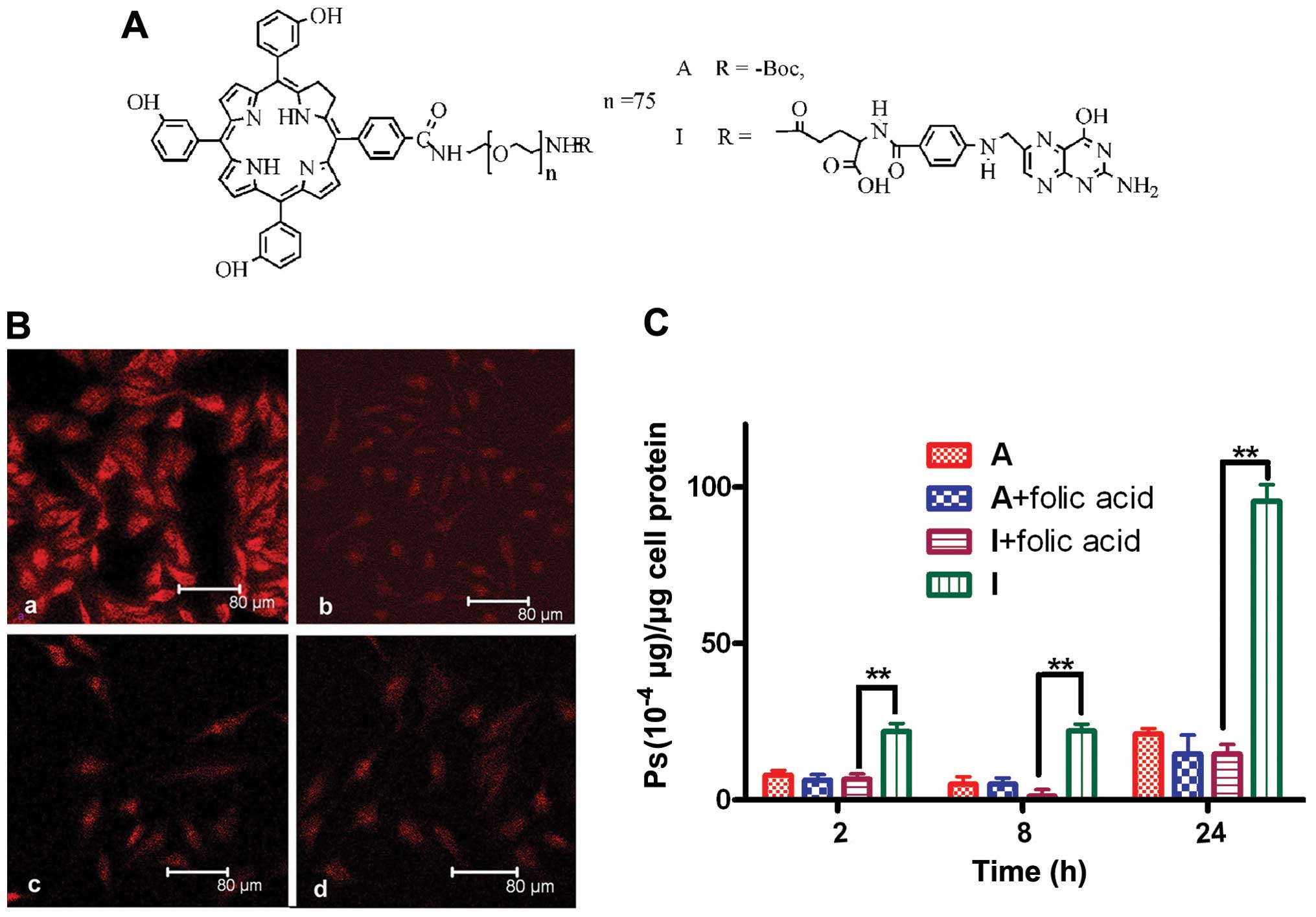
Cellular uptake of Ps I
The cellular uptake of Ps I and A by HeLa cells was
first investigated using laser scanning confocal microscopy. After
incubation with Ps I, strong red fluorescence of Ps I was observed
in the cytoplasm of HeLa cells (Fig.
1B–a), and the fluorescence intensity became weaker with the
addition of excess free folic acid (Fig. 1B–b). However, for HeLa cells
incubated with Ps A, a slight amount of Ps A aggregated on the cell
surface (Fig. 1B–c). No visible
differences between Fig. 1B–c and
–d were observed.
The cellular uptake of Ps I and A by HeLa cells was
quantitatively determined by fluorescence analysis. With the
extension of incubation time, the cellular uptake of Ps I by HeLa
cells increased, and, the disparity between cells with or without
folic acid was more obvious (Fig.
1C). For example, when the incubation time was extended from 2
to 24 h, the disparity of cellular uptake of Ps I with or without
the presence of folic acid increased from 3.1- to 6.5-fold.
However, the presence of folic acid did not affect the uptake of Ps
A by HeLa cells, although the cellular uptake slightly increased
with incubation time.
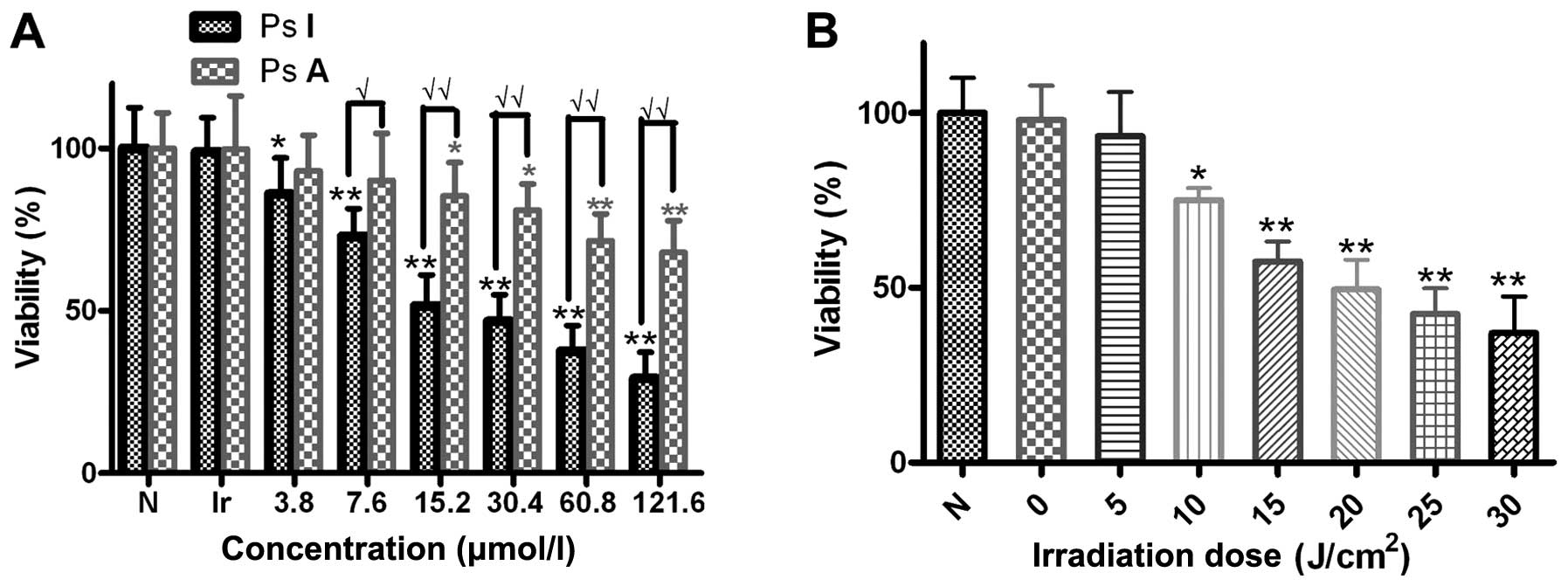
Photodynamic activity of Ps I in
vitro
The photodynamic activity of Ps I and Ps A in HeLa
cells was measured by MTT assay. The viability of HeLa cells in the
Ps I-PDT group was lower than that of cells in the Ps A-PDT group
at all concentrations of photosensitizer tested in our assay
(Fig. 2A). However, the cells
illuminated without Ps I (Fig. 2A)
and the cells kept in the dark in the presence of Ps I (Fig. 2B) did not present any significant
loss of viability in our system. Additionally, the cytotoxicity
induced by Ps I-PDT had a positive correlation with the
concentration of Ps I (R=0.763 and P=0.017) (Fig. 2A) and the irraditation dose (R=0.962
and P=0.001) (Fig. 2B).
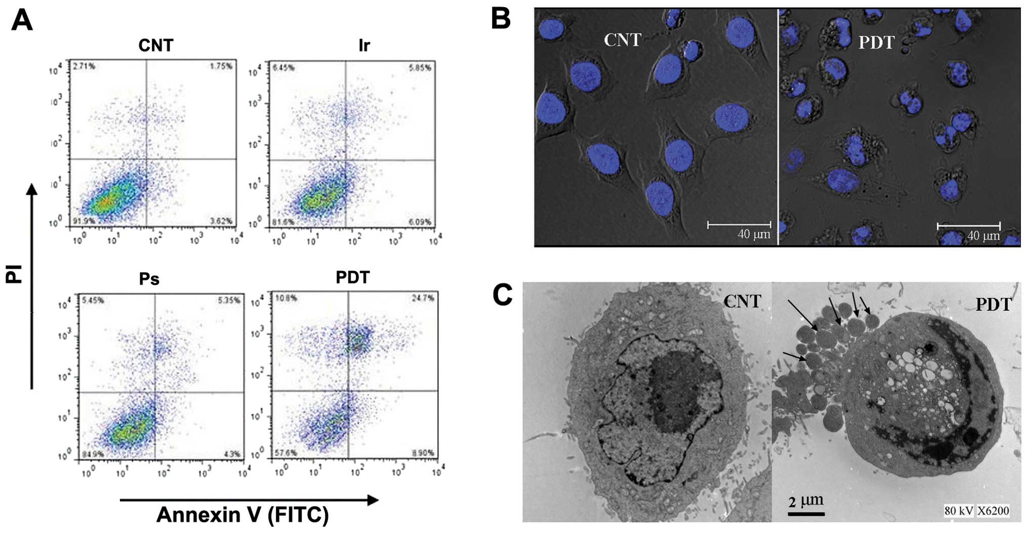
Apoptosis of HeLa cells after Ps
I-PDT
The percentage of apoptotic and necrotic cells was
first quantified via flow cytometric analysis. Compared with the
control group, the number of apoptotic and necrotic cells only
slightly increased in the irradiation alone group and the Ps I
without light group. However, the numbers significantly increased
in the PDT group, especially the number of apoptotic cells
(Fig. 3A). After PDT, cell
rounding, shrinkage and deformation and typical nuclear
fragmentation were observed (Fig.
3B). Under an electron microscope, the untreated control cells
exhibited normal morphology. However, HeLa cells treated with Ps
I-PDT displayed an apoptotic morphology, including the loss of
microvilli and blebbing of the cell membrane, chromatin
condensation around the nuclear membrane into dense half-moon
lumps, and the occurrence of representative apoptotic bodies
alongside the HeLa cells with lobulated protuberance of cytoplasm
(Fig. 3C, arrow).
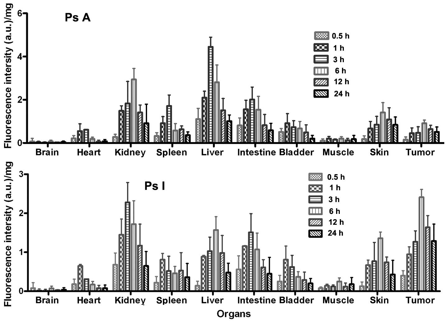
Tissue distribution of Ps I and A
Ps I and A exhibited a different biodistribution in
normal tissues, and the largest accumulations were found to occur
in the kidney and liver, respectively, followed by the liver (or
kidney) and intestine, and the lowest amounts were found in the
brain for tumor and normal tissues (Fig. 4). However, the amount of Ps I
accumulation was clearly reduced in the normal tissues as compared
to that of Ps A at all time-points. By contrast, as early as 0.5 h
after injection, Ps I exhibited, on average, a 2.7-fold higher
tumor uptake than Ps A, and the increased tumor uptake persisted.
As a consequence, the tumor to normal tissue ratios were >2-fold
higher for Ps I than for Ps A at all the tested time-points
(Table I).
 | Table IComparison of tumor to normal tissue
ratios in various organs after intravenous administration of 5
mg/kg Ps A and I. |
Table I
Comparison of tumor to normal tissue
ratios in various organs after intravenous administration of 5
mg/kg Ps A and I.
| Tissue | 0.5 h | 1 h | 3 h | 6 h | 12 h | 24 h |
|---|
| Ps A |
| Heart | 0.71±0.23 | 0.82±0.20 | 0.77±0.13 | 4.79±0.49 | 10.67±1.13 | 6.38±0.20 |
| Liver | 0.14±0.17 | 0.21±0.07 | 0.11±0.08 | 0.32± 0.05 | 0.42±0.03 | 0.50±0.08 |
| Spleen | 0.47±0.13 | 0.49±0.08 | 0.27±0.10 | 1.57±0.43 | 1.02±0.23 | 1.42±0.33 |
| Kidney | 0.54±0.09 | 0.30±0.06 | 0.26±0.03 | 0.31±0.04 | 0.45±0.07 | 0.56±0.04 |
| Intestine | 0.19±0.10 | 0.29±0.12 | 0.23±0.05 | 0.59±0.23 | 0.78±0.13 | 0.86±0.07 |
| Bladder | 0.29±0.07 | 0.49±0.11 | 0.64±0.19 | 1.36±0.25 | 1.31±0.13 | 2.55±0.14 |
| Muscle | 1.50±0.12 | 2.25±0.20 | 3.13±0.45 | 4.55±0.41 | 5.33±0.39 | 2.83±0.09 |
| Skin | 0.83±0.10 | 0.67±0.15 | 0.55±0.16 | 0.65±0.03 | 0.59±0.07 | 0.61±0.05 |
| Ps I |
| Heart | 2.22±0.37 | 1.46±0.17 | 4.10±0.37 | 14.18±0.77 | 20.50±1.63 | 16.13±0.58 |
| Liver | 2.86±0.17 | 1.07±0.07 | 1.23±0.07 | 1.54±0.17 | 1.67±0.10 | 2.69±0.23 |
| Spleen | 1.82±0.27 | 1.17±0.13 | 2.44±0.13 | 5.24±0.23 | 3.09±0.29 | 3.58±0.21 |
| Kidney | 0.59±0.29 | 0.66±0.11 | 0.56±0.11 | 1.40±0.31 | 1.40± 0.21 | 1.98±0.17 |
| Intestine | 0.71±0.19 | 0.82±0.12 | 0.84±0.10 | 2.25±0.23 | 2.69±0.37 | 2.87±0.14 |
| Bladder | 1.60±0.21 | 1.17±0.17 | 2.05±0.27 | 6.51±0.58 | 5.66±0.41 | 6.45±0.57 |
| Muscle | 5.00±0.57 | 6.79±0.69 | 10.58±0.67 | 9.64±0.63 | 13.67±1.04 | 7.17±0.83 |
| Skin | 3.08±0.23 | 1.42±0.12 | 1.65±0.12 | 1.77±0.09 | 2.22±0.19 | 3.00±0.36 |
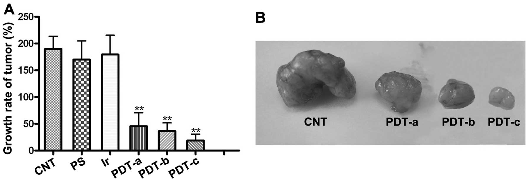
PDT efficacy of Ps I in vivo
The antitumor efficiency of Ps I-PDT against
xenograft tumors in vivo is shown in Fig. 5. In the negative control groups, no
treatment (CNT), the light alone and Ps I alone, the tumor rapidly
grew, while no significant difference among these groups was
observed (Fig. 5A) (P>0.05). Of
note, the Ps I-PDT was found to cause strong suppression of tumor
growth, and the inhibition of Ps I-PDT was dependent on the
concentration of Ps I and irradiation dose. The tumor volume in the
control groups was much larger than that in the PDT groups
(P<0.001), and the smallest tumor was found in the Ps I-PDT
group treated with 7 mg/kg of Ps I and 80 J/cm2 of
irradiation. On the 14th day, the tumor volume in the CNT group was
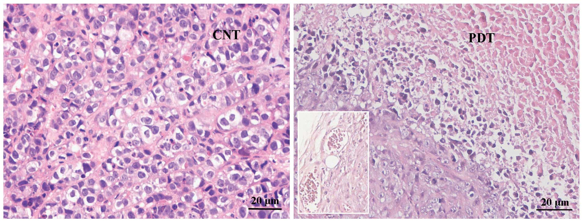
4- and 10-fold larger than that of the PDT groups treated with 5
and 7 mg/kg of Ps I, respectively. The large area of amorphous
necrosis and disorganization of tumor cells, reduction in the size
and number of cell nuclei, fusion of cytoplasm, and congestion of
blood vessels (inset) were evident in the Ps I-PDT group, although
almost no effects were observed in the control groups (Fig. 6). However, the cell damage caused by
PDT was not observed in the liver, spleen or kidney (data not
shown).
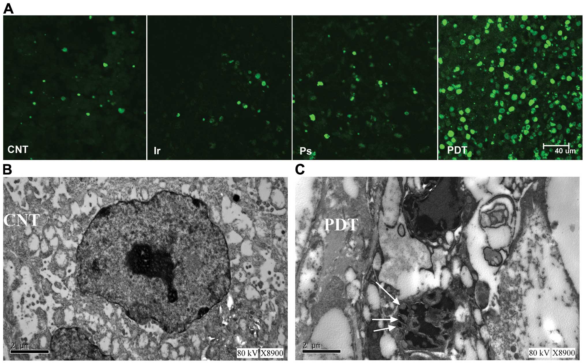
Apoptosis of tumor cells
The TUNEL-positive apoptotic cells (green) in tumor
sections were significantly increased in the Ps I-PDT group,
whereas no significant TUNEL fluorescence was observed in the three
control groups (Fig. 7A). The
nuclear fragmentation and the apoptotic bodies were observed in the
Ps I-PDT group under electron microscopy (Fig. 7B and C). These results strongly
suggested that cell apoptosis was only stimulated by Ps I-PDT, and
this apoptosis inhibited tumor growth in the HeLa xenograft animal
model.
Discussion
Cervical cancer is a considerable threat to public
health. However, the currently available therapies are a
combination of pelvic radiotherapy, radical surgery, and
chemotherapy (21), which
inevitably damage the surrounding normal tissues when destroying
the tumors. As a conservative therapy, PDT can preserve complete
organic structure for the patients and be repeated numerous times
(22). Given the positive
expression of the folate receptor on human cervical carcinoma HeLa
cells (23) and the strong demand
for conservative therapy methods to treat cervical carcinoma, HeLa
cells and nude mice-bearing human cervical carcinoma xenografts
were used as experimental models to evaluate the anticancer
efficiency of Ps I-PDT in the present experimental study.
As Ps I is intravenously administered and is
supposed to localize and kill only the malignant cells after light
irradiation, the tumor targeting of Ps I is extremely important for
the application of Ps I-PDT in cancer. Thus, the tumor targeting of
Ps I was first analyzed in vitro and in vivo. The
results in vitro indicated that there was no obvious
difference in the cellular uptake of Ps A with or without folic
acid present. By contrast, the cellular uptake of Ps I by HeLa
cells was much higher than that of Ps A, and the cellular uptake of
Ps I was markedly inhibited by the presence of excess free folic
acid, suggesting that the free folic acid molecules inhibited the
cellular uptake of Ps I by competitively binding with the folate
receptors on the surface of HeLa cells. In vivo, the tumor
uptake of Ps I was enhanced, and the tumor to normal tissue ratio
was considerably increased compared with Ps A. The results showed
that as compared with Ps A, with the increased accumulation of Ps I
in the tumor and the decrease in the skin, the ratio of tumor to
skin for Ps I significantly increased. For example, the ratio of
tumor to skin for Ps I was 2.72- and 4.91-fold to Ps A at 6 and 24
h post-injection, respectively. This finding was crucial for the
reduction of cutaneous photosensitivity. However, we do not have a
reasonable explanation at this time for the tumor targeting of Ps I
being more significant in vitro than in vivo, and
this finding requires further investigation.
Westermann et al (24) reported that the tumor selectivity of
chlorin could be improved by conjugation with such a macromolecule
as PEG because of the extended circulation half-life. To determine
whether PEG improves the tumor targeting of Ps I and A, we compared
the cellular uptake of Ps I and A by HeLa cells with their
precursor, 5, 10, 15-tris (3-hydroxyphenyl)-20-(4-carboxyphenyl)
chlorin. However, as the precursor is only soluble in DMSO and due
to the membrane penetrant-carrier properties of DMSO (18), the cellular uptake of the precursor
was higher than that of Ps I (data not shown).
The largest accumulation of Ps I in normal tissues
was found in the kidney and not in the liver. This observation is
not in agreement with the known tendency of most Ps used in the
clinic, which preferentially accumulate in the liver, similar to Ps
A (25). This may be due to the
expression of the folate receptor on the apical membranes of kidney
proximal tubule cells (26)
resulting in more Ps I being linked to the kidney prior to
excretion and then transcellularly returned back into
circulation.
The antitumor activity of Ps I was first evaluated
by MTT assay in vitro. Compared with Ps A, Ps I exhibited a
much higher proliferation inhibition rate for HeLa cells, and the
higher the concentration of the photosensitizer, the more obvious
was the difference (Fig. 2A). For
example, when the concentration of the photosensitizer increased
from 3.8 to 60.8 μmol/l, the difference in the inhibition rate
between Ps I and A increased from 7.1 to 34.0%. Given the much
higher cellular uptake of HeLa cells for Ps I than for Ps A, the
result may be explained. The phototoxicity of Ps I was then
examined in vivo using human cervical tumor-bearing mice.
Based on our results of the metabolism of Ps I, phototherapy was
performed 6 h after the intravenous (i.v.) injection of Ps I (PDT
groups) and saline (irradiation control groups). Ps I-PDT
significantly inhibited tumor growth without causing any side
effects to the mice post-PDT. Tumor growth inhibition in
vitro and in vivo exhibited an obvious dependency on the
photosensitizer concentration and light energy intensity.
Apoptosis is of particular importance in cancer
treatment. To investigate the death mode of tumor cells induced by
Ps I-PDT in cervical carcinoma, cell apoptosis was analyzed in
vitro and in vivo. The exposure of plasma membrane
phosphatidylserine is an early marker of apoptosis and can be
detected by Annexin V staining. Accordingly, the percentage of
apoptosis of HeLa cells post-PDT in vitro was analyzed first
by flow cytometry with Annexin V and PI double-staining. Compared
with the control groups, the number of apoptotic cells in Ps I-PDT
group clearly increased, and the HeLa cell apoptosis displayed an
obvious dependency on Ps I concentration and light energy intensity
(data not shown). Given that the upper left quadrant represents
cell fragments (27), the presence
and increase in the cell fragment population in the upper left
quadrant indicated that the Ps I-mediated PDT was able to damage
the target cells. The apoptosis of HeLa cells post-Ps I-PDT was
determined by the characteristic nuclear fragmentation and the
representative apoptotic bodies observed with confocal and electron
microscopy, respectively. Moreover, cell apoptosis was demonstrated
in vivo by TUNEL assay and electron microscopy observation.
These results confirm that Ps I-PDT induced HeLa cell death through
apoptosis. However, the results of the histological examination
revealed the congestion of blood vessels. Therefore, the Ps I-PDT
antitumor effect may also cause vascular impairment, resulting in a
deficient nutrient supply, which directly induces tumor cell
apoptosis.
Taken together, the findings reported in the present
study have demonstrated that Ps I shows higher targeting for HeLa
cells and xenograft cervical carcinoma because of the special
binding between folic acid with folate receptor and the endocytosis
mediated by the folate receptor. As a consequence, Ps I-PDT
exhibited much higher antitumor efficiency against the
proliferation of HeLa cells and the growth of xenograft tumors.
Apoptosis is therefore the main mode of HeLa cell death induced by
Ps I-PDT.
Acknowledgements
The authors are grateful for the financial support
from the National Nature Science Foundation of China (project no.
21072227).
References
|
1
|
Downs LS, Smith JS, Scarinci I, Flowers L
and Parham G: The disparity of cervical cancer in diverse
populations. Gynecol Oncol. 109:22–30. 2008. View Article : Google Scholar
|
|
2
|
Wells SF: Cervical cancer: an overview
with suggested practice and policy goals. Medsurg Nurs. 17:43–50.
2008.PubMed/NCBI
|
|
3
|
He GF, Bian ML, Zhao YW, Xiang Q, Li HY
and Xiao C: A study on the mechanism of 5-aminolevulinic acid
photodynamic therapy in vitro and in vivo in cervical cancer. Oncol
Rep. 21:861–868. 2009.PubMed/NCBI
|
|
4
|
Fayter D, Corbett M, Heirs M, Fox D and
Eastwood A: A systematic review of photodynamic therapy in the
treatment of pre-cancerous skin conditions, Barrett’s oesophagus
and cancers of the biliary tract, brain, head and neck, lung,
oesophagus and skin. Health Technol Asses. 14:1–288. 2010.
|
|
5
|
Benov L: Photodynamic therapy: current
status and future directions. Med Princ Pract. May 10–2014.(Epub
ahead of print). View Article : Google Scholar : PubMed/NCBI
|
|
6
|
Syu WJ, Yu HP, Hsu CY, et al: Improved
photodynamic cancer treatment by folate conjugated polymeric
micelles in a KB xeno-grafted animal model. Small. 8:2060–2069.
2012. View Article : Google Scholar : PubMed/NCBI
|
|
7
|
Dabrowski JM, Arnaut LG, Pereira MM,
Monteiro CJ, Urbanska K, Simoes S and Stochel G: New halogenated
water-soluble chlorin and bacteriochlorin as photostable PDT
sensitizers: synthesis, spectroscopy, photophysics, and in vitro
photosensitizing efficacy. Chem Med Chem. 5:1770–1780. 2010.
View Article : Google Scholar
|
|
8
|
Eshghi H, Sazgarnia MSc A, Rahimizadeh M,
Attaran N, Bakavoli M and Soudmand S: Protoporphydin IX-gold
nanoparticle conjugates as an efficient photosensitizer in cervial
cancer therapy. Photodiagnosis Photodyn Ther. 10:304–312. 2013.
View Article : Google Scholar : PubMed/NCBI
|
|
9
|
Shupin-Mrugalska P, Piskorz J, Goslinski
T, Mielcarek J, Konopka K and Düzgüneş N: Current status of
liposomal porphyrinoid photosensitizers. Drug Discov Today.
18:776–784. 2013. View Article : Google Scholar
|
|
10
|
Gijsens A, Missiaen L, Merlevede W and de
Witte P: Epidermal growth factor-mediated targeting of chlorin e6
selectively potentiates its photodynamic activity. Cancer Res.
60:2197–2202. 2000.PubMed/NCBI
|
|
11
|
Stefflova K, Li H, Chen J and Zheng G:
Peptide-based pharmaco-modulation of a cancer-targeted optical
imaging and photodynamic therapy agent. Bioconjug Chem. 18:379–388.
2007. View Article : Google Scholar : PubMed/NCBI
|
|
12
|
Vrouenraets MB, Visser GWM, Loup C, et al:
Targeting of a hydrophilic photosensitizer by use of internalizing
monoclonal antibodies: A new possibility for use in photodynamic
therapy. Int J Cancer. 88:108–114. 2000. View Article : Google Scholar : PubMed/NCBI
|
|
13
|
McCarthy JR, Bhaumik J, Merbouh N and
Weissleder R: High-yielding syntheses of hydrophilic, conjugatable
chlorins and bacteriochlorins. Org Biomol Chem. 7:3430–3436. 2009.
View Article : Google Scholar : PubMed/NCBI
|
|
14
|
Vlahov IR and Leamon CP: Engineering
folate-drug conjugates to target cancer: from chemistry to clinic.
Bioconjug Chem. 23:1357–1369. 2012. View Article : Google Scholar : PubMed/NCBI
|
|
15
|
Bugaj AM: Targeted photodynamic therapy -
a promising strategy of tumor treatment. Photochem Photobiol Sci.
10:1097–1109. 2011. View Article : Google Scholar : PubMed/NCBI
|
|
16
|
Leung WN, Sun X, Mak NK and Yow CM:
Photodynamic effects of mTHPC on human colon adenocarcinoma cells:
photo-cytotoxicity, subcellular localization and apoptosis.
Photochem Photobiol. 75:406–411. 2002. View Article : Google Scholar : PubMed/NCBI
|
|
17
|
Wang L, Li M and Zhang N: Folate-targeted
docetaxel-lipid-based-nanosuspensions for active-targeted cancer
therapy. Int J Nanomedicine. 7:3281–3294. 2012.PubMed/NCBI
|
|
18
|
Li D, LI P, Lin H, Jiang Z, Guo L and Li
B: A novel chlorin-PEG-folate conjugate with higher water
solubility, lower cytotoxicity, better tumor targeting and
photodynamic activity. J Photochem Photobiol B. 127:28–37. 2013.
View Article : Google Scholar : PubMed/NCBI
|
|
19
|
Vaidya A, Sun Y, Feng Y, Emerson L, Jeong
EK and Lu ZR: Contrast-enhanced MRI-guided photodynamic cancer
therapy with a pegylated bifunctional polymer conjugate. Pharm Res.
25:2002–2011. 2008. View Article : Google Scholar : PubMed/NCBI
|
|
20
|
Chin WWL, Heng PWS, Thong PSP, et al:
Improved formulation of photosensitizer chlorin e6
polyvinylpyrrolidone for fluorescence diagnostic imaging and
photodynamic therapy of human cancer. Eur J Pharm Biopharm.
69:1083–1093. 2008. View Article : Google Scholar : PubMed/NCBI
|
|
21
|
Gui T, Wang Y, Mao Y, Liu J, Sun S, Cao D,
Yang J and Shen K: Comparisons of 5-aminolevulinic acid
photodynamic therapy and after-loading radiotherapy in vivo in
cervical cancer. Clin Transl Oncol. 15:434–442. 2013. View Article : Google Scholar
|
|
22
|
Hiorth M, Liereng L, Reinertsen R and Tho
I: Formulation of bioadhesive hexylaminolevulinate pellets intended
for photo-dynamic therapy in treatment of cervical cancer. Int J
Pharm. 441:544–554. 2013. View Article : Google Scholar
|
|
23
|
Saul JM, Annapragada A, Natarajan JV and
Bellamkonda RV: Controlled targeting of liposomal doxorubicin via
the folate receptor in vitro. J Control Release. 92:49–67. 2003.
View Article : Google Scholar : PubMed/NCBI
|
|
24
|
Westermann P, Glanzmann T, Andrejevic S,
et al: Long circulating half-life and high tumor selectivity of the
photosensitizer meta-tetrahydroxyphenylchlorin conjugated to
polyethylene glycol in nude mice grafted with a human colon
carcinoma. Int J Cancer. 76:842–850. 1998. View Article : Google Scholar
|
|
25
|
Evensen JF, Moan J, Hindar A and Sommer S:
Tissue distribution of 3H-hematoporphyrin derivative and
its main components, 67Ga- and 131I-albumin
in mice bearing Lewis lung carcinoma. Prog Clin Biol Res.
170:541–562. 1984.
|
|
26
|
Leamon CP and Reddy JA: Folate-targeted
chemotherapy. Adv Drug Deliv Rev. 56:1127–1141. 2004. View Article : Google Scholar : PubMed/NCBI
|
|
27
|
Zhang JL, Deng L, Yao JZ, et al: Synthesis
and photobiological study of a novel chlorin photosensitizer
BCPD-18MA for photo-dynamic therapy. Bioorg Med Chem. 19:5520–5528.
2011. View Article : Google Scholar : PubMed/NCBI
|