Introduction
Pancreatic cancer is the fourth leading cause of
cancer-related mortality with a median overall 5-year survival of
5% worldwide (1). Patients at
advanced stages face a dire median overall survival of less than
one year (2). The lethal nature of
pancreatic cancer is marked by its high potential for metastasis to
the lymphatic system and distant organs (3), which prompted us to investigate the
mechanisms involved in motility and metastasis.
v-ets erythroblastosis virus E26 oncogene homolog 1
(ETS-1) is the founding member of the ETS oncogene family, members
of which possess a characteristic DNA-binding domain (ETS domain)
of 85 amino acids (4,5). The ETS-1 protein controls the
expression level of a multitude of genes including other
transcription factors, proteases, cell cycle regulation genes,
apoptosis-related genes, cytokines and growth factors (6). Upregulated ETS-1 has been observed in
breast cancer, lung cancer, ovarian cancer, colorectal cancer and
malignant melanoma (7–11). ETS-1 is overexpressed in invasive
breast cancer and is correlated with the poor prognosis of breast
cancer patients (12,13). High expression of ETS-1 promotes
cell migration, invasion and anchorage-independent growth, while
low ETS-1 expression reduces adherence of HeLa cells (14). One study showed that
fibronectin-stimulated cell adhesion and migration of glioma U251
cells were suppressed by the expression of a dominant-negative form
of ETS-1 (15). These studies
demonstrated that ETS-1 plays major roles in the migration and
invasion of cancer cells. Notably, ETS-1 is barely detectable in
normal human pancreatic tissue, but high levels of expression are
found in samples from human pancreatic cancer biopsies (16). Thus, there is compelling evidence
showing that ETS-1 is involved in the migration and invasion of
pancreatic cancer cells; however, the mechanisms by which ETS-1
mediates these effects have not been fully elucidated.
Epithelial-mesenchymal transition (EMT) facilitates
malignant tumor progression and metastatic spread by enabling
cancer cells to depart from the primary tumor, invade surrounding
tissue and disseminate to distant organs (17). EMT is characterized by reduced
E-cadherin expression and increased N-cadherin expression (17–19).
E-cadherin is an adhesion molecule of epithelial cells, whose
expression is frequently downregulated in invasive cancers.
N-cadherin is associated with higher invasive potential, and is
typically expressed by mesenchymal cells (20). It has been shown that expression of
E-cadherin is increased in stably ETS-1-overexpressing cells,
indicating that ETS-1 by itself has no activity to induce EMT in
human squamous carcinoma cells (21). However, the results indicate that
ETS-1 functions as one of the effectors of EMT (21). Additionally, EMT is usually
accompanied by aberrant expression of vascular endothelial growth
factor (VEGF) (22,23). Mukherjee et al (24) showed that high ETS-1 and VEGF
expression is correlated with tumor angiogenesis, lymph node
metastasis and poor patient survival in esophageal squamous cell
carcinoma.
In the present study, we examined mRNA expression
levels of ETS-1, E-cadherin and N-cadherin in
five pancreatic cancer cell lines. We probed the influence of ETS-1
silencing on the expression of the EMT-related molecules,
E-cadherin, N-cadherin and VEGF, and we investigated the effects of
transcription factor ETS-1 on the motility of Panc-1 pancreatic
cancer cells. Our data showed that ETS-1 functions as a regulator
of EMT in pancreatic cancer cells, and suggest that analysis of
ETS-1 expression levels may provide an avenue for evaluating
prognosis in pancreatic cancer.
Materials and methods
Pancreatic cancer cell lines and cell
culture
Human pancreatic cancer cell lines, Panc-1,
PaTu-8988t, SW1990, Capan2 and BxPC3, were cultured under standard
conditions (37°C in a humidified atmosphere with 5% CO2)
in Dulbecco’s modified Eagle’s medium (DMEM) (Gibco, Carlsbad, CA,
USA) supplemented with 0.1 mM non-essential amino acid, 10% fetal
bovine serum (FBS) and 1% penicillin-streptomycin (all from
Invitrogen, Carlsbad, CA, USA).
Reverse transcription-polymerase chain
reaction (RT-PCR)
Total RNA was extracted from stably transfected
cells with TRIzol reagent (15596-026; Invitrogen), and first-strand
cDNA was synthesized according to the manufacturer’s instructions
(DRR036A; Takara, Tokyo, Japan). RT-PCR analysis was carried out
using 2X Power Taq PCR Master Mix (PR1700; BioTeke, Beijing, China)
under the following conditions: 95°C for 5 min to denature cDNA,
followed by 30 cycles of 95°C for 30 sec, 57°C for 40 sec, and 72°C
for 45 sec, followed by a terminal extension for 10 min at 72°C.
The products were analyzed by electrophoresis on 2% agarose gels
(16550-100; Invitrogen) at 120 V for 40 min, and the bands were
visualized by an UltraPower™ Gel Imaging System (EP2018; BioTeke).
The primer sequences are listed in Table I. All of the primers were obtained
from Invitrogen.
 | Table ISequences of the oligonucleotide
primers. |
Table I
Sequences of the oligonucleotide
primers.
| Gene | Sequence
(5′-3′) | Product (bp) |
|---|
| ETS-1 | Forward:
5′-GTCGTGGTAAACTCGG-3′
Reverse: 5′-CAGCAGGAATGACAGG-3′ | 246 |
|
N-cadherin | Forward:
5′-AGTGAGCCTGCAGATTTTAAGGTGGATG-3′
Reverse: 5′-CACTTGCCACTTTTCCTGGGTCTCTT-3′ | 132 |
|
E-cadherin | Forward:
5′-TTGCACCGGTCGACAAAGGAC-3′
Reverse: 5′-TGGAGTCCCAGGCGTAGACCAA-3′ | 140 |
| VEGF | Forward:
5′-AACCAGCAGAAAGAGGAAAGAGG-3′
Reverse: 5′-CCAAAAGCAGGTCACTCACTTTG-3′ | 133 |
| GAPDH | Forward:
5′-CCACCCATGGCAAATTCCATGGCA-3′
Reverse: 5′-TCTAGACGGCAGGTCAGGTCCACC-3′ | 251 |
Short hairpin RNA transfection
Panc-1 cells (5×10⁵) were seeded in each
well of a 6-well plate and were transfected with 500 ng/ml of
either pSi-ETS1 (PIEE102075355) or control plasmids (both from
Genechen, Shanghai, China) coupled with Lipofectamine 2000
(11668-019; Invitrogen). Following transfection, the culture medium
was replaced by Opti-MEM medium (31985-062; Invitrogen) according
to the manufacturer’s instructions.
Real-time reverse transcription PCR
(qRT-PCR)
Total RNA was extracted from stably transfected
cells with the TRIzol reagent (15596-026; Invitrogen), and
first-strand cDNA was synthesized according to the manufacturer’s
instruction (DRR036A; Takara). qRT-PCR was carried out using
LightCycler® DNA Master SYBR-Green I as reaction mix
(12015099001; Roche, Branchburg, NJ, USA) on the ABI 7300 Real-Time
PCR system (Applied Biosystems, Foster City, CA, USA) under the
following conditions: 95°C for 30 sec to denature cDNA and primers,
followed by 40 cycles at 95°C for 5 sec, 60°C for 20 sec and 72°C
for 30 sec, followed by a terminal extension for 7 min at 72°C.
Gene expression was calculated with the comparative Ct method and
normalized against the endogenous levels of
glyceraldehyde-3-phosphate dehydrogenase (GAPDH). The primer
sequences are listed in Table
I.
Western blot analysis
The transfected cells were washed with
phosphate-buffered solution (PBS) and lysed with RIPA buffer
containing a protease inhibitor cocktail (11873580001; Roche). The
supernatant was collected after centrifuging the cell lysate
(13,300 × g for 10 min), and the concentration of the cellular
protein was measured using a BCA detection kit (P0010S; Beyotime,
Jiangsu, China). The protein concentration was adjusted to 2 μg/μl
for electrophoresis on a 10% SDS-PAGE gel. Cellular proteins
separated on the gel were transferred to a polyvinylidene
difluoride membrane (03010040001; Roche). After blocking with 5%
nonfat milk in Tris-buffered saline containing 0.1% Tween-20, the
membrane was incubated with specific primary antibodies: anti-GAPDH
antibody (MB001, 1:5,000 diluted; Bioworld, St. Louis, MN, USA),
anti-VEGF antibody (ab46154, 1:1,000 diluted), anti-ETS-1 antibody
(ab26096, 1:1,000 diluted) (both from Abcam, Cambridge, UK),
anti-N-cadherin antibody (BS2224, 1:1,000 diluted) and
anti-E-cadherin antibody (BS1098, 1:1,000 diluted) (both from
Bioworld). After incubation with the appropriate horseradish
peroxidase-conjugated secondary antibody (LK-RAG007; Multi Sciences
Biotech Co., Hangzhou, China), signals were detected using an
enhanced chemiluminescence reagent (WBKLS0500; Millipore,
Darmstadt, Germany) and subjected to the Alpha Innotech FluorChem
FC2 Imaging system (Alpha Innotech Corp., San Leandro, CA,
USA).
In vitro cell migration assay
A scratch assay was utilized to examine in
vitro cell migration of pancreatic cancer cells. After being
transfected for 24 h, Panc-1 cells (5×10⁵ cells) were
seeded in a 6-well plate and allowed to form a confluent cell
monolayer, which was then scratched by a sterile pipette tip. The
monolayer was washed with PBS, and fresh medium supplemented with
1% FBS (Invitrogen) was added. An inverted fluorescence microscope
system (DMI3000B; Leica Microsystems, Heerbrugg, China) was used
for imaging immediately after wounding (0 h) and after 12 h. The
distances between the cell boundaries were measured using TPView
(Shanghai Weitu Optics and Electron Technology Co., Ltd., Shanghai,
China).
In vitro cell adhesion assay
Cell adhesion assay was performed to quantify the
ability of the cancer cells to adhere to fibronectin (F2006;
Sigma-Aldrich, St. Louis, MO, USA). After 24 h of transfection, the
ETS-1 short hairpin RNA (shRNA)-transfected and control cells were
collected and seeded into a 96-well plate at 5×10³
cells/well and into a 12-well plate/well, which were both
pre-coated with 20 μg/ml fibronectin for 30 min. Cells were allowed
to adhere for 2 h at 37°C in a humidified atmosphere of 5%
CO2. Unbound cells were removed by inverting and gentle
washing in PBS. Cells in 96-well plates were stained with crystal
violet solution containing 0.4 mg/ml crystal violet and 5%
formaldehyde in PBS for 20 min at room temperature. The plate was
then washed twice with de-ionized water and air-dried for scanning.
The number of adherent cells at the bottom of each well of the
12-well plate was determined by a Countstar Automated cell counter
(IC 1000; Inno-Alliance Biotech Inc., Wilmington, DE, USA).
Statistical analysis
The Student’s t-test (two-tailed) was applied in the
analysis of statistical significance. All data are presented as
mean ± standard deviation. A P-value <0.05 was considered to
indicate a statistically significant difference.
Results
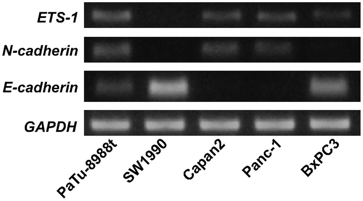
Analysis of ETS-1, N-cadherin and
E-cadherin mRNA expression in pancreatic cancer cell lines
To evaluate the correlation between ETS-1 and
EMT-related molecules, we analyzed the expression levels of
ETS-1, N-cadherin and E-cadherin by RT-PCR in
the following pancreatic cancer cell lines: PaTu-8988t, SW1990,
Capan2, Panc-1 and BxPC3. The result showed that only SW1990 cells
were negative for ETS-1 mRNA expression. High ETS-1
expression was positively associated with the expression levels of
N-cadherin and negatively with the expression of
E-cadherin in the PaTu-8988t, Capan2 and Panc-1 cells. In
the SW1990 and BxPC3 cells, low ETS-1 expression was
correlated with high levels of E-cadherin but low levels of
N-cadherin (Fig. 1). These
results are consistent with a role for ETS-1 in EMT.
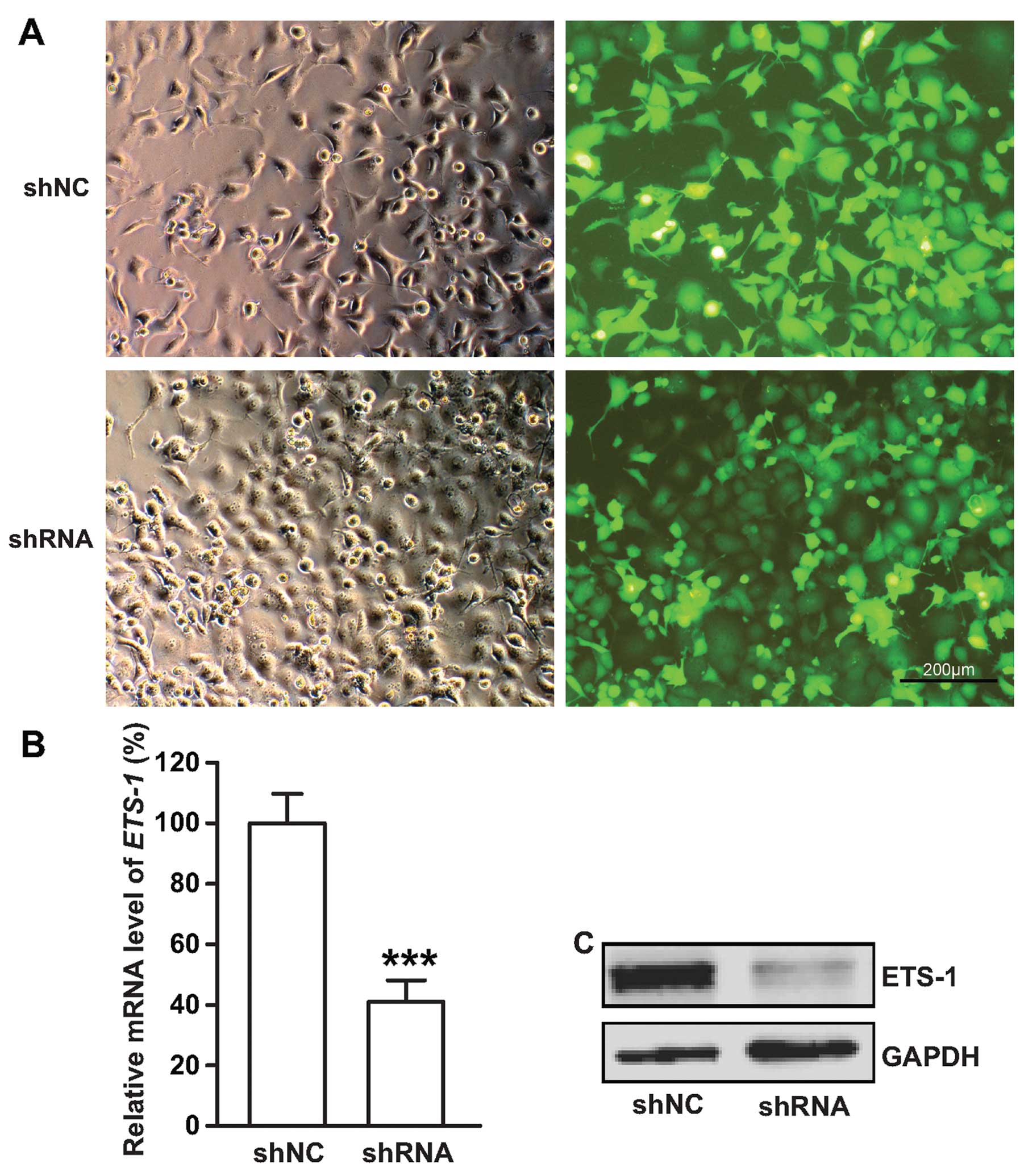
Efficacy of the ETS-1 transcriptional
silencing
We performed ETS-1 inhibition in the Panc-1 cancer
cell line, which exhibited relatively high expression of ETS-1
(Fig. 1). ETS-1 inhibition was
performed using a green fluorescent protein (GFP)-expressing
adenoviral vector carrying an shRNA targeting the ETS-1
gene. The success of the plasmid transfection was monitored by
green fluorescence (Fig. 2A). The
silencing efficiency of the ETS-1 shRNA was determined using
qRT-PCR and western blot analysis. qRT-PCR showed a 70% reduction
in mRNA expression in the ETS-1 shRNA-transfected cells (Fig. 2B). The level of ETS-1 protein was
also significantly decreased in the ETS-1-silenced cells (Fig. 2C).
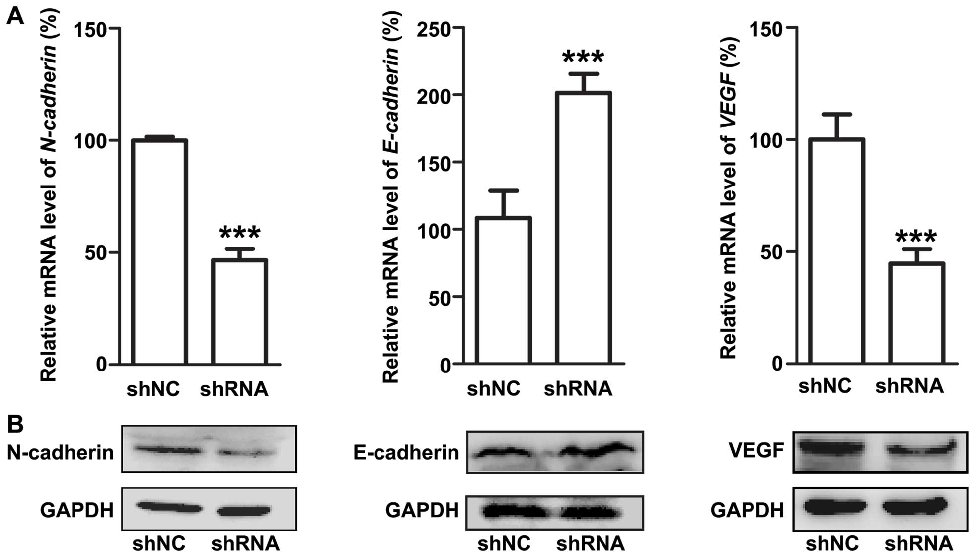
Transcriptional silencing of ETS-1
inhibits epithelial-mesenchymal transition
We investigated the effect of ETS-1 silencing on EMT
by examining the expression levels of EMT-related proteins. In the
ETS-1 shRNA-transfected cells, the level of N-cadherin mRNA
expression was reduced to half, while the level of
E-cadherin mRNA expression was increased as much as 2-fold
(Fig. 3A). Consistently, western
blot analysis revealed that ETS-1-shRNA plasmid transfection
significantly decreased N-cadherin expression and increased
E-cadherin expression in the Panc-1 cells (Fig. 3B). Our results also showed decreased
expression of VEGF in the ETS-1 shRNA-transfected cells both at the
mRNA (Fig. 3A) and protein
expression levels (Fig. 3B).
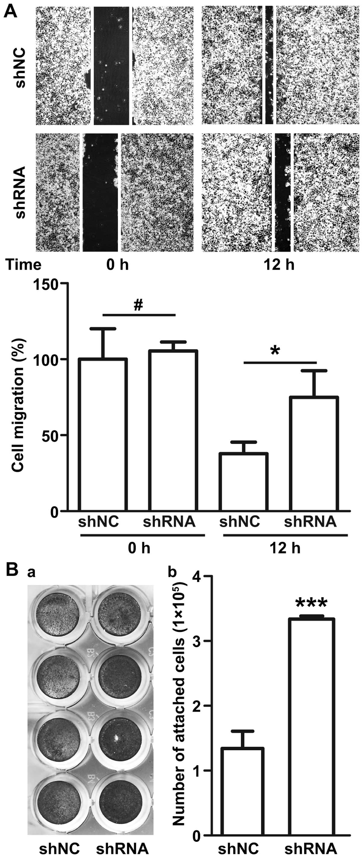
Transcriptional silencing of ETS-1
reduces cell migration, but increases cell adhesion
Migration and invasion of cancer cells are key steps
in tumor metastasis. The effect of transcriptional silencing of
ETS-1 on the motility of Panc-1 cancer cells was measured in
scratch and adhesion assays. The results of the scratch assay
showed that ETS-1 silencing led to decreased cell migration. At 12
h, the distance between the scratch boundaries was 37% wider in the
ETS-1 shRNA-transfected cells (Fig.
4A). Crystal violet staining assay showed that adhesion
increased in the ETS-1 shRNA-infected cells (Fig. 4B-a). Following washing, an ~2.5-fold
more ETS-1 shRNA transfected cells than control cells remained
attached to the plate (Fig. 4B-b).
These results showed that ETS-1 inhibition was highly effective in
increasing the adhesion and reducing the motility of Panc-1
cells.
Discussion
Patients with pancreatic cancer have an extremely
poor prognosis due to the malignant behaviors of this disease.
Invasion and metastasis are two main events associated with the
poor prognosis. The prognosis of patients with clinically
aggressive pancreatic cancer cannot be accurately predicted by
standard variables such as pathologic stage; thus other biomarkers
are needed. Expression of transcription factors associated with
metastasis have shown promise as prognostic biomarkers (25,26).
Little is known, however, regarding the expression of transcription
factors involved in the processes regulating invasion and
metastasis in pancreatic cancer. In the present study, we showed
that the transcription factor ETS-1 plays important roles in
regulating EMT and may have prognostic value.
EMT is characterized by downregulation of E-cadherin
expression and acquisition of mesenchymal markers including
N-cadherin, vimentin and fibronectin, which lead to loss of cell
adhesion facilitating cell motility (27,28).
Studies investigating the migration and invasion of malignant
cancers have mostly associated ETS-1 with the expression of
extracellular matrix metalloproteinase (13,21),
integrins (29,30) and cadherins (27,31).
One study showed that expression of c-ets-1 mRNA was associated
with the EMT-derived phenotype typified by the expression of
vimentin and the lack of E-cadherin in breast carcinoma cell lines
(31). However, the influence of
ETS-1 on the expression of N-cadherin and E-cadherin involved in
the EMT-derived phenotype has not been investigated in pancreatic
cancer cells. In this study, associations between expression of
ETS-1 and EMT-related molecules N-cadherin and
E-cadherin were investigated. We found that ETS-1 mRNA was
expressed in four of five pancreatic cancer cell lines.
Furthermore, the ETS-1 mRNA expression level was positively
associated with the mRNA expression level of N-cadherin but
was negatively associated with that of E-cadherin. To
further evaluate the role of ETS-1 during EMT, we inhibited
ETS-1 expression with an ETS-1-specific shRNA and assessed
its effects on the expression of N-cadherin, E-cadherin and VEGF in
the Panc-1 cell line, a highly metastatic epithelial-pancreatic
adenocarcinoma cell line. Our data showed that transcriptional
silencing of ETS-1 induced a switch from N-cadherin to E-cadherin
expression in Panc-1 cells, which indicates a block in EMT. In
addition, our results showed that ETS-1 inhibition reduced VEGF
expression. This is consistent with the previous finding that VEGF
expression is significantly associated with ETS-1 expression
(32). Hence, by demonstrating a
close relationship between ETS-1 and EMT-related molecules, we
provide strong evidence that ETS-1 plays a crucial role in the
process of EMT in pancreatic cancer cells.
Aberrant expression of ETS-1 was found to be
correlated with malignant cancer behaviors such as tumor
proliferation, invasion, migration and angiogenesis (9,33,34).
Our previous study also showed that gambogic acid-loaded magnetic
Fe3O4 nanoparticles could inhibit
proliferation and migration of Panc-1 cells via inactivating ETS-1
(35). Expression of ETS-1 has also
been shown to decrease adhesion in endothelial (36), HeLa (14) and U251 glioma cells (15). However, the mechanisms of ETS-1
involvement in the EMT process have not been investigated in
pancreatic cancer cells. Our results revealed that ETS-1 inhibition
led to EMT downregulation. Cells undergoing EMT become invasive and
migratory. Therefore, to determine whether ETS-1 silencing would
reduce the motility of Panc-1 pancreatic cancer cells, we performed
scratch and adhesion assays. We demonstrated, as shown in Fig. 4, that ETS-1 silencing reduced cell
migration and increased cell adhesion. Our results suggest that
ETS-1 promotes cell migration and invasion by promoting EMT.
In conclusion, our data support that ETS-1 plays
functionally significant roles in the metastasis of pancreatic
cancer cells by regulating the expression of N-cadherin and
E-cadherin involved in epithelial-mesenchymal transition. We also
found that ETS-1 silencing inhibits the expression of VEGF, which
has been reported to be a probable marker for poor prognosis
(37). Although its mechanism
remains incompletely understood, our results suggest the potential
utility of ETS-1 as an adverse prognostic factor, and highlight the
need for further research to elucidate the role and importance of
ETS-1 during the progression of pancreatic cancer.
Acknowledgements
This study was supported by the Natural Science
Foundation of Jiangsu Province (BK2011604) and the National Nature
Science Foundation of China (81271699). We are grateful for the
excellent experimental guidance from Professor Jiwu Wei (Jiangsu
Key Laboratory of Molecular Medicine, Medical School of Nanjing
University). We also appreciate Ke Xu (Department of Molecular
Cytology of the Memorial Sloan-Kettering Cancer Center) for the
revision and editing of this manuscript.
References
|
1
|
Saika K and Sobue T: Cancer statistics in
the world. Gan To Kagaku Ryoho. 40:2475–2480. 2013.(In Japanese).
PubMed/NCBI
|
|
2
|
Hidalgo M: Pancreatic cancer. N Engl J
Med. 362:1605–1617. 2010. View Article : Google Scholar : PubMed/NCBI
|
|
3
|
Li Y, Kong D, Ahmad A, Bao B and Sarkar
FH: Pancreatic cancer stem cells: emerging target for designing
novel therapy. Cancer Lett. 338:94–100. 2013. View Article : Google Scholar
|
|
4
|
Laudet V, Niel C, Duterque-Coquillaud M,
Leprince D and Stehelin D: Evolution of the ets gene family.
Biochem Biophys Res Commun. 190:8–14. 1993. View Article : Google Scholar : PubMed/NCBI
|
|
5
|
Laudet V, Hänni C, Stéhelin D and
Duterque-Coquillaud M: Molecular phylogeny of the ETS gene family.
Oncogene. 18:1351–1359. 1999. View Article : Google Scholar : PubMed/NCBI
|
|
6
|
Sementchenko VI and Watson DK: Ets target
genes: past, present and future. Oncogene. 19:6533–6548. 2000.
View Article : Google Scholar
|
|
7
|
Fujimoto J, Aoki I, Toyoki H, et al:
Clinical implications of expression of ETS-1 related to
angiogenesis in metastatic lesions of ovarian cancers. Oncology.
66:420–428. 2004. View Article : Google Scholar : PubMed/NCBI
|
|
8
|
Yamaguchi E, Nakayama T, Nanashima A, et
al: Ets-1 proto-oncogene as a potential predictor for poor
prognosis of lung adenocarcinoma. Tohoku J Exp Med. 213:41–50.
2007. View Article : Google Scholar : PubMed/NCBI
|
|
9
|
Rothhammer T, Hahne JC, Florin A, et al:
The Ets-1 transcription factor is involved in the development and
invasion of malignant melanoma. Cell Mol Life Sci. 61:118–128.
2004. View Article : Google Scholar : PubMed/NCBI
|
|
10
|
Nakayama T, Ito M, Ohtsuru A, Naito S and
Sekine I: Expression of the ets-1 proto-oncogene in human
colorectal carcinoma. Mod Pathol. 14:415–422. 2001. View Article : Google Scholar : PubMed/NCBI
|
|
11
|
Span PN, Manders P, Heuvel JJ, et al:
Expression of the transcription factor Ets-1 is an independent
prognostic marker for relapse-free survival in breast cancer.
Oncogene. 21:8506–8509. 2002. View Article : Google Scholar : PubMed/NCBI
|
|
12
|
Zhang Y, Yan LX, Wu QN, et al: miR-125b is
methylated and functions as a tumor suppressor by regulating the
ETS1 proto-oncogene in human invasive breast cancer. Cancer Res.
71:3552–3562. 2011. View Article : Google Scholar : PubMed/NCBI
|
|
13
|
Puzovic V, Brcic I, Ranogajec I and
Jakic-Razumovic J: Prognostic values of ETS-1, MMP-2 and MMP-9
expression and co-expression in breast cancer patients. Neoplasma.
61:439–446. 2014. View Article : Google Scholar : PubMed/NCBI
|
|
14
|
Hahne JC, Okuducu AF, Kaminski A, Florin
A, Soncin F and Wernert N: Ets-1 expression promotes epithelial
cell transformation by inducing migration, invasion and
anchorage-independent growth. Oncogene. 24:5384–5388. 2005.
View Article : Google Scholar : PubMed/NCBI
|
|
15
|
Kita D, Takino T, Nakada M, Takahashi T,
Yamashita J and Sato H: Expression of dominant-negative form of
Ets-1 suppresses fibronectin-stimulated cell adhesion and migration
through down-regulation of integrin alpha5 expression in U251
glioma cell line. Cancer Res. 61:7985–7991. 2001.PubMed/NCBI
|
|
16
|
Duda DG, Sunamura M, Lefter LP, et al:
Restoration of SMAD4 by gene therapy reverses the invasive
phenotype in pancreatic adenocarcinoma cells. Oncogene.
22:6857–6864. 2003. View Article : Google Scholar : PubMed/NCBI
|
|
17
|
Kang Y and Massague J:
Epithelial-mesenchymal transitions: twist in development and
metastasis. Cell. 118:277–279. 2004. View Article : Google Scholar : PubMed/NCBI
|
|
18
|
Christiansen JJ and Rajasekaran AK:
Reassessing epithelial to mesenchymal transition as a prerequisite
for carcinoma invasion and metastasis. Cancer Res. 66:8319–8326.
2006. View Article : Google Scholar : PubMed/NCBI
|
|
19
|
Gravdal K, Halvorsen OJ, Haukaas SA and
Akslen LA: A switch from E-cadherin to N-cadherin expression
indicates epithelial to mesenchymal transition and is of strong and
independent importance for the progress of prostate cancer. Clin
Cancer Res. 13:7003–7011. 2007. View Article : Google Scholar : PubMed/NCBI
|
|
20
|
Nakajima S, Doi R, Toyoda E, et al:
N-cadherin expression and epithelial-mesenchymal transition in
pancreatic carcinoma. Clin Cancer Res. 10:4125–4133. 2004.
View Article : Google Scholar : PubMed/NCBI
|
|
21
|
Taki M, Verschueren K, Yokoyama K,
Nagayama M and Kamata N: Involvement of Ets-1 transcription factor
in inducing matrix metalloproteinase-2 expression by
epithelial-mesenchymal transition in human squamous carcinoma
cells. Int J Oncol. 28:487–496. 2006.PubMed/NCBI
|
|
22
|
Tokuhara K, Ogata Y, Nakagawa M and
Shirouzu K: Ets-1 expression in vascular endothelial cells as an
angiogenic and prognostic factor in colorectal carcinoma. Int Surg.
88:25–33. 2003.PubMed/NCBI
|
|
23
|
Fantozzi A, Gruber DC, Pisarsky L, et al:
VEGF-mediated angiogenesis links EMT-induced cancer stemness to
tumor initiation. Cancer Res. 74:1566–1575. 2014. View Article : Google Scholar : PubMed/NCBI
|
|
24
|
Mukherjee T, Kumar A, Mathur M,
Chattopadhyay TK and Ralhan R: Ets-1 and VEGF expression correlates
with tumor angiogenesis, lymph node metastasis, and patient
survival in esophageal squamous cell carcinoma. J Cancer Res Clin
Oncol. 129:430–436. 2003. View Article : Google Scholar : PubMed/NCBI
|
|
25
|
Rabbani F, Richon VM, Orlow I, et al:
Prognostic significance of transcription factor E2F-1 in bladder
cancer: genotypic and phenotypic characterization. J Natl Cancer
Inst. 91:874–881. 1999. View Article : Google Scholar : PubMed/NCBI
|
|
26
|
Szasz AM, Majoros A, Rosen P, et al:
Prognostic potential of ERG (ETS-related gene) expression in
prostatic adenocarcinoma. Int Urol Nephrol. 45:727–733. 2013.
View Article : Google Scholar : PubMed/NCBI
|
|
27
|
Pap Z, Pavai Z, Denes L, Kovalszky I and
Jung J: An immunohistochemical study of colon adenomas and
carcinomas: E-cadherin, Syndecan-1, Ets-1. Pathol Oncol Res.
15:579–587. 2009. View Article : Google Scholar : PubMed/NCBI
|
|
28
|
Maeda M, Johnson KR and Wheelock MJ:
Cadherin switching: essential for behavioral but not morphological
changes during an epithelium-to-mesenchyme transition. J Cell Sci.
118:873–887. 2005. View Article : Google Scholar : PubMed/NCBI
|
|
29
|
Tang N, Wang X, Huang T, Wu Y and Chen Y:
Role of Ets-1 in fibronectin-derived heparin-binding domain
polypeptides alleviating melanoma cell invasiveness and
chemoresistance. Exp Dermatol. 23:512–513. 2014. View Article : Google Scholar : PubMed/NCBI
|
|
30
|
Oda N, Abe M and Sato Y: ETS-1 converts
endothelial cells to the angiogenic phenotype by inducing the
expression of matrix metalloproteinases and integrin beta3. J Cell
Physiol. 178:121–132. 1999. View Article : Google Scholar : PubMed/NCBI
|
|
31
|
Gilles C, Polette M, Birembaut P, Brünner
N and Thompson EW: Expression of c-ets-1 mRNA is associated with an
invasive, EMT-derived phenotype in breast carcinoma cell lines.
Clin Exp Metastasis. 15:519–526. 1997. View Article : Google Scholar : PubMed/NCBI
|
|
32
|
Hashiya N, Jo N, Aoki M, et al: In vivo
evidence of angiogenesis induced by transcription factor Ets-1:
Ets-1 is located upstream of angiogenesis cascade. Circulation.
109:3035–3041. 2004. View Article : Google Scholar : PubMed/NCBI
|
|
33
|
Holterman CE, Franovic A, Payette J and
Lee S: ETS-1 oncogenic activity mediated by transforming growth
factor alpha. Cancer Res. 70:730–740. 2010. View Article : Google Scholar : PubMed/NCBI
|
|
34
|
Lefter LP, Dima S, Sunamura M, et al:
Transcriptional silencing of ETS-1 efficiently suppresses
angiogenesis of pancreatic cancer. Cancer Gene Ther. 16:137–148.
2009. View Article : Google Scholar
|
|
35
|
Wang C, Zhang H, Chen Y, Shi F and Chen B:
Gambogic acid-loaded magnetic Fe(3)O(4) nanoparticles inhibit
Panc-1 pancreatic cancer cell proliferation and migration by
inactivating transcription factor ETS1. Int J Nanomed. 7:781–787.
2012.
|
|
36
|
Naito S, Shimizu S, Matsuu M, et al: Ets-1
upregulates matrix metalloproteinase-1 expression through
extracellular matrix adhesion in vascular endothelial cells.
Biochem Biophys Res Commun. 291:130–138. 2002. View Article : Google Scholar : PubMed/NCBI
|
|
37
|
Hogendorf P, Durczynski A, Kumor A and
Strzelczyk J: Pancreatic head carcinoma and vascular endothelial
growth factor (VEGF-A) concentration in portal blood: its
association with cancer grade, tumor size and probably poor
prognosis. Arch Med Sci. 10:288–293. 2014. View Article : Google Scholar : PubMed/NCBI
|