Introduction
Gastric cancer is the fourth most common cancer and
is the second leading cause of cancer-related mortality worldwide.
Although there have been marked decreases in the incidence and
mortality in some regions of the world, gastric cancer remains an
important public health burden, particularly in developing
countries, and patient prognosis is generally rather poor, with a
5-year relative survival less than 30% in most countries (1,2).
Extragastric lymph node metastasis is a critical independent
prognostic factor, which leads to the failure of surgery,
chemotherapy or radiotherapy (3,4).
Therefore, prevention of metastatic gastric cancer is an important
therapeutic goal; however, the underlying mechanism of gastric
cancer metastasis remains unclear.
Chemokines are a large subfamily of chemoattractant
cytokines which are classified into 4 highly conserved groups: CXC,
CC, C and CX3C. Chemokines have been mostly studied for their
potent effect on the recruitment of leukocytes at sites of
inflammation (5). However, it is
now established that migrating malignant cells may exploit
chemokine receptors to invade surrounding tissues leading to
distant metastasis (6–25).
CX3CL1 is a structurally unique chemokine and is
currently the only known member of the CX3C family of chemokines
(26,27). Unlike several other chemokines,
CX3CL1 has both a membrane-bound and a soluble form. Membrane-bound
CX3CL1 can act as an adhesion molecule and can be cleaved by
metalloproteinases to create circulating soluble CX3CL1 acting as a
chemoattractant (26,28,29).
Both transmembrane and soluble forms of CX3CL1 bind to the only
known G protein-coupled seven-transmembrane receptor CX3CR1
(30,31). The CX3CR1/CX3CL1 axis has been
demonstrated to be involved in the proliferation, survival and
metastasis of various malignant tumor types, including clear cell
renal cell carcinoma (11),
prostate cancer (21), breast
cancer (19), pancreatic ductal
adenocarcinoma (16) and glioma
tumors (32). However, to our
knowledge, whether the CX3CR1/CX3CL1 axis is involved in gastric
cancer metastasis remains unknown.
In the present study, we investigated CX3CR1
expression in gastric cancer tissues and gastric cancer cell lines
compared with non-neoplastic gastric tissues and a gastric
epithelial cell line, and further analyzed the functional role of
the CX3CR1/CX3CL1 axis in gastric cancer cell lines and a gastric
epithelial cell line in vitro. We demonstrated that the
CX3CR1/CX3CL1 axis plays an important role in gastric cancer
metastasis, proliferation and survival, and additionally might play
a physiological role in normal gastric tissue renewal and/or tissue
remodeling after injury.
Materials and methods
Patients
A total of 89 patients diagnosed with gastric cancer
as confirmed by pathology at the Peking University People’s
Hospital from January 2000 to December 2004 were enrolled in the
study. The mean patient age was 61.5±11.6 years. Among the
patients, 65 were men and 24 were women. None had previously
received radiotherapy, chemotherapy or other medical interventions
before surgery. The control group consisted of 30 contemporaneous
patients who had chronic superficial gastritis diagnosed by
gastroscopy at the Peking University People’s Hospital selected
randomly (simple random sampling). The mean age was 59.3±10.4
years. Among the control group, 16 individuals were men and 14 were
women. This study was approved by The Ethics Committee of the
Peking University, and written informed consent was obtained from
all patients at study entry.
Cell lines and cell culture
Gastric cancer cell lines MKN-28, SGC-7901, MKN-45
and the immortalized gastric epithelial cell line GES-1 were
obtained from the China Center For Type Culture Collection (Wuhan,
China). All cells were cultured in RPMI-1640 (Life Technologies,
USA) supplemented with 10% fetal bovine serum (FBS; Life
Technologies, USA) in a humid atmosphere of 5% CO2 and
95% air at 37°C.
Quantitative real-time PCR
Total RNA was isolated using the RNeasy Mini kit
(Qiagen, USA), according to the manufacturer’s instructions. The
first cDNA strand was synthesized from total RNA with the
SuperScript III First-Strand synthesis system for RT-PCR (Life
Technologies, USA). The following primers were used for the
subsequent PCR: human CX3CR1 sense, 5′-TTGAGTACGATGATTTGGCTGA-3′
and antisense, 5′-GGCTTTGGCTTTCTTGTGG-3′; human GAPDH sense,
5′-TGTTGCCATCAATGACCCCTT-3′ and antisense,
5′-CTCCACGACGTACTCAGCG-3′. Quantitative real-time PCR was conducted
with RealMasterMix™ (SYBR-Green) (Tiangen, China) and GAPDH to
normalize data. PCR conditions were 30 sec at 94°C, 30 sec at 60°C
and 1 min at 72°C for 40 cycles.
Western blot analysis
The cells were collected and washed with cold PBS
three times, and then lysed at 4°C for 30 min in lysis buffer [50
mM Tris, (pH 7.4), 100 mM NaCl2, 1 mM MgCl2,
2.5 mM Na3VO4, 1 mM PMSF, 2.5 mM
ethylenediaminetetraacetic acid, 0.5% Triton X-100, 0.5% NP-40, 5
mg/ml of aprotinin, pepstatin A and leupeptin]. The lysates were
centrifuged at 10,000 × g for 20 min at 4°C. The protein
concentration was determined using a bicinchoninic acid protein
assay reagent kit (Pierce, USA) according to the manufacturer’s
protocol. Forty micrograms of total proteins was electrophoresed on
a 10% denaturing SDS gel and transferred onto a polyvinylidene
difluoride (PVDF) membrane. The PVDF membrane was then incubated
with blocking buffer (PBS containing 5% non-fat milk) for 2 h at
room temperature, followed by incubation with rabbit polyclonal
antibodies against total Akt (1:1,000; Cell Signaling Tech, USA),
phospho-Akt (ser473; 1:1,000; Cell Signaling Tech), and CX3CR1
(1:1,000; Origene, USA) overnight with gentle shaking. As a loading
control, GAPDH (1:1,000) or β-actin (1:1,000) was detected using a
mouse monoclonal antibody (Cell Signaling Tech). The membrane was
washed twice with PBS for 5 min, and then incubated with
horseradish peroxidase-conjugated goat anti-rabbit/mouse
immunoglobulin G (Cell Signaling Tech) as secondary antibody
diluted at 1:2,000 for 2 h at room temperature. The protein bands
were detected using a western blotting detection system (Bio-Rad,
USA). Experiments were repeated three times.
Immunohistochemistry
Paraffin-embedded, formalin-fixed gastric cancer
tumor tissues and healthy control tissues of the gastric mucosa
were cut into 4-μm sections and placed onto polylysine-coated
slides. Sections were incubated overnight with the diluted rabbit
anti-human CX3CR1 antibody (Origene) at 1:100 in PBS in a
humidified chamber at 4°C. Tissue sections were counterstained with
haematoxylin and permanently mounted. Under an ordinary optical
microscope, 5 different perspectives were randomly selected at a
×400 magnification. The expression and distribution of CX3CR1 were
analyzed systematically and quantitatively by Image- Pro-Plus
software 6.0. Integral optical density (IOD) for each perspective
was recorded.
Immunocytochemistry
After MKN28, SGC-7901, MKN-45 and GES-1 cells were
cultured in a 24-well plate (1×10⁴ cells/well) for 24 h,
the cell culture was poured and the cells were washed three times
with PBS. Following cell fixing with 4% paraformaldehyde for 10 min
and washing with PBS, 0.5% Triton X-100 in 10% blocking serum was
applied for 20 min at room temperature. Cells were incubated with
the diluted rabbit anti-human CX3CR1 antibody at 1:100 in PBS
overnight at 4°C, and then the cells were washed three times with
PBS for 5 min, and incubated with EnVision™ Detection kit,
Peroxidase/DAB, Rabbit/Mouse (Dako, USA) according to the
manufacturer’s instructions.
Cell transfection
MKN28, SGC-7901, MKN-45 and GES-1 cells were seeded
in a 6 well-plate (2×10⁵ cells/well), and were
transfected for 4 h with 1 μg of plasmid DNA encoding CX3CR1 short
hairpin RNA (pRFP-c-RS; TF313635; Origene) or plasmid DNA encoding
irrelevant shRNA with random nucleotides as a control, or plasmid
DNA encoding CX3CR1 cDNA (pCMV6-AC-GFP; RG207022; Origene) or empty
vector as a control using Lipofectamine 2000 (Invitrogen Life
Tecnologies, USA), according to the manufacturer’s instructions.
Stable clones were selected in complete RPMI-1640 medium containing
2 μg/ml puromycin (Merck, Germany) or 500 μg/ml G418 (Merck,
Germany) and used for the subsequent experiments. Following
transfection of the cells with shRNA or cDNA, the transfected cells
were conveniently monitored by fluorescence microscopy for red
fluorescent protein or green fluorescent protein expression. Stable
transfected cells were identified at the RNA and protein level.
CCK-8 proliferation assay
Cellular proliferation was measured using the
Cell-Counting Kit-8 (CCK-8; Dojindo, USA) according to the
manufacturer’s instructions. Briefly, the cells were treated for 0,
24, 48, 72, or 96 h with complete RPMI-1640 medium containing 200
ng/ml recombinant protein of human CX3CL1 (Origene). At the end of
the culture period, 10 μl CCK-8 reagent was added to each well, and
the plates were placed at 37°C for 3 h. Absorbance was measured at
450 nm using a multiwell spectrophotometer.
Apoptosis assay
Cells were plated in 12-well plates and cultured for
24 h. They were then incubated under apoptosis-inducing conditions
(serum deprivation) with 200 ng/ml recombinant protein of human
CX3CL1 for 24 h. The cells were collected and resuspended in 100 μl
binding buffer, and 5 μl FITC-Annexin-V (eBioscience, USA) was
added and incubated in the dark for 15 min at room temperature.
Subsequently, 5 μl of 7-AAD (eBioscience, USA) was added and
incubation was carried out for 5 min at room temperature in the
dark. Annexin-V-positive cells were considered to be apoptotic
cells.
Chemotaxis and invasion assay
Migration and invasion assays were performed in
24-well cell culture chambers using inserts with 8-μm pore size
(Becton Dickinson, USA). For the invasion assays, the inserts were
coated with Matrigel (100 μg/cm2; Becton Dickinson,
USA). Gastric cancer cells were suspended in the chemotaxis buffer
(RPMI-1640, 0.1% BSA and 12 mM HEPES) at 5×10⁴/ml and
added to the inserts, which were transferred to wells containing
buffer with recombinant protein of human CX3CL1. After incubation
for 6 or 24 h for the chemotaxis or the chemoinvasion assay,
respectively, cells on the lower surface of the membrane were
stained and counted under a light microscope in five different
fields (x200). Assays were performed in triplicate.
Statistical analysis
All data are expressed as mean ± SD. Statistical
comparisons between groups were performed using one-way ANOVA or
the two-tailed Student’s t-test. P<0.05 was considered to
indicate a statistically significant difference.
Results
CX3CR1 is expressed in gastric cancer and
non-neoplastic gastric tissues
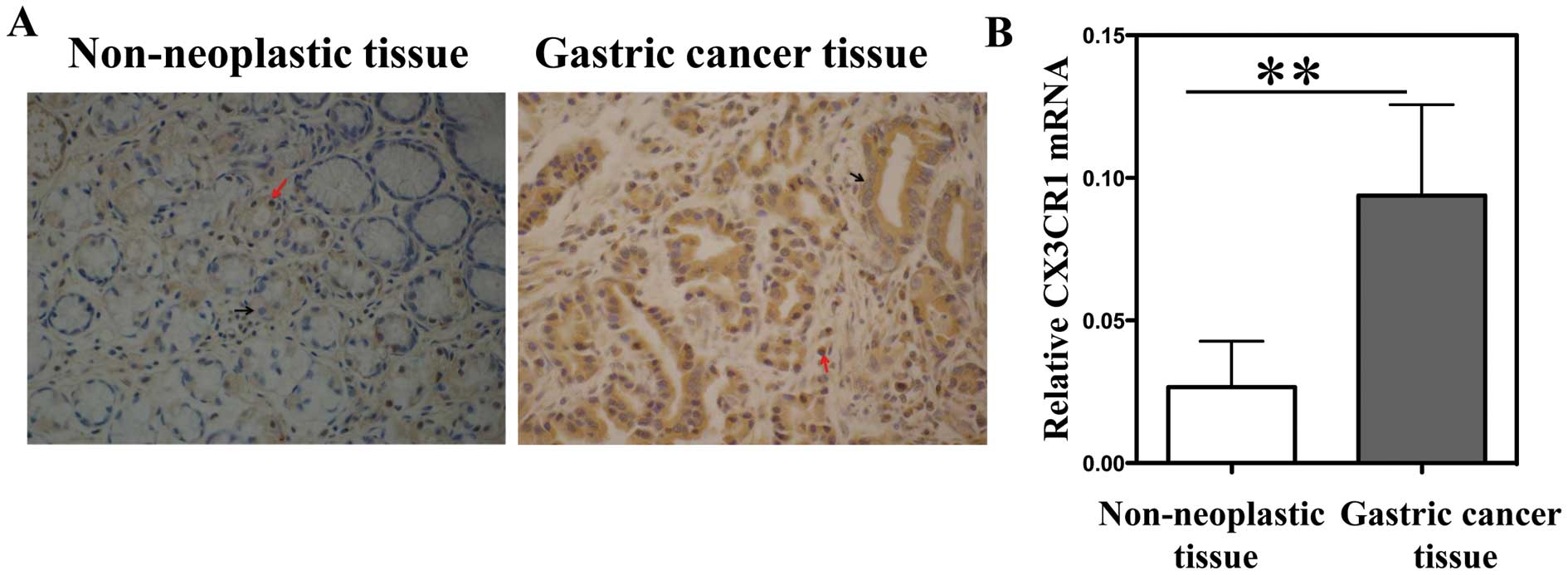
Immunohistochemical staining and quantitative
real-time PCR showed that both gastric cancer and non-neoplastic
gastric tissues expressed CX3CR1, and compared with the
non-neoplastic gastric tissues, CX3CR1 expression was significantly
increased in the gastric cancer tissues (P<0.01, Fig. 1A and B). We then analyzed the
relationship between the CX3CR1 expression level in the primary
tumor and the clinicopathological characteristics by comparing the
counted IOD (integrated optical density) in five fields at a ×400
magnification. Increased CX3CR1 expression was significantly
related to lymph node metastasis (P=0.029), higher clinical TNM
stage (P=0.021) and larger tumor size (P=0.011); however, the
CX3CR1 protein expression level had no association with age,
gender, tumor differentiation or tumor location (Table I).
 | Table IRelationship between the CX3CR1
expression level and clinicopathological features of the gastric
cancer patients. |
Table I
Relationship between the CX3CR1
expression level and clinicopathological features of the gastric
cancer patients.
| Clinicopathological
features | n | Primary
tumor
IOD for CX3CR1 (mean ± SD) | P-value |
|---|
| Age (years) |
| ≤60 | 30 |
25,255.03±12,812.09 | NS |
| >60 | 59 |
34,613.17±20,400.92 | |
| Gender |
| Male | 65 |
35,782.98±23,382.74 | NS |
| Female | 24 |
29,862.10±10,908.13 | |
| Tumor size
(cm) |
| <4 | 29 |
24,362.98±10,226.68 | 0.011 |
| ≥4 | 60 |
43,725.84±26,560.86 | |
| Tumor location |
| Cardia | 16 |
29,717.94±16,315.59 | NS |
| Non-cardia | 73 |
33,073.17±19,477.31 | |
| Tissue
differentiation |
| High | 29 |
28,243.80±15,216.78 | NS |
| Middle | 35 |
31,870.63±23,488.66 | |
| Low | 25 |
32,053.73±23,683.80 | 0.021 |
| TNM stage |
| I+II | 33 |
24,228.20±10,866.49 | |
| III+IV | 56 |
39,486.33±14,841.06 | |
| Lymph node
metastasis |
| Absent | 27 |
20,233.44±8,167.99 | 0.029 |
| Present | 62 |
34,065.75±18,939.28 | |
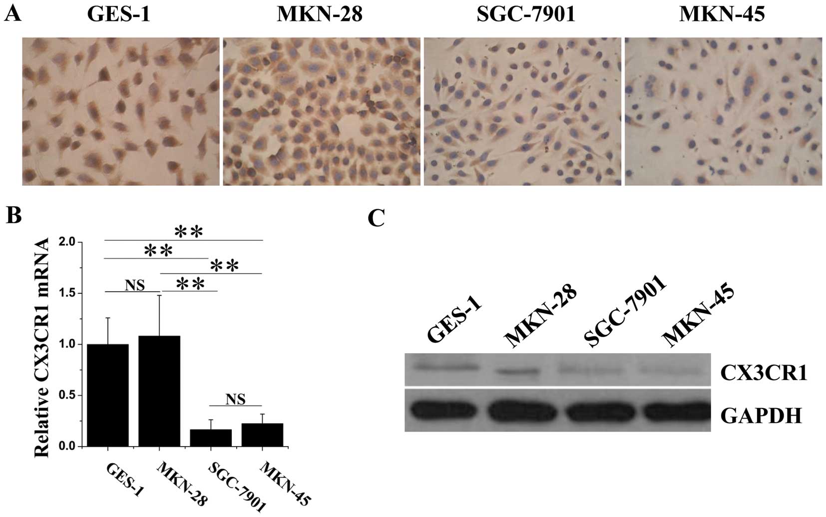
CX3CR1 is expressed in several different
human gastric cancer cell lines and in a gastric epithelial cell
line
As the results from the clinical tissues showed that
CX3CR1 was expressed in both gastric cancer tissues and
non-neoplastic gastric tissues, we aimed to ascertain whether the
expression of CX3CR1 in non-neoplastic gastric epithelial cells
and/or gastric cancer cells was constitutive or inducible. We
selected three gastric cancer cell lines and one immortalized
gastric epithelial cell line to investigate CX3CR1 expression in
vitro. Fig. 2 shows that, in
line with the clinical results, although the expression level in
all cell lines was evidently lower compared with the GAPDH
expression level, the gastric epithelial cell line (GES-1) and the
three gastric cancer cell lines (MKN-28, SGC-7901 and MKN-45)
expressed CX3CR1. In contrast to the results in vivo,
SGC-7901 and MKN-45 cells produced less CX3CR1 protein and MKN-28
cells produced an equal level of CX3CR1 when compared with the
GES-1 cells. We speculated that there possibly unknown mechanisms
existing in the tumor microenvironment in vivo, which could
increase the expression of CX3CR1 in malignant cells, while gastric
cancer cell lines in vitro lacked this tumor
microenvironment. According to these results, we hypothesized that
CX3CR1 is constitutively expressed in normal gastric epithelial
cells at a low level and is induced to express in gastric cancer
cells at a higher level in the tumor microenvironment.
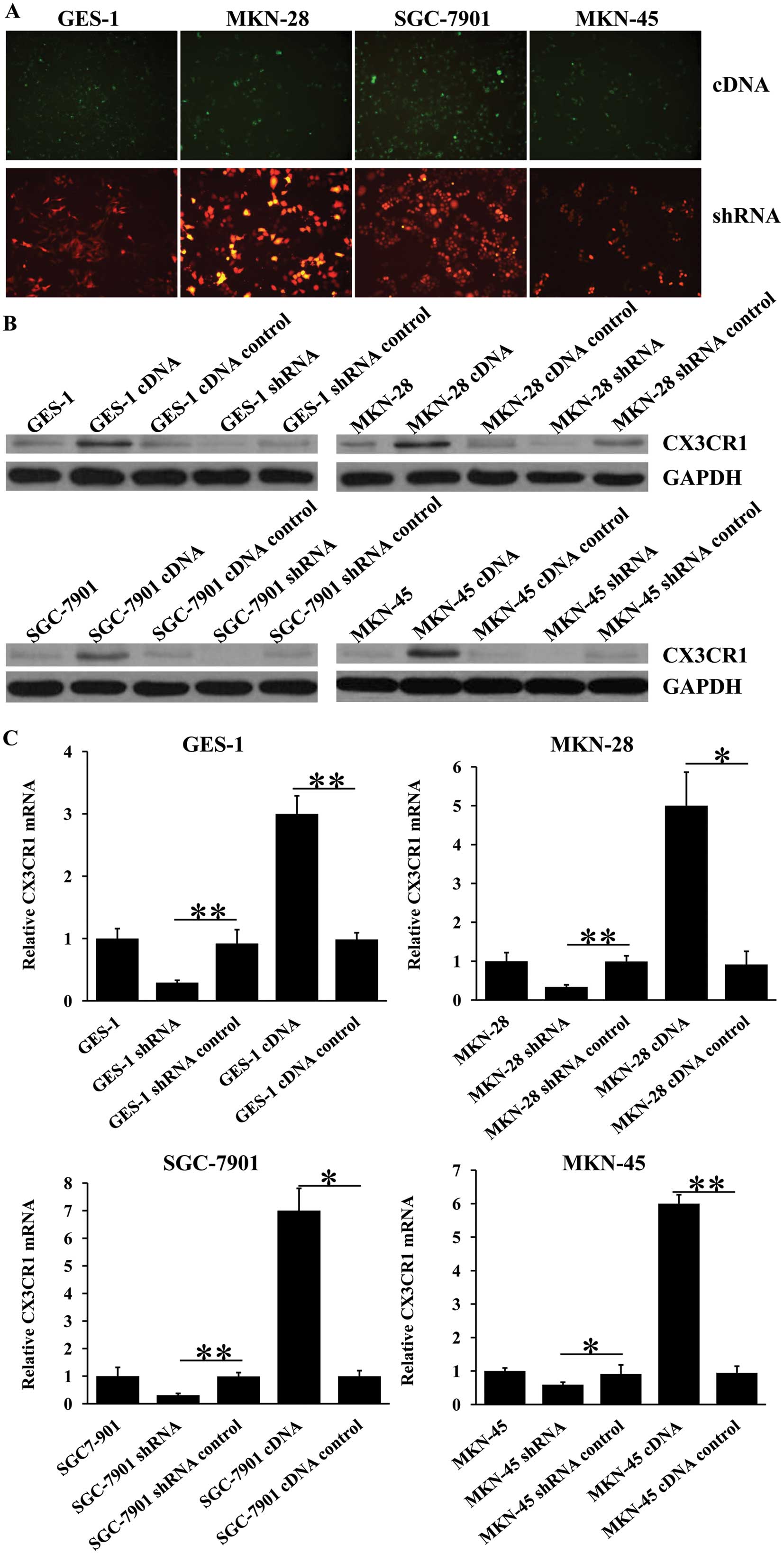
Identification of stable
CX3CR1-overexpressing or -knockdown cells
To simulate the high expression of CX3CR1 in gastric
cancer tissues in vivo and clarify the functional role of
CX3CR1 in gastric normal epithelial and gastric cancer cells, we
transfected three gastric cancer cell lines and one gastric
epithelial cell line with CX3CR1 cDNA or CX3CR1 short hairpin
(sh)RNA to obtain cDNA-mediated CX3CR1- overexpressing or
shRNA-mediated CX3CR1-knockdown cell lines. The stable transfected
cells were monitored by fluorescence microscopy for red fluorescent
protein (a marker for plasmid pRFP-c-RS) or green fluorescent
protein (a marker for plasmid pCMV6-AC-GFP) expression (Fig. 3A). In order to determine whether
shRNA knockdown and cDNA overexpression are correlated with a
change in RNA and protein, we measured the RNA and protein levels
of CX3CR1 following transfection by quantitative real-time PCR and
western blot analysis. As shown in Fig.
3B and C, cDNA transfection efficiently inceased CX3CR1
production compared with cells transfected with the empty vector
(i.e. cDNA control); shRNA transfection decreased the CX3CR1
production compared with the cells transfected with irrelevant
shRNA (i.e. shRNA control). Importantly, none of these shRNAs and
cDNAs affected the transcription of the housekeeping gene GAPDH.
Stable transfected cells were further used in the subsequent
functional experiments.
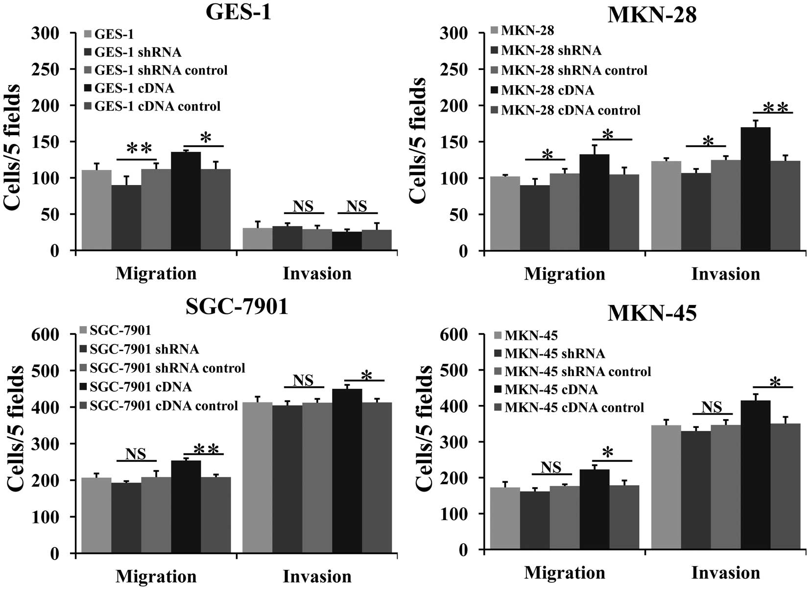
The CX3CR1/CX3CL1 axis stimulates gastric
cancer cell migration and invasion
Transwell migration and invasion assays were
performed to examine the mobilizing effect of the CX3CR1/CX3CL1
axis on the selected cell lines. Fig.
4 shows the number of cells that migrated or invaded per five
different fields in response to CX3CL1. In all gastric cancer cell
lines, cells overexpressing CX3CR1 exhibited significant migratory
and invasive responses to CX3CL1 compared with the empty
vector-transfected cells, while cells with CX3CR1 knockdown showed
almost no impact on migratory and invasive responses to the CX3CL1
cytokine except for MKN-28 cells. We proposed that the minor change
in CX3CR1 expression at the protein level after shRNA transfection
was due to the fact that the CX3CR1 expression level in the
parental SGC-7901 and MKN-45 cells was already quite low. In
addition, an important finding was that CX3CR1 expressed in the
GES-1 cells also stimulated GES-1 cell migration although the
number of migrated cells was evidently fewer than that noted in the
SGC-7901 and MKN-45 cells. Altogether, these findings suggest that
on the one hand, increased expression of CX3CR1 in gastric cancer
cells might play a role in migration and invasion; on the other
hand, the CX3CR1-CX3CL1 axis also stimulated GES-1 cell
migration.
The CX3CR1/CX3CL1 axis stimulates both
gastric cancer and GES-1 cell proliferation
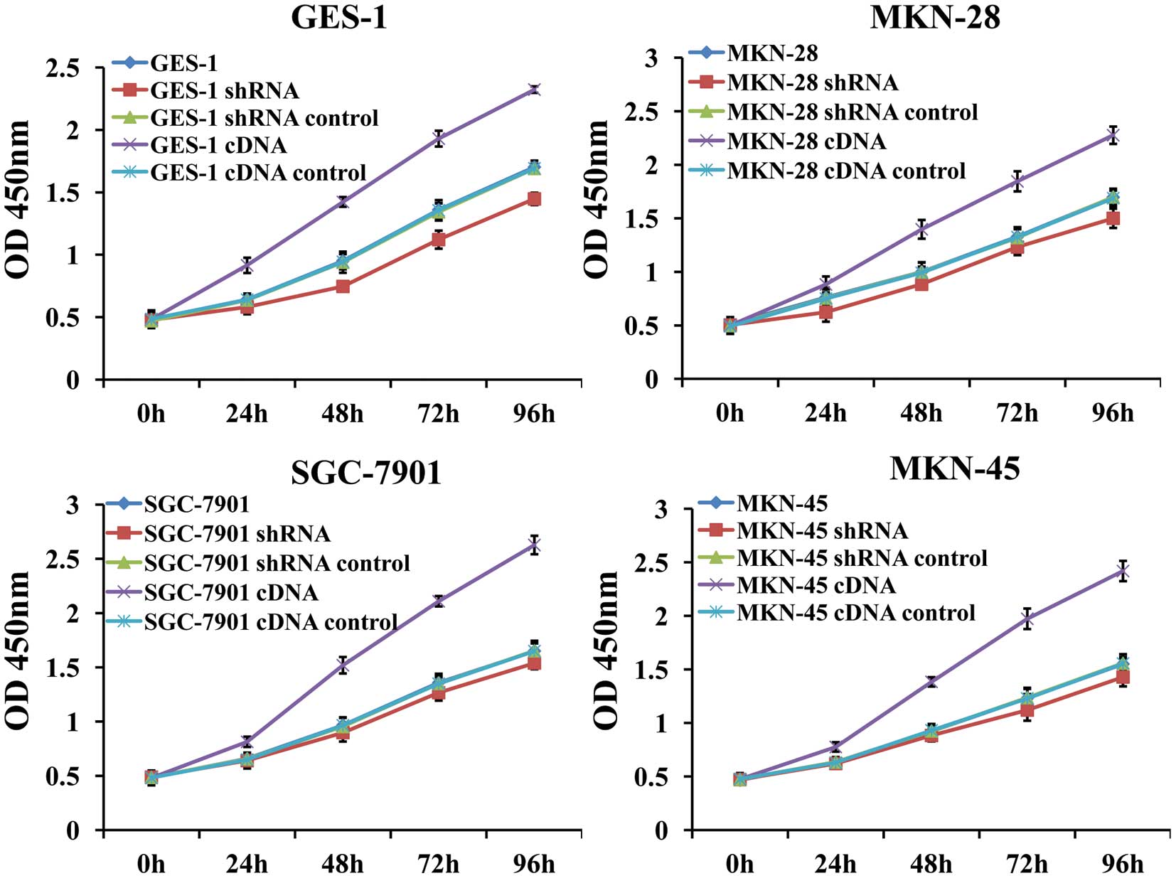
The effects of the CX3CR1/CX3CL1 axis on MKN-28,
SGC-7901, MKN-45 and GES-1 cell proliferation were assessed by the
CCK-8 assay. Under optimal culture conditions (in the presence of
10% FBS), addition of CX3CL1 (200 ng/ml) significantly increased
the proliferation of CX3CR1-overexpressing cells compared with that
of the empty vector-transfected cells, Whereas, we found that
knockdown of CX3CR1 production had no obvious impact on the
proliferation of gastric cancer cells, and only shRNA-transfected
GES-1 cells showed an inhibited proliferation compared with the
irrelevant shRNA-transfected GES-1 cells (Fig. 5). In addition to our findings on
CX3CR1, CCR7 has also been proven to promote the growth of gastric
carcinoma (20,33).
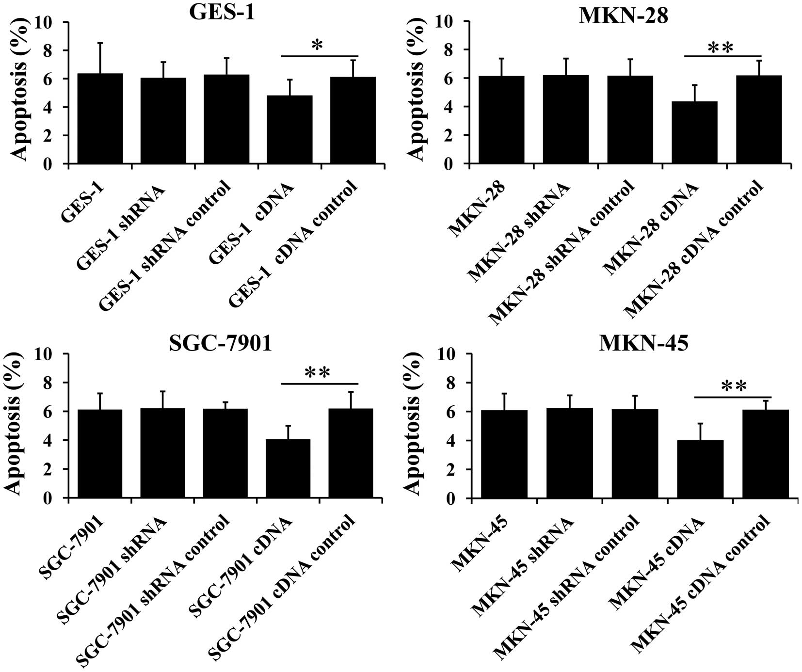
The CX3CR1/CX3CL1 axis promotes gastric
cancer and GES-1 cell survival
An important feature of metastatic cells is the
ability to regulate their survival. We, therefore, tested whether
the CX3CR1/CX3CL1 axis rescues gastric cancer and GES-1 cells from
serum deprivation-induced death. All cells were cultured in
serum-free medium with CX3CL1 (200 ng/ml) for 24 h before flow
cytometric analysis. Fig. 6 shows
that CX3CR1 overexpressed in gastric cancer and GES-1 cells
significantly decreased the percentage of Annexin V-positive cells.
The data suggest that the CX3CR1/CX3CL1 axis plays an antiapoptotic
role in gastric cancer and GES-1 cells.
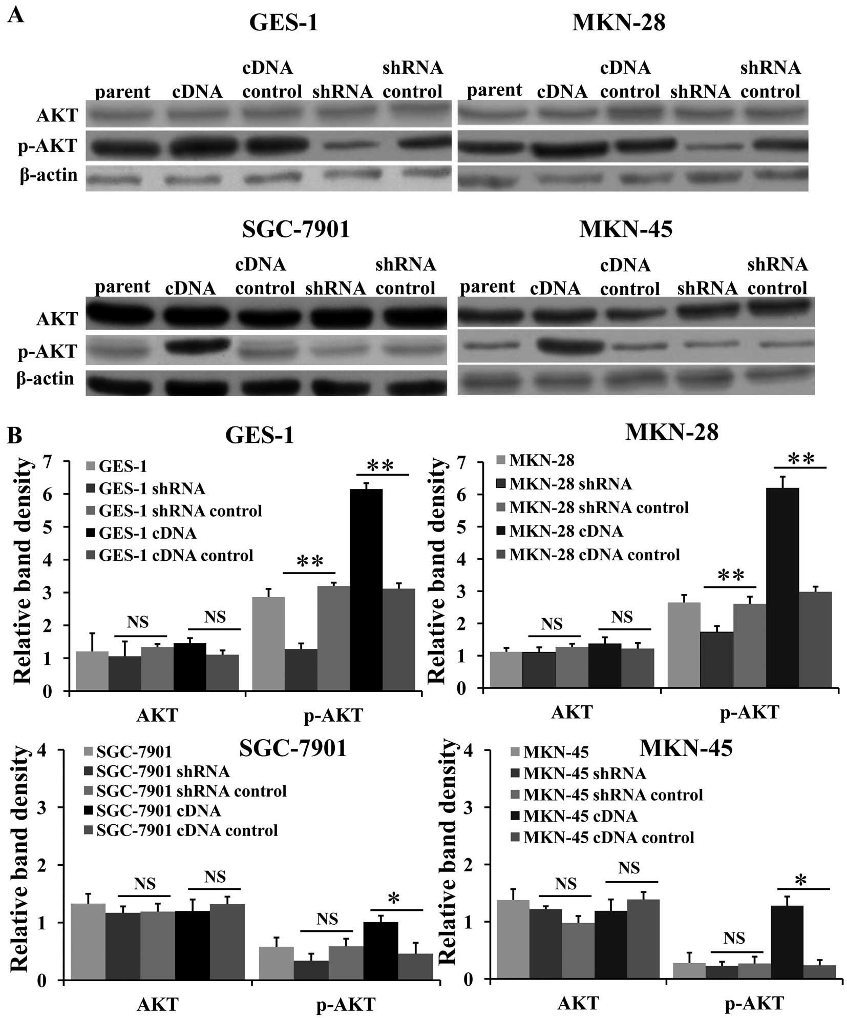
The CX3CR1/CX3CL1 axis activates Akt
kinase in gastric cancer and gastric epithelial cells in vitro
To investigate the mechanisms underlying the
CX3CR1/CX3CL1 axis-induced proliferation and survival of gastric
cancer and gastric epithelial cells, signal transduction
experiments were next performed. Phosphorylated (p)-Akt (Ser-473)
kinase has been found to be an important intermediary in the
control of proliferation and apoptosis in many types of cells
(21,34,35).
We, therefore, studied the effect of the CX3CR1/CX3CL1 axis on Akt
activation by measuring the levels of p-Akt (Ser-473) in protein
extracts from cells incubated with CX3CL1 for 24 h. Quantitative
analysis of the bands was performed by densitometry. As shown in
Fig. 7, Akt was significantly
phosphorylated in the CX3CR1-overexpressing cells compared with the
empty vector-transfected cells. The CX3CR1/CX3CL1 axis has been
previously identified as a proliferative factor activating Akt in
epithelial ovarian cancer (36),
endothelial cells (37) and neurons
(34). In conclusion, these results
strongly suggest that the proliferation and survival effect of the
CX3CL1/CX3CR1 axis in gastric cancer and gastric epithelial cells
is associated with Akt activation.
Discussion
In the present study we demonstrated that CX3CR1 was
expressed not only in gastric carcinoma, but also in non-neoplastic
gastric epithelium, and upregulated expression of CX3CR1 was
associated with the metastasis, proliferation and survival of
gastric cancer, and a parallel increase was observed in p-Akt
levels. In addition to CCR7 (12,23),
CXCR4 (33,38) and CCR4 (14), we demonstrated that the chemokine
receptor CX3CR1 is involved in metastasis and growth of gastric
cancer. These findings suggest that gastric cancer metastasis is a
complex and synergistic process involving multiple chemokine
receptors and chemokines.
An intriguing finding was that CX3CR1 was expressed
in non-neoplastic gastric tissues in vivo and GES-1 cells
in vitro, and played a functional role in stimulating
migration, promoting proliferation and inhibiting apoptosis of
GES-1 cells in vitro. In addition to CX3CR1, other chemokine
receptors were demonstrated to be expressed in normal tissue cells
and play a physiological role. Murdoch and colleagues demonstrated
that colon epithelium expresses CX3CR1 which regulates epithelial
maintenance and renewal (39).
Banas et al also found that CCR7 expression in mesangial
cells (MC) promoted MC proliferation, migration and survival and
enhanced ‘wound healing’ in vitro (40). Therefore we hypothesized that CX3CR1
expressed in normal gastric epithelial cells may have some
biological function. Further studies are required to confirm the
biological function of CX3CR1 in vivo.
In addition, contradictory CX3CR1 expression in
gastric cancer cells in vivo and in vitro was found,
that is, gastric cancer cells expressed a higher level of CX3CR1
than that in the non-neoplastic gastric epithelial cells in
vivo, but expressed a lower or equal CX3CR1 protein level
compared with the gastric epithelial cells in vitro. We
hypothesized that there was some unknown mechanisms which could
upregulate the expression of CX3CR1 in gastric cancer cells in
vivo. Gaudin et al found that a decreased concentration
of FBS in culture medium led to increased membrane expression of
CX3CR1 in epithelial ovarian carcinoma BG1 cells (36). What is more, hypoxia enhanced CXCR4
expression, which was demonstrated in melanoma and oral squamous
cell carcinoma (41,42). Thus, we speculated that hypoxia and
the lack of nutrients in the tumor microenvironment might increase
the expression of CX3CR1 in gastric cancer cells, thus promoting
gastric cancer metastasis, proliferation and survival. Further
studies are needed to confirm this hypothesis.
In summary, we demonstrated that CX3CR1 was
expressed not only in gastric cancer tissues and gastric cancer
cell lines, but also in non-neoplastic gastric tissues and a
gastric epithelial cell line. Increased expression of CX3CR1 in
gastric cancer cells promoted cancer cell metastasis, proliferation
and survival, and an appropriate expression level of CX3CR1 in
gastric tissues might be beneficial to cell renewal and/or tissue
remodeling after injury. In addition, the tumor microenvironment
may play an important role in the increased expression of CX3CR1 in
gastric cancer cells. Further studies are warranted to clarify the
mechanisms responsible for inducing overexpression of CX3CR1 in
gastric cancer cells.
Acknowledgements
This study was supported by The Beijing Natural
Science Foundation (no. 7112134).
References
|
1
|
Brenner H, Rothenbacher D and Arndt V:
Epidemiology of stomach cancer. Methods Mol Biol. 472:467–477.
2009. View Article : Google Scholar
|
|
2
|
Herszenyi L and Tulassay Z: Epidemiology
of gastrointestinal and liver tumors. Eur Rev Med Pharmacol Sci.
14:249–258. 2010.PubMed/NCBI
|
|
3
|
Hyung WJ, Noh SH, Yoo CH, et al:
Prognostic significance of metastatic lymph node ratio in T3
gastric cancer. World J Surg. 26:323–329. 2002. View Article : Google Scholar : PubMed/NCBI
|
|
4
|
Feng J, Wu YF, Xu HM, Wang SB and Chen JQ:
Prognostic significance of the metastatic lymph node ratio in T3
gastric cancer patients undergoing total gastrectomy. Asian Pac J
Cancer Prev. 12:3289–3292. 2011.PubMed/NCBI
|
|
5
|
Zlotnik A and Yoshie O: Chemokines: a new
classification system and their role in immunity. Immunity.
12:121–127. 2000. View Article : Google Scholar : PubMed/NCBI
|
|
6
|
Balkwill F: Cancer and the chemokine
network. Nat Rev Cancer. 4:540–550. 2004. View Article : Google Scholar : PubMed/NCBI
|
|
7
|
Zlotnik A: Chemokines in neoplastic
progression. Semin Cancer Biol. 14:181–185. 2004. View Article : Google Scholar : PubMed/NCBI
|
|
8
|
Righi E, Kashiwagi S, Yuan J, et al:
CXCL12/CXCR4 blockade induces multimodal antitumor effects that
prolong survival in an immunocompetent mouse model of ovarian
cancer. Cancer Res. 71:5522–5534. 2011. View Article : Google Scholar : PubMed/NCBI
|
|
9
|
Marchesi F, Grizzi F, Laghi L, Mantovani A
and Allavena P: Molecular mechanisms of pancreatic cancer
dissemination: the role of the chemokine system. Curr Pharm Des.
18:2432–2438. 2012. View Article : Google Scholar : PubMed/NCBI
|
|
10
|
Balkwill FR: The chemokine system and
cancer. J Pathol. 226:148–157. 2012. View Article : Google Scholar
|
|
11
|
Yao X, Qi L, Chen X, Du J, Zhang Z and Liu
S: Expression of CX3CR1 associates with cellular migration,
metastasis, and prognosis in human clear cell renal cell carcinoma.
Urol Oncol. 32:162–170. 2014. View Article : Google Scholar
|
|
12
|
Wang WN, Chen Y, Zhang YD and Hu TH: The
regulatory mechanism of CCR7 gene expression and its involvement in
the metastasis and progression of gastric cancer. Tumour Biol.
34:1865–1871. 2013. View Article : Google Scholar : PubMed/NCBI
|
|
13
|
Marchesi F, Piemonti L, Mantovani A and
Allavena P: Molecular mechanisms of perineural invasion, a
forgotten pathway of dissemination and metastasis. Cytokine Growth
Factor Rev. 21:77–82. 2010. View Article : Google Scholar : PubMed/NCBI
|
|
14
|
Lee JH, Cho YS, Lee JY, et al: The
chemokine receptor CCR4 is expressed and associated with a poor
prognosis in patients with gastric cancer. Ann Surg. 249:933–941.
2009. View Article : Google Scholar : PubMed/NCBI
|
|
15
|
Jamieson WL, Shimizu S, D’Ambrosio JA,
Meucci O and Fatatis A: CX3CR1 is expressed by prostate epithelial
cells and androgens regulate the levels of CX3CL1/fractalkine in
the bone marrow: potential role in prostate cancer bone tropism.
Cancer Res. 68:1715–1722. 2008. View Article : Google Scholar : PubMed/NCBI
|
|
16
|
Marchesi F, Piemonti L, Fedele G, et al:
The chemokine receptor CX3CR1 is involved in the neural tropism and
malignant behavior of pancreatic ductal adenocarcinoma. Cancer Res.
68:9060–9069. 2008. View Article : Google Scholar : PubMed/NCBI
|
|
17
|
Kodama J, Hasengaowa, Kusumoto T, et al:
Association of CXCR4 and CCR7 chemokine receptor expression and
lymph node metastasis in human cervical cancer. Ann Oncol.
18:70–76. 2007. View Article : Google Scholar
|
|
18
|
Miao Z, Luker KE, Summers BC, et al: CXCR7
(RDC1) promotes breast and lung tumor growth in vivo and is
expressed on tumor-associated vasculature. Proc Natl Acad Sci USA.
104:15735–15740. 2007. View Article : Google Scholar : PubMed/NCBI
|
|
19
|
Andre F, Cabioglu N, Assi H, et al:
Expression of chemokine receptors predicts the site of metastatic
relapse in patients with axillary node positive primary breast
cancer. Ann Oncol. 17:945–951. 2006. View Article : Google Scholar : PubMed/NCBI
|
|
20
|
Meijer J, Zeelenberg IS, Sipos B and Roos
E: The CXCR5 chemokine receptor is expressed by carcinoma cells and
promotes growth of colon carcinoma in the liver. Cancer Res.
66:9576–9582. 2006. View Article : Google Scholar : PubMed/NCBI
|
|
21
|
Shulby SA, Dolloff NG, Stearns ME, Meucci
O and Fatatis A: CX3CR1-fractalkine expression regulates cellular
mechanisms involved in adhesion, migration, and survival of human
prostate cancer cells. Cancer Res. 64:4693–4698. 2004. View Article : Google Scholar : PubMed/NCBI
|
|
22
|
Marchesi F, Monti P, Leone BE, et al:
Increased survival, proliferation, and migration in metastatic
human pancreatic tumor cells expressing functional CXCR4. Cancer
Res. 64:8420–8427. 2004. View Article : Google Scholar : PubMed/NCBI
|
|
23
|
Mashino K, Sadanaga N, Yamaguchi H, et al:
Expression of chemokine receptor CCR7 is associated with lymph node
metastasis of gastric carcinoma. Cancer Res. 62:2937–2941.
2002.PubMed/NCBI
|
|
24
|
Brand S, Sakaguchi T, Gu X, Colgan SP and
Reinecker HC: Fractalkine-mediated signals regulate cell-survival
and immune-modulatory responses in intestinal epithelial cells.
Gastroenterology. 122:166–177. 2002. View Article : Google Scholar : PubMed/NCBI
|
|
25
|
Muller A, Homey B, Soto H, et al:
Involvement of chemokine receptors in breast cancer metastasis.
Nature. 410:50–56. 2001. View
Article : Google Scholar : PubMed/NCBI
|
|
26
|
White GE and Greaves DR: Fractalkine: one
chemokine, many functions. Blood. 113:767–768. 2009. View Article : Google Scholar : PubMed/NCBI
|
|
27
|
Bazan JF, Bacon KB, Hardiman G, et al: A
new class of membrane-bound chemokine with a CX3C motif. Nature.
385:640–644. 1997. View
Article : Google Scholar : PubMed/NCBI
|
|
28
|
Lucas AD, Chadwick N, Warren BF, et al:
The transmembrane form of the CX3CL1 chemokine fractalkine is
expressed predominantly by epithelial cells in vivo. Am J Pathol.
158:855–866. 2001. View Article : Google Scholar : PubMed/NCBI
|
|
29
|
Hundhausen C, Misztela D, Berkhout TA, et
al: The disintegrin-like metalloproteinase ADAM10 is involved in
constitutive cleavage of CX3CL1 (fractalkine) and regulates
CX3CL1-mediated cell-cell adhesion. Blood. 102:1186–1195. 2003.
View Article : Google Scholar : PubMed/NCBI
|
|
30
|
Imai T, Hieshima K, Haskell C, et al:
Identification and molecular characterization of fractalkine
receptor CX3CR1, which mediates both leukocyte migration and
adhesion. Cell. 91:521–530. 1997. View Article : Google Scholar : PubMed/NCBI
|
|
31
|
Nevo I, Sagi-Assif O, Meshel T, et al: The
involvement of the fractalkine receptor in the transmigration of
neuroblastoma cells through bone-marrow endothelial cells. Cancer
Lett. 273:127–139. 2009. View Article : Google Scholar
|
|
32
|
Locatelli M, Boiocchi L, Ferrero S, et al:
Human glioma tumors express high levels of the chemokine receptor
CX3CR1. Eur Cytokine Netw. 21:27–33. 2010.PubMed/NCBI
|
|
33
|
Zhao BC, Wang ZJ, Mao WZ, et al:
CXCR4/SDF-1 axis is involved in lymph node metastasis of gastric
carcinoma. World J Gastroenterol. 17:2389–2396. 2011. View Article : Google Scholar : PubMed/NCBI
|
|
34
|
Meucci O, Fatatis A, Simen AA and Miller
RJ: Expression of CX3CR1 chemokine receptors on neurons and their
role in neuronal survival. Proc Natl Acad Sci USA. 97:8075–8080.
2000. View Article : Google Scholar : PubMed/NCBI
|
|
35
|
Alessi DR, Andjelkovic M, Caudwell B, et
al: Mechanism of activation of protein kinase B by insulin and
IGF-1. EMBO J. 15:6541–6551. 1996.PubMed/NCBI
|
|
36
|
Gaudin F, Nasreddine S, Donnadieu AC, et
al: Identification of the chemokine CX3CL1 as a new regulator of
malignant cell proliferation in epithelial ovarian cancer. PLoS
One. 6:e215462011. View Article : Google Scholar : PubMed/NCBI
|
|
37
|
Lee SJ, Namkoong S, Kim YM, et al:
Fractalkine stimulates angiogenesis by activating the
Raf-1/MEK/ERK- and PI3K/Akt/eNOS-dependent signal pathways. Am J
Physiol Heart Circ Physiol. 291:H2836–H2846. 2006. View Article : Google Scholar : PubMed/NCBI
|
|
38
|
Ishigami S, Natsugoe S, Okumura H, et al:
Clinical implication of CXCL12 expression in gastric cancer. Ann
Surg Oncol. 14:3154–3158. 2007. View Article : Google Scholar : PubMed/NCBI
|
|
39
|
Murdoch C, Monk PN and Finn A: Functional
expression of chemokine receptor CXCR4 on human epithelial cells.
Immunology. 98:36–41. 1999. View Article : Google Scholar : PubMed/NCBI
|
|
40
|
Banas B, Wornle M, Berger T, et al: Roles
of SLC/CCL21 and CCR7 in human kidney for mesangial proliferation,
migration, apoptosis, and tissue homeostasis. J Immunol.
168:4301–4307. 2002. View Article : Google Scholar : PubMed/NCBI
|
|
41
|
Schutyser E, Su Y, Yu Y, et al: Hypoxia
enhances CXCR4 expression in human microvascular endothelial cells
and human melanoma cells. Eur Cytokine Netw. 18:59–70.
2007.PubMed/NCBI
|
|
42
|
Ishikawa T, Nakashiro K, Klosek SK, et al:
Hypoxia enhances CXCR4 expression by activating HIF-1 in oral
squamous cell carcinoma. Oncol Rep. 21:707–712. 2009.PubMed/NCBI
|