Comparison of 2D- and 3D-culture models as drug-testing platforms in breast cancer

  • Authors:
    • Yoshinori Imamura
    • Toru Mukohara
    • Yohei Shimono
    • Yohei Funakoshi
    • Naoko Chayahara
    • Masanori Toyoda
    • Naomi Kiyota
    • Shintaro Takao
    • Seishi Kono
    • Tetsuya Nakatsura
    • Hironobu Minami
  • View Affiliations

  • Published online on: January 29, 2015     https://doi.org/10.3892/or.2015.3767
  • Pages: 1837-1843
Metrics: Total Views: 0 (Spandidos Publications: | PMC Statistics: )
Total PDF Downloads: 0 (Spandidos Publications: | PMC Statistics: )


Abstract

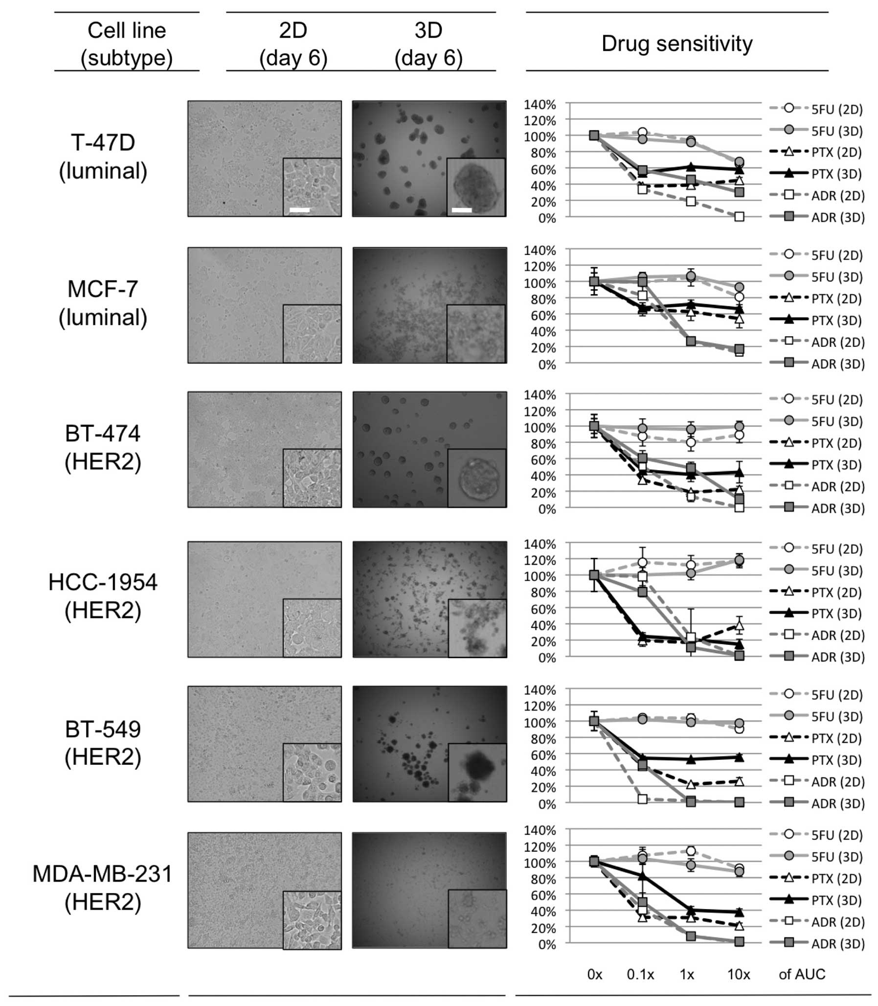
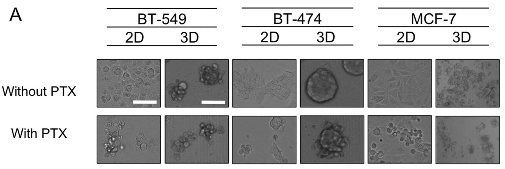
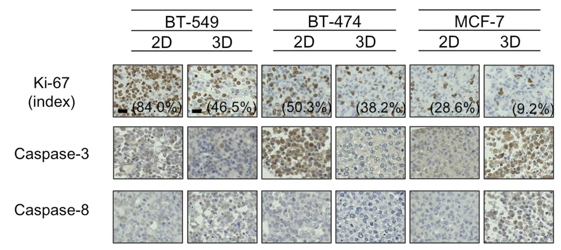
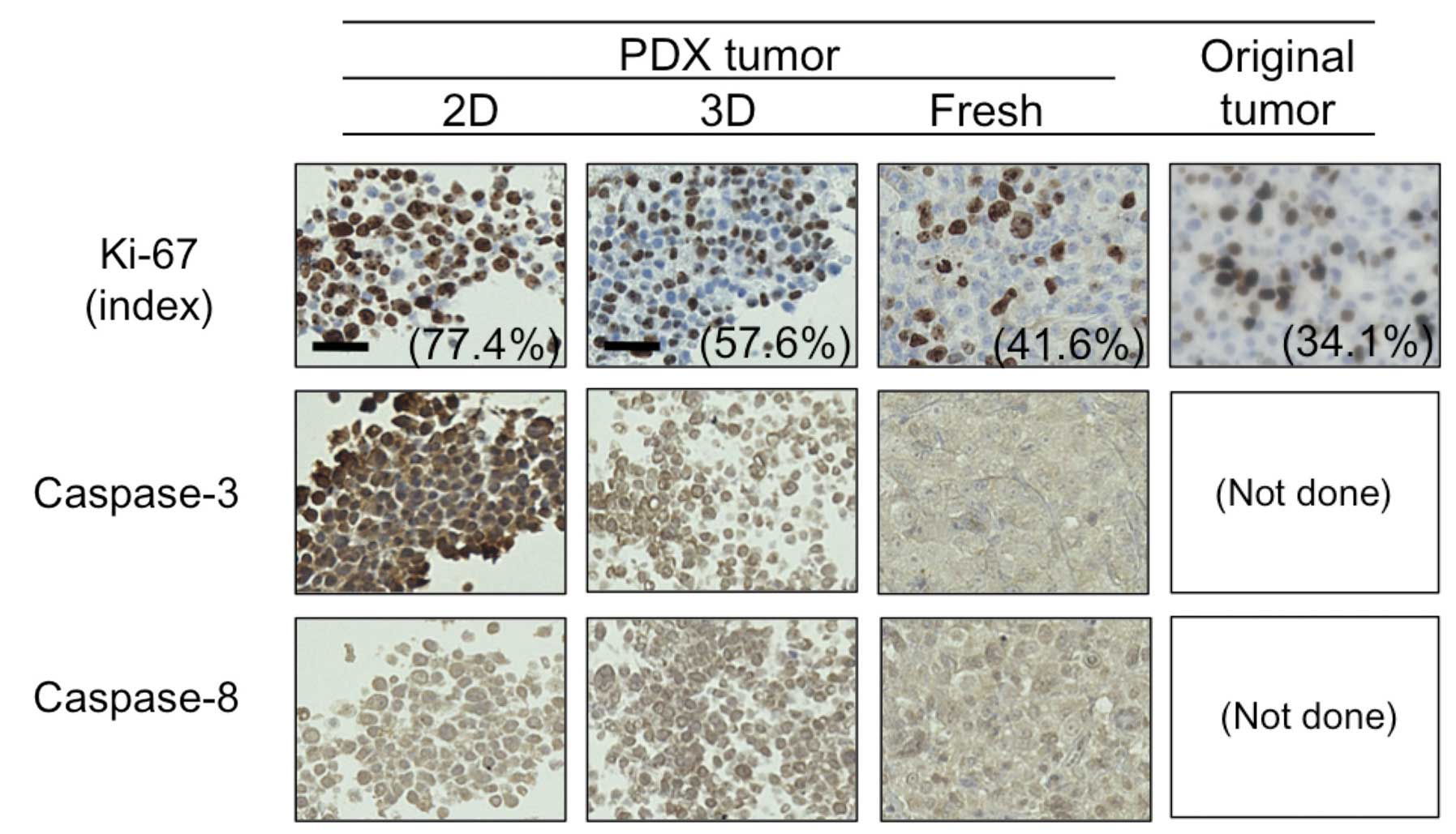
It is becoming recognized that screening of oncology drugs on a platform using two-dimensionally (2D)-cultured cell lines is unable to precisely select clinically active drugs; therefore three-dimensional (3D)-culture systems are emerging and show potential for better simulating the in vivo tumor microenvironment. The purpose of this study was to reveal the differential effects of chemotherapeutic drugs between 2D- and 3D-cultures and to explore their underlying mechanisms. We evaluated differences between 2D- and 3D-cultured breast cancer cell lines by assessing drug sensitivity, oxygen status and expression of Ki-67 and caspases. Three cell lines (BT-549, BT-474 and T-47D) developed dense multicellular spheroids (MCSs) in 3D-culture, and showed greater resistance to paclitaxel and doxorubicin compared to the 2D-cultured cells. An additional three cell lines (MCF-7, HCC-1954, and MDA-MB‑231) developed only loose MCSs in 3D, and showed drug sensitivities similar to those found in the 2D-culture. Treatment with paclitaxel resulted in greater increases in cleaved-PARP expression in the 2D-culture compared with the 3D-culture, but only in cell lines forming dense 3D-MCSs, suggesting that MCS formation protected the cells from paclitaxel-induced apoptosis. Hypoxia was observed only in the dense 3D-MCSs. BT-549 had fewer cells positive for Ki-67 in 3D- than in 2D-culture, suggesting that the greater G0-dormant subpopulation was responsible for its drug resistance in the 3D-culture. BT-474 had a lower level of caspase-3 in the 3D- than in the 2D-culture, suggesting that the 3D-environment was anti-apoptotic. Finally, we compared staining for Ki-67 and caspases in the 2D- and 3D-primary‑cultured cells originating from a patient-derived xenograft (PDX), fresh PDX tumor, and the patient's original tumor; 2D-cultured cells showed greater proportions of Ki-67-positive and caspase-3-positive cells, in agreement with the view that 3D-primary culture better represents characteristics of tumors in vivo. In conclusion, 3D-cultured cells forming dense MCSs may be better than 2D-cultured cells in simulating important tumor characteristics in vivo, namely hypoxia, dormancy, anti-apoptotic features and their resulting drug resistance.
View Figures
View References

Related Articles

Journal Cover

April-2015
Volume 33 Issue 4

Print ISSN: 1021-335X
Online ISSN:1791-2431

Sign up for eToc alerts

Recommend to Library

Copy and paste a formatted citation
x
Spandidos Publications style
Imamura Y, Mukohara T, Shimono Y, Funakoshi Y, Chayahara N, Toyoda M, Kiyota N, Takao S, Kono S, Nakatsura T, Nakatsura T, et al: Comparison of 2D- and 3D-culture models as drug-testing platforms in breast cancer. Oncol Rep 33: 1837-1843, 2015
APA
Imamura, Y., Mukohara, T., Shimono, Y., Funakoshi, Y., Chayahara, N., Toyoda, M. ... Minami, H. (2015). Comparison of 2D- and 3D-culture models as drug-testing platforms in breast cancer. Oncology Reports, 33, 1837-1843. https://doi.org/10.3892/or.2015.3767
MLA
Imamura, Y., Mukohara, T., Shimono, Y., Funakoshi, Y., Chayahara, N., Toyoda, M., Kiyota, N., Takao, S., Kono, S., Nakatsura, T., Minami, H."Comparison of 2D- and 3D-culture models as drug-testing platforms in breast cancer". Oncology Reports 33.4 (2015): 1837-1843.
Chicago
Imamura, Y., Mukohara, T., Shimono, Y., Funakoshi, Y., Chayahara, N., Toyoda, M., Kiyota, N., Takao, S., Kono, S., Nakatsura, T., Minami, H."Comparison of 2D- and 3D-culture models as drug-testing platforms in breast cancer". Oncology Reports 33, no. 4 (2015): 1837-1843. https://doi.org/10.3892/or.2015.3767