Introduction
Colorectal cancer (CRC) is a major cause of cancer
incidence and mortality worldwide (1) and its incidence has increased
particularly among adults <50 years (2). Due to the high recurrence and distant
metastases, the long-term outcome of CRC patients remains
unsatisfactory. Although by improving access to and use of
screening and standard treatment have led to significant advances
in early detection and reduction of the death rates of CRC, these
methods are not cost-effective. Molecular genetics have provided
evidence that the occurrence of CRC is a series of molecular
events, including the accumulation of genetic and epigenetic
changes (3). Sensitive biomarkers
can contribute to early diagnosis and prognosis prediction,
therefore, novel factors for predicting tumor recurrence following
surgery remain to be defined and novel therapeutic strategies
should be identified.
Signal peptide-CUB-epidermal growth factor-like
domain-containing protein 2 (SCUBE2) belongs to a novel, small and
evolutionarily conserved family that comprises three different
members have been designated as SCUBE1 to SCUBE3 in the sequence of
identification (4,5). These SCUBE proteins contain ~1,000
amino acids and share an organized protein domain structure with
five motifs: an NH2 terminal signal peptide sequence, nine copies
of EGF-like repeats, a spacer region followed by three repeated
stretches of 6-cysteine residues and one CUB domain at the COOH
terminus (6–9). SCUBE2 is predominantly expressed in
vascular endothelial cells (4) and
is a secreted cell-surface glycoprotein which is a novel positive
component of Sonic hedgehog (SHH) signaling and can specifically
interact with SHH and its receptor PTCH1 to enhance the SHH
signaling activity acting upstream of ligand binding at the plasma
membrane (7). Increased
angiogenesis (10,11) and dysregulation of the SHH pathway
(12,13) have been shown to contribute to
colorectal carcinogenesis.
Mounting evidence suggested that SCUBE2 act as a
novel breast tumor-suppressor gene that serves as a useful
prognostic marker (8,9). Furthermore, SCUBE2 expression is
associated with better prognosis and longer disease-free survival
in breast cancer (8) and is part of
the 8-gene expression score, which has a prognostic value for early
breast cancer (14). Similarly,
ectopic overexpression of the full-length SCUBE2 protein resulted
in the suppression of breast cancer cell proliferation in
vitro and in vivo through co-ordinated regulation of the
suppression of the bone morphogenetic protein and β-catenin
signaling pathways (8,9). Furthermore, SCUBE2 plays a key role in
the suppression of breast cancer cell migration and invasion by
increasing the formation of epithelial E-cadherin-containing
adherens junctions and driving the reversal of
epithelial-mesenchymal transition (EMT) (15). In prostate cancer and endometrial
cancer, SCUBE2 expression is reduced in high-grade tumors (16,17).
In the present study, we investigated SCUBE2
expression (mRNA and protein level) in CRC and then analyzed the
correlations between SCUBE2 expression and its clinicopathological
parameters. The effect of SCUBE2 expression on RKO CRC cell growth,
migration, invasion and apoptosis was also analyzed.
Materials and methods
Clinical samples and cell lines
The present study was approved by the Ethics
Committee of Shanghai Jiaotong University Affiliated First People’s
Hospital. A total of 120 patient-derived specimens were collected
between January, 2001 and December, 2003 and archived under
protocols approved by our Institutional Review Board. None of the
patients underwent therapy prior to surgery. The diagnoses were
confirmed by two pathologists, and the tumor stage was determined
on the basis of pathological findings in accordance with the
American Joint Committee on Cancer (AJCC). Disease-free survival
(DFS) and overall survival (OS) durations were defined as the
interval from initial surgery to clinically or radiologically
proven metastasis or recurrence and death, respectively. There were
48 male and 72 female patients, with a mean age of 64±14 years
(range, 22–85 years) at the time of surgery. The median patient
follow-up time was 61 months after surgery (range, 10–81 months).
Each patient provided informed consent for the use of their tissue
samples in the present study.
The LoVo, RKO, HCT8, HT29, HCT116, SW480 and SW620
CRC cell lines were obtained from the Cell Resource Center of
Shanghai Institutes for Biological Sciences, Type Culture
Collection of the Chinese Academy of Sciences (Shanghai, China).
The cells were cultured according to the manufacturer’s
instructions.
RNA extraction, reverse transcription PCR
and quantitative PCR
Total RNA in 40 pairs of frozen primary tumor and
adjacent normal mucosa of CRC specimens were extracted according to
the manufacturer’s instructions (Qiagen, Hilden, Germany).
First-strand cDNA was synthesized from 1 μg of RNA using the A3500
RT-PCR System (Promega, Madison, WI, USA). The primers used for
quantitative PCR were: SCUBE2, forward: 5′-CCCCCAAGCGCCGCATCCTGA-3′
and reverse: 5′-TATTGAGTGGCACGTGGGCTGAGT-3′; GAPDH, forward:
5′-GGAGCGAGATCCCTCCAAAAT-3′ and reverse:
5′-GGCTGTTGTCATACTTCTCATGG-3′. Quantitative SCUBE2 mRNA levels were
assessed using Mastercycler ep realplex® (Eppendorf,
Hamburg, Germany) with a SYBR-Green RNA PCR kit (Fermentas,
Waltham, MA, USA) according to the manufacturer’s instructions. The
cycling conditions used were: initial denaturation (10 min at
95°C), 40 cycles of denaturation (10 sec at 95°C) followed by
annealing (30 sec at 60°C), and a final elongation (30 sec at
72°C). Each reaction was repeated three times and the average
SCUBE2 mRNA level for each tumor was compared with the level of its
matched normal mucosa. The fold-change (2−ΔΔCt) of
SCUBE2 expression was calculated for each group (18).
Western blot analysis
Total proteins of four randomly selected, paired,
frozen CRC tissues and adjacent normal mucosa specimens were
extracted and measured using the BCA protein assay kit (Beyotime
Biotechnology Co., Jiangsu, China). Equivalent amounts of protein
were separated on a 10% polyacrylamide gel and transferred onto
polyvinylidene difluoride membranes, which were blocked in 5%
non-fat milk for 1 h at room temperature and incubated overnight
with the appropriate primary antibodies: SCUBE2 (1:300 dilution)
and TUBB2C (1:3,000 dilution) (both from Abgent, San Diego, CA,
USA). After washing with TBST buffer, the membranes were incubated
with a HRP-conjugated goat anti-rabbit (1:5,000; Santa Cruz
Biotechnology, Inc., Santa Cruz, CA, USA) secondary antibody. The
membranes were visualized using the ECL Plus enhanced
chemiluminescence kit (Pierce Biotechnology, Rockford, IL, USA) and
exposed to X-ray film. TUBB2C expression was used to confirm and
normalize equal loading of the samples.
Tissue microarray construction and
immunohistochemistry
Tissue microarray (TMA) was made from paired tumor
and adjacent normal mucosa from the 120 patients in our archive (in
collaboration with Shanghai Biochip, Shanghai, China). Two cores,
which were validated as having high accordance with the whole
archived section, were obtained from each specimen of
formalin-fixed, paraffin-embedded CRC tissue and normal mucosa
using punch cores measuring 2.0 mm in greatest dimension from the
non-necrotic area of the tissue (19).
The primary antibody against SCUBE2 (1:50;
Sigma-Aldrich, St. Louis, MO, USA), which was produced primarily by
the Human Protein Atlas (HPA), as HPA performs antigen microarray,
was chosen for use in the present study. The sections were
incubated with primary antibody at 4°C overnight and then incubated
with the secondary antibody (Gene Tech, Shanghai, China) for 30 min
at 37°C. After rinsing in PBS, the sections were incubated with
3,3′-diaminobenzidine (DAB) liquid, counterstained with Mayer’s
hematoxylin, dehydrated and then mounted.
Immunoreactivity was evaluated independently by two
investigators who were blinded to patient outcome according to the
intensity and extent of staining. Staining intensity for SCUBE2 was
scored as: 0 (negative), 1 (weak), 2 (moderate) and 3 (strong), and
the staining extent was scored as: 0 (0%), 1 (<10%), 2 (10–50%)
and 3 (>50%), on the basis of the percentage of positively
stained cells. The sum of the intensity and extent scores was used
as the final staining score, which was defined as: 0–2, negative;
3–4, weakly positive; and 5–6, strongly positive.
Transfection of SCUBE2 in CRC RKO cell
line
RKO cells (5×10⁵/well) were seeded in
6-well plates and cultured to 70–80% confluence. The cells were
then transiently transfected with GV144-SCUBE2 (Jikai Gene Chemical
Co., Ltd., Shanghai, China) or empty vector controls using
Lipofectamine 2000 (Invitrogen). SCUBE2 expression was confirmed
using quantitative PCR and western blot analysis.
Cell proliferation assay and plate colony
formation
Exponentially growing RKO, RKO-vector and RKO-SCUBE2
cells were trypsinized and re-suspended in DMEM supplemented with
10% fetal bovine serum (FBS), and then seeded in 96-well plates
(2×10³ cells/well). The cells were incubated for 7 days,
and the number of living cells in each well was determined daily
using a Cell Counting Kit (Rainbio, Shanghai, China). The
experiments were independently repeated three times.
Colony formation was determined by preparing
single-cell suspension solutions and seeding in 6-well plates with
1×10³ cells. Following incubation for 14 days, the
colonies were washed three times with phosphate-buffered saline
(PBS), fixed with paraformaldehyde for 30 min, and stained with
crystal violet for 10 min. The stained colonies were counted, and
the plates were photographed. The experiments were performed in
triplicate.
Apoptosis assay
Apoptosis was analyzed using the Annexin V-FITC
Apoptosis kit (Rainbio). After a 48-h transfection, the cells were
collected, rinsed with PBS, and stained with Annexin V-FITC and PI.
Apoptotic rates were determined using a Accuri™ C6 flow cytometer
(BD Biosciences, San Jose, CA, USA). At least 1×10⁴
cells were captured in each sample.
Cell migration assay and invasion
assay
The migration and invasion assays were performed
using 24-well Transwell chambers with polycarbonate membranes of
8-μm pore size (Corning Inc., New York, NY, USA) that were uncoated
or coated with Matrigel (BD Biosciences). After a 24-h
transfection, cells were cultured for 12 h in serum-free medium. A
cell suspension containing 5×10⁵ cells/ml in serum-free
medium was prepared, and 200 μl were added to the upper chamber and
500 μl medium with 20% FBS was added to the lower chamber. After 48
h for the migration assay and 72 h for the invasion assay, the
incubation was terminated, and the cells were fixed with 95%
ethanol and stained with crystal violet. The images were captured
using a microscope (Nikon, Tokyo, Japan) at a magnification of
×200. The experiments were carried out in triplicate.
Statistical analysis
SPSS 19.0 statistical software (SPSS, Inc., Chicago,
IL, USA) was used to perform the statistical analyses. Data were
presented as means ± standard deviations for continuous variables
or frequencies and percentages for categorical data. The Student’s
t-test was used for comparisons of means between two groups and the
three-group comparisons were conducted using one-way analysis of
variance (ANOVA). The χ2 or Fisher’s exact tests were
used to determine the significance of differences between SCUBE2
and clinicopathological variables. Kaplan-Meier curves with
log-rank tests were used to calculate the cumulative survival
proportion for OS and DFS by SCUBE2 expression level. A Cox
proportional hazards model was applied to investigate the
univariate and multivariate hazard ratios for the study variables.
P<0.05 was considered to indicate a statistically significant
result.
Results
Expression of SCUBE2 mRNA and protein in
CRC tissues
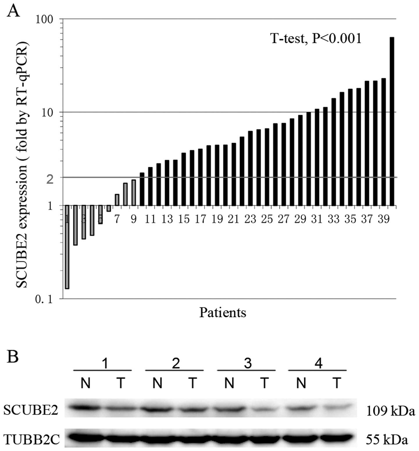
SCUBE2 gene expression in mRNA level was confirmed
by the quantitative PCR analysis of 40 pairs of CRC tissue and
matched adjacent normal mucosa. As shown in Fig. 1A, the relative level of SCUBE2 mRNA
in 31/40 (77.5%) tumor tissues showed a ≥2-fold decrease in the
SCUBE2 mRNA level, compared with that of the adjacent normal mucosa
(P<0.001). Subsequent western blot analysis also confirmed that
SCUBE2 protein levels were decreased in tumor tissue as compared
with the paired normal mucosa (Fig.
1B), consistent with the results of quantitative PCR.
Association of SCUBE2 immunohistochemical
staining with clinicopathological characteristics of CRC
To analyze the clinicopathological characteristics
of SCUBE2 expression, immunohistochemistry was used to detect the
SCUBE2 protein expression in TMA containing 120 cases of primary
CRC and paired adjacent normal mucosa.
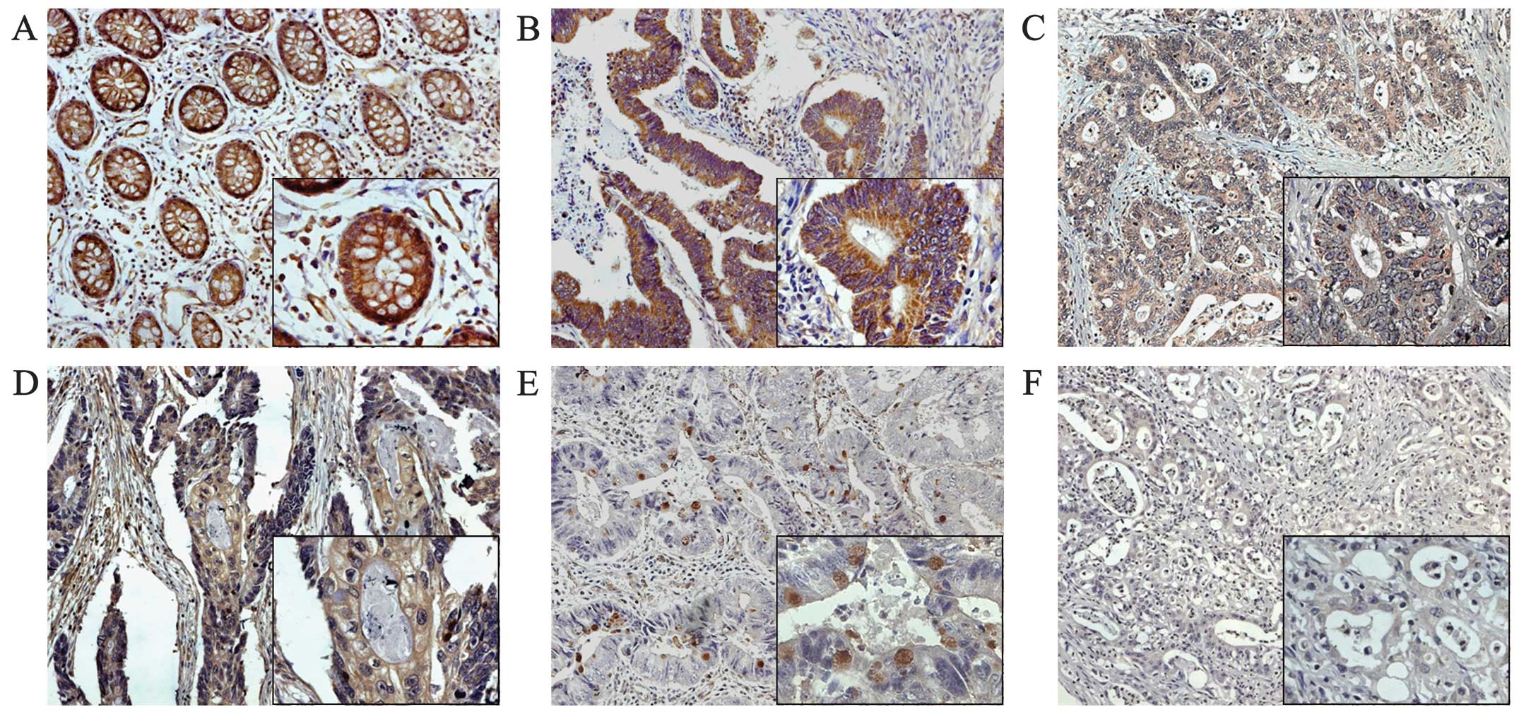
Consistent with its endothelial origin, SCUBE2
protein was detected in the endothelial cells of small vasculars
(Fig. 2A and E). As shown in
Fig. 2, SCUBE2 was mainly expressed
in the membrane of colorectal epithelium, with cytoplasmic staining
only observed with strong positive staining. Notably, in some
well-differentiated CRC tissues, positive staining was detected in
the membrane of colorectal mucosa (Fig.
2D) and was expressed in the cytoplasm of goblet cells
(Fig. 2E).
Of the 120 normal mucosa specimens in the TMA 16/120
(13.33%) showed a negative SCUBE2 expression, 56/120 (46.67%)
specimens had weak staining, and 48/120 (40%) specimens exhibited
strong staining. By contrast, the immunoreactive patterns of SCUBE2
were not predominantly positively identified in the majority of CRC
specimens. Of these CRC tissues, 57/120 (47.50%) showed negative
SCUBE2 expression, 39 (32.50%) cases showed weak staining and 24
(20%) exhibited strong staining (Table
I). The distribution of SCUBE2 expression was significantly
different between normal mucosa and tumor tissues and SCUBE2 was
significantly downregulated in the cancer tissues compared with the
corresponding non-cancer mucosas (P<0.001).
 | Table IExpression of SCUBE2 in normal colonic
mucosa and cancerous tissues. |
Table I
Expression of SCUBE2 in normal colonic
mucosa and cancerous tissues.
| | Expression of
SCUBE2 | |
|---|
| |
| |
|---|
| Tissue sample | n | Negative (n, %) | Weak (n, %) | Strong (n, %) | P-value |
|---|
| Normal mucosa | 120 | 16 (13.33) | 56 (46.67) | 48 (40) | <0.001a |
| Cancer tissue | 120 | 57 (47.50) | 39 (32.50) | 24 (20) | |
Associations between SCUBE2 expression and
clinicopathological characteristics for the 120 subjects are shown
in Table II. The downregulation of
SCUBE2 was significantly correlated with that of the American Joint
Committee on cancer (AJCC) stage (P<0.001), depth of tumor
invasion (P<0.001), nodal involvement (P=0.016), distant
metastasis (P=0.044) and histological differentiation (P=0.031). No
correlations were identified between SCUBE2 expression and age,
gender, tumor location and vascular invasion.
| Table IIAssociation between SCUBE2 expression
and the clinicopathological characteristics in CRC tissues. |
Table II
Association between SCUBE2 expression
and the clinicopathological characteristics in CRC tissues.
| SCUBE2
expression | |
|---|
|
| |
|---|
|
Characteristics | Negative
(n=57) | Weak (n=39) | Strong (n=24) | P-value |
|---|
| Age (years) | | | | 0.211 |
| <65 | 27 | 14 | 14 | |
| ≥65 | 30 | 25 | 10 | |
| Gender | | | | 0.708 |
| Male | 25 | 14 | 9 | |
| Female | 32 | 25 | 15 | |
| Location | | | | 0.560 |
| Right | 22 | 9 | 8 | |
| Transverse | 9 | 10 | 3 | |
| Left | 12 | 7 | 4 | |
| Sigmoid | 14 | 13 | 9 | |
| AJCC stage | | | | <0.001a |
| I | 12 | 10 | 15 | |
| II | 13 | 21 | 7 | |
| III | 18 | 5 | 1 | |
| IV | 14 | 3 | 1 | |
| T stage | | | | <0.001a |
| T1 | 2 | 5 | 9 | |
| T2 | 4 | 6 | 7 | |
| T3 | 22 | 16 | 5 | |
| T4 | 29 | 12 | 3 | |
| N stage | | | | 0.016a |
| N0 | 25 | 28 | 19 | |
| N1 | 19 | 7 | 3 | |
| N2 | 13 | 4 | 2 | |
| M stage | | | | 0.044a |
| M0 | 48 | 37 | 23 | |
| M1 | 9 | 2 | 1 | |
|
Differentiation | | | | 0.031a |
| Well | 19 | 17 | 17 | |
| Moderate | 23 | 14 | 6 | |
| Poor | 15 | 8 | 1 | |
| Vascular
invasion | | | | 0.079 |
| Yes | 8 | 2 | 1 | |
| No | 49 | 37 | 23 | |
Survival analysis and prognostic
significance of SCUBE2 expression
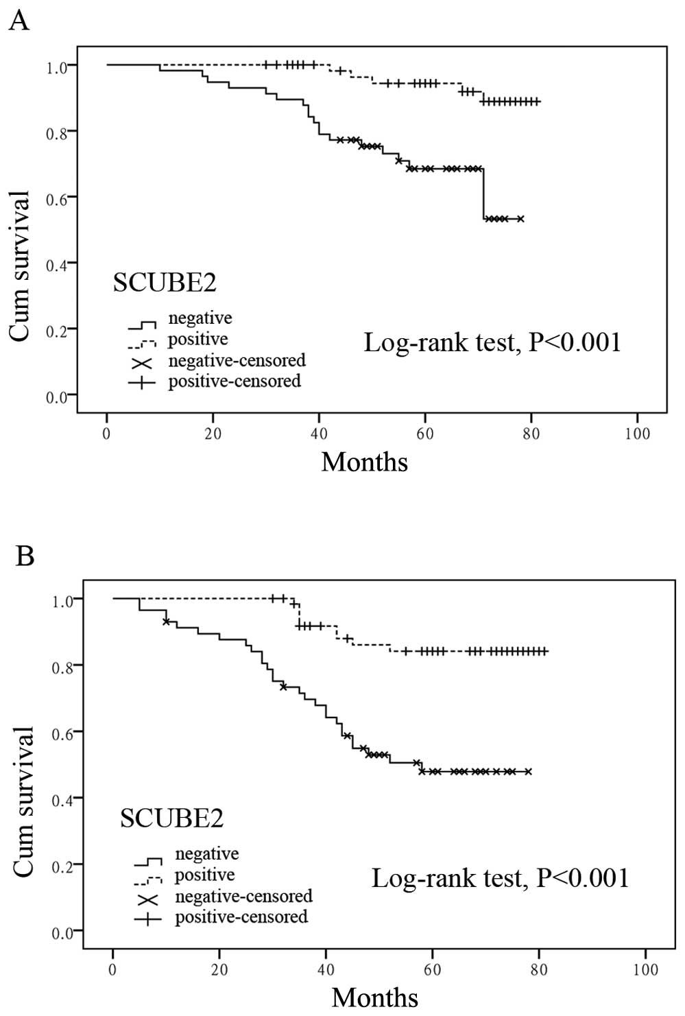
To assess the possible association between CRC
SCUBE2 expression and patient survival, Kaplan-Meier curves with a
log-rank test for OS and DFS were undertaken. As shown in Fig. 3, the estimated mean OS time was
significantly different between patients with SCUBE2-positive and
-negative tumors (63.38±1.86 and 53.98±2.09 months, respectively;
P=0.001). The estimated mean DFS time was 61.05±2.11 and 45.32±2.66
months for subjects with SCUBE2-positive and SCUBE2-negative tumors
(P<0.001). Kaplan-Meier curves showed that the rate of
recurrence was significantly elevated with the negative SCUBE2
expression. In the 37 recurrence cases, patients with a negative
SCUBE2 expression had a higher recurrence rate than patients with a
positive expression (negative, 75.68% and positive, 24.32%;
P<0.001).
The results of the univariate analysis revealed that
patients with positive tumor SCUBE2 expression had a significantly
higher overall survival (OS) and disease-free survival (DFS) rate
than patients with a negative SCUBE2 expression (HR 0.356, 95% CI
0.178–0.714, P=0.004; HR 0.226, 95% CI 0.107–0.481, P<0.001,
respectively; Tables III and
IV). In addition, both OS and DFS
were significantly associated with AJCC stage, LNM (P<0.001),
distant metastasis (P<0.001), histological differentiation, and
vascular invasion (Tables III and
IV). However, the multivariate
analysis revealed that a positive SCUBE2 expression could not be a
significant independent prognostic factor for decreased disease
recurrence and increased survival (Tables III and IV).
| Table IIIAssociation between
clinicopathological characteristics and OS by COX regression model
analysis. |
Table III
Association between
clinicopathological characteristics and OS by COX regression model
analysis.
| Univariate | Multivariate |
|---|
|
|
|
|---|
| Overall
survival | HR | CI (95%) | P-value | HR | CI (95%) | P-value |
|---|
| Age (years) |
| <65 | 1 | | | | | |
| ≥ 65 | 1.988 | 0.850–4.649 | 0.113 | | | |
| Gender |
| Male | 1 | | | | | |
| Female | 1.399 | 0.598–3.270 | 0.439 | | | |
| Vascular
invasion |
| No | 1 | | | 1 | | |
| Yes | 4.417 | 1.632–11.953 | 0.003a | 3.645 | 1.165–11.401 | 0.026a |
| AJCC stage |
| I–II | 1 | | | 1 | | |
| III–IV | 5.042 | 2.130–11.939 | <0.001a | 1.948 | 0.776–4.893 | 0.156 |
| T stage |
| T1–T2 | 1 | | | 1 | | |
| T3–T4 | 2.468 | 0.840–7.249 | 0.100 | 1.669 | 0.421–6.612 | 0.466 |
| N stage |
| N0 | 1 | | | 1 | | |
| N1–N2 | 12.465 | 3.715–41.818 | <0.001a | 13.661 | 1.463–127.602 | 0.022a |
| M stage |
| M0 | 1 | | | 1 | | |
| M1 | 6.684 | 2.844–15.705 | <0.001a | 3.140 | 1.223–8.062 | 0.017a |
|
Differentiation |
| Well | 1 | | | 1 | | |
| Moderate/poor | 5.875 | 1.752–19.703 | 0.004a | 0.416 | 0.042–4.103 | 0.453 |
| SCUBE2
expression |
| Negative | 1 | | | 1 | | |
| Positive | 0.356 | 0.178–0.714 | 0.004a | 0.465 | 0.128–1.682 | 0.243 |
| Table IVAssociation between
clinicopathological characteristics and DFS by COX regression model
analysis. |
Table IV
Association between
clinicopathological characteristics and DFS by COX regression model
analysis.
| Univariate | Multivariate |
|---|
|
|
|
|---|
| Disease-free
survival | HR | CI (95%) | P-value | HR | CI (95%) | P-value |
|---|
| Age (years) |
| <65 | 1 | | | | | |
| ≥65 | 1.274 | 0.665–2.444 | 0.465 | | | |
| Gender |
| Male | 1 | | | | | |
| Female | 1.115 | 0.574–2.168 | 0.747 | | | |
| Vascular
invasion |
| No | 1 | | | 1 | | |
| Yes | 4.753 | 2.162–10.452 | <0.001a | 4.327 | 1.781–10.510 | 0.001a |
| AJCC stage |
| I–II | 1 | | | 1 | | |
| III–IV | 2.506 | 1.314–4.781 | 0.005a | 1.410 | 0.683–2.915 | 0.353 |
| T stage |
| T1–T2 | 1 | | | 1 | | |
| T3–T4 | 2.454 | 1.023–5.887 | 0.044a | 2.246 | 0.813–6.208 | 0.119 |
| N stage |
| N0 | 1 | | | 1 | | |
| N1–N2 | 3.673 | 1.868–7.224 | <0.001a | 1.896 | 0.637–5.645 | 0.250 |
| M stage |
| M0 | 1 | | | 1 | | |
| M1 | 6.136 | 2.856–13.181 | <0.001a | 4.207 | 1.729–10.239 | 0.002a |
|
Differentiation |
| Well | 1 | | | 1 | | |
| Moderate/poor | 3.008 | 1.419–6.377 | 0.004a | 1.460 | 0.452–4.711 | 0.527 |
| SCUBE2
expression |
| Negative | | | | 1 | | |
| Positive | 0.226 | 0.107–0.481 | <0.001a | 0.438 | 0.183–1.049 | 0.064 |
Overexpression of SCUBE2 protein
suppresses proliferation and inhibits cell migration and invasion
of RKO CRC cell line, but does not increase apoptosis
To examine the effect of SCUBE2 on CRC, its mRNA and
protein expression was evaluated in the CRC cell lines (data not
shown). The expression level of the RKO CRC cell line was decreased
as compared to the remaining cell lines and was more easily
transfected. Thus, RKO cell line was selected for the subsequent
in vitro experiments.
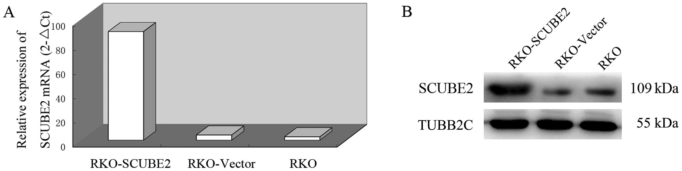
RKO was transfected with a SCUBE2 expression plasmid
and a series of functional experiments concerning cell
proliferation, apoptosis and metastasis was performed. Upregulation
of SCUBE2 expression was confirmed in transfected RKO cells by
quantitative PCR and western blot analysis (Fig. 4).
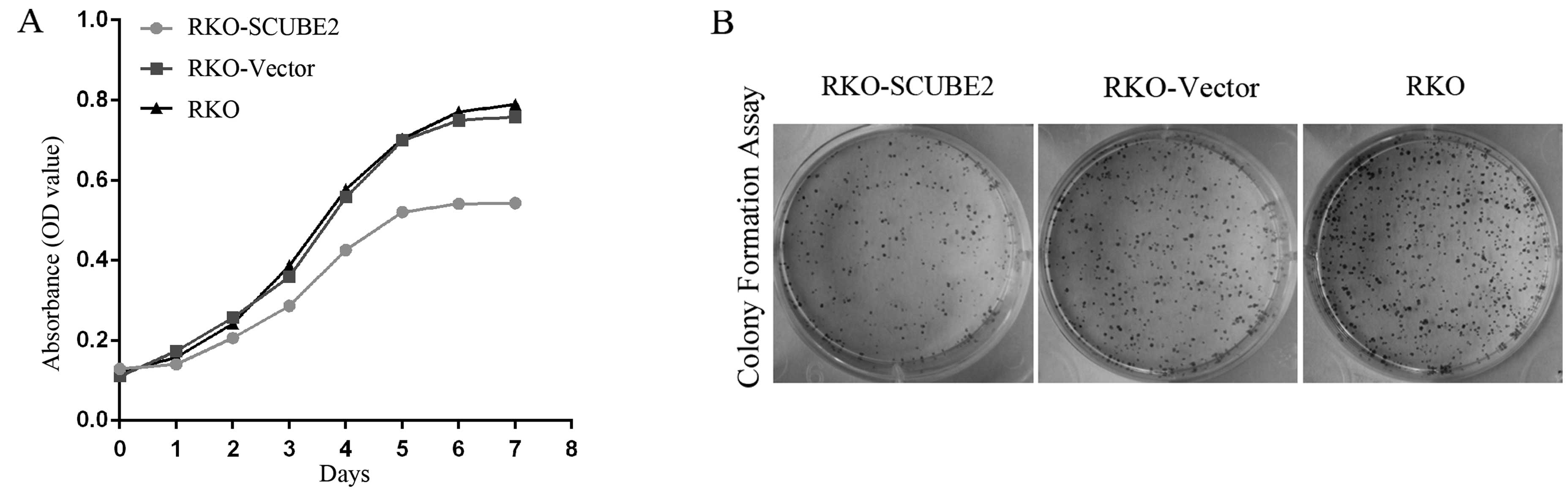
Significantly decreased proliferation of
SCUBE2-transfected RKO cells was observed after 3–7 days as
compared to the two control groups (Fig. 5A). In addition, RKO colony formation
was significantly decreased upon SCUBE2 expression (P<0.05,
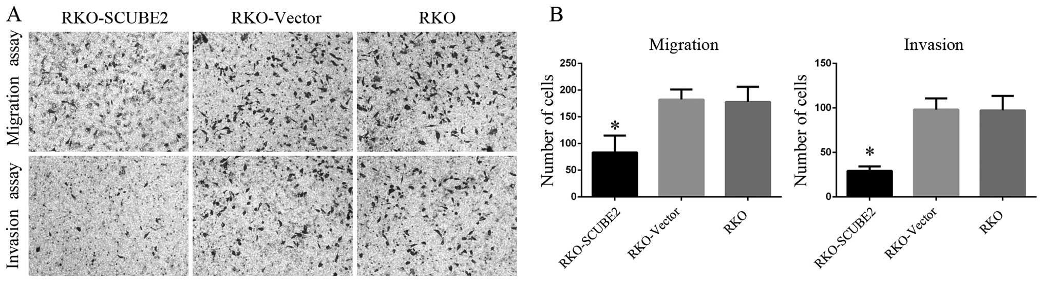
Fig. 5B). Cell migration and
invasion were significantly reduced in SCUBE2-transfected cells as
compared to parental and empty vector-transfected cells (P<0.05,
Fig. 6A and B). Furthermore, the
RKO cell apoptotic rate was not significantly increased from 0.2
and 0.1% among parental and empty vector-transfected cells,
respectively, to 0.5% in SCUBE2-transfected cells (data not
shown).
Discussion
In the present study, we used a variety of
experimental approaches to examine protein localization, function
and clinical implications of a newly described human gene,
SCUBE2, and its a role in human CRC. Our findings have
demonstrated the importance of SCUBE2 as a potential tumor
suppressor in CRC and correlations were observed between the
downregulated SCUBE2 expression and advanced cancer biology, OS and
DFS.
SCUBE2 belongs to a small, evolutionarily conserved
SCUBE protein family comprising the members SCUBE1 to SCUBE3 by
sequence of identification (4,5). These
proteins, containing ~1,000 amino acids, are characterized by nine
tandem copies of EGF-like repeats, a spacer region, three repeated
stretches of 6-cysteine residues and one CUB domain at the COOH
terminus. The EGF motif is identified in many types of proteins
functioning as adhesion molecules, secreted growth factors,
signaling molecules, transmembrane receptors, and important
components of the extracellular matrix. The CUB domain regulates
cell-cell communication and has been found in several proteins
involved in developmental processes (4). SCUBE2 is known to enhance the HH
signaling pathway (6,20,21),
the dysregulation of which is known to contribute to carcinogenesis
(12,13). SCUBE2 expression has been observed
in vascular endothelium primarily and thus may affect CRC
angiogenesis (10,11,22).
SCUBE2 has been reported to act as a tumor suppressor in human
breast cancer (8,9,14,15).
To the best of our knowledg, the present study is the first to
examine SCUBE2 expression in CRC, suggesting that SCUBE2 has a
similar role in this tumor type.
Consistent with its endothelial origin, SCUBE2
immuno-reactive staining was localized in vascular endothelial
cells, and can be detected in colorectal mucosa epithelium.
Notably, we found that the SCUBE2 transcript and translational
levels were significantly decreased in CRC tissues compared with
adjacent normal tissues. The relative level of SCUBE2 mRNA in 34/40
(85%) tumor tissues showed a ≥2-fold decrease in the corresponding
non-cancer mucosa. These results suggest that similar to breast
cancer (14,23) and endometrial cancer (16), the transcript levels of SCUBE2 are
an essential component of predictive gene signatures. Moreover,
immunohistochemistry results revealed that only 20% (24/120) of
primary CRC had strong positive-SCUBE2 protein staining and 40%
(48/120) of normal colorectal epithelium. These data indicate that
SCUBE2 is invovled in the progression of colorectal carcinogenesis.
Negative correlations were observed between SCUBE2 expression and
clinicopathological characteristics of the patients, including AJCC
stage (P<0.001), tumor invasion depth (P<0.001), lymph-node
metastasis (P=0.016), distant metastasis (P=0.044) and tumor
differentiation (P=0.031). The data suggest that downregulated
SCUBE2 may contribute to tumor invasion and metastasis. Therefore,
SCUBE2 is a potential biomarker for the identification of subsets
of CRC with a less aggressive phenotype. In breast cancer, SCUBE2
is reduced in high-grade tumors and is a biomarker for a good
prognosis (24–27). Parris et al (28) identified that SCUBE2 is a putative
target for clinical management and drug development of squamous
cell carcinoma of the oral cavity.
Inaddition, we found that individuals with
SCUBE2-positive tumors have higher OS and DFS as compared to those
with SCUBE2-negative tumors according to the Kaplan-Meier curves
and univariate analysis. These results are partly consistent with
those of a previous report (8)
whereby SCUBE2 acted as an independent prognostic factor for DFS in
breast cancer. The multivariate analyses revealed that SCUBE2
expression in CRC was not an independent prognostic factor for
survival. This may be due to the relatively small number of CRC
patients and requires further confirmation in a larger group of CRC
patients from multicenter trials.
In the present study, restoration of SCUBE2 gene
expression in RKO cells was reduced in in vitro
proliferation and in vitro colony formation. The mechanism
by which SCUBE2 suppresses CRC progression remains to be
determined. According to previous reports, SCUBE2 suppresses cancer
cell growth through, at least in part, a coordinated regulation of
two distinct mechanisms: antagonizing bone morphogenetic protein
activity by releasing an active COOH terminal fragment and
suppressing the β-catenin pathway by the NH2-terminal EGF-like
repeats (8,9). However, the molecular mechanism of
SCUBE2 governing the effects on CRC cell growth needs further
exploration. SCUBE2 expression was significantly correlated with N
stage and distant metastasis, suggesting that SCUBE2 may modulate
the metastatic process. In concordance with findings of those
reports, restoration of SCUBE2 inhibited RKO cell migration and
invasion in vitro. According to previous reports, SCUBE2
increased the formation of epithelial E-cadherin-containing
adherens junctions to promote epithelial differentiation and drive
the reversal of EMT (15). The
molecular mechanism of SCUBE2 governing the effects on CRC cell
migration and invasion needs further exploration.
In conclusion, a specific downregulated expression
of SCUBE2 and its association with multiple clinicopathological
factors in CRC patients has been identified in the present study.
This downregulation may possess an important role in promoting CRC.
Moreover, it is closely correlated with aggressive malignant
behavior and predicted poor survival in CRC patients. In future
studies, large clinical sample validation and mechanism
investigations are required to fully elucidate the molecular
mechanisms and the role of SCUBE2 in CRC progression.
References
|
1
|
Siegel R, Ma J, Zou Z and Jemal A: Cancer
statistics, 2014. CA Cancer J Clin. 64:9–29. 2014. View Article : Google Scholar : PubMed/NCBI
|
|
2
|
Siegel R, Desantis C and Jemal A:
Colorectal cancer statistics, 2014. CA Cancer J Clin. 64:104–117.
2014. View Article : Google Scholar : PubMed/NCBI
|
|
3
|
Fearon ER: Molecular genetics of
colorectal cancer. Annu Rev Pathol. 6:479–507. 2011. View Article : Google Scholar
|
|
4
|
Yang RB, Ng CK, Wasserman SM, et al:
Identification of a novel family of cell-surface proteins expressed
in human vascular endothelium. J Biol Chem. 277:46364–46373. 2002.
View Article : Google Scholar : PubMed/NCBI
|
|
5
|
Wu BT, Su YH, Tsai MT, Wasserman SM,
Topper JN and Yang RB: A novel secreted, cell-surface glycoprotein
containing multiple epidermal growth factor-like repeats and one
CUB domain is highly expressed in primary osteoblasts and bones. J
Biol Chem. 279:37485–37490. 2004. View Article : Google Scholar : PubMed/NCBI
|
|
6
|
Hollway GE, Maule J, Gautier P, et al:
Scube2 mediates Hedgehog signalling in the zebrafish embryo. Dev
Biol. 294:104–118. 2006. View Article : Google Scholar : PubMed/NCBI
|
|
7
|
Tsai MT, Cheng CJ, Lin YC, et al:
Isolation and characterization of a secreted, cell-surface
glycoprotein SCUBE2 from humans. Biochem J. 422:119–128. 2009.
View Article : Google Scholar : PubMed/NCBI
|
|
8
|
Cheng CJ, Lin YC, Tsai MT, et al: SCUBE2
suppresses breast tumor cell proliferation and confers a favorable
prognosis in invasive breast cancer. Cancer Res. 69:3634–3641.
2009. View Article : Google Scholar : PubMed/NCBI
|
|
9
|
Lin YC, Chen CC, Cheng CJ and Yang RB:
Domain and functional analysis of a novel breast tumor suppressor
protein, SCUBE2. J Biol Chem. 286:27039–27047. 2011. View Article : Google Scholar : PubMed/NCBI
|
|
10
|
Rmali KA, Puntis MC and Jiang WG:
Tumour-associated angio-genesis in human colorectal cancer.
Colorectal Dis. 9:3–14. 2007. View Article : Google Scholar
|
|
11
|
Najib S, Kowalski-Chauvel A, Do C, Roche
S, Cohen-Jonathan-Moyal E and Seva C: Progastrin a new
pro-angiogenic factor in colorectal cancer. Oncogene. Aug
11–2014.(Epub ahead of print). View Article : Google Scholar : PubMed/NCBI
|
|
12
|
Hu X, Lai D, Chen W, et al: Differential
expression profiles of the Hedgehog signaling pathway between
microsatellite-stable and microsatellite-unstable colorectal
cancers. Mol Med Rep. 4:873–877. 2011.PubMed/NCBI
|
|
13
|
Wang H, Li YY, Wu YY and Nie YQ:
Expression and clinical significance of hedgehog signaling pathway
related components in colorectal cancer. Asian Pac J Cancer Prev.
13:2319–2324. 2012. View Article : Google Scholar : PubMed/NCBI
|
|
14
|
Sánchez-Navarro I, Gamez-Pozo A, Pinto A,
et al: An 8-gene qRT-PCR-based gene expression score that has
prognostic value in early breast cancer. BMC Cancer. 10:3362010.
View Article : Google Scholar : PubMed/NCBI
|
|
15
|
Lin YC, Lee YC, Li LH, Cheng CJ and Yang
RB: Tumor suppressor SCUBE2 inhibits breast-cancer cell migration
and invasion through the reversal of epithelial-mesenchymal
transition. J Cell Sci. 127:85–100. 2014. View Article : Google Scholar
|
|
16
|
Skrzypczak M, Lattrich C, Häring J,
Schüler S, Ortmann O and Treeck O: Expression of SCUBE2 gene
declines in high grade endometrial cancer and associates with
expression of steroid hormone receptors and tumor suppressor PTEN.
Gynecol Endocrinol. 29:1031–1035. 2013. View Article : Google Scholar : PubMed/NCBI
|
|
17
|
Penney KL, Sinnott JA, Fall K, et al: mRNA
expression signature of Gleason grade predicts lethal prostate
cancer. J Clin Oncol. 29:2391–2396. 2011. View Article : Google Scholar : PubMed/NCBI
|
|
18
|
Livak KJ and Schmittgen TD: Analysis of
relative gene expression data using real-time quantitative PCR and
the 2−ΔΔCT method.
Methods. 25:402–408. 2001. View Article : Google Scholar
|
|
19
|
Li DW, Tang HM, Fan JW, et al: Expression
level of Bmi-1 onco-protein is associated with progression and
prognosis in colon cancer. J Cancer Res Clin Oncol. 136:997–1006.
2010. View Article : Google Scholar
|
|
20
|
Creanga A, Glenn TD, Mann RK, Saunders AM,
Talbot WS and Beachy PA: Scube/You activity mediates release of
dually lipid-modified Hedgehog signal in soluble form. Genes Dev.
26:1312–1325. 2012. View Article : Google Scholar : PubMed/NCBI
|
|
21
|
Kawakami A, Nojima Y, Toyoda A, et al: The
zebrafish-secreted matrix protein you/scube2 is implicated in
long-range regulation of hedgehog signaling. Curr Biol. 15:480–488.
2005. View Article : Google Scholar : PubMed/NCBI
|
|
22
|
Yang M, Guo M, Hu Y and Jiang Y: Scube
regulates synovial angiogenesis-related signaling. Med Hypotheses.
81:948–953. 2013. View Article : Google Scholar : PubMed/NCBI
|
|
23
|
Abba MC, Hu Y, Sun H, et al: Gene
expression signature of estrogen receptor α status in breast
cancer. BMC Genomics. 6:372005. View Article : Google Scholar
|
|
24
|
Parris TZ, Danielsson A, Nemes S, et al:
Clinical implications of gene dosage and gene expression patterns
in diploid breast carcinoma. Clin Cancer Res. 16:3860–3874. 2010.
View Article : Google Scholar : PubMed/NCBI
|
|
25
|
Sørlie T, Wang Y, Xiao C, et al: Distinct
molecular mechanisms underlying clinically relevant subtypes of
breast cancer: gene expression analyses across three different
platforms. BMC Genomics. 7:1272006. View Article : Google Scholar : PubMed/NCBI
|
|
26
|
Sørlie T, Perou CM, Tibshirani R, et al:
Gene expression patterns of breast carcinomas distinguish tumor
subclasses with clinical implications. Proc Natl Acad Sci USA.
98:10869–10874. 2001. View Article : Google Scholar : PubMed/NCBI
|
|
27
|
Calza S, Hall P, Auer G, et al: Intrinsic
molecular signature of breast cancer in a population-based cohort
of 412 patients. Breast Cancer Res. 8:R342006. View Article : Google Scholar : PubMed/NCBI
|
|
28
|
Parris TZ, Aziz L, Kovacs A, et al:
Clinical relevance of breast cancer-related genes as potential
biomarkers for oral squamous cell carcinoma. BMC Cancer.
14:3242014. View Article : Google Scholar : PubMed/NCBI
|