Introduction
Hepatocellular carcinoma (HCC) is the fifth most
common malignant tumor due to increases in the rates of viral
hepatitis and adiposis hepatica. HCC is also the third leading
cause of cancer mortality worldwide (1,2). Each
year, ~650,000 HCC cases are reported globally, and according to
statistics, China alone accounts for 55% of HCC cases worldwide
(3,4). Hepatic resection is the gold standard
therapy for HCC (5). Although the
survival of patients with HCC has been greatly improved owing to
advances in surgical improvements and perioperative management,
long-term survival after surgical resection remains low due to the
high rates of recurrence and metastasis (6,7). The
recurrence and metastasis of HCC are multistep processes that
involve complex biological and pathological events (8,9).
Several molecular pathways and abnormal genetic changes are
involved (10).
In daily clinical practice, the prediction of
prognosis plays an important role in the assessment of HCC patients
and in identifying the optimal therapeutic options. To estimate
malignant biological behavior or patient prognosis, sensitive tumor
markers are necessary. As a classical and specific marker for HCC,
α-fetoprotein (AFP) has been widely used for the diagnosis and
evaluation of tumor aggressiveness and to determine the prognosis
of HCC (11). When a cut-off value
of 20 ng/ml is used, AFP has a sensitivity and specificity for
detecting HCC in the ranges of 41–65 and 80–90%, respectively
(12). However, up to 50% of
patients with HCC have AFP levels <20 ng/ml (13), thus AFP cannot be used as a unique
target to screen for HCC recurrence and to predict patient
prognosis. Therefore, it is important to identify useful prognostic
biomarkers to predict the risk of this devastating disease and to
identify effective therapeutic target molecules that are strongly
associated with a poor prognosis in HCC.
Neural precursor cell expressed, developmentally
downregulated 9 (NEDD9), also known as Crk-associated substrate
lymphocyte type (Cas-L) (14) or
human enhancer of filamentation-1 (HEF1) (15), is a focal adhesion scaffold protein
that was initially identified by its developmentally regulated
expression pattern in the early embryonic, but not adult, mouse
brain (16). It also plays an
integral role in natural and pathological cell biology (17). Overexpression of NEDD9 protein is
correlated with poor prognosis in various types of cancer,
including breast cancer (18),
glioblastoma (19) and melanoma
(20). However, to the best of our
knowledge, few available data address the precise function of
NEDD9 genes in HCC.
In the present study, we investigated NEDD9
expression in primary human HCC tissues compared with matched
adjacent non-tumor hepatic tissues (ANHTs). Immunohistochemistry
was performed to analyze the correlation of NEDD9 expression with
the clinicopathological factors of patients. Additionally, the
potential prognostic utility of NEDD9 expression in HCC was
analyzed, including in HCC patients with normal serum AFP levels
and with early-stage HCC. The results suggest that NEDD9 may be a
valuable prognostic biomarker for HCC.
Materials and methods
Patients and tissue specimens
In the present study, HCC tissue specimens were
obtained from 164 patients with HCC who underwent hepatectomy in
the Department of Hepatobiliary and Pancreaticosplenic Surgery,
Xijing Hospital (Xi’an, China) from 2004 to 2007. Specimens were
obtained from patients who had not received preoperative
treatments, such as chemotherapy, ethanol injection, radiofrequency
ablation or transarterial chemoembolization. These patients
included 133 men and 31 women, and the median patient age was 49
years (range, 29–81 years).
The present study was approved by the Ethics
Committee of the Fourth Military Medical University, and it
conformed to the ethical guidelines of the 2004 Declaration of
Helsinki. Written informed consent was obtained from each patient
or from his/her legal guardians. Of the 164 patients with HCC,
matched fresh specimens of HCC tissues and ANHTs were collected
from 40 random patients, immediately frozen in liquid nitrogen, and
subsequently stored at −80°C for reverse-transcription quantitative
polymerase chain reaction (RT-qPCR) and western blot analysis.
Before the study was initiated, histopathological examinations were
performed to confirm that there were sufficient cancer cells in the
tumor samples and that no cancer cells had contaminated the ANHTs.
All of the specimens were embedded in paraffin and stained with
hematoxylin and eosin. The diagnosis in each patient was confirmed
by a histopathologist who was blinded to our examination. The data
for 10 conventional clinical and pathologic variables were
collected for analysis, including gender, age, tumor size, number
of tumor nodules, tumor grade, metastasis, venous invasion, UICC
TNM stage, HbsAg status and serum AFP level.
RNA extraction, reverse transcription and
quantitative PCR
Total RNA was extracted from fresh tissues using
TRIzol reagent (Invitrogen, Carlsbad, CA, USA), according to the
manufacturer’s instructions. cDNA was synthesized from 2 μg
of RNA using an iScript™ cDNA Synthesis kit (Bio-Rad, Hercules, CA,
USA). Quantitative PCR (qPCR) was performed using a MyiQ2 Two-Color
Real-Time PCR Detection System and SYBR-Green dye (both from
Bio-Rad), according to the manufacturer’s instructions.
Oligonucleotide primers were synthesized by Sangon Biotech Co.,
Ltd. (Shanghai, China). The primers used for the PCR reaction were:
NEDD9, forward: 5′-ATG TCC ACG TCT TCC ACC TCC-3′ and reverse:
5′-AGT GAC CAG TGC CAT TAG GCT G-3′; and GAPDH, forward: 5′-GAC AGT
CAG CCG CAT CTT CT-3′ and reverse: 5′-TTA AAA GCA GCC CTG GTG
AC-3′. The primer sequences were verified by running a virtual PCR,
and the primer concentrations were optimized to avoid primer-dimer
formation. Additionally, dissociation curves were evaluated to
avoid non-specific amplification. The data were analyzed according
to the comparative Ct method and were normalized to GAPDH
expression in each sample. All of the experiments were performed in
triplicate.
Protein extraction and western
blotting
Total protein was extracted using a lysis buffer (20
mmol/l Tris-HCl, pH 7.4; 10 mmol/l NaCl; 1 mmol/l ethylene diamine
tetraacetic acid, pH 8.0; 1 mmol/l MgCl2; 1% NP-40; 0.1%
sodium dodecyl sulfate; 0.01% phenylmethylsulfonyl fluoride (Sigma,
St. Louis, MO, USA); and 10 μl/ml protease inhibitor
cocktail (Promega, Madison, WI, USA) incubated for 20 min at 4°C.
The protein concentration was determined using the Bio-Rad assay
system, and proteins were separated by SDS-polyacrylamide gel
electrophoresis and then transferred onto polyvinylidine fluoride
membranes (Millipore, Bedford, MA, USA). The blotted membranes were
incubated with anti-human NEDD9 antibody (Abcam, Cambridge, UK),
followed by a secondary antibody (KPL, Gaithersburg, MD, USA). The
β-actin protein levels were also determined, using this specific
antibody (Sigma) as a loading control. Subsequently, the protein
bands were detected using the enhanced chemiluminescence detection
system (Amersham Pharmacia Biotech, Piscataway, NJ, USA). The
western blot analyses were quantified using laser densitometry, and
the relative protein expression was then normalized to the β-actin
levels.
Immunohistochemistry and evaluation of
staining
Immunohistochemical analysis was performed using the
avidin-biotin-peroxidase method for all tissue specimens. Sections
were deparaffinized in xylene and dehydrated using a graded alcohol
series before endogenous peroxidase activity was blocked with 0.5%
hydrogen peroxide in methanol for 10 min. Non-specific binding was
blocked by incubating sections with 10% normal goat serum in
phosphate-buffered saline (PBS) for 1 h at room temperature.
Without washing, the sections were incubated with a monoclonal
antibody against human NEDD9 (1:1,000; Abcam; ab18056) in PBS at
4°C overnight. Biotinylated goat anti-mouse immunoglobulin G (IgG;
1:400; Abcam; ab128976) was then incubated with the sections for 1
h at room temperature, and this antibody was detected using
streptavidin-peroxidase. Brown color, indicative of peroxidase
activity, was developed by incubating sections with 0.1%
3,3′-diaminobenzidine (Sigma; D3939) in PBS with 0.05% hydrogen
peroxide for 5 min at room temperature. Tissue specimens were
assessed separately by two pathologists under double-blind
conditions, in which they had no prior knowledge of the clinical or
clinicopathological status of the specimens. NEDD9 expression in
HCC specimens was evaluated by scanning the entire tissue specimen
under low magnification (×40) and was confirmed under high
magnification (×200 and ×400). An immunoreactivity score system was
applied as previously described (21). The percentage of positive cells was
scored as: 0, <5%, negative; 1, 5–25%, sporadic; 2, 25–50%,
focal; or 3, >50%, diffuse. The staining intensity was scored
as: 0, no; 1, weak; 2, moderate; or 3, strong staining. The NEDD9
immunostaining score was calculated by multiplying the positive
cell score by the staining intensity score and thus ranged from 0
to 9. A high NEDD9 expression was defined as a total score of ≥4,
and a low NEDD9 expression level was defined as a total score of
<4.
Follow-up and prognostic study
Follow-up data were obtained for all the 164
patients. The follow-up period was defined as the interval between
the date of surgery and the date of patient death or the last
follow up. Death from other causes was treated as censored events.
Recurrence was diagnosed by ultrasonography or computed tomography
scans. Recurrence-free survival was defined as the length of time
after hepatectomy during which a patient survived without signs of
HCC.
Statistical analysis
Statistical analysis was performed using SPSS
software, version 17.0 (IBM SPSS, Chicago, IL, USA). Each
experiment was repeated at least three times, and all data are
summarized and presented as the mean ± SD. The differences between
means were statistically analyzed using the t-test. The
χ2 test for proportions was used to analyze the
relationships between NEDD9 expression and various
clinicopathological factors. Survival curves were calculated using
the Kaplan-Meier method and were compared using the log-rank test.
Cox proportional hazard analysis was used to explore the effects of
clinicopathological factors and NEDD9 expression on survival. SPSS
statistical software was used to perform the univariate analysis of
each variance and identify the factors that were significantly
different in the two groups, followed by a multivariate analysis.
P-values were two-sided, and P<0.05 was considered to indicate a
statistically significant result.
Results
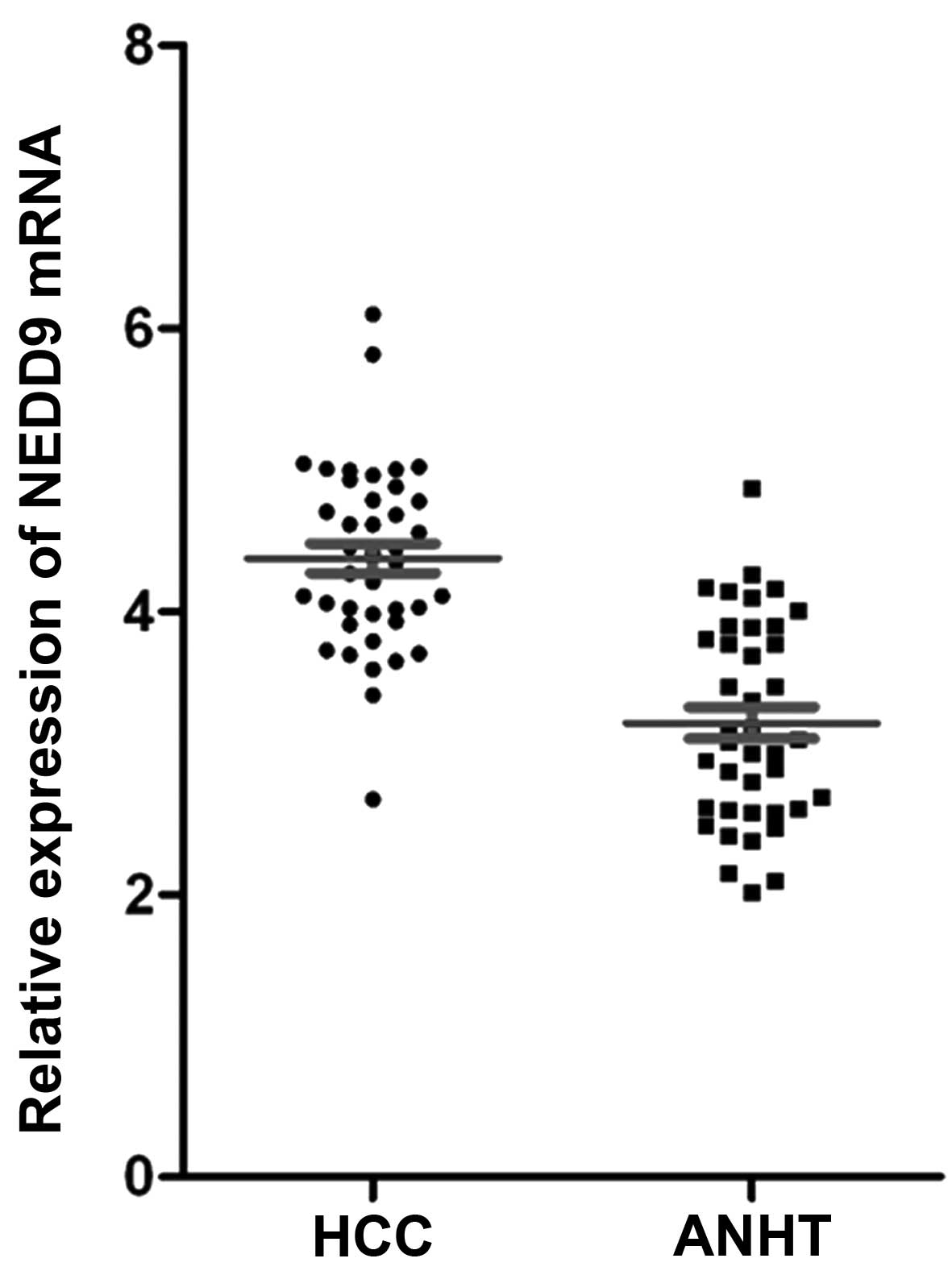
Analysis of NEDD9 mRNA expression
mRNA expression levels of NEDD9 were evaluated in 40
pairs of resected specimens (consisting of tumor tissue samples and
matched ANHT samples) from patients with HCC using RT-qPCR. We
found that 27 of the 40 patients (67.5%) had higher NEDD9 mRNA
expression levels in HCC tissues than in ANHTs. The results
indicated that the relative expression levels of NEDD9 mRNA in HCC
tissues were significantly higher than those in ANHTs (4.38±0.65
vs. 3.21±0.71, P<0.01; Fig.
1).
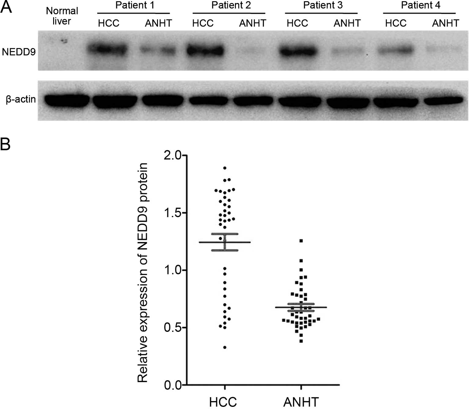
Analysis of NEDD9 protein expression
NEDD9 protein levels in resected HCC tissues and
ANHT samples were measured by western blot analysis. NEDD9
expression was increased in 26 of the 40 tumor tissue samples
(65%), compared with the matched ANHT samples. The relative
expression levels of NEDD9 protein in HCC tissues were
significantly higher than in matched ANHT samples (1.24±0.45 vs.
0.68±0.19, P<0.01; Fig. 2).
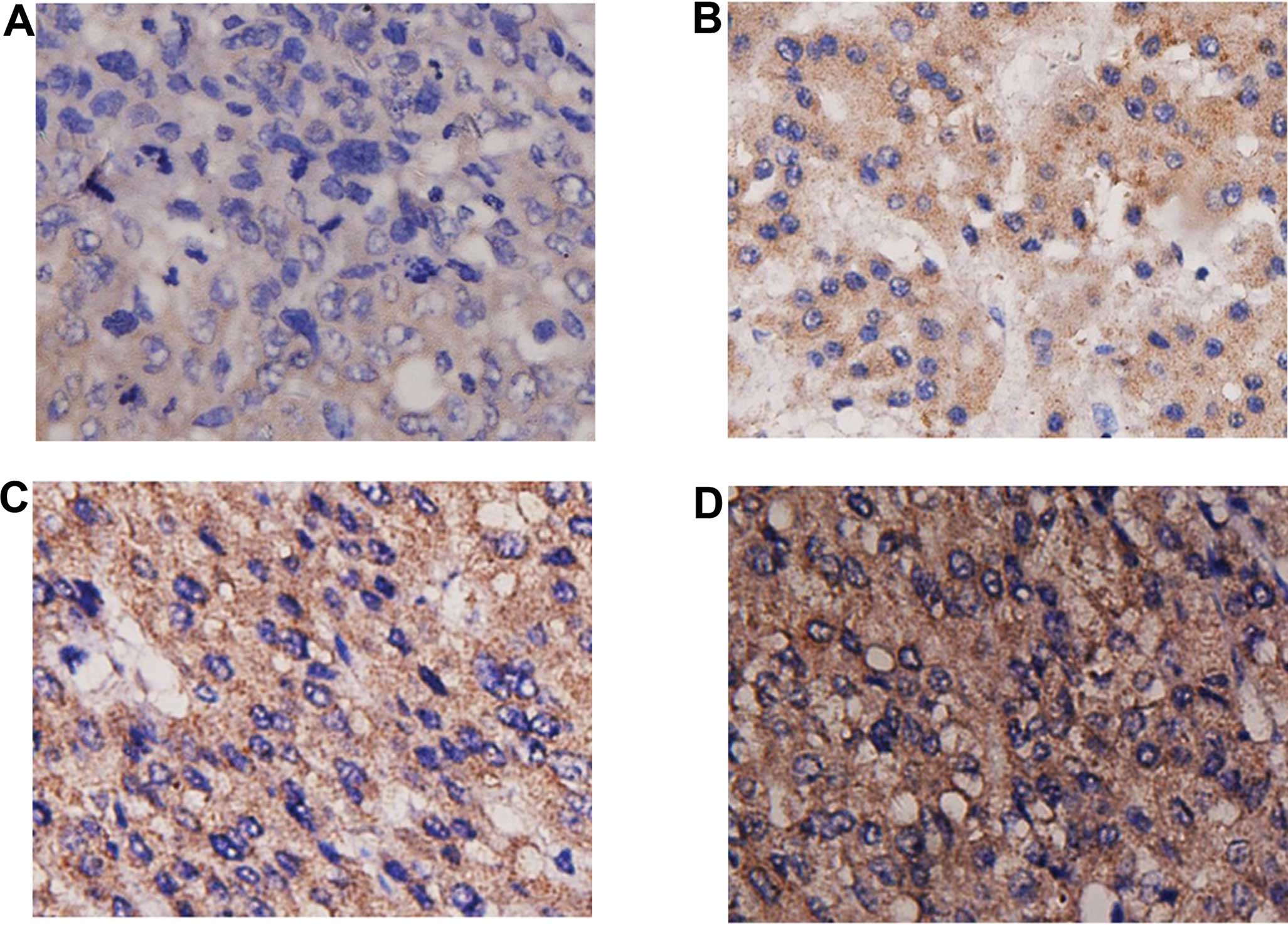
NEDD9 immunohistochemistry
NEDD9 expression was mainly localized within the
cytoplasm. NEDD9 immunoreactivity varied between HCC tissue samples
and matched ANHT samples. There was no significant NEDD9 expression
in ANHTs or only weak staining in the cytoplasm. As shown in
Fig. 3, the NEDD9 expression
patterns differed among HCC tissues. NEDD9 staining was negative
(score of 0) in 19 HCC samples, whereas weak positive staining
(score of >0 and <4) was detected in 61 HCC samples, moderate
positive staining (score of ≥4 and <6) was detected in 49 HCC
samples, and strong positive staining (score of ≥6) was detected in
35 HCC samples.
Relationship between NEDD9 expression and
clinicopathological characteristics
According to the immunohistochemical results
(Fig. 3), the HCC tissues were
divided into two groups based on the NEDD9 expression: the low
expression group (negative + weak, n=80) and the high expression
group (moderate + strong, n=84). The correlations between NEDD9
expression and HCC patient clinicopathological characteristics were
analyzed (Table I). We demonstrated
that tumor size was larger (P=0.017), tumor grade was more advanced
(P=0.021), metastasis and intrahepatic venous invasion were more
frequent (P=0.003 and P<0.001), and UICC TNM stage was higher
(P=0.034) in the high expression group than in the low expression
group. However, there were no significant differences of genders
(P=0.726), ages (P=0.831), numbers of tumor nodules (P=0.312),
HbsAg status (P=0.672) or AFP level (P=0.336) between the high and
low expression groups. These results indicated that NEDD9 may be
involved in the differentiation, metastasis, invasion and
progression of HCC.
 | Table ICorrelations between NEDD9 expression
and clinicopathological characteristics in 164 patients with
hepatocellular carcinoma. |
Table I
Correlations between NEDD9 expression
and clinicopathological characteristics in 164 patients with
hepatocellular carcinoma.
| Clinicopathological
characteristics | n | NEDD9 expression
levels
| P-value |
|---|
| Low | High |
|---|
| Gender | | | | 0.726 |
| Male | 133 | 64 | 69 | |
| Female | 31 | 16 | 15 | |
| Age (years) | | | | 0.831 |
| ≤55 | 112 | 54 | 58 | |
| >55 | 52 | 26 | 26 | |
| Tumor size
(cm) | | | | 0.017a |
| ≤5 | 89 | 51 | 38 | |
| >5 | 75 | 29 | 46 | |
| No. of tumor
nodules | | | | 0.312 |
| Solitary | 117 | 60 | 57 | |
| Multiple (≥2) | 47 | 20 | 27 | |
| Tumor grade
(differentiation) | | | | 0.021a |
| Well | 44 | 28 | 16 | |
| Moderate or
poor | 120 | 52 | 68 | |
| Metastasis | | | | 0.003a |
| Absence | 120 | 67 | 53 | |
| Presence | 44 | 13 | 31 | |
| Venous
invasion | | | | <0.001a |
| Absence | 112 | 69 | 43 | |
| Presence | 52 | 11 | 41 | |
| UICC TNM stage | | | | 0.034a |
| I+II | 97 | 54 | 43 | |
| III+IV | 67 | 26 | 41 | |
| HBsAg | | | | 0.672 |
| Positive | 148 | 73 | 75 | |
| Negative | 16 | 7 | 9 | |
| Serum AFP
(ng/ml) | | | | 0.336 |
| ≥20 | 88 | 46 | 42 | |
| <20 | 76 | 34 | 42 | |
Correlations of NEDD9 expression with the
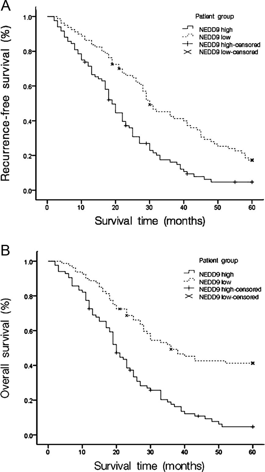
recurrence and prognosis of HCC
Since the level of NEDD9 expression was correlated
with tumor size, tumor grade, metastasis, intrahepatic venous
invasion and UICC TNM stage, we further hypothesized that the level
of NEDD9 expression would influence the recurrence and prognosis of
patients suffering from HCC. Subsequently, post-operative
Kaplan-Meier survival curves were used to evaluate the
recurrence-free survival and overall survival rates of patients
with HCC relative with their levels of NEDD9 expression. The
log-rank test showed that both recurrence-free and overall survival
rates were significantly different between low and high NEDD9
expression groups (P<0.01 each). The low NEDD9 expression group
demonstrated increased recurrence-free and overall survival rates
compared with the high NEDD9 expression group (Fig. 4). The cumulative 5-year
recurrence-free survival rate was 2.4% in the high NEDD9 expression
group but 16.3% in the low NEDD9 expression group. The cumulative
5-year overall survival rate was 3.6% in the high NEDD9 expression
group but 38.8% in the low NEDD9 expression group.
Univariate and multivariate analyses
Univariate Cox regression analysis showed that tumor
size, metastasis, venous invasion, UICC TNM stage and NEDD9 protein
expression levels were significantly associated with HCC patient
overall survival. Furthermore, to evaluate the potential role of
high NEDD9 expression levels as an independent predictor of overall
survival among HCC patients, multivariate Cox regression analyses
were performed. The results indicated that only metastasis, venous
invasion, UICC TNM stage and NEDD9 expression independently
predicted overall survival among HCC patients (Table II).
| Table IIUnivariate and multivariate Cox
regression analysis for the overall survival of 164 patients with
HCC. |
Table II
Univariate and multivariate Cox
regression analysis for the overall survival of 164 patients with
HCC.
| Clinicopathological
characteristics | RR | Univariate analyses
| Multivariate
analyses
| P-value |
|---|
| 95% CI | P-value | RR | 95% CI |
|---|
| Gender | 1.193 | 0.769–1.852 | 0.431 | 0.751 | 0.453–1.243 | 0.265 |
| Age (years) | 0.885 | 0.599–1.305 | 0.537 | 1.085 | 0.703–1.675 | 0.713 |
| Tumor size | 1.552 | 1.085–2.220 | 0.016a | 1.109 | 0.756–1.627 | 0.579 |
| No. of tumor
nodules | 1.082 | 0.733–1.597 | 0.692 | 0.688 | 0.424–1.115 | 0.129 |
| Tumor grade | 1.057 | 0.709–1.576 | 0.786 | 0.784 | 0.517–1.186 | 0.249 |
| Metastasis | 2.174 | 1.482–3.190 | <0.01a | 0.917 | 1.171–3.139 | 0.01a |
| Venous
invasion | 3.423 | 2.346–4.995 | <0.01a | 2.735 | 1.750–4.274 | <0.01a |
| UICC TNM stage | 2.040 | 1.423–2.923 | <0.01a | 1.806 | 1.222–2.670 | <0.01a |
| HBsAg | 1.144 | 0.644–2.035 | 0.646 | 1.278 | 0.641–2.547 | 0.485 |
| Serum AFP | 1.260 | 0.883–1.798 | 0.203 | 1.032 | 0.704–1.513 | 0.873 |
| NEDD9 level | 0.365 | 0.251–0.532 | <0.01a | 1.618 | 1.050–2.493 | 0.029a |
Prognostic values of NEDD9 in different
HCC subgroups
To demonstrate the value of NEDD9 expression in
predicting the survival of HCC patients, multiple analysis methods
were performed in the present study. We proved that the
recurrence-free and overall survival times of patients expressing
low levels of NEDD9 in their HCC lesions were much longer than
those with high NEDD9 expression (Fig.
4). Notably, these patients with different recurrence-free and
overall survival could not be distinguished by conventional AFP
testing. In other words, we could not use negative serum AFP to
predict HCC patients’ recurrence and prognosis. A validation cohort
was employed to evaluate further the prognostic value of NEDD9 for
specific subgroups of patients. We used a cut-off level of 20 ng/ml
AFP to subgroup the 164 HCC patients, and we evaluated the
prognostic significance of NEDD9 in the patient subgroups. Our data
suggested that the AFP cut-off value of 20 ng/ml was not
significantly predictive of patient survival (P=0.195), whereas the
NEDD9 levels were significantly associated with overall survival in
patients with serum AFP that was either positive (≥20 ng/ml) or
negative (<20 ng/ml) (P<0.01) (Fig. 5).
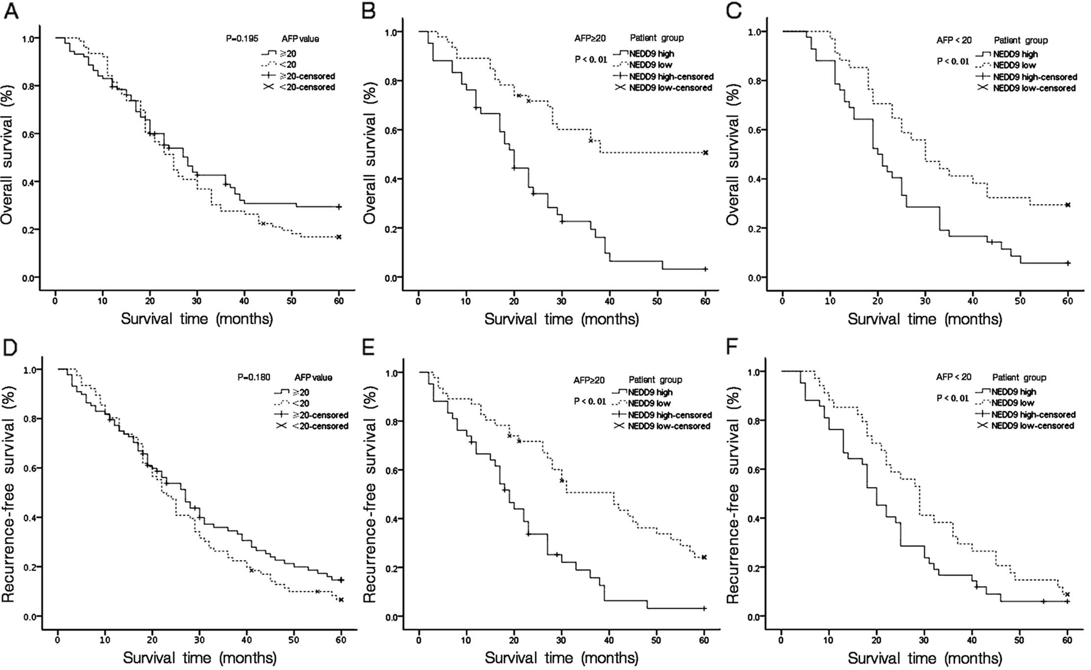 | Figure 5Kaplan-Meier analysis of overall and
recurrence-free survival in 164 patients, based on NEDD9
expressions in HCC subgroups with different AFP levels. Using AFP
level (20 ng/ml) as the cut-off could not separate patients with
different overall (A, P=0.195) and recurrence-free (D, P=0.180)
rates in the study cohort. In contrast, the NEDD9 expression level
predicted different overall survival rates in the subgroups with
AFP ≥20 ng/ml (B, P≤0.01) and AFP <20 ng/ml (C, P≤0.01). The
NEDD9 expression level also predicted different recurrence-free
survival rates of patients with AFP ≥20 ng/ml (E, P≤0.01) and AFP
<20 ng/ml (F, P≤0.01). NEDD9, neural precursor cell expressed,
developmentally downregulated 9; HCC, hepatocellular carcinoma;
AFP, α-fetoprotein. |
In the clinic, it is difficult to predict survival
in HCC patients with tumors <5 cm in diameter, single tumor
nodules and early tumor stages (UICC TNM stages I-II), and in these
early-stage HCC subgroups of our entire study cohort, NEDD9
expression level presented efficient predictive power for
predicting patient prognosis. In the subgroup with tumor size <5
cm in diameter, the 5-year overall survival rate was 45.1% in the
low NEDD9 group, compared with 5.26% for patients exhibiting a high
NEDD9 expression (P<0.01, Fig.
6B). In the clinical subgroup with single neoplastic nodules,
low NEDD9 expression patients revealed a 5-year survival rate of
38.3%, whereas the overall survival rate decreased to 5.26% in the
high NEDD9 group (P<0.01, Fig.
6E). In early TNM stage HCC patients (TNM stages), the 5-year
overall survival rates were 51.9 and 6.98%, respectively, for low
and high NEDD9 expression patients (P<0.01, Fig. 6H). Collectively, our results
indicate that, in various HCC clinical subgroups or even with
difficult-to-predict survival, NEDD9 expression is a promising
prognostic factor.
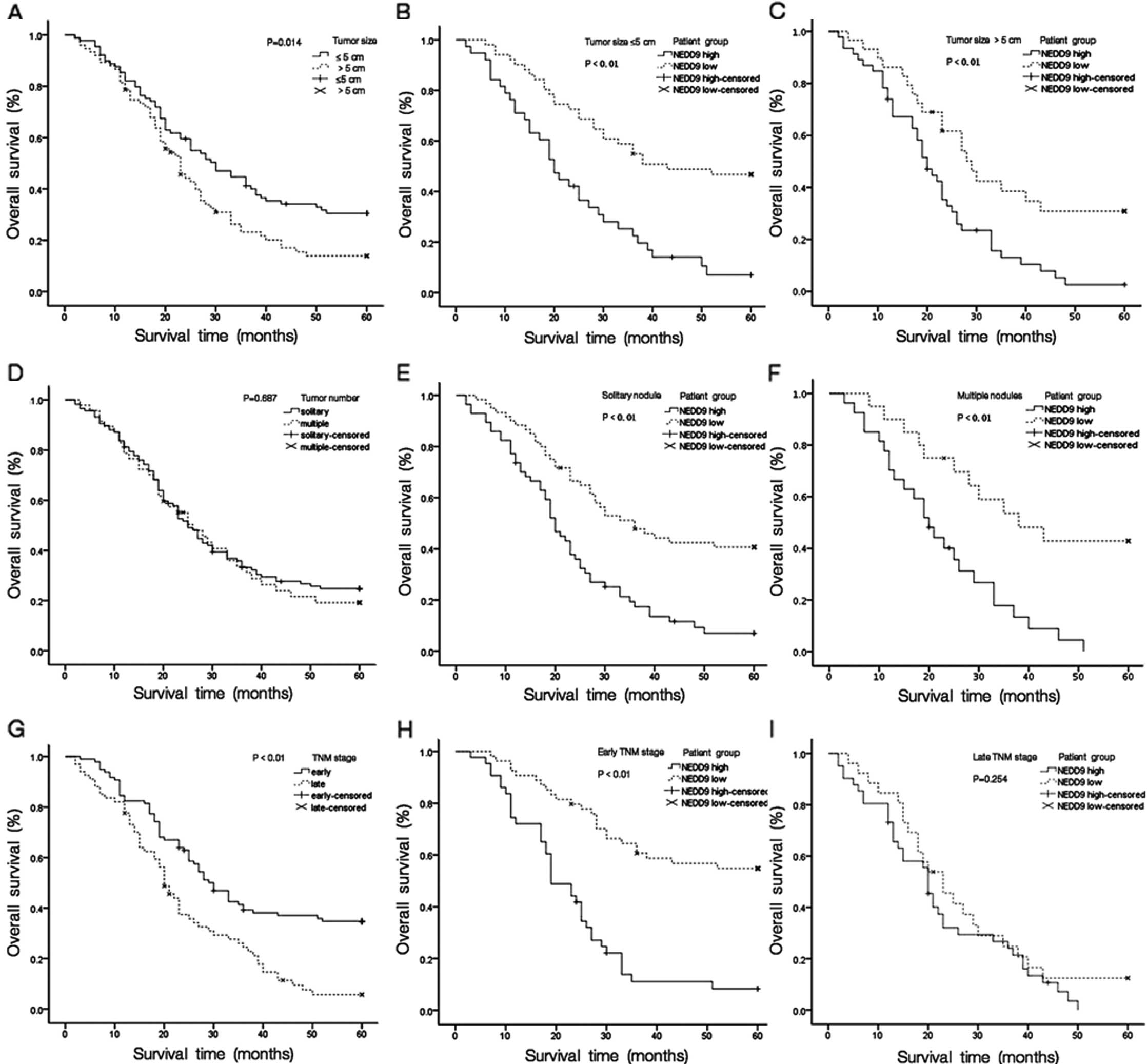 | Figure 6Kaplan-Meier analysis of overall
survival in 164 patients, based on NEDD9 expression in the HCC
clinical subgroups. (A) Tumor size and (G) TNM stage, but not the
number of (D) tumor nodules, distinguished patients with different
overall survival rates in the study cohort. (B and C) When patients
were divided into subclinical groups according to tumor size, the
probabilities of survival with either HCC lesion diameter >5 or
≤5 cm and high and low NEDD9 expression, distinguished lower and
higher overall survival rates, respectively. (E and F) Overall
survival in patients with single tumor lesions and multi-tumor
lesions. (H and I) Overall survival rates in patients classified
into early TNM stages (I+II) and late TNM stages (III+IV), as
differentiated by a high or low NEDD9 expression. NEDD9, neural
precursor cell expressed, developmentally downregulated 9; HCC,
hepatocellular carcinoma. |
Discussion
NEDD9 is one of a four-member family of protein
scaffold and adaptor molecules, and it plays a central role in
transducing signals from cell surface receptors and cytoplasmic
protein tyrosine kinases (PTKs). NEDD9 has no catalytic activity
but instead exerts its influence on cell behavior by functioning as
a platform for the assembly of multiprotein complexes (22). The expression of NEDD9 is
tissue-specific, and high levels of NEDD9 mRNA and protein
expression are observed in the lung, kidney, fetal brain and
tissues, which are rich in immature lymphoid cells (15). NEDD9 is abundant in tumor cell lines
derived from epithelial lineages, in glioblastomas (19) and in lymphoma cell lines (23). Findings of previous studies have
shown that NEDD9 overexpression, as caused by oncogenic signaling
abnormalities, is associated with metastasis in different types of
carcinomas (19,20,25,26)
and has been associated with drug resistance in gastrointestinal
stromal tumors (27). However, to
the best of our knowledge, few available data address the
involvement of the NEDD9 gene in HCC.
In the present study, we first showed that the
expression of NEDD9 mRNA and protein in HCC tissues was
significantly higher than that in matched ANHTs. Additionally, high
NEDD9 expression was correlated with larger tumor size, advanced
tumor grade, metastasis, intrahepatic venous invasion and high UICC
TNM stages in HCC patients. Survival analyses indicated that
patients with a high NEDD9 expression showed poorer recurrence-free
and overall survival than those with a low NEDD9 expression, and
the status of NEDD9 expression was an independent prognostic
factor.
AFP is most commonly employed in clinics for HCC
screening worldwide and as an important predictor of survival and
tumor recurrence following tumor resection among serum AFP-positive
HCC patients (28–30). When used independently, the
diagnostic sensitivity of AFP for HCC is not satisfactory, leading
to a large number of HCC patients without AFP elevation not being
diagnosed and subsequently progressing to late-stage HCC, when they
become clinically symptomatic and detectable (29). In the present study cohort, only
53.7% of HCC patients were AFP-positive. Due to its poor
sensitivity in screening for new HCC cases that have not been
detected by imaging technology previously, AFP is only
unsatisfactorily effective in specific patient populations
(29). By comparison, in the
patient group in which AFP levels did not predict prognosis or
cancer recurrence, NEDD9 appeared to be a powerful indicator of
overall and recurrence-free survival times among different
patients. Thus, the present study has demonstrated the potential
value of NEDD9 in predicting patient survival and recurrence in
subgroups with normal AFP levels, which may have proven difficult
using the current clinically available biomarkers.
The classification of HCC by anatomic disease
extent, such as UICC TNM stage, is commonly used in clinics to
determine the appropriate treatment and prognosis. It is an
increasingly important component of cancer surveillance and control
and an endpoint for the evaluation of population-based screening
and early detection efforts. Small tumor size (≤5 cm), single tumor
nodules, and other factors contribute to the early staging of HCC,
most cases of which can be effectively treated by strategies
including surgical anatomic hepatectomy or liver transplantation.
Nevertheless, some HCC patients have the potential to progress to
poor survival even though they have employed curative treatment. In
our validation cohort, early-stage HCC patients (tumor size ≤5 cm
and TNM stage I-II) showed relatively higher overall survival rates
than late-stage HCC patients (tumor size ≥5 cm and TNM stage
III-IV) (P<0.01 each). Notably, early-stage HCC patients (tumor
size ≤5 cm, single tumor nodules and TNM stage I-II) expressing low
levels of NEDD9 exhibited significantly improved overall survival
than those expressing high levels of NEDD9, which was consistent
with our observations in late-stage HCC patients (tumor size >5
cm, multiple tumor nodules and TNM stage III-IV).
Using biomarkers to identify patients who are at
higher risk of developing worse prognosis may reduce mortality and
medical costs. In conclusion, to the best of our knowledge, we have
demonstrated for the first time that the NEDD9 expression level was
strongly associated with the prognoses of HCC patients. The
available evaluation systems for HCC remain limited in providing
predictive information for determining therapeutic strategies in
subgroups of HCC patients, such as predicting the prognosis of
early-stage HCC and AFP-negative HCC. The present findings provide
new evidence that a higher expression of NEDD9 in HCC may be
important for detecting an aggressive phenotype or a phenotype
predicting poor prognosis. The present study indicates that NEDD9
serves as a diagnostic biomarker for HCC that may improve prospects
for the early detection of HCC and that an improved rate of
detection may have important prognostic implications for patients
with HCC. Of note, the current study was of a retrospective nature,
and the number of patients was small. Clearly, further prospective
studies designed to include a larger number of HCC lesions are
needed to validate the conclusions of the present study. Moreover,
it would be of great clinical value to determine the relationships
of NEDD9 with other signaling molecules and pathways, which may be
useful in obtaining a better understanding of the molecular
pathogenesis of these tumors and to develop more effective targeted
therapeutic strategies.
Acknowledgments
This study was edited for proper English language,
grammar, punctuation, spelling and overall style by one or more of
the highly qualified native English speaking editors at American
Journal Experts. This study was supported by grants from the
National Natural Science Foundation of China (81172291 and
81101818).
Abbreviations:
|
HCC
|
hepatocellular carcinoma
|
|
NEDD9
|
neural precursor cell expressed,
developmentally downregulated 9
|
|
AFP
|
α-fetoprotein
|
|
Cas-L
|
Crk-associated substrate lymphocyte
type
|
|
HEF1
|
human enhancer of filamentation-1
|
|
ANHT
|
adjacent non-tumor hepatic tissues
|
|
RT-qPCR
|
reverse-transcription quantitative
polymerase chain reaction
|
|
GAPDH
|
glyceraldehyde-3-phosphate
dehydrogenase
|
|
RR
|
relative risk
|
|
95% CI
|
95% confidence interval
|
References
|
1
|
Nordenstedt H, White DL and El-Serag HB:
The changing pattern of epidemiology in hepatocellular carcinoma.
Dig Liver Dis. 42(Suppl 3): S206–S214. 2010. View Article : Google Scholar : PubMed/NCBI
|
|
2
|
Parkin DM, Bray F, Ferlay J and Pisani P:
Global cancer statistics, 2002. CA Cancer J Clin. 55:74–108. 2005.
View Article : Google Scholar : PubMed/NCBI
|
|
3
|
Asia-Pacific Working Party on Prevention
of Hepatocellular Carcinoma: Prevention of hepatocellular carcinoma
in the Asia-Pacifc region: consensus statements. J Gastroenterol
Hepatol. 25:657–663. 2010. View Article : Google Scholar
|
|
4
|
Yuen MF, Hou JL and Chutaputti A; Asia
Pacific Working Party on Prevention of Hepatocellular Carcinoma:
Hepatocellular carcinoma in the Asia pacific region. J
Gastroenterol Hepatol. 24:346–353. 2009. View Article : Google Scholar : PubMed/NCBI
|
|
5
|
Schwartz ME and Shrager B: Surgical
resection for hepatocellular carcinoma in the noncirrhotic: the
Western experience. Recent Results Cancer Res. 190:85–100. 2013.
View Article : Google Scholar
|
|
6
|
Yang Y, Nagano H, Ota H, et al: Patterns
and clinicopathologic features of extrahepatic recurrence of
hepatocellular carcinoma after curative resection. Surgery.
141:196–202. 2007. View Article : Google Scholar : PubMed/NCBI
|
|
7
|
Tang ZY, Ye SL, Liu YK, et al: A decade’s
studies on metastasis of hepatocellular carcinoma. J Cancer Res
Clin Oncol. 130:187–196. 2004. View Article : Google Scholar
|
|
8
|
Yang LY, Tao YM, Ou DP, Wang W, Chang ZG
and Wu F: Increased expression of Wiskott-Aldrich syndrome protein
family verprolin-homologous protein 2 correlated with poor
prognosis of hepatocellular carcinoma. Clin Cancer Res.
12:5673–5679. 2006. View Article : Google Scholar : PubMed/NCBI
|
|
9
|
Okuda K: Hepatocellular carcinoma:
clinicopathological aspects. J Gastroenterol Hepatol. 12:S314–S318.
1997. View Article : Google Scholar
|
|
10
|
Tandon P and Garcia-Tsao G: Prognostic
indicators in hepatocellular carcinoma: a systematic review of 72
studies. Liver Int. 29:502–510. 2009. View Article : Google Scholar : PubMed/NCBI
|
|
11
|
Di Bisceglie AM: Issues in screening and
surveillance for hepatocellular carcinoma. Gastroenterology.
127(Suppl 1): S104–S107. 2004. View Article : Google Scholar : PubMed/NCBI
|
|
12
|
Daniele B, Bencivenga A, Megna AS and
Tinessa V: α-fetoprotein and ultrasonography screening for
hepatocellular carcinoma. Gastroenterology. 127(Suppl 1):
S108–S112. 2004. View Article : Google Scholar : PubMed/NCBI
|
|
13
|
Farinati F, Marino D, De Giorgio M, et al:
Diagnostic and prognostic role of α-fetoprotein in hepatocellular
carcinoma: both or neither? Am J Gastroenterol. 101:524–532. 2006.
View Article : Google Scholar : PubMed/NCBI
|
|
14
|
Minegishi M, Tachibana K, Sato T, Iwata S,
Nojima Y and Morimoto C: Structure and function of Cas-L, a 105-kD
Crk-associated substrate-related protein that is involved in β1
integrin-mediated signaling in lymphocytes. J Exp Med.
184:1365–1375. 1996. View Article : Google Scholar : PubMed/NCBI
|
|
15
|
Law SF, Estojak J, Wang B, Mysliwiec T,
Kruh G and Golemis EA: Human enhancer of filamentation 1, a novel
p130cas-like docking protein, associates with focal
adhesion kinase and induces pseudohyphal growth in Saccharomyces
cerevisiae. Mol Cell Biol. 16:3327–3337. 1996.PubMed/NCBI
|
|
16
|
Kumar S, Tomooka Y and Noda M:
Identification of a set of genes with developmentally
down-regulated expression in the mouse brain. Biochem Biophys Res
Commun. 185:1155–1161. 1992. View Article : Google Scholar : PubMed/NCBI
|
|
17
|
Tikhmyanova N, Little JL and Golemis EA:
CAS proteins in normal and pathological cell growth control. Cell
Mol Life Sci. 67:1025–1048. 2010. View Article : Google Scholar :
|
|
18
|
Minn AJ, Gupta GP, Siegel PM, et al: Genes
that mediate breast cancer metastasis to lung. Nature. 436:518–524.
2005. View Article : Google Scholar : PubMed/NCBI
|
|
19
|
Natarajan M, Stewart JE, Golemis EA, et
al: HEF1 is a necessary and specific downstream effector of FAK
that promotes the migration of glioblastoma cells. Oncogene.
25:1721–1732. 2006. View Article : Google Scholar
|
|
20
|
Kim M, Gans JD, Nogueira C, et al:
Comparative oncogenomics identifies NEDD9 as a melanoma metastasis
gene. Cell. 125:1269–1281. 2006. View Article : Google Scholar : PubMed/NCBI
|
|
21
|
Xue YZ, Sheng YY, Liu ZL, et al:
Expression of NEDD9 in pancreatic ductal adenocarcinoma and its
clinical significance. Tumour Biol. 34:895–899. 2013. View Article : Google Scholar
|
|
22
|
Guerrero MS, Parsons JT and Bouton AH: Cas
and NEDD9 contribute to tumor progression through dynamic
regulation of the cytoskeleton. Genes Cancer. 3:371–381. 2012.
View Article : Google Scholar : PubMed/NCBI
|
|
23
|
Astier A, Manié SN, Law SF, et al:
Association of the Cas-like molecule HEF1 with CrkL following
integrin and antigen receptor signaling in human B-cells: potential
relevance to neoplastic lymphohematopoietic cells. Leuk Lymphoma.
28:65–72. 1997.
|
|
24
|
Lucas JT Jr, Salimath BP, Slomiany MG and
Rosenzweig SA: Regulation of invasive behavior by vascular
endothelial growth factor is HEF1-dependent. Oncogene.
29:4449–4459. 2010. View Article : Google Scholar : PubMed/NCBI
|
|
25
|
Kong C, Wang C, Wang L, et al: NEDD9 is a
positive regulator of epithelial-mesenchymal transition and
promotes invasion in aggressive breast cancer. PLoS One.
6:e226662011. View Article : Google Scholar : PubMed/NCBI
|
|
26
|
Tikhmyanova N and Golemis EA: NEDD9 and
BCAR1 negatively regulate E-cadherin membrane localization, and
promote E-cadherin degradation. PLoS One. 6:e221022011. View Article : Google Scholar : PubMed/NCBI
|
|
27
|
Thao le B, Vu HA, Yasuda K, et al: Cas-L
was over-expressed in imatinib-resistant gastrointestinal stromal
tumor cells. Cancer Biol Ther. 8:683–688. 2009. View Article : Google Scholar : PubMed/NCBI
|
|
28
|
Nagasue N: Liver resection for
hepatocellular carcinoma: indications, techniques, complications,
and prognostic factors. J Hepatobiliary Pancreat Surg. 5:7–13.
1998. View Article : Google Scholar : PubMed/NCBI
|
|
29
|
Peng SY, Chen WJ, Lai PL, Jeng YM, Sheu JC
and Hsu HC: High α-fetoprotein level correlates with high stage,
early recurrence and poor prognosis of hepatocellular carcinoma:
significance of hepatitis virus infection, age, p53 and β-catenin
mutations. Int J Cancer. 112:44–50. 2004. View Article : Google Scholar : PubMed/NCBI
|
|
30
|
Trevisani F, D’Intino PE, Morselli-Labate
AM, et al: Serum α-fetoprotein for diagnosis of hepatocellular
carcinoma in patients with chronic liver disease: influence of
HBsAg and anti-HCV status. J Hepatol. 34:570–575. 2001. View Article : Google Scholar : PubMed/NCBI
|