Introduction
Colon cancer is one of the most common cancers in
the world (1). Regarding treatment,
surgical resection is frequently limited due to metastasis such as
in most other cancers. Although several chemotherapeutic drugs are
available for the treatment of metastatic lesions, the toxic
effects are serious. Recently, with the advancement in science, the
life-span has been increasing, and the elderly population with
cancer is also increasing. However, these patients cannot tolerate
the cytotoxic effects of chemotherapies. Therefore, new treatment
strategies are required for elderly patients. Arsenic trioxide
(As2O3) had been used in Chinese medicine for
cancer treatment, and is now used as a standard treatment for
refractory acute promyelocytic leukemia (2,3).
Several clinical trials have been performed in certain types of
solid cancers (4,5), yet they failed to prove clinical
efficacy due to high toxicities (6,7).
Tetraarsenic hexoxide (As4O6) has been used
as a Korean folk remedy for the management of cancer since the late
1980’s and shows no serious toxicities. However, little research
regarding the anticancer effects of As4O6 has
been conducted even though previous studies have shown that the
anticancer effects of As4O6 are more potent
than those of As2O3 in human cancer cells
in vitro, and that the signaling pathways of
As4O6-induced cell death are different from
those of As2O3 (8,9). We
previously demonstrated that As4O6 has
synergistic effects with tumor necrosis factor (TNF). TNF is known
as a stimulator of nuclear factor (NF)-κB and NF-κB is a
transcription factor closely linked to cell survival, proliferation
and metastasis (10). In the
present study, we explored the anticancer effects of
As4O6 with special focus on the NF-κB
pathway, on NF-κB-regulated gene products and on NF-κB-mediated
cellular responses.
Materials and methods
Cells and reagents
SW620 human colon cancer cells purchased from the
American Type Culture Collection (Rockville, MD, USA) were cultured
in RPMI-1640 medium (Invitrogen Corp., Carlsbad, CA, USA)
supplemented with 10% (v/v) fetal bovine serum (FBS) (Gibco-BRL,
Grand Island, NY, USA), 1 mM L-glutamine, 100 U/ml penicillin and
100 μg/ml streptomycin at 37° C in a humidified atmosphere
of 95% air and 5% CO2. As4O6 was
provided by the Chonjisan Institute (Seoul, Korea). Antibodies
against NF-κB (p65), cyclin D1, Bcl-2, Bcl-xL, XIAP, cIAP-1,
cIAP-2, MMP-2, MMP-9, VEGF, p-NF-κB, transglutaminase 2 (TG-2),
Ki-67 and CD34 were purchased from Santa Cruz Biotechnology, Inc.
(Santa Cruz, CA, USA). An antibody against β-actin was from Sigma
(Beverly, MA, USA). Peroxidase-labeled donkey anti-rabbit and sheep
anti-mouse immunoglobulins, and an enhanced chemiluminescence (ECL)
kit were purchased from Amersham (Arlington Heights, IL, USA). All
other chemicals not specifically cited here were purchased from
Sigma Chemical Co. (St. Louis, MO, USA). All of these solutions
were stored at −20° C. Stock solutions of 4′,
6-diamidino-2-phenylindole (DAPI) (100 μg/ml) and propidium
iodide (PI; 1 mg/ml) were prepared in phosphate-buffered saline
(PBS).
Cell viability assay
For the cell viability assay, the cells were seeded
onto 24-well plates at a concentration of 5×10⁵
cells/ml, and then treated with the indicated concentration of
As4O6 for 24 or 48 h.
3-(4,5-Dimethylthiazol-2-yl)-2,5-diphenyltetrazolium bromide (MTT)
(0.5 mg/ml) was subsequently added to each well. After 3 h of
additional incubation, 100 μl of a solution containing 10%
SDS (pH 4.8) plus 0.01 N HCl was added to dissolve the crystals.
The absorption values at 570 nm were determined with an ELISA plate
reader.
Western blotting
Total cell lysates were obtained using lysis buffer
containing 0.5% SDS, 1% NP-40, 1% sodium deoxycholate, 150 mM NaCl,
50 mM Tris-Cl (pH 7.5) and protease inhibitors. The concentrations
of cell lysate proteins were determined by the Bradford protein
assay (Bio-Rad Laboratories, Richmond, CA, USA) using bovine serum
albumin as the standard. To determine the protein expression of
NF-κB in the cytoplasm and the nuclei, we prepared separate
extracts. The cells were washed with ice-cold PBS (pH 7.4) and
lysed in buffer A [10 mM HEPES (pH 7.9), 1.5 mM MgCl2,
0.5 mM dithiothreitol (DTT), 5 μM leupeptin, 2 μM
pepstatin A, 1 μM aprotinin and 20 μM
phenylmethylsulfonyl fluoride] by repeated freezing and thawing.
Nuclear and cytoplasmic fractions were separated by centrifugation
at 1,000 × g for 20 min. The cytoplasmic extract (supernatant) was
obtained. The pellets were washed with buffer A, and resuspended in
buffer B [10 mM Tris-Cl (pH 7.5), 0.5% deoxycholate, 1% NP-40, 5 mM
EDTA, 0.5 mM DTT, 5 μM leupeptin, 2 μM pepstatin A, 1
μM aprotinin and 20 μM phenylmethylsulfonyl
fluoride]. The suspension was agitated for 30 min at 4° C and
centrifuged at 10,000 × g for 20 min. The supernatant fraction
containing nuclear proteins was collected. Molecular mass markers
for proteins were obtained from Pharmacia Biotech (Saclay, France).
Thirty micrograms of the lysate proteins were resolved by
electrophoresis, electrotransferred to polyvinylidene difluoride
membranes (Millipore, Bedford, MA, USA), and then incubated with
primary antibodies followed by a secondary antibody conjugated to
peroxidase. Blots were developed with an ECL detection system.
Immunocytochemistry
The cells were placed on coverslips coated with
poly-L-lysine (1 mg/ml) in 6-well plates. They were fixed in 4%
paraformaldehyde for 10 min followed by 1. 0 % H
2O2/0.1 M PBS treatment for 30 min after
washing twice in PBS. Then, cells were treated with 0.3% Triton/0.1
M PBS for 5 min and then washed twice in buffered saline. They were
incubated in 5% serum solution for 30 min at room temperature and
then serum solution was removed with suction. The cells were
incubated in buffered saline with a 1:50 dilution of primary
antibodies for p65 NF-κB (Santa Cruz Biotechnology, Inc.) for 2 h
and then washed in buffered saline three times for 10 min each at
room temperature. They were incubated in buffered saline with a
1:250 dilution of biotinylated secondary antibodies (Vector
Laboratories, Burlingame, CA, USA). Positive staining was
visualized with diaminobenzidine, followed by a light hematoxylin
counter-staining.
Transfection
NF-κB-luciferase constructs (consensus NF-κB binding
sequence was cloned into the pGL3 basic luciferase expression
vector) were kindly provided by Dr G. Koretzky (University of
Pennsylvania). Transient transfection was performed using
Lipofectamine (Gibco-BRL) according to the manufacturer’s
protocol.
Luciferase assay
After experimental treatments, the cells were washed
twice with cold PBS, lysed in a passive lysis buffer provided in
the Dual-Luciferase kit (Promega, Madison, WI, USA), and assayed
for luciferase activity using a TD-20/20 luminometer (Turner
Designs, Sunnyvale, CA, USA) according to the manufacturer’s
protocol. Data are presented as a ratio between firefly and
Renilla luciferase activities.
Generation of xenograft tumors and
immunohistochemical staining
All animal procedures were performed in accordance
with a protocol approved by the Ethics Committee for Animal
Experimentation, Gyeongsang National University. We followed animal
science guidelines for animal experimentation. Xenograft tumors
were generated by subcutaneous injection of SW620 cells, as
described elsewhere (11). Briefly,
nude mice were injected in a single dorsal flank site with
5×10⁷ SW620 cells (n=12 mice) in 100 μl of PBS.
Injection of these cells into nude mice induced exponentially
growing tumors. When tumors reached a volume of 50–100
mm3 (termed day 0 for our experiments), the mice were
treated intraperitoneally with vehicle (1 μl of normal
saline) or As4O6 at 5 mg/kg once a day for 12
days. Tumor size was measured every 3–4 days, and tumor growth was
quantified by measuring the tumors in two dimensions. Volumes were
calculated by the formula: 0.5 × a × b, where a and b are the
longest and the greatest perpendicular diameters, respectively.
Tumor volumes were expressed as the mean and 95% confidence
interval (CI) and expressed as relative change vs. time.
Histopathologic evidence of pulmonary toxicity (i.e., edema or
inflammation of the bronchial epithelium and alveoli), inflammation
or injury in other organs, such as liver, and kidney were evaluated
by a pathologist. Tumors were fixed in 10% buffered formalin,
embedded in paraffin, and sectioned for hematoxylin and eosin
(H&E) and immunohistochemical staining. Immunohistochemical
staining for p-NF-κB, TG, Ki-67 and tumor vessel density was
performed as previously described (12).
Statistical analysis
Each experiment was performed in triplicate. The
results are expressed as means ± SD. Significant differences were
determined using the one-way ANOVA with post-hoc
Neuman-Keuls test in the case of at least three treatment groups
and Student’s t-test for two group comparison. Statistical
significance was defined as P<0.05.
Results
As4O6 suppresses
cell proliferation of SW620 human colon cancer cells in a
dose-dependent manner
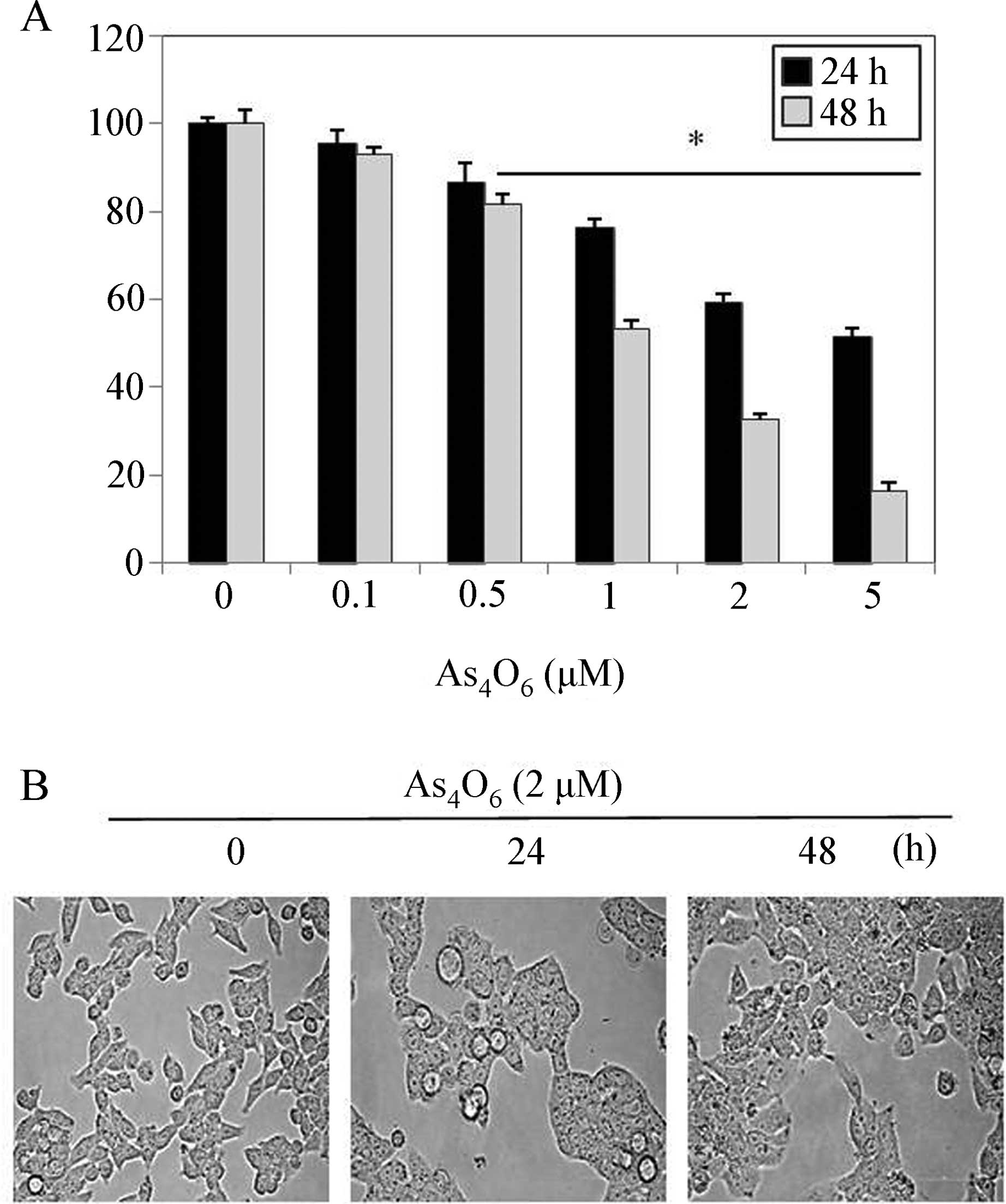
To investigate the antitumor activity of
As4O6 in SW620 cells, the cells were treated
for 24 and 48 h with various concentrations of
As4O6 (0.1–5 μM), and the cell growth
was assessed by MTT assay. The MTT assay revealed that
As4O6 inhibited the growth of SW620 cells in
a dose-dependent manner at 24 and 48 h. As4O6
had a strong inhibitory effect after 48 h of treatment and the half
maximal inhibitory concentration (IC50) was ~1 μM
(Fig. 1A). Next, we assessed the
changes in cellular morphology of the
As4O6-treated cells under microscopy. The
light microscopy results revealed that cell shrinkage and
cytoplasmic blebs were observed after 24 and 48 h of incubation
(Fig. 1B).
As4O6 suppresses
NF-κB activity at least in part through inhibition of IκBα
phosphorylation
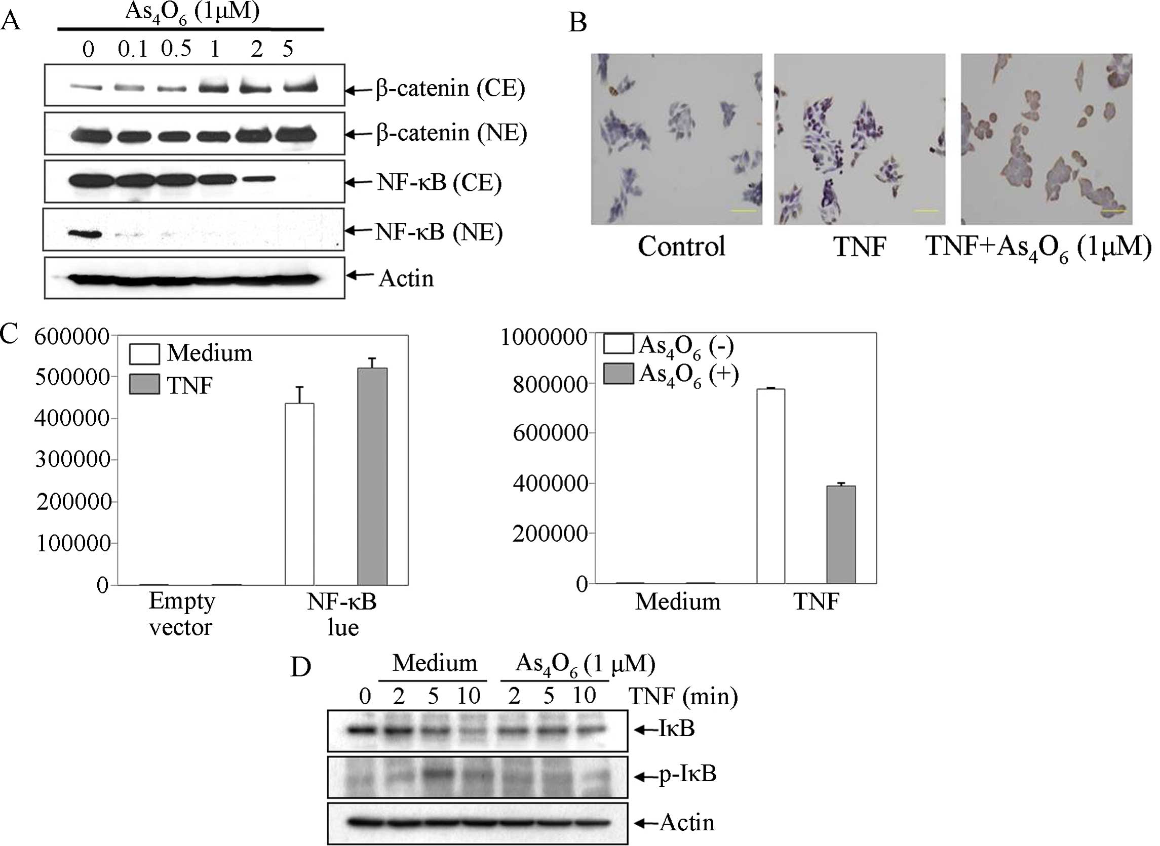
To determine whether As4O6
inhibits NF-κB activity of SW620 cells, we used western
blotting, immunohistocytochemistry and luciferase assay. Under
resting conditions, NF-κB mostly consists of a heterotrimer of p50,
p65 and inhibitory κBα (IκBα) in the cytoplasm; when activated, the
heterodimer of p50 and p65 is translocated into the nucleus after
separating from p-IκBα. Hence, we performed western blot analysis,
which revealed that As4O6 reduced both the
translocation of NF-κB into the nucleus and the levels of NF-κB in
the cytoplasm (Fig. 2A). One
advantage of immunohistochemistry is the ability to confirm NF-κB
(p65) translocation into the nucleus on activation. As expected,
TNF enhanced the NF-κB translocation into the nucleus and
As4O6 inhibited the TNF-induced NF-κB
activation (Fig. 2B). To confirm
the effects of As4O6 on NF-κB activity, we
performed a luciferase assay. As shown in Fig. 2C, the NF-κB gene was successfully
transfected into the cells and the NF-κB-luciferase activity was
augmented by TNF The NF-κB-luciferase activity induced by TNF was
inhibited by As4O6 (Fig. 2C). As mentioned, NF-κB activation is
required for the degradation of IκBα through phosphorylation by
kinases. We also tested whether As4O6
suppressed TNF-induced phosphorylation of IκBα. Western blot
analysis revealed that As4O6 prevented
TNF-induced IκBα phosphorylation (Fig.
2D). This result suggested that As4O6
suppressed NF-κB activity at least in part through inhibition of
IκBα phosphorylation.
As4O6 suppresses
NF-κB-regulated proteins involved in anti-apoptosis, proliferation,
invasion and angiogenesis
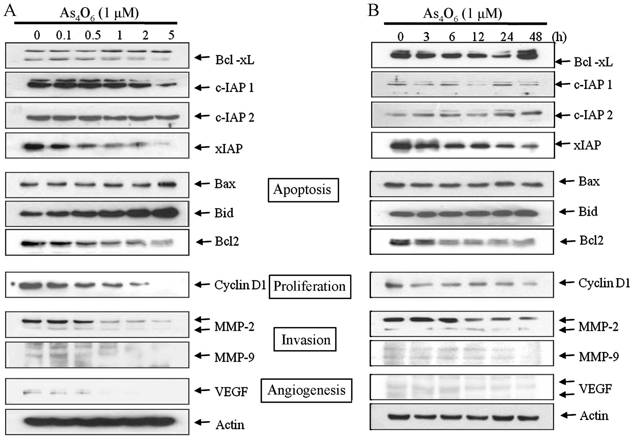
NF-κB activation leads to activation of several
genes involved in anti-apoptosis, proliferation, invasion and
angiogenesis in cancer. NF-κB regulates expression of
anti-apoptotic proteins (c-IAP1/2, XIAP and Bcl-xL) (13), cyclin D1 for cell proliferation
(14), MMP-2, MMP-9 for invasion
and VEGF for angiogenesis of cancer (13,15).
Hence, we investigated the effect of As4O6 on
these molecules. Western blot analysis revealed that
As4O6 suppressed the protein expression of
XIAP, Bcl-2, Bcl-xL, cIAP-1, cyclin D1, MMP-2, MMP-9 and VEGF in a
dose-and time-dependent manner (Fig.
3). These findings revealed that As4O6
suppressed the NF-κB-mediated cellular responses regarding cancer
apoptosis, proliferation, invasion and angiogenesis in the SW620
cells.
As4O6 marginally
suppresses the tumor growth of SW620 cells
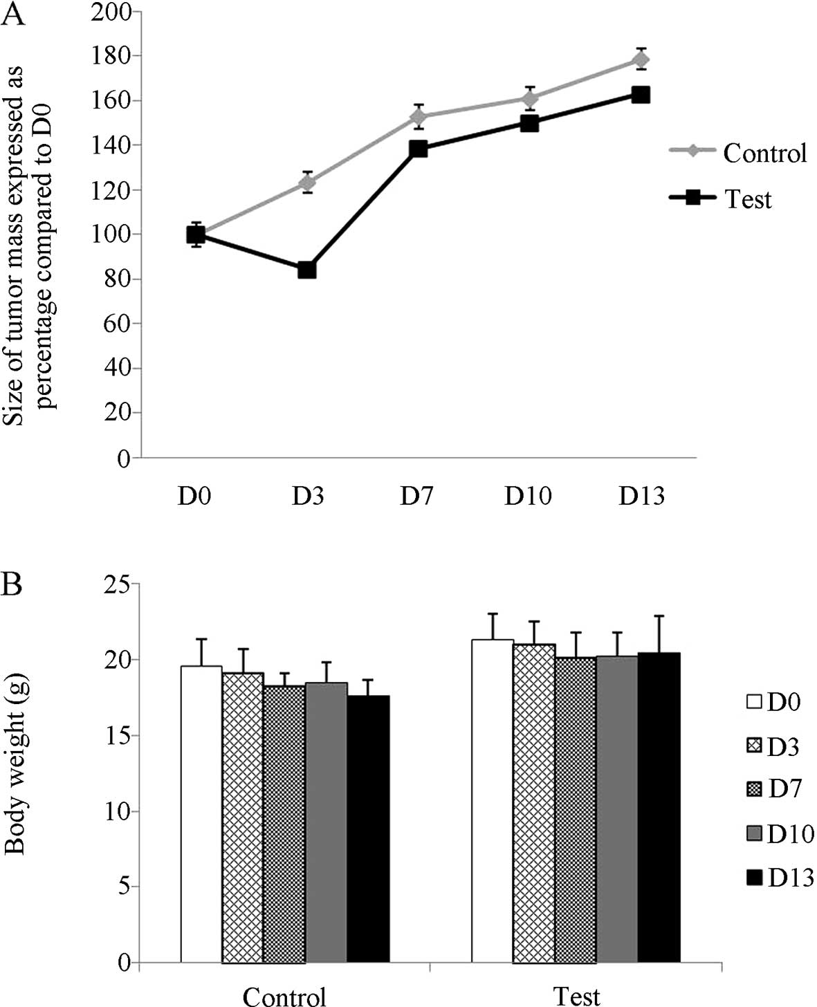
Next, we evaluated the effect of
As4O6 treatment on the growth of SW620 cells
(Fig. 4). Tumor growth was
marginally suppressed by As4O6 treatment
throughout the 12-day treatment regimen, indicating the potent
therapeutic efficacy of As4O6 in SW620 cancer
cells (Fig. 4A). The volume of the
control SW620 xenografts was 798 mm3 and that of the
xenografts treated with As4O6 at 5 mg/kg was
115.9 mm3 (difference, 682.1 mm3; 95% CI,
480.4–883.9 mm3; P<0.001). Also, there were no
significant difference in body weight between the control and
treatment groups (Fig. 4B).
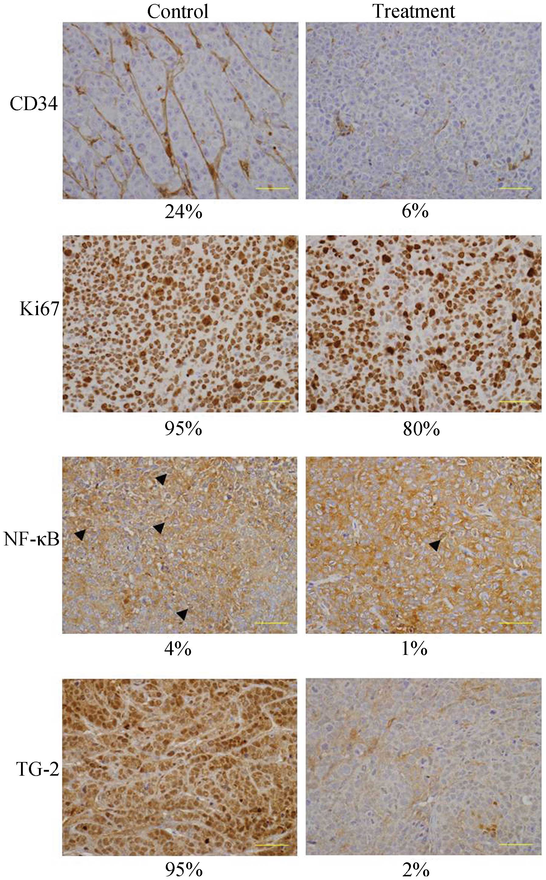
As4O6 suppresses
NF-κB activity and NF-κB-mediated cellular phenotype such as cancer
proliferation and angiogenesis in the in vivo xenograft mouse
model
We further investigated the in vivo effect of
As4O6 treatment on NF-κB activity and
NF-κB-regulated proteins in the SW620 xenograft tumors.
Immunohistochemical studies revealed that the expression of p-NF-κB
in the tumors from the As4O6-treated mice was
lower than that in the control tumors from the untreated mice
(Fig. 5). Here, we also tested TG-2
since TG-2 expression has a good correlation with NF-κB activity
(16), and a difference in p-NF-κB
expression is not easily observed. The result indicated that
As4O6 significantly suppressed TG-2
expression. In addition As4O6 also clearly
suppressed CD34, a protein which is involved in angiogenesis and
Ki-67, a nuclear protein that is associated with cellular
proliferation. These findings were consistent with p-NF-κB
expression and suggest that As4O6 may
suppress NF-κB activity and NF-κB-regulated cellular phenotype.
Discussion
The present study was designed to investigate the
anticancer effects of As4O6 with special
focus on the NF-κB pathway, and NF-κB-regulated gene products, in
in vitro and in vivo models. We found that
As4O6 inhibited the growth of SW620 cells in
a dose-dependent manner at 24 and 48 h. Furthermore,
As4O6 inhibited NF-κB activity and
NF-κB-regulated proteins involved in anti-apoptosis, cell
proliferation, invasion and angiogenesis. Even though this finding
is novel for As4O6, there is previous
supporting evidence showing that arsenic trioxide
(As2O3) suppresses NF-κB-mediated cellular
activities (17). NF-κB is a
well-known transcription factor involved in cancer proliferation,
invasion, metastasis and drug resistance. We found that
As4O6 suppressed MMP-2 and MMP-9 activity.
MMP-2 and MMP-9 are key molecules in cancer cell invasion (18,19)
which have been used as targets for drug development against cancer
invasion (20). We also found that
As4O6 suppressed cyclin D1 which is
associated with cancer cell proliferation (13,14),
and XIAP, Bcl-2, Bcl-xL and cIAP-1 that are involved in cancer cell
survival and drug resistance (13).
In addition, the role of VEGF in the angiogenesis of cancer is well
known (21). All of these gene
products are known to be regulated by NF-κB (13,15).
Here, we used TNF to clearly demonstrate that
As4O6 inhibits NF-κB. Plasma TNF is usually
increased in patients with advanced and metastatic cancers
(22). The pathophysiological
relevance between TNF and NF-κB activation in advanced and
metastatic cancers suggests that the use of TNF is also similar to
the cancer environment in the human body. IκBα is the best-studied
and a major IκB protein of the IκB family. When activated by
signals, the IκB kinase phosphorylates two serine residues located
in an IκBα regulatory domain. When IκBα is phosphorylated at
serines 32 and 36, IκBα is degraded by ubiquitination (23). Here, we found that
As4O6 suppressed phosphorylation of IκBα
induced by TNF. This finding suggests that the anti-NF-κB
activities of As4O6 are contributed to
suppression of IκBα phosphorylation. In addition, we demonstrated
that As4O6 inhibited NF-κB activity in an
in vivo animal model even though the anticancer effects were
marginal. One weak point is that although
As4O6 suppressed the whole expression of
NF-κB (Fig. 2A), we could not
exactly elucidate the mechanisms. We also found that
As4O6 suppressed the whole expression of
NF-κB (data not shown). We need to further investigate this
mechanism.
In conclusion, the present study demonstrated that
As4O6 exerts anticancer effects by
suppressing NF-κB and NF-κB-regulated genes involved in
anti-apoptosis, proliferation, invasion and angiogenesis in cancer
(Fig. 6). The present study
provides evidence that As4O6 may have
anticancer effects on human colon cancer.
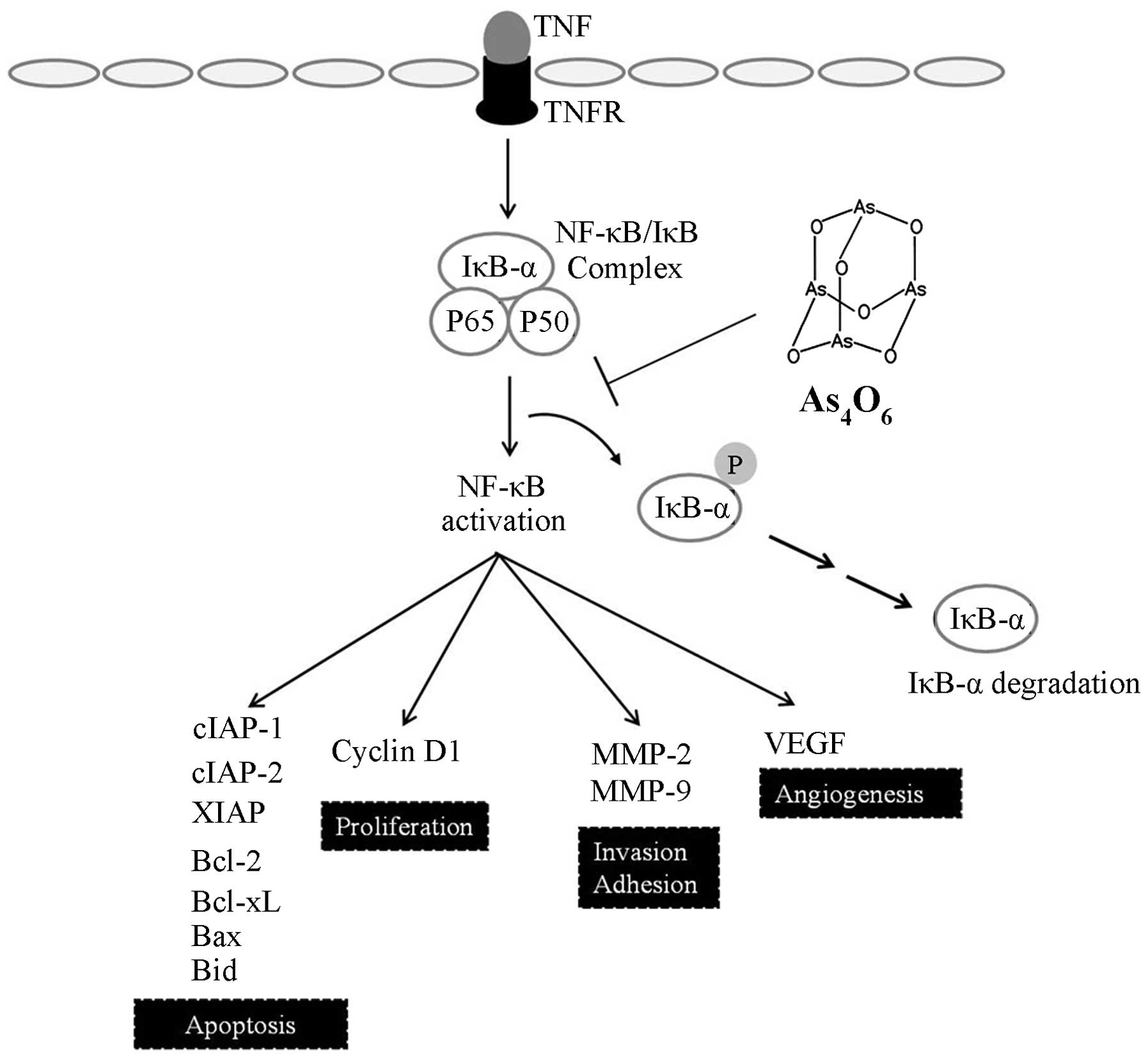 | Figure 6Schematic representation of the
anticancer effects of As4O6 on SW620 human
colon cancer cells. As4O6 suppressed the
invasive effects of SW620 cells by suppression of NF-κB through
inhibition of IκB phosphorylation stimulated by TNF. In addition,
TNF participated in induction of NF-κB-regulated proteins involved
in cancer cell proliferation (cyclin D1), anti-apoptosis (XIAP,
IAP1, IAP2, Bcl-xL, Bcl-2, Bax and Bid), and invasion and
angiogenesis (MMP-2, MMP-9 and VEGF). Taken together, the present
study suggests that As4O6 has anticancer
properties through suppression of NF-κB activity and NF-κB-mediated
cellular responses. As4O6, tetraarsenic
hexoxide; NF-κB, nuclear factor-κB. |
Acknowledgments
This study was supported by a grant from the
National Research Foundation of Korea (NRF) funded by the Korea
government (MEST) (no. 2014-012154).
References
|
1
|
El-Serag HB and Mason AC: Rising incidence
of hepatocellular carcinoma in the United States. N Engl J Med.
340:745–750. 1999. View Article : Google Scholar : PubMed/NCBI
|
|
2
|
Shen ZX, Chen GQ, Ni JH, Li XS, Xiong SM,
Qiu QY, Zhu J, Tang W, Sun GL, Yang KQ, et al: use of arsenic
trioxide (As2O3) in the treatment of acute
promyelocytic leukemia (APL). II Clinical efficacy and
pharmacokinetics in relapsed patients. Blood. 89:3354–3360.
1997.PubMed/NCBI
|
|
3
|
Niu C, Yan H, Yu T, Sun HP, Liu JX, Li XS,
Wu W, Zhang FQ, Chen Y, Zhou L, et al: Studies on treatment of
acute promyelocytic leukemia with arsenic trioxide: Remission
induction, follow-up, and molecular monitoring in 11 newly
diagnosed and 47 relapsed acute promyelocytic leukemia patients.
Blood. 94:3315–3324. 1999.PubMed/NCBI
|
|
4
|
Munshi NC, Tricot G, Desikan R, Badros A,
Zangari M, Toor A, Morris C, Anaissie E and Barlogie B: Clinical
activity of arsenic trioxide for the treatment of multiple myeloma.
Leukemia. 16:1835–1837. 2002. View Article : Google Scholar : PubMed/NCBI
|
|
5
|
Lin YC, Li DR and Lin W: Relationship
between radiotherapy enhancing effect of arsenic trioxide and the
proliferation and apoptosis of related protein in nasopharyngeal
carcinoma patients. Zhongguo Zhong Xi Yi Jie He Za Zhi. 27:704–707.
2007.In Chinese. PubMed/NCBI
|
|
6
|
Welch JS, Klco JM, Gao F, Procknow E, Uy
GL, Stockerl-Goldstein KE, Abboud CN, Westervelt P, DiPersio JF,
Hassan A, et al: Combination decitabine, arsenic trioxide, and
ascorbic acid for the treatment of myelodysplastic syndrome and
acute myeloid leukemia: A phase I study. Am J Hematol. 86:796–800.
2011. View Article : Google Scholar : PubMed/NCBI
|
|
7
|
Beer TM, Tangen CM, Nichols CR, Margolin
KA, Dreicer R, Stephenson WT, Quinn DI, Raghavan D and Crawford ED:
Southwest Oncology Group phase II study of arsenic trioxide in
patients with refractory germ cell malignancies. Cancer.
106:2624–2629. 2006. View Article : Google Scholar : PubMed/NCBI
|
|
8
|
Chang HS, Bae SM, Kim YW, Kwak SY, Min HJ,
Bae IJ, Lee YJ, Shin JC, Kim CK and Ahn WS: Comparison of diarsenic
oxide and tetraarsenic oxide on anticancer effects: Relation to the
apoptosis molecular pathway. Int J Oncol. 30:1129–1135.
2007.PubMed/NCBI
|
|
9
|
Han MH, Lee WS, Lu JN, Yun JW, Kim G, Jung
JM, Kim GY, Lee SJ, Kim WJ and Choi YH: Tetraarsenic hexoxide
induces Beclin-1-induced autophagic cell death as well as
caspase-dependent apoptosis in u937 human leukemic cells. Evid
Based Complement Alternat Med. 2012:2014142012. View Article : Google Scholar
|
|
10
|
Guttridge DC, Albanese C, Reuther JY,
Pestell RG and Baldwin AS Jr: NF-kappaB controls cell growth and
differentiation through transcriptional regulation of cyclin D1.
Mol Cell Biol. 19:5785–5799. 1999.PubMed/NCBI
|
|
11
|
Lee HY, Moon H, Chun KH, Chang YS, Hassan
K, Ji L, Lotan R, Khuri FR and Hong WK: Effects of insulin-like
growth factor binding protein-3 and farnesyltransferase inhibitor
SCH66336 on Akt expression and apoptosis in non-small-cell lung
cancer cells. J Natl Cancer Inst. 96:1536–1548. 2004. View Article : Google Scholar : PubMed/NCBI
|
|
12
|
Jang JS, Lee WS, Lee JS, Kim HW, Ko GH and
Ha WS: The expression of thymidine phosphorylase in
cancer-infiltrating inflammatory cells in stomach cancer. J Korean
Med Sci. 22(Suppl 22): S109–S114. 2007. View Article : Google Scholar : PubMed/NCBI
|
|
13
|
Aggarwal BB: Nuclear factor-kappaB: The
enemy within. Cancer Cell. 6:203–208. 2004. View Article : Google Scholar : PubMed/NCBI
|
|
14
|
Motokura T and Arnold A: PRAD1/cyclin D1
proto-oncogene: Genomic organization, 5′ DNA sequence, and sequence
of a tumor-specific rearrangement breakpoint. Genes Chromosomes
Cancer. 7:89–95. 1993. View Article : Google Scholar : PubMed/NCBI
|
|
15
|
Gilmore TD: Introduction to NF-kappaB:
Players, pathways, perspectives. Oncogene. 25:6680–6684. 2006.
View Article : Google Scholar : PubMed/NCBI
|
|
16
|
Kim DS, Park SS, Nam BH, Kim IH and Kim
SY: Reversal of drug resistance in breast cancer cells by
transglutaminase 2 inhibition and nuclear factor-kappaB
inactivation. Cancer Res. 66:10936–10943. 2006. View Article : Google Scholar : PubMed/NCBI
|
|
17
|
Kerbauy DM, Lesnikov V, Abbasi N, Seal S,
Scott B and Deeg HJ: NF-kappaB and FLIP in arsenic trioxide
(ATO)-induced apoptosis in myelodysplastic syndromes (MDSs). Blood.
106:3917–3925. 2005. View Article : Google Scholar : PubMed/NCBI
|
|
18
|
Davies B, Waxman J, Wasan H, Abel P,
Williams G, Krausz T, Neal D, Thomas D, Hanby A and Balkwill F:
Levels of matrix metalloproteases in bladder cancer correlate with
tumor grade and invasion. Cancer Res. 53:5365–5369. 1993.PubMed/NCBI
|
|
19
|
Bogenrieder T and Herlyn M: Axis of evil:
Molecular mechanisms of cancer metastasis. Oncogene. 22:6524–6536.
2003. View Article : Google Scholar : PubMed/NCBI
|
|
20
|
Vihinen P and Kähäri VM: Matrix
metalloproteinases in cancer: Prognostic markers and therapeutic
targets. Int J Cancer. 99:157–166. 2002. View Article : Google Scholar : PubMed/NCBI
|
|
21
|
Nishida N, Yano H, Nishida T, Kamura T and
Kojiro M: Angiogenesis in cancer. Vasc Health Risk Manag.
2:213–219. 2006. View Article : Google Scholar
|
|
22
|
Correia M, Cravo M, Marques-Vidal P,
Grimble R, Dias-Pereira A, Faias S and Nobre-Leitão C: Serum
concentrations of TNF-alpha as a surrogate marker for malnutrition
and worse quality of life in patients with gastric cancer. Clin
Nutr. 26:728–735. 2007. View Article : Google Scholar : PubMed/NCBI
|
|
23
|
Chen ZJ, Parent L and Maniatis T:
Site-specific phosphorylation of IkappaBalpha by a novel
ubiquitination-dependent protein kinase activity. Cell. 84:853–862.
1996. View Article : Google Scholar : PubMed/NCBI
|