Introduction
Signal transducer and activator of transcription
(STAT) proteins have garnered more attention recently as latent
cytoplasmic transcription factors. Six STAT family members have
been identified thus far: STAT 1, STAT 2, STAT 3, STAT 4, STAT 5
(including STAT 5a and STAT 5b), and STAT 6. They are believed to
regulate many cellular process including cell proliferation,
differentiation, apoptosis and survival (1). STAT 1, STAT 3 and STAT 5 (including
STAT 5a and STAT 5b) were reported to play important roles in the
malignant biological behavior of tumors (2–4).
Regarding STAT 5a and STAT 5b, evidence has identified some of
their similarities and characteristics, including their gene
encoding, distinct regulatory features and functions. Based on
recent studies, despite some overlapping functions, STAT 5a and
STAT 5b generally play different physiological functions (5). However, the roles of STAT 5a and STAT
5b in malignant tumors, especially in gastric cancer, which is one
of the major causes of cancer-associated mortalities worldwide and
has become one of the great social harms to human health due to its
poor prognosis (6,7), remain largely unknown.
Gefitinib, also known as Iressa, is commonly used to
treat locally advanced or metastatic non-small-cell lung cancer
(NSCLC). Use of gefitinib has been widely promoted for its
anticancer effects (8). Gefitinib
is a selective inhibitor of the EGFR tyrosine kinase, which is
usually expressed in the solid tumors of epithelial cells.
Inhibition of the EGFR tyrosine kinase can disrupt tumor growth,
metastasis, and angiogenesis and can promote apoptosis of cancer
cells (9). Thus, gefitinib is
effective in treating certain types of epithelial-derived solid
tumors rich in tyrosine kinase, including gastric cancers. However,
the clinical significance and molecular mechanism of gefitinib in
gastric cancers remain largely unknown.
In this study, we aimed to investigate the
expression of STAT 5b in gastric cancer tissues and to analyze the
role and possible mechanism of STAT 5b in the chemo-sensitivity of
gastric cancers to gefitinib. The results showed the increased
expression of STAT 5b in gastric carcinoma clinical samples but not
in para-carcinoma samples. The expression of STAT 5b was
downregulated significantly by treatment with gefitinib (4 mM, 24
h). Interference of STAT 5b expression by siRNA targeting enhanced
the chemo-sensitivity of gastric cancer cells to gefitinib, the
mechanism of which involved promoting mitochondrial
pathway-mediated apoptosis. The results provide important
theoretical guidance for the clinical prevention and treatment of
gastric cancers.
Materials and methods
Patients and specimens
A total of 69 patients with gastric carcinomas (48
male and 21 female cases) obtained from Shandong Provincial
Qianfoshan Hospital Affiliated with Shandong University during the
period March, 2013 to December, 2013 were studied. All the cases in
this study were treated with primary carcinoma and para-carcinoma
resection. Written informed consent was obtained from all the
patients prior to surgery. The study was approved by the Ethics
Committees of Shandong University. Resected tissues were fixed by
immediate immersion in formalin for immunohistochemistry (IHC) or
were rapidly frozen by immediate immersion in liquid nitrogen, and
stored at −80°C for protein extraction. The mean age of the
patients was 65.8 years (range, 46–83 years). Tissue specimens were
embedded in paraffin for the experiments by the same investigator.
Clinicopathological characteristics, including gender, age, T
stage, N stage, clinical stage and pathological grading were
studied. T stage, N stage, clinical stage and pathological grading
were defined based on the TNM Staging Classification for Carcinoma
of the Stomach (7th edition, 2010) from the American Joint
Committee on Cancer (AJCC).
Immunohistochemistry
Paraffin-embedded gastric carcinoma sections (4 µm)
were dewaxed and subjected to antigen retrieval by water bathing in
0.01 M citric buffer (pH 6.0) at 95–98°C for 15 min. To quench
endogenous peroxidase, the sections were incubated in 3% hydrogen
peroxide solution (H2O2) for 30 min.
Non-specific binding was prevented by incubating the samples in 10%
normal goat serum for 30 min in a humid chamber. The slides were
incubated overnight at 4°C in antibodies for STAT 5b (Epitomics,
Burlingame, CA, USA; rabbit polyclonal antibody, 1:300). For the
negative control, an equal volume of PBS was used instead of the
primary antibody solution. After washing, the primary antibodies
were detected with appropriate secondary antibodies for 30 min at
37°C. A solution of 3,3′-diamino-benzidine tetrahydrochloride (DAB)
was used to visualize positive staining, whereas hematoxylin was
used to counterstain the nucleoli.
Interpretation of immunohistochemical
staining
The expression of STAT 5b as visualized by
immunohistochemical staining was evaluated in five random areas of
the slide sections at a magnification of ×100. Validation was
performed blindly and without knowledge of the eventual clinical
parameters. When differences between inter-observers occurred, the
slides in question were jointly re-examined by two
investigators.
Cell culture
MGC-803 and MKN-45 cells were cultured in Dulbecco’s
modified Eagle’s medium (DMEM) containing 10% fetal calf serum, 100
U/ml penicillin, and 100 mg streptomycin at 37°C in a humidified
atmosphere composed of 95% air and 5% CO2. Passage
digestion was conducted using a 0.25% trypsin-0.02% EDTA digestive
solution.
Cell treatment and assessment of cell
viability
To determine the optimal gefitinib co-culture
conditions, cells (5×10⁴/ml) were subcultured in a
96-well cell culture cluster (Corning, N Y, USA) and were treated
with different concentrations of gefitinib (0, 1, 2, 4 and 8 mM)
for 24, 48 and 72 h. MTT (5 mg/ml, 20 µl) was added to each
well 4 h before each of the indicated time-points. After 4 h of
incubation at 37°C, the cells were resuspended in dimethylsulfoxide
(DMSO). The effects of gefitinib on the two cell lines were
measured using the Cell Counting Kit-8 (CCK-8). Optical density
(OD) values were measured at 570 nm using an ELISA reader
(Multiskan MK3, Shanghai Bio-excellent, Shanghai, China). The
relative cell viability was calculated according to the formula:
Relative cell viability (%) = OD experiment/OD control × 100% (OD
blank was tared to zero).
Interference of STAT 5b expression
STAT 5b siRNA (10 µM) was purchased from
Santa Cruz Biotechnology, Inc. (Santa Cruz, CA, USA), and
interference of STAT 5b expression was conducted according to the
manufacturer’s instructions. Briefly, 1, 2, or 3 µl of 10
µM STAT 5b siRNA was added to each well of confluent cells.
After a 6-h incubation, the transfection complexes were discarded,
and the cells were cultured in growth medium for 48 h. Protein
extracts were used to detect STAT 5b expression levels in the
different STAT 5b siRNA (10 µM) dosages.
Flow cytometry
Cells incubated with or without gefitinib (4 mM, 24
h) or with the gefitinib+siRNA combination were trypsinized. After
washing, the cells were resuspended in 200 µl binding buffer
containing Annexin V FITC (5 µl) and PI (10 µl) for
15 min at room temperature. Then, 300 µl binding buffer was
added before analysis with a FACScan flow cytometer
(Becton-Dickinson, Mountain View, CA, USA).
Protein extraction and western blot
analysis
Total protein was extracted from primary tissues or
cultured cells using radioimmune precipitation (RIPA) protein lysis
buffer according to appropriate protocols. The Bradford method was
used to determine the protein concentration of the supernatant. The
samples (40 µg of total protein each) were used for western
blot analysis with primary antibodies (STAT 5b, 1:1,000; β-actin,
1:2,500). The STAT 5b and β-actin bands were visualized at apparent
molecular weights of 90 and 43 kDa, respectively.
Statistical analysis
Statistical analyses were performed using SPSS
statistical software (version 13.0; SPSS Inc., Chicago, IL, USA).
To test the associations between STAT 5b expression and various
clinicopathological characteristics, including gender, age, T
stage, N stage, clinical stage and pathological grading stages, we
used a non-parametric test for the comparisons of differences
between groups. A χ2 test was applied for trend
variables. P<0.05 was considered to indicate statistical
significance.
Results
Increased expression of STAT 5b is
detected in clinical gastric carcinomas but not in
para-carcinomas
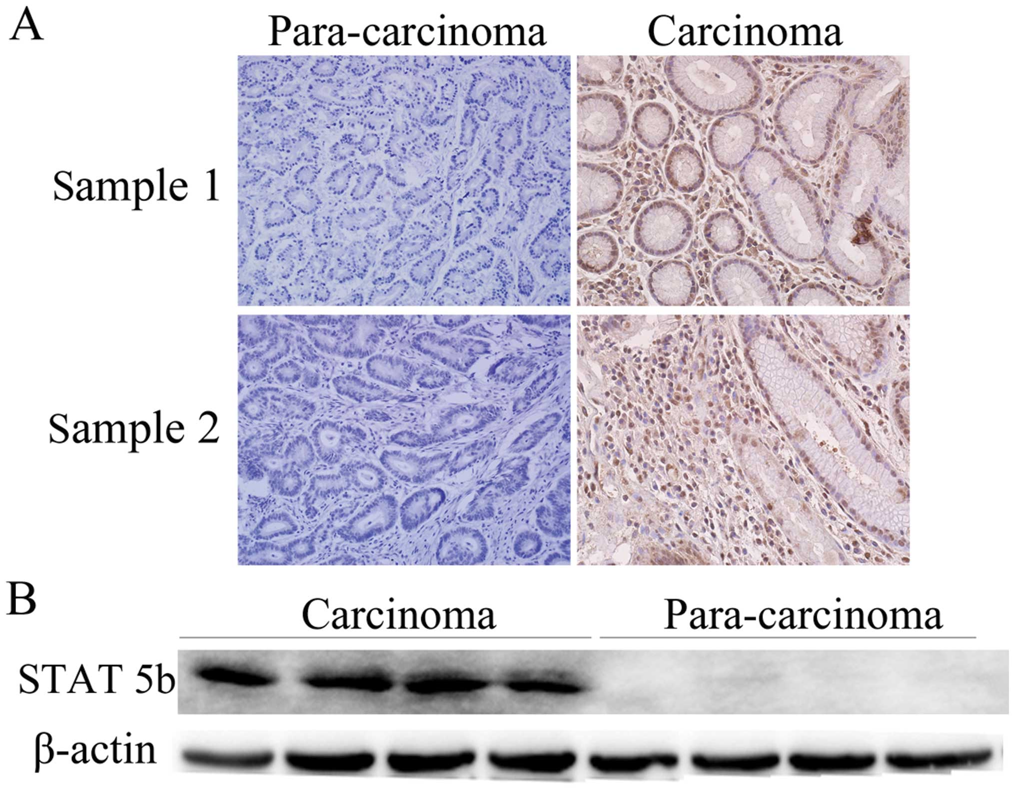
Sixty-nine gastric carcinoma and corresponding
para-carcinoma clinical samples were collected in this study. IHC
was conducted to evaluate STAT 5b expression between carcinomas and
para-carcinomas. Fig. 1A shows
images from two randomly selected samples, for which STAT 5b
expression was significantly increased in gastric carcinomas
compared with para-carcinomas. As shown in Table I, the statistical analysis revealed
that this difference in STAT 5b expression was significant, with
49/69 carcinomas and 27/69 para-carcinomas positively staining for
STAT 5b (P = 0.001).
 | Table IProtein expression of STAT 5b in
gastric carcinomas as assessed by immunohistochemical staining. |
Table I
Protein expression of STAT 5b in
gastric carcinomas as assessed by immunohistochemical staining.
| Variables | Negative | Positive | Total | P-value |
|---|
| Carcinoma | 20 | 49 | 69 | 0.001 |
| Para-carcinoma | 42 | 27 | 69 | |
Western blot analysis was also conducted to evaluate
STAT 5b expression in the protein extracts of gastric carcinomas or
para-carcinomas. As shown in Fig.
1B, STAT 5b was also found to be significantly upregulated in
carcinomas compared with para-carcinomas when β-actin was used as a
loading control.
Associations between STAT 5b expression and
clinicopathological parameters, including gender, age, T stage, N
stage, clinical stage and pathological grading, in gastric
carcinomas are shown in Table II.
The results showed no significant association between STAT 5b
expression and gender (P=0.774) or age (P=1.001). Significant
associations were found between STAT 5b expression and T stage
(P=0.037), N stage (P=0.014), clinical stage (P=0.014) and
pathological grading (P=0.005).
| Table IIAssociations between STAT 5b
expression and clinicopathological parameters in gastric
carcinomas. |
Table II
Associations between STAT 5b
expression and clinicopathological parameters in gastric
carcinomas.
| Characteristics | No. | STAT 5b
| P-value |
|---|
| Negative | Positive (%) |
|---|
| Gender | | | | |
| Male | 48 | 13 | 35 (72.9) | 0.774 |
| Female | 21 | 7 | 14 (66.7) | |
| Age | | | | |
| <60 | 21 | 6 | 15 (71.4) | 1.001 |
| ≥60 | 48 | 14 | 34 (70.8) | |
| T stage | | | | |
| T1 | 18 | 8 | 10 (55.6) | 0.037 |
| T2 | 14 | 6 | 8 (57.1) | |
| T3 | 25 | 6 | 19 (76.0) | |
| T4 | 12 | 0 | 12 (100) | |
| N stage | | | | |
| N0 | 24 | 11 | 13 (54.2) | 0.014 |
| N1 | 10 | 5 | 5 (50.0) | |
| N2 | 18 | 2 | 16 (88.9) | |
| N3 | 17 | 2 | 15 (88.2) | |
| Clinical stage | | | | |
| 1 | 12 | 6 | 6 (50.0) | 0.014 |
| 2 | 22 | 10 | 12 (54.5) | |
| 3 | 34 | 4 | 30 (88.2) | |
| 4 | 1 | 0 | 1 (100) | |
| Pathological
grading | | | | |
| 1 | 11 | 8 | 3 (27.3) | 0.005 |
| 2 | 22 | 3 | 19 (86.4) | |
| 3 | 29 | 7 | 22 (75.9) | |
| 4 | 7 | 2 | 5 (71.4) | |
Gefitinib reduces the relative viability
of gastric cancer cells in a concentration- and time-dependent
manner
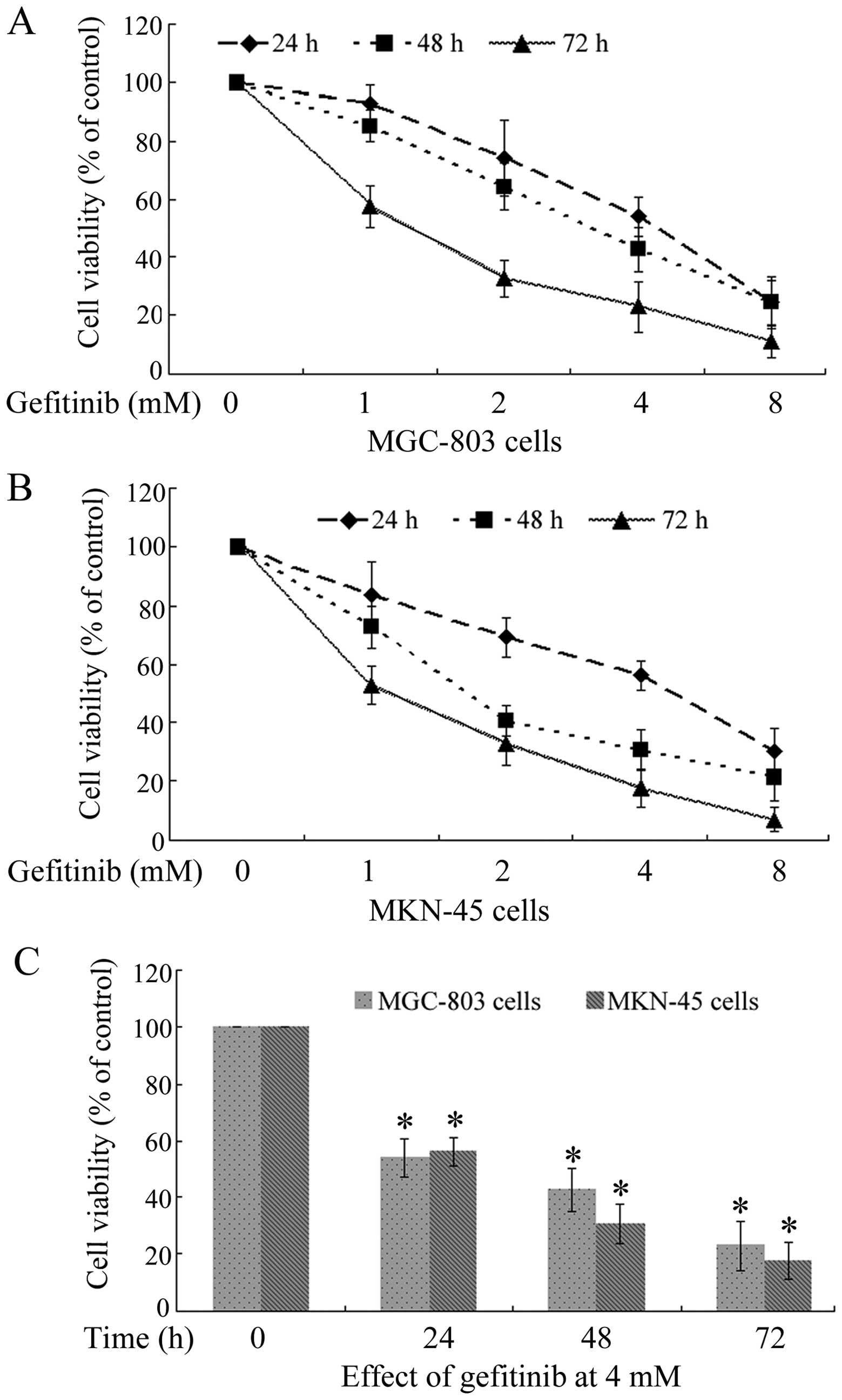
The effects of gefitinib on cultured MGC-803 and
MKN-45 gastric cancer cells, were measured using the CCK-8 kit.
Gefitinib, a common anticancer chemotherapeutic, was shown to
reduce the relative viability of gastric cancer cells. As shown in
Fig. 2A and B, cultured MGC-803 and
MKN-45 cells were exposed to different concentrations of gefitinib
(0, 1, 2, 4 and 8 mM) for 24, 48 and 72 h to assay their relative
cell viabilities using the CCK-8 kit. Taking the cell viability of
the control sample (without gefitinib) as 100%, the cell
viabilities of MGC-803 and MKN-45 cells were reduced in a
concentration- and time-dependent manner. The cell viabilities of
MGC-803 and MKN-45 cells treated with gefitinib (4 mM, 24 h) were
(54.01±6.72)% and (56.13±5.02)%, respectively.
The effects of 4 mM gefitinib on MGC-803 and MKN-45
cells were also studied in cells incubated with gefitinib for 0,
24, 48 and 72 h. The cell viabilities markedly decreased after 24 h
(P<0.05) (Fig. 2C).
Cell apoptosis of MGC-803 and MKN-45
cells treated with gefitinib (4 mM, 24 h) increases
significantly
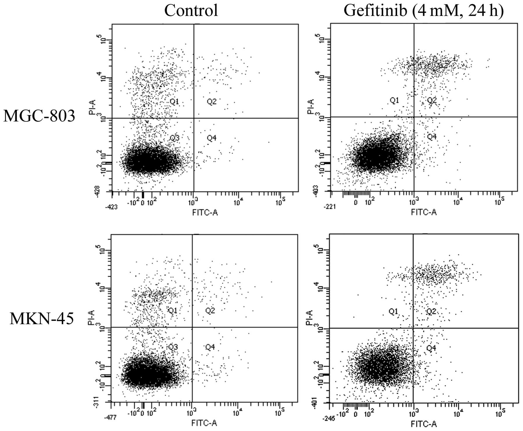
Cell apoptosis of MGC-803 and MKN-45 cells treated
with gefitinib (4 mM, 24 h) and assayed using flow cytometry (FCM).
The apoptotic rates of normal MGC-803 cells and normal MKN-45 cells
were (1.53±0.31)% and (2.2±0.53)%, respectively. Cell apoptotic
rates increased significantly in MGC-803 and MKN-45 cells treated
with gefitinib, with rates of (9.33±1.45)% in MGC-803 cells and
(10.17±1.66)% in MKN-45 cells (Fig.
3).
STAT 5b expression is downregulated
significantly in MGC-803 and MKN-45 cells treated with gefitinib (4
mM, 24 h)
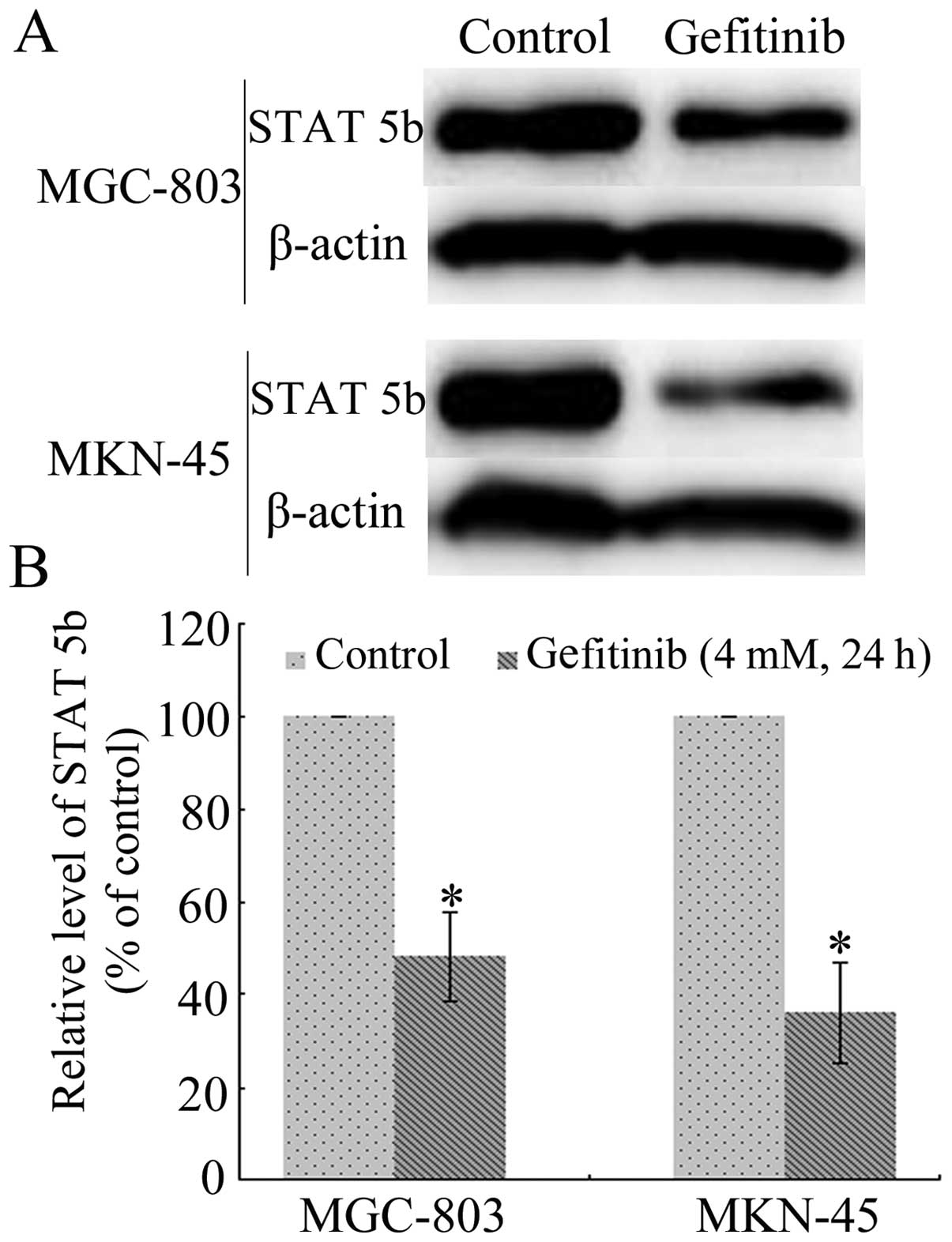
MGC-803 and MKN-45 cells were exposed to gefitinib
(4 mM) for 24 h. Protein extracts were collected, and the
expression of STAT 5b was assayed by western blot analysis
(Fig. 4A). Taking the expression in
the control cells as 100%, the relative expression levels of STAT
5b in MGC-803 and MKN-45 cells treated with gefitinib (4 mM, 24 h)
were (48.2±9.68)% and (35.78±10.85)%, respectively, as assessed by
a quantitative analysis using ImageJ software (Fig. 4B). The differences in expression
were statistically significant (P<0.05).
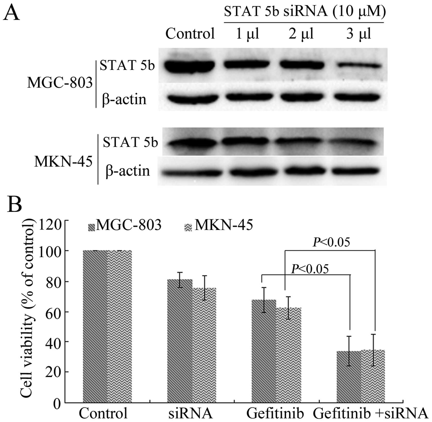
Interference of STAT 5b expression by
siRNA targeting enhanced the chemo-sensitivity of gastric cancer
cells to gefitinib
To assess the role of STAT 5b in the
chemo-sensitivity of gastric cancer cells to gefitinib, we
interfered with the expression of STAT 5b by siRNA (10 µM)
targeting. Optimal conditions for interference were assessed by
incubating MGC-803 and MKN-45 cells with 1, 2 and 3 µl STAT
5b siRNA. The results indicated that incubation with 3 µl of
10 µM STAT 5b siRNA significantly interfered with STAT 5b
expression (Fig. 5A).
The chemo-sensitivity of gastric cancer cells to
gefitinib (4 mM, 24 h) with or without STAT 5b siRNA (10 µM,
3 µl) was detected using the CCK-8 kit. Fig. 5B showed that the cell viabilities
significantly decreased in the combined treatment group
(gefitinib+siRNA) compared with the gefitinib (4 mM, 24 h) only
group in MGC-803 and MKN-45 cells (P<0.05).
Interference of STAT 5b expression by
siRNA targeting enhances gefitinib-induced apoptosis in gastric
cancer cells
As shown in Fig. 6,
the cell apoptosis of MGC-803 and MKN-45 cells in the combined
treatment group (gefitinib+siRNA) increased significantly compared
with the gefitinib (4 mM, 24 h) only group, as determined by flow
cytometry.
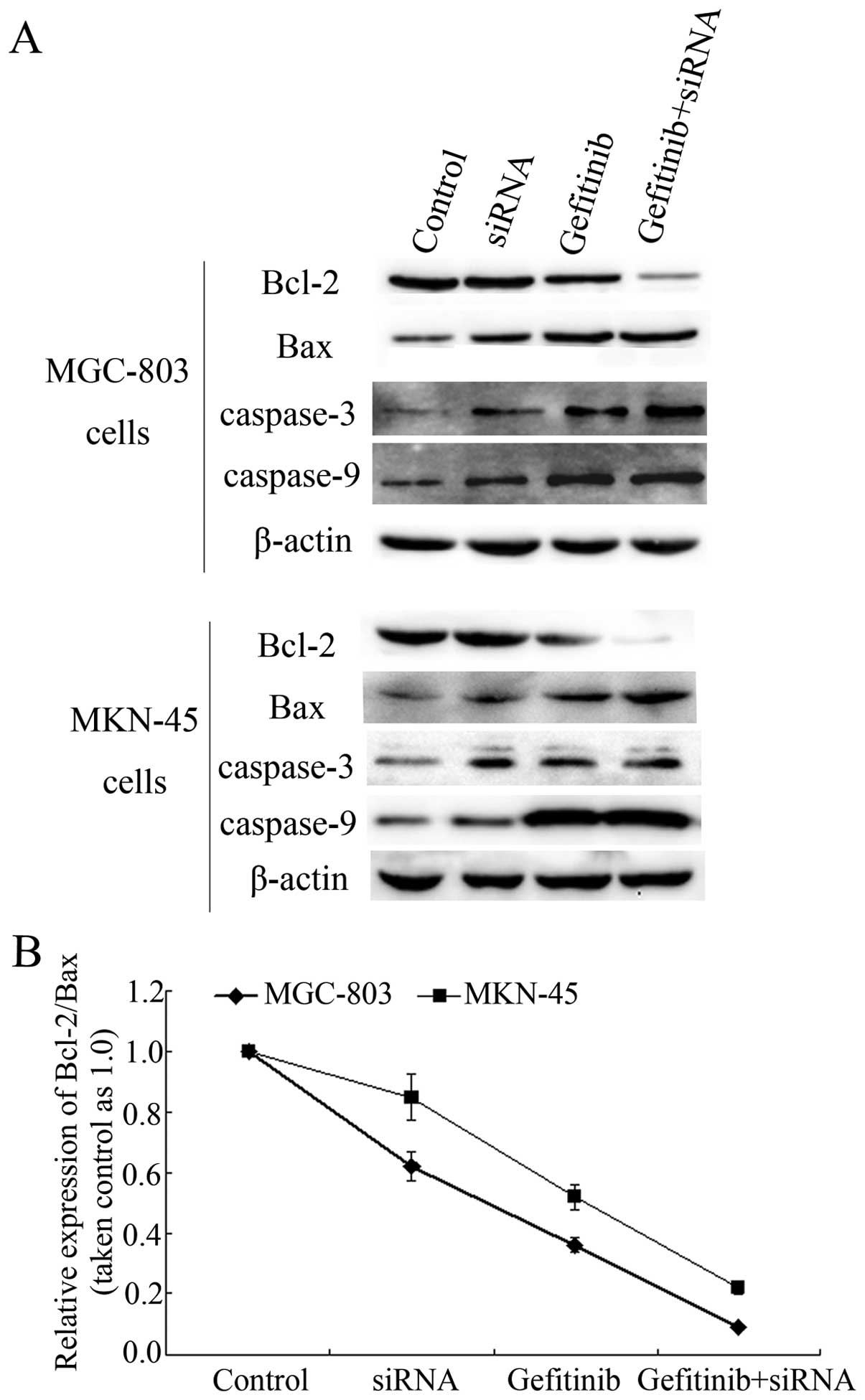
Interference of STAT 5b expression
influences the expression of mitochondrial pathway-related
apoptosis proteins in MGC-803 and MKN-45 cells
The mitochondrial pathway is a major apoptosis
mechanism. Mitochondrial proteins were extracted using a special
kit for extracting mitochondrial proteins. Mitochondrial
pathway-related apoptosis proteins, including Bcl-2, Bax, caspase-3
and caspase-9, were assayed. The results showed that the
interference of STAT 5b expression by siRNA (10 µM, 3
µl) targeting upregulated the expression of Bax, caspase-3
and caspase-9 and downregulated the expression of Bcl-2. The
expression of these proteins was enhanced significantly in the
combined treatment group (gefitinib+siRNA) compared with the
gefitinib (4 mM, 24 h) only group in the MGC-803 and MKN-45 cells
(Fig. 7A).
The relative expression of Bcl-2 and Bax is usually
used to assess sensitivity to apoptosis. In the present study, we
analyzed Bcl-2/Bax levels under gefitinib with or without STAT 5b
siRNA. The results showed that interference of STAT 5b expression
significantly decreased the Bcl-2/Bax expression ratio (P<0.05)
(Fig. 7B).
Discussion
In this study, we found evidence that a molecular
mechanism links STAT 5b expression to gastric cancers. We found an
increased expression of STAT 5b in both gastric carcinomas and
MGC-803 and MKN-45 cells treated with gefitinib. Additionally, we
found that interference of STAT 5b expression by siRNA targeting
enhances the chemo-sensitivity of gastric cancer cells to gefitinib
by promoting mitochondrial pathway-mediated cell apoptosis,
indicating a potential role and mechanism for STAT 5b in the
chemo-sensitivity of gastric cancers.
As a main member of the STAT family, STAT 5 has been
reported to play an important role in many biological processes.
For example, STAT 5 plays essential roles in body growth, lipid
metabolism, and the cell cycle pertaining to hepatosteatosis,
fibrosis, and hepatocellular carcinoma through its action in the
GH-STAT 5 axis. STAT 5 has been reported to mediate GH signals, and
GH-STAT 5 signaling has been found to be important in hepatic
physiology and pathophysiology (10). Additionally, STAT 5, including STAT
5a and STAT 5b, has been shown to be important in some types of
solid tumors (11–13). In preclinical prostate cancer (PCa)
models, STAT 5a/b promotes PCa growth and progression. STAT 5a/b is
critical for PCa cell viability in vitro and for tumor
growth in vivo and promotes the metastatic dissemination of
cancer in nude mice (14). We
reported the upegulation of STAT 5b in gastric cancers compared
with the para-carcinomas and found a significant association
between STAT 5b expression and the clinicopathological parameters,
including T stage, N stage, clinical stage and pathological
grading. These results indicate the critical importance of STAT 5b
in gastric cancers. To the best of our knowledge, this study is the
first to describe a specific role of STAT 5b in gastric cancers.
However, findings of other studies on the function of STAT 5 are
inconsistent with our results. According to studies based on 100
patients who underwent gastrectomy due to gastric adenocarcinoma,
there was no statistically significant association between STAT 5
expression and TNM staging and survival, as assessed by IHC
(15). Those authors did not refer
specifically to STAT 5a or STAT 5b but to a general STAT 5, which
may account for this inconsistency. Mounting evidence indicates
that the role of STAT 5a or STAT 5b is different in various tumors.
Studies in breast cancer reported that loss of STAT 5a, but not
STAT 5b, represents a new independent marker of poor prognosis in
node-negative breast cancer and may be a predictor of response to
antiestrogen therapy, if validated in randomized clinical trials
(16). Of note is that STAT 5a was
also detected in our gastric carcinomas and para-carcinomas, and an
increased expression was found in gastric carcinomas. However, no
significant association was observed between STAT 5a expression and
the above clinicopathological parameters (data not shown). This
result indicates the different effects of STAT 5a and STAT 5b in
gastric cancers. Results of the present study have shown that, STAT
5b, but not STAT 5a, plays an important role in the progression of
gastric cancer.
Apoptosis, known as programmed cell death, is a
programmed mechanism of cell death that ensures normal development.
STAT 5 is believed to be critical in the process of cell apoptosis.
Repression of STAT 5a and STAT 5b expression in the chronic
myelogenous leukemia (CML) cell lines, K-562, with unmodified or
chemically modified siRNAs has been shown to induce apoptosis and
is believed to constitute a potential new and alternative curative
method for supporting therapy of the CML-diagnosed patients
(17). Avoidance of apoptosis is an
important contributor to the survival of tumor cells, and an
important aim in cancer treatment is targeted towards tumor cell
apoptosis. In this study, the gastric cancer cell lines MGC-803 and
MKN-45 were exposed to gefinitib, a selective inhibitor of the EGFR
tyrosine kinase. Cell apoptosis was induced and expression of STAT
5b was upregulated in gefinitib-treated cells, indicating the
possible importance of STAT 5b in the chemo-sensitivity of gastric
cancers by promoting apoptosis. Previous studies have reported the
effect of STAT 5b in the apoptotic-sensitivity of many solid
tumors, such as colorectal cancer (18), breast cancer (19), and lymphoid and non-lymphoid
malignancies (20). These studies
on other cancer types are consistent with our novel finding of an
effect of STAT 5b on the apoptotic-sensitivity of gastric
cancers.
Two apoptotic pathways have been described in
mammalian cells, the extrinsic pathway, also known as the death
receptor pathway, and the intrinsic pathway, known as the
mitochondrial pathway (21,22). In the extrinsic pathway, an
extracellular signal such as Fas ligand, a member of the tumor
necrosis factor (TNF) family, is targeted to cell surface
receptors. In the intrinsic pathway, intracellular signals such as
DNA damage and mitochondrial injury are involved. At the same time,
many proteins, mostly in the mitochondrial cytoplasm, are involved
in the intrinsic pathway. The B-cell lymphoma-2 (Bcl-2) and caspase
family members are important in the intrinsic pathway, and the
Bcl-2 family is the major target of STAT 5 in anti-apoptotic
pathways (23,24). Most Bcl-2 family members, including
Bcl-2, Bcl-XL, Bcl-w, Mcl-l, Bfl/A-1 and Bcl-B, play anti-apoptotic
roles, with a subset classified as pro-apoptotic (BAX, BAK and
BID). The pro-apoptotic BAX was first identified as an inhibitory
binding partner of Bcl-2, and the ratio of Bcl-2 to BAX expression
is usually used as an index for predicting apoptosis (25). In this study, we detected the
expression of Bcl-2 and BAX in mitochondria, and the Bcl-2/BAX
ratio was also calculated. The results show that Bax expression was
upregulated and Bcl-2 expression was downregulated in
gefitinib-treated cells and in the gefitinib+STAT 5b siRNA-combined
treatment group, indicating the pro-apoptotic role of STAT 5b in
the gefitinib treatment of gastric cancer. Caspase-3 and caspase-9
are also two common pro-apoptotic molecules (24,26).
Our study, based on the detection of caspase-3 and caspase-9, also
confirmed the pro-apoptotic role of STAT 5b in the
gefitinib-mediated treatment of gastric cancer. To the best of our
knowledge, this is the first study to describe the pro-apoptotic
role of STAT 5b in the gefitinib-mediated treatment of gastric
cancer, especially in the mitochondrial apoptotic pathway.
This study had some limitations. For example,
gefitinib is as a type of molecular-targeted drugs, however, its
targeted molecular EGFR was not studied. Additionally, the change
of phosphorylated STAT 5b (p-STAT 5b) was not completed in the
present study, because phosphorylation is the main approach to play
its role for STAT 5b. It is not clear whether p-STAT 5b was changed
accordingly. These limitations indicate potential avenues for
future studies.
We showed the increased expression of STAT 5b in
both gastric carcinomas and in MGC-803 and MKN-45 cells treated
with gefitinib. We found that interference of STAT 5b expression by
siRNA targeting enhances the chemo-sensitivity of gastric cancer
cells to gefitinib by promoting mitochondrial pathway-mediated cell
apoptosis, indicating a potential role and mechanism for STAT 5b in
the chemo-sensitivity of gastric cancers. This study has the
potential to increase our understanding concerning the molecular
pathophysiology of gastric cancer and introduces gefitinib as a
potential medication to be included in the treatment regimen of
gastric cancer chemotherapy.
Acknowledgments
We would like to acknowledge the reviewers for their
helpful comments on this manuscript.
References
|
1
|
Böhmer FD and Friedrich K: Protein
tyrosine phosphatases as wardens of STAT signaling. JAK-STAT.
3:e280872014. View Article : Google Scholar : PubMed/NCBI
|
|
2
|
Becerra-Díaz M, Valderrama-Carvajal H and
Terrazas LI: Signal transducers and activators of transcription
(STAT) family members in helminth infections. Int J Biol Sci.
7:1371–1381. 2011. View Article : Google Scholar : PubMed/NCBI
|
|
3
|
Calò V, Migliavacca M, Bazan V, Macaluso
M, Buscemi M, Gebbia N and Russo A: STAT proteins: From normal
control of cellular events to tumorigenesis. J Cell Physiol.
197:157–168. 2003. View Article : Google Scholar : PubMed/NCBI
|
|
4
|
Gao B, Wang H, Lafdil F and Feng D: STAT
proteins - key regulators of anti-viral responses, inflammation,
and tumorigenesis in the liver. J Hepatol. 57:430–441. 2012.
View Article : Google Scholar : PubMed/NCBI
|
|
5
|
Sobti RC, Singh N, Hussain S, Suri V,
Bharadwaj M and Das BC: Deregulation of STAT-5 isoforms in the
development of HPV-mediated cervical carcinogenesis. J Recept
Signal Transduct Res. 30:178–188. 2010. View Article : Google Scholar : PubMed/NCBI
|
|
6
|
Yan S, Li B, Bai ZZ, Wu JQ, Xie DW, Ma YC,
Ma XX, Zhao JH and Guo XJ: Clinical epidemiology of gastric cancer
in Hehuang valley of China: A 10-year epidemiological study of
gastric cancer. World J Gastroenterol. 20:10486–10494. 2014.
View Article : Google Scholar : PubMed/NCBI
|
|
7
|
Shen L, Shan YS, Hu HM, Price TJ, Sirohi
B, Yeh KH, Yang YH, Sano T, Yang HK, Zhang X, et al: Management of
gastric cancer in Asia: Resource-stratified guidelines. Lancet
Oncol. 14:e535–e547. 2013. View Article : Google Scholar : PubMed/NCBI
|
|
8
|
Rahman AF, Korashy HM and Kassem MG:
Gefitinib. Profiles Drug Subst Excip Relat Methodol. 39:239–264.
2014. View Article : Google Scholar : PubMed/NCBI
|
|
9
|
Blumenberg M: Differential transcriptional
effects of EGFR inhibitors. PLoS One. 9:e1024662014. View Article : Google Scholar : PubMed/NCBI
|
|
10
|
Baik M, Yu JH and Hennighausen L: Growth
hormone-STAT 5 regulation of growth, hepatocellular carcinoma, and
liver metabolism. Ann NY Acad Sci. 1229:29–37. 2011. View Article : Google Scholar
|
|
11
|
Bourgeais J, Gouilleux-Gruart V and
Gouilleux F: Oxidative metabolism in cancer: A STAT affair?
JAK-STAT. 2:e257642013. View Article : Google Scholar
|
|
12
|
Quintás-Cardama A and Verstovsek S:
Molecular pathways: Jak/STAT pathway: mutations, inhibitors, and
resistance. Clin Cancer Res. 19:1933–1940. 2013. View Article : Google Scholar : PubMed/NCBI
|
|
13
|
Hennighausen L and Robinson GW:
Interpretation of cytokine signaling through the transcription
factors STAT 5A and STAT 5B. Genes Dev. 22:711–721. 2008.
View Article : Google Scholar : PubMed/NCBI
|
|
14
|
Mirtti T, Leiby BE, Abdulghani J, Aaltonen
E, Pavela M, Mamtani A, Alanen K, Egevad L, Granfors T, Josefsson
A, et al: Nuclear STAT 5a/b predicts early recurrence and prostate
cancer-specific death in patients treated by radical prostatectomy.
Hum Pathol. 44:310–319. 2013. View Article : Google Scholar :
|
|
15
|
Kim DY, Cha ST, Ahn DH, Kang HY, Kwon CI,
Ko KH, Hwang SG, Park PW, Rim KS and Hong SP: STAT3 expression in
gastric cancer indicates a poor prognosis. J Gastroenterol Hepatol.
24:646–651. 2009. View Article : Google Scholar : PubMed/NCBI
|
|
16
|
Peck AR, Witkiewicz AK, Liu C, Klimowicz
AC, Stringer GA, Pequignot E, Freydin B, Yang N, Ertel A, Tran TH,
et al: Low levels of STAT 5a protein in breast cancer are
associated with tumor progression and unfavorable clinical
outcomes. Breast Cancer Res. 14:R1302012. View Article : Google Scholar
|
|
17
|
Kaymaz BT, Selvi N, Gündüz C, Aktan C,
Dalmızrak A, Saydam G and Kosova B: Repression of STAT3, STAT 5A,
and STAT 5B expressions in chronic myelogenous leukemia cell line
K-562 with unmodified or chemically modified siRNAs and induction
of apoptosis. Ann Hematol. 92:151–162. 2013. View Article : Google Scholar
|
|
18
|
Du W, Wang YC, Hong J, Su WY, Lin YW, Lu
R, Xiong H and Fang JY: STAT 5 isoforms regulate colorectal cancer
cell apoptosis via reduction of mitochondrial membrane potential
and generation of reactive oxygen species. J Cell Physiol.
227:2421–2429. 2012. View Article : Google Scholar
|
|
19
|
Riggins RB, Thomas KS, Ta HQ, Wen J, Davis
RJ, Schuh NR, Donelan SS, Owen KA, Gibson MA, Shupnik MA, et al:
Physical and functional interactions between Cas and c-Src induce
tamoxifen resistance of breast cancer cells through pathways
involving epidermal growth factor receptor and signal transducer
and activator of transcription 5b. Cancer Res. 66:7007–7015. 2006.
View Article : Google Scholar : PubMed/NCBI
|
|
20
|
Shi M, Cooper JC and Yu CL: A
constitutively active Lck kinase promotes cell proliferation and
resistance to apoptosis through signal transducer and activator of
transcription 5b activation. Mol Cancer Res. 4:39–45. 2006.
View Article : Google Scholar : PubMed/NCBI
|
|
21
|
Ledgerwood EC and Morison IM: Targeting
the apoptosome for cancer therapy. Clin Cancer Res. 15:420–424.
2009. View Article : Google Scholar : PubMed/NCBI
|
|
22
|
McCracken JM and Allen LA: Regulation of
human neutrophil apoptosis and lifespan in health and disease. J
Cell Death. 7:15–23. 2014.PubMed/NCBI
|
|
23
|
Pajak B, Gajkowska B and Orzechowski A:
Molecular basis of parthenolide-dependent proapoptotic activity in
cancer cells. Folia Histochem Cytobiol. 46:129–135. 2008.
View Article : Google Scholar : PubMed/NCBI
|
|
24
|
Debierre-Grockiego F: Anti-apoptotic role
of STAT 5 in haematopoietic cells and in the pathogenesis of
malignancies. Apoptosis. 9:717–728. 2004. View Article : Google Scholar : PubMed/NCBI
|
|
25
|
Hardwick JM and Soane L: Multiple
functions of BCL-2 family proteins. Cold Spring Harb Perspect Biol.
5:a0087222013. View Article : Google Scholar : PubMed/NCBI
|
|
26
|
Qin S, Yang C, Wang X, Xu C, Li S, Zhang B
and Ren H: Overexpression of Smac promotes Cisplatin-induced
apoptosis by activating caspase-3 and caspase-9 in lung cancer A549
cells. Cancer Biother Radiopharm. 28:177–182. 2013. View Article : Google Scholar
|