Introduction
The past decade has brought significant advances in
our understanding of the pathogenesis of chronic lymphocytic
leukemia (CLL), accompanied by a significant increase in the number
and range of treatment options. However, despite these
opportunities, the cure for CLL is still unavailable (1). Both intrinsic defects affecting the
regulation of programmed cell death (apoptosis) and an altered,
survival-stimulating microenvironment are considered to be the
major pathogenic factors for CLL (1–3). Thus,
it is now clear that the expansion of the malignant clone depends
not only on its intrinsic characteristics (such as the expression
of anti-apoptotic molecules), yet also on delivery of stimulating
signals from stromal cells infiltrating neoplastic cells. This
tumor microenvironment is constituted mostly by mononuclear
phagocytes, namely monocytes and monocyte-derived macrophages.
Monocytes are a heterogeneous population that comprises cells at
different maturation levels and with different immunomodulatory
potential (4,5). As demonstrated by numerous studies
including ours, monocytes can be divided into 3 distinct subsets
defined by differential expression of CD14 and CD16 molecules
(6–8). In healthy conditions, the majority of
monocytes are referred to as classical monocytes that can be
delineated by the CD14++CD16− phenotype.
These classical monocytes play largely phagocytic and antitumor
roles. Importantly, this monocyte subset gives rise to so called M1
macrophages that, in some contrast to M2 macrophages, exert
numerous potent antitumor effects (9,10). The
other two subsets of monocytes are named intermediate
CD14++CD16+ and non-classical
CD14+CD16++ monocytes (6). Notably, we and others demonstrated
that these two monocyte subsets with CD16 expression were
significantly expanded in patients with numerous inflammatory
and/or malignant disorders such as (but not only) asthma, HIV,
atherosclerosis and breast cancer (7,8,11–13).
Moreover, we recently proved that non-classical
CD14+CD16++ monocytes are capable of
secreting significantly higher TNF-α levels than classical
CD14++CD16− and intermediate
CD14++CD16+ monocytes (8). Notably, TNF-α promotes the
proliferation of leukemic B cells and plays an important role in
the progression of B-CLL (14).
Indeed, circulating monocytes derived from CLL patients have been
recently shown to play an important role in leukemic cell survival
(15). In addition, monocytes from
CLL patients were shown to differentiate in vitro into
large, adherent cells capable of protecting leukemic cells from
spontaneous and drug-induced apoptosis (16,17).
Notably, higher numbers of non-classical
CD14+CD16++ have been recently detected in
CLL patients. This anomaly was observed as more prominent in CLL
cases with adverse genomic aberrations (18). However, to date, it is still unknown
whether enhanced numbers of non-classical or classical monocytes in
CLL patients could be related to an unfavorable prognosis. To date,
it also remains unclear whether immune chemotherapy is capable of
affecting the numbers of these pro-inflammatory monocyte
subsets.
Monocytes/macrophages are important for tumor cell
migration, invasion and metastasis (19). Fusion between tumor-associated
macrophages (TAMs), particularly M2 type, which express CD163 and
cancer cells causes hybrids with an increased metastatic potential.
Indeed, recent studies have proven the significance of
CD163-expressing M2 TAMs in the growth of tumor cells (20). Enhanced levels of M2 TAMs were
reportedly associated with a worse prognosis in patients with
numerous malignant tumors, including lymphomas (20). To date, however, the role of CD163
was not investigated in the context of CLL and no evaluation of
CD163 expression was performed in patients subjected to standard
immune chemotherapy. Knowing that CD163 can be shed from
monocytes/macrophages into the bloodstream as a soluble form
(referred to as sCD163), we wished to investigate here whether
soluble CD163 can be associated with CLL stage or response to
therapy. CD163 is a monocyte/macrophage-restricted receptor
involved in the clearance of hemoglobin-haptoglobin complexes and
regulation of inflammatory processes (21–23).
CD163 is widely considered as a marker of low-grade inflammation
that is enhanced in such disorders as sepsis, coronary
atherosclerosis and myeloid leukemia (24,25).
Elevation of sCD163 can reflect the status of inappropriate
activation of macrophages. In the present study, we performed both
cross-sectional and time-course analysis of different monocyte
subsets in newly diagnosed CLL patients who were further subjected
to either 'watch and wait' strategy or immune chemotherapy. We
demonstrated that newly diagnosed CLL patients who qualified for
the 'wait and watch' strategy that presented with higher absolute
numbers of classical monocytes had a longer time to treatment.
Notably, we found that CLL patients who had progressive disease at
diagnosis and thus required immediate treatment had a lower
baseline expression of CD163 as compared to these CLL patients who
were qualified for the 'wait and watch' strategy. Notably, we
showed that immune chemotherapy resulted in a significant
enhancement of membrane-associated CD163 expression and a
significant decrease in the pro-inflammatory non-classical
CD14+CD16++ monocytes and soluble CD163
levels.
Patients and methods
Patients
A total of 56 patients with newly diagnosed B
lineage CLL were enrolled in the present study (Table I). Their median age at the time of
sample collection was 64 years and the range was 55–69. There were
23 male and 35 female subjects. Patients with an acute or chronic
infection, inflammatory processes and liver or kidney diseases
(creatinine >2.0 mg/dl or creatinine clearance rate CrCl <60
ml/min), or those who received corticosteroids before the beginning
of the treatment course, or whose co-morbid conditions possibly
required systemic corticosteroids were excluded from the study.
 | Table IClinical and molecular
characteristics of the studied patients. |
Table I
Clinical and molecular
characteristics of the studied patients.
|
Characteristics | Data |
|---|
| Patients, n | 56 |
| Age, mean (range)
in years | 64 (55–69) |
| Rai stage, n
(%) |
| 0 | 7 (12.5) |
| I | 11 (19.64) |
| II | 24 (41.85) |
| III | 8 (14.28) |
| IV | 6 (10.71) |
| WBC
(×103), median (range) | 80.9
(9.87–360.1) |
| Lymphocytes
(×103), median (range) | 58.930
(5.580–229.000) |
| Hemoglobin (mg/dl),
median (range) | 13 (6.6–16) |
| Platelets
(×103), median (range) | 160 (32–312) |
| β2m (g/l), median
(range) | 3.94
(2.094–5.357) |
| LDH (IU/l), median
(range) | 221 (7.3–477) |
| Creatinine level
(mg/dl), median (range) | 0.91
(0.55–239) |
| Hierarchical
cytogenetic subgroup (%) |
| Sole 13q
deletion | 30.1 |
| Normal | 34.0 |
| Trisomy 12 (no
17p13 or 11q22 deletion) | 10.7 |
| 11q22 deletion (no
17p13 deletion) | 14.3 |
| 17p13
deletion | 10.7 |
| ZAP70 >30% | 30.4 |
| Patients at 'wait
and watch' strategy, n | 30 |
| Patients qualified
to immune chemotherapy, n | 26 |
| Response rate after
treatment, n |
| Patients with CR
response | 7 |
| Patients with PR
response | 11 |
| Patients with SD
response | 5 |
The diagnosis of CLL was based on clinical
observation, morphological composition of the peripheral blood
(flow cytometry to identify immunophenotype of leukemic cells),
bone marrow aspiration, trephine biopsy and computer tomography
from neck to pelvis, according to the Hallek recommendation
(26). Patient characteristics at
the time of CLL diagnosis are summarized in Table I.
At the time of diagnosis, patients were staged
according to the Rai staging system (27) as follows: stage 0 (7 cases), stage I
(11 cases), stage II (24 cases), stage III (8 cases) and stage IV
(6 cases). Thirty patients with stable disease did not receive
chemotherapy and 24 patients with progressive disease (including 10
cases at stage 2, with massive lymphadenopathy confirmed by CT
scan) were treated at the Department of Hematology of the Medical
University of Bialystok from 2010 to 2014.
Patients who were qualified for treatment received
FCR therapy: intravenous (iv) fludarabine 25 mg/m2/day
and cyclophosphamide 250 mg/m2/day for 3 days, repeated
every 28 days for a total of 6 cycles and rituximab 375
mg/m2 by iv infusion on day 1 of the first cycle and 500
mg/m2 iv on day 1 of the subsequent cycles with
premedication (oral acetaminophen and an antihistamine).
Prophylaxis for tumor lysis syndrome (including allopurinol) and
prophylactic antimicrobials (sulfa-metoksazol + trimetoprim and
acyclovir) were required for all our patients.
Disease status after the treatment was assessed by
regular blood counts, clinical examination and computed tomography
(CT) scans in all cases according to The International Workshop on
Chronic Lymphocytic Leukemia (IWCLL) 2008. Cases of complete
remission (CR) were confirmed by bone marrow biopsy and trephine
biopsy.
For the patients who qualified for the 'wait and
watch' strategy, the follow-up relied on the assessment of the
evaluation of each patient every 3 months until progression.
Additionally, 21 age-matched healthy blood donors
were enrolled in the study. There were 13 male and 8 female
subjects (median age 66.6 years; range 60–79).
All samples were collected following informed
consent and upon approval of the Ethics Committee of the Medical
University of Bialystok, who approved the research protocol.
Methods
Flow cytometry
Freshly obtained EDTA-anti-coagulated whole blood
samples were stained by means of mouse anti-human monoclonal
antibodies, according to stain and then lyse and wash protocol.
Briefly, 100 µl of whole blood was stained with 5 µl
of the following murine anti-human monoclonal antibodies: anti-CD16
FITC (clone, 3G8), anti-CD14 PE (clone, M5E2) and anti-HLA-DR APC
(clone, TU36) (all from BD Biosciences) and incubated for 30 min at
room temperature in the dark. Thereafter, erythrocytes were lysed
by adding 2 ml of FACS lysing solution (BD), followed by incubation
for 15 min in the dark. Cells were washed twice with cold
phosphate-buffered saline (PBS) and fixed with Cell Fix (BD
Biosciences). Fluorescence minus one (FMO) controls were used for
setting compensation and to assure correct gating. Specimen
acquisition was performed using a FACSCalibur flow cytometer
equipped with CellQuest software (BD Biosciences). The obtained
data were analyzed with FlowJo version 7.6.5 software (Tree
Star).
Cytokine assay
sCD163 levels were quantified by means of
commercially available enzyme-linked immunosorbent assays (ELISA).
Initially, all samples were diluted 1,000-fold with reagent diluent
[1% BSA (Sigma-Aldrich) in PBS]. Next, the specimens were assayed
using sCD163 DuoSet ELISA kit (R&D Systems), according to the
manufacturer's instruction. Finally, the protein levels in the
diluted specimens were calculated from a reference curve generated
using reference standards (range 156–10,000 pg/ml), and the final
results were obtained by an appropriate multiplication. The samples
were analyzed with automated light absorbance reader (LEDETEC 96
system) at 450 nm wavelength, and the results were calculated by
MicroWin 2000 software.
Statistical analysis
Statistical analysis was carried out using GraphPad
Prism 6 (GraphPad software). Categorical variables were analyzed
with the Fisher's exact test while continuous variables were
analyzed with the Mann-Whitney U test. Survival curves were created
by the application of the Kaplan-Meyer method, and the log-rank
test was used to determine differences between survival
proportions. Spearman correlation coefficient was used to determine
correlations between variables. The differences were considered
statistically significant at p<0.05. The results are presented
as medians with interquartile range (IQR).
Results
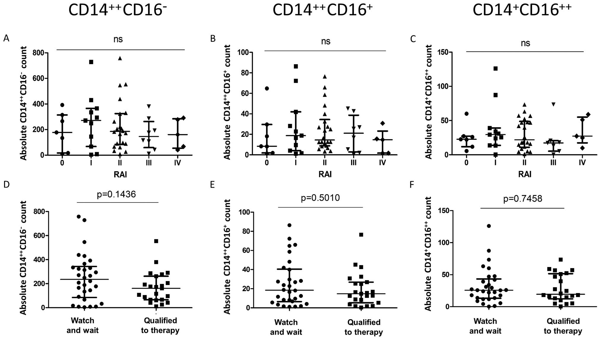
First, we demonstrated that the quantitative
distribution of different monocyte subsets (classical
CD14++CD16−, intermediate
CD14++CD16+ and non-classical
CD14+CD16++) in newly diagnosed CLL patients
was comparable at different stages of the disease (for all
p>0.05) (Fig. 1).
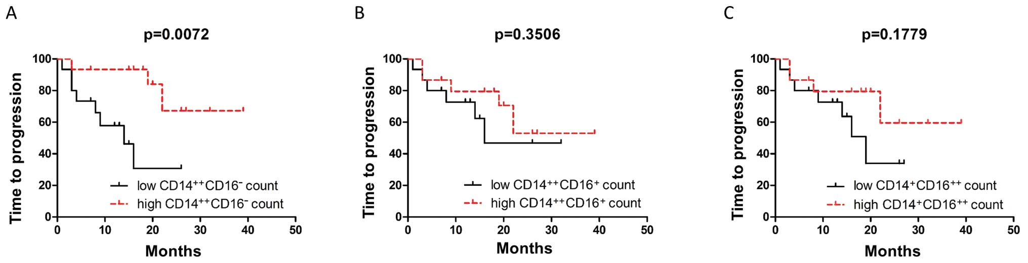
Notably, in the group of CLL patients under the
'wait and watch' strategy, the study established a significantly
longer time to initial treatment in the patients with lower than
median absolute counts of CD14++CD16−
compared to those with initially higher
CD14++CD16− amounts (Fig. 2A). There were no differences in the
time to initial treatment in patients with higher amounts of
non-classical and intermediate monocytes values compared to
patients with lower counts of the above mentioned subsets (Fig. 2B and C).
Next, we correlated the numbers of each monocyte
subset with a number of widely acknowledged disease progression
parameters of prognosis and tumor load in CLL. Our analysis
demonstrated that numbers of classical, intermediate and
non-classical monocytes were positively correlated with the total
absolute numbers of neutrophils and monocytes (Table II). In some contrast, we did not
find significant correlations between C-protein levels and any
subsets of monocytes (for all, p>0.05).
| Table IICorrelations between pre-treatment
counts of monocyte subsets and selected hematological and
biochemical parameters in 'wait and watch' patients, and patients
qualified for immediate treatment. |
Table II
Correlations between pre-treatment
counts of monocyte subsets and selected hematological and
biochemical parameters in 'wait and watch' patients, and patients
qualified for immediate treatment.
| Parameters | CLL patients
subjected to 'watch and wait' strategy
n=30
| CLL patients
qualified for immediate treatment
n=22
|
|---|
Absolute
CD14++CD16− count
| Absolute
CD14++CD16+ count
| Absolute
CD14+CD16++ count
| Absolute
CD14++CD16− count
| Absolute
CD14++CD16+ count
| Absolute
CD14+CD16++ count
|
|---|
| r | p-value | r | p-value | r | p-value | r | p-value | r | p-value | r | p-value |
|---|
| WBC
(103/µl) | 0.06251 | 0.7428 | 0.1390 | 0.4637 | 0.1889 | 0.3175 | −0.2006 | 0.3588 | 0.08696 | 0.6932 | 0.2388 | 0.2844 |
| HGB (mg/dl) | −0.3072 | 0.0986 | −0.2140 | 0.2562 | −0.09707 | 0.6098 | −0.01236 | 0.9554 | 0.1706 | 0.4365 | 0.08701 | 0.7002 |
| PLT
(103/µl) | 0.2935 | 0.1223 | 0.1072 | 0.5799 | 0.1759 | 0.3613 | 0.1749 | 0.4246 | 0.2753 | 0.2036 | 0.07851 | 0.7284 |
| Frequencies of
lymphocytes (%) | −0.1454 | 0.4434 | −0.1767 | 0.3502 | −0.2544 | 0.1749 | −0.2525 | 0.2451 | 0.04208 | 0.8488 | −0.002264 | 0.9920 |
| Absolute lymphocyte
count (103/µl) | 0.03270 | 0.8638 | 0.1261 | 0.5066 | 0.1591 | 0.4011 | −0.2332 | 0.2842 | 0.09980 | 0.6505 | 0.2196 | 0.3260 |
| Frequencies of
neutrophils (%) | −0.003 | 0.9879 | −0.1595 | 0.4174 | −0.07237 | 0.7144 | 0.4635 | 0.0259 | 0.1817 | 0.4066 | 0.08095 | 0.7203 |
| Absolute neutrophil
count (103/µl) | 0.5610 | 0.0029 | 0.4776 | 0.0136 | 0.4605 | 0.0179 | 0.4794 | 0.0602 | 0.3765 | 0.1506 | 0.6714 | 0.0061 |
| Frequencies of
monocytes (%) | 0.5034 | 0.0088 | 0.3264 | 0.1036 | 0.4188 | 0.0332 | 0.6773 | 0.0007 | 0.3689 | 0.0998 | 0.3979 | 0.0740 |
| Absolute monocyte
count (103/µl) | 0.8549 |
<0.0001 | 0.7621 |
<0.0001 | 0.7474 |
<0.0001 | 0.4998 | 0.0152 | 0.5818 | 0.0036 | 0.8831 |
<0.0001 |
| IgG (mg/dl) | −0.2802 | 0.3538 | −0.3297 | 0.2713 | 0.07143 | 0.8166 | −0.3941 | 0.1309 | −0.5618 | 0.0235 | −0.1536 | 0.5848 |
| LDH (IU/l) | 0.009032 | 0.9636 | 0.1642 | 0.4037 | 0.2472 | 0.2048 | 0.03313 | 0.8866 | 0.1351 | 0.5593 | 0.07898 | 0.7407 |
| β2m (g/l) | 0.6029 | 0.0134 | 0.6824 | 0.0036 | 0.6471 | 0.0067 | 0.2527 | 0.4048 | −0.07143 | 0.8166 | −0.2363 | 0.4371 |
| CRP (mg/l) | 0.2119 | 0.2698 | 0.04218 | 0.8280 | 0.05649 | 0.7710 | 0.1261 | 0.5859 | −0.08583 | 0.7115 | 0.003251 | 0.9888 |
| TP (g/dl) | −0.08893 | 0.6939 | −0.2118 | 0.3439 | 0.02322 | 0.9183 | 0.04425 | 0.8707 | −0.5133 | 0.0420 | −0.1209 | 0.6555 |
| Creatinine
(mg/dl) | −0.1010 | 0.6021 | −0.1927 | 0.3166 | 0.09708 | 0.6164 | −0.2429 | 0.2641 | −0.04353 | 0.8437 | −0.1787 | 0.4263 |
| % of lymphocytic
cells in smear BM | 0.2484 | 0.3919 | 0.1473 | 0.6154 | 0.01099 | 0.9703 | −0.4037 | 0.0966 | −0.6402 | 0.0042 | −0.1925 | 0.4591 |
| % of lymphocytic
cells in TB | 0.5593 | 0.8113 | 0.3647 | 0.4483 | 0.3040 | 0.2475 | −0.01879 | 0.9673 | −0.3147 | 0.3415 | −0.05592 | 0.3869 |
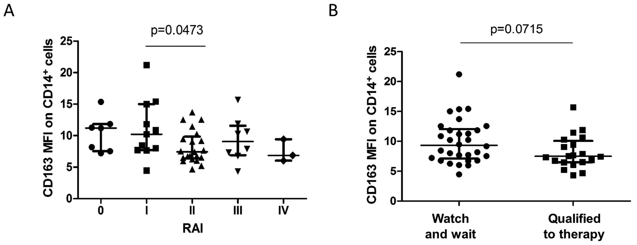
Furthermore, we demonstrated decreased levels of the
CD163 membrane-associated monocyte expression in newly diagnosed
CLL patients in advanced disease stages according to Rai
classification (Fig. 3A). However,
we found that these CLL patients who had progressive disease at the
time of diagnosis and were qualified for immediate treatment tended
to have lower CD163 expression as compared to those patients who,
due to the stable character of their disease, were qualified for
the 'wait and watch' strategy (Fig.
3B).
In some contrast to membrane-associated CD163,
levels of the soluble form of CD163 (sCD163) were significantly
increased in the CLL patients (622.3 µg/ml) (428.3–832.2) as
compared to the healthy controls (386.6 µg/ml) (322.3–473.8)
(p=0.0053, Fig. 4A). Moreover,
patients that were qualified to immediate treatment due to advanced
stage of disease were found with significantly higher sCD163 levels
as compared to patients subjected to 'watch and wait' therapy
[1,036 µg/ml (672.6–1,592) vs. 622.3 µg/ml
(428.3–832.2), p=0.0083, respectively, Fig. 4A]. However, analysis of baseline
levels of sCD163 in 'watch and wait' patients did not reveal
differences between patients with different times to initial
treatment (p>0.05, Fig. 4B).
Quite surprisingly, we did not find an expected negative
correlation between levels of soluble and membrane-associated CD163
(p>0.05, Table III).
| Table IIICorrelations between pre-treatment
serum sCD163 levels and selected hematological and biochemical
parameters in 'wait and watch' patients, and patients qualified for
immediate treatment. |
Table III
Correlations between pre-treatment
serum sCD163 levels and selected hematological and biochemical
parameters in 'wait and watch' patients, and patients qualified for
immediate treatment.
| Parameters | sCD163
|
|---|
All
patients
n=44
| Untreated 'watch
and wait'
n=22
| Qualified for
treatment
n=22
|
|---|
| r | p-value | r | p-value | r | p-value |
|---|
| WBC
(103/µl) | 0.2895 | 0.0597 | 0.05391 | 0.8165 | 0.3362 | 0.1261 |
| HGB (mg/dl) | −0.3907 | 0.0096 |
−0.1103 | 0.6342 | −0.4820 | 0.0231 |
| PLT
(103/µl) | −0.2763 | 0.0766 | −0.1667 | 0.4823 | −0.3051 | 0.1674 |
| Frequencies of
lymphocytes (%) | −0.04357 | 0.7815 | 0.1504 | 0.5152 | −0.1939 | 0.3873 |
| Absolute lymphocyte
count (103/µl) | 0.2430 | 0.1164 | 0.01104 | 0.9621 | 0.3288 | 0.1351 |
| Frequencies of
neutrophils (%) | −0.4101 | 0.0077 | −0.2855 | 0.2360 | −0.4822 | 0.0231 |
| Absolute
neutrophils count (103/µl) | −0.2577 | 0.1350 | −0.2405 | 0.3214 | −0.2104 | 0.4340 |
| Frequencies of
monocytes (%) | −0.1567 | 0.3474 | −0.1019 | 0.6874 | −0.1558 | 0.5118 |
| Absolute monocyte
count (103/µl) | −0.01534 | 0.9222 | −0.03769 | 0.8712 | 0.2326 | 0.2975 |
| IgG (mg/dl) | 0.1853 | 0.3972 | −0.4048 | 0.3268 | 0.1769 | 0.5281 |
| LDH (IU/l) | 0.3500 | 0.0290 | 0.3872 | 0.1015 | 0.1686 | 0.4773 |
| β2m (g/l) | 0.4884 | 0.0211 | −0.05455 | 0.8916 | 0.3217 | 0.3079 |
| CRP (mg/l) | 0.1187 | 0.4658 | 0.1974 | 0.4043 | 0.02260 | 0.9247 |
| TP (g/dl) | 0.1188 | 0.5471 | −0.06630 | 0.8296 | −0.01166 | 0.9671 |
| Creatinine
(mg/dl) | 0.07183 | 0.6512 | 0.05156 | 0.8291 | −0.1758 | 0.4339 |
| % of lymphocytic
cells in smear BM | 0.5096 | 0.0093 | 0.3571 | 0.3894 | 0.5267 | 0.0298 |
| % of lymphocytic
cells in TB | 0.4498 | 0.0804 | −0.2571 | 0.6583 | 0.9119 | 0.0002 |
| Absolute
CD14++CD16− count
(103/µl) | −0.2537 | 0.0966 | −0.1457 | 0.5176 | −0.07175 | 0.7510 |
| Absolute
CD14++CD16+ count
(103/µl) | −0.1649 | 0.2847 | −0.1265 | 0.5748 | −0.1480 | 0.5109 |
| Absolute
CD14+CD16++ count
(103/µl) | −0.05452 | 0.7284 | −0.03954 | 0.8613 | 0.1462 | 0.5273 |
| CD163 MFI on
monocytes | −0.206 | 0.1962 | −0.2129 | 0.3414 | −0.1659 | 0.4972 |
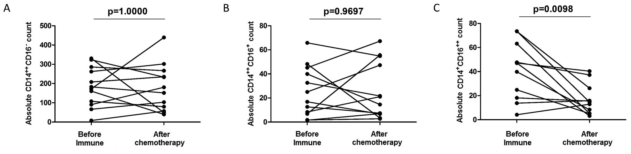
Next, we set out to analyze the effects of immune
chemotherapy on monocyte subsets and surface and soluble CD163. We
demonstrated that applied chemotherapy (FCR) resulted in a
statistically significant reduction in the absolute numbers of
non-classical CD14+CD16++ monocytes in a
group of patients who achieved at least partial remission (Fig. 5C). There were no significant changes
in the numbers of classical CD14++CD16− and
intermediate CD14++CD16+ monocyte subsets
(Fig. 5A and B). Importantly, the
study established that initial numbers of classical monocytes
predicted better response to immune chemotherapy as the patients
who achieved CR had significantly (p=0.0297) higher baseline
numbers of CD14++CD16− monocytes as compared
to patients who achieved only PR [182.7 (115.5–262.4) vs. 66.65
(40.89–160.7), respectively].
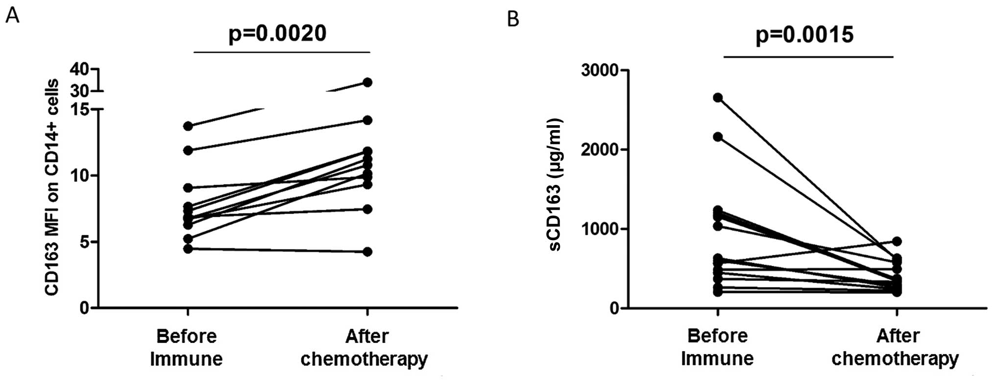
Notably, we found that monocytes of CLL patients who
underwent immune chemotherapy and achieved at least partial
response had significantly upregulated expression of the
anti-inflammatory surface protein CD163 (Fig. 6A). In clear contrast to
membrane-associated CD163, standard immune chemotherapy resulted in
a significant decrease in soluble CD163 levels (Fig. 6B).
Discussion
During tumorigenesis, monocytes are destined to
provide the antitumor response of the host and act both as cells
presenting tumor-associated antigens to tumor-infiltrating
lymphocytes and as cytotoxic effector cells (28). However, cancer cells have developed
mechanisms that inhibit immune surveillance (27). Indeed, it was shown that circulating
monocytes are actively recruited to tumor beds, where they are
'conditioned' to promote the survival of malignant cells both
directly and indirectly via the suppression of host immunity
(29,30). Practically, extensive DNA microarray
and cytokine antibody array data, on the basis of survival-inducing
CLL cultures, identified a variety of inflammatory cytokines and
signaling pathways which upregulated the expression and increased
the secretion or activation of B-CLL (31,32).
Moreover, experiments with normal B cells isolated from peripheral
blood samples of healthy donors further indicate that monocytes
harbor the survival-inducing activity for B cells in general
(31).
In the present study, we demonstrated that
quantification of different monocyte subsets served as a novel
predictive marker of time that elapses before the treatment of CLL
patients was needed. We found that detection of higher baseline
numbers of classical CD14++CD16− monocytes
(but not non-classical and intermediate monocytes) in patients
subjected to 'watch and wait' strategy was associated with
significantly longer time to treatment. This finding was supported
by identification of a negative correlation between the levels of
lymphocytosis and absolute counts of classical monocytes. As above
mentioned, classical CD14++CD16− monocytes
exhibit high antitumor and phagocytic capacities confirmed by both
functional assays (9,33,34)
and genomic analyses (35).
Moreover, classical monocytes represent the first line of immune
defense against microbial pathogens (36). Thus, our data suggest that monocyte
subsets with high antitumor and phagocytic potential can play an
important role in a series of processes that can either protect
from or delay CLL progression. It remains unclear however whether
the putative beneficial effects of classical monocytes in CLL are
indeed directly associated with their phagocytic potential or
rather some immunomodulatory actions directed at either neoplastic
cells or other cell types. Nevertheless, our findings warrant
further functional studies on the role of classical monocytes in
the pathogenesis of CLL as these cells may appear as potential
novel targets of immune therapy of CLL.
In concert with putative protective roles of
classical monocytes, we showed in the present study that CLL
patients who qualified for immediate treatment due to progressive
disease that presented with higher baseline levels of classical and
lower baseline levels of non-classical monocytes, demonstrated
better clinical response to applied therapy. It is difficult now to
conclude whether these differential relationships of classical and
non-classical monocytes were dependent directly on these monocyte
subsets or rather their subsequent developmental fates. In fact,
classical monocytes mainly differentiate into M1 macrophages that
are known to play pro-inflammatory and tumor-suppressive roles
(9,37). In contrast, non-classical monocytes
differentiate into M2 macrophages that exert potent
immunosuppressive and tumor-promoting effects (25). Moreover, it remains unknown whether
the positive correlation of non-classical monocytes with
lymphocytosis reflects direct B-CLL driven activation of monocytes
or rather their involvement in the regulation of inflammatory
reaction induced by the growth of neoplastic cells. Regardless of
the exact mechanism, our findings point to the differential roles
of classical and non-classical monocytes not only in the course of
CLL but also in modulation of response to immune chemotherapy.
To the best of our knowledge, the present study is
the first to investigate changes in monocyte subsets in CLL in the
context of applied immune chemotherapy. Our standard therapy
regimen included both chemotherapeutic agents
(fludarabine-cyclophosphamide) and rituximab. For ethical reasons,
we could not introduce treatment with either component administered
alone. To date, it is known that rituximab treatment of CLL induced
in vitro a substantial loss of CD20 on B cells that are
stripped out of B cells by monocytes/macrophages in a reaction
mediated by FcγR (38–40). Such antigenic modulation mediated by
monocytes/macrophages, if occurring in vivo, severely
compromised the therapeutic efficacy of rituximab treatment. In the
present study, we found that higher numbers of classical and lower
numbers of non-classical monocytes allow for prediction of the type
of clinical response to immune chemotherapy. Our data suggest
indirectly that classical monocytes could play certain roles in
promoting the beneficial effects of anti-CLL therapy whereas
non-classical monocytes can be linked to actions compromising
efficacy of immune chemotherapy. On the other hand, however, the
opposite actions, namely the effects of rituximab on mono-cyte
subsets were not sufficiently studied. Here, we showed a
significant treatment-related reduction in the absolute numbers of
pro-inflammatory non-classical CD14+CD16++
monocytes. This suggests that this cell subset can be identified as
a novel target of CLL-specific immune chemotherapy. Thus, our data
indicate that chemotherapy, beyond its anti-neoplastic actions,
also exerts anti-inflammatory effects via elimination of the
CD14+CD16++ pro-inflammatory cell subset,
known to constitute an effective source of TNF-α (8). In support of this notion, the present
study, in line with other studies, confirmed the anti-inflammatory
effects of CLL-specific therapy (significant reduction in CRP
levels following chemotherapy).
In order to further the explore putative
anti-inflammatory effects of immune chemotherapy, we investigated
changes in the expression of monocyte protein CD163 whose
anti-inflammatory functions have been previously identified by
numerous research group including ours (4,7,22,41–44).
In addition, CD163-expressing macrophages are involved in the
resolution of inflammation by limiting free-hemoglobin associated
damage (45), secreting
anti-inflammatory cytokines in response to inflammation (46,47)
and inhibiting T cell-mediated responses. The role of CD163 in the
pathogenesis of solid tumors seems quite complex. In breast cancer,
monocyte/macrophage CD163 expression was found to be significantly
decreased as compared to healthy controls (48–50).
In contrast, in a recent study, Tiainen et al (51) showed that detection of
CD163-positive macrophages in breast cancer patients was related to
poor prognosis. Similarly, accumulation of CD163-postive
macrophages was associated with poor outcome of a few types of
solid tumors (52–54). To date, the role of the differential
patterns of monocyte CD163 expression in the pathogenesis of CLL
were not studied. In the present study, we found that these CLL
patients who had progressive disease at diagnosis and were
therefore qualified for immediate treatment presented with lower
levels of surface CD163 expression yet higher levels of soluble
CD163 as compared to the patients who were qualified for the 'wait
and watch' strategy due to the stable character of their disease.
In concordance with this notion, the application of immune
chemotherapy resulted in significant upregulation of surface CD163
expression and downregulation of soluble CD163 in a group of
patients with at least partial remission. This pattern of
CD163-related alterations reflects the dynamic monocyte response to
intensity and extent of inflammatory processes driven by expanding
B-CLL cells. In that case, a decrease in inflammation initiated by
immune chemotherapy could have resulted in decreasing the intensity
of CD163-related anti-inflammatory compensatory mechanisms. On the
other hand, one cannot exclude that in CLL, in contrast to breast
cancer, CD163 exerts some protective antitumor effects limiting the
growth and expansion of neoplastic cells. Similarly, the literature
on soluble CD163 in the pathogenesis of neoplastic disorders is
very scarce and its role in malignancy is not yet elucidated
(55,56). The present study is the first to our
knowledge to investigate the role of soluble CD163 in CLL. It
remains unclear whether our notion of elevated levels of sCD163 in
CLL patients (particularly in those with more advanced disease)
reflects yet unknown mechanisms fueling the progression of CLL or
it rather represents the compensatory counter-action against
malignant process and on-going inflammation. Regardless of the
exact mechanism, our data indicate that the role of CD163 in the
regulation of the development and progression of CLL warrants
further studies.
In summary, we presented in the present study a
number of significant relationships between classical monocytes
(but not the other subsets) and markers of more favorable CLL
prognosis or positive response to anti-CLL treatment. Our data
warrant further studies that could explore in more detail the
beneficial potential of this cell subset in CLL treatment.
Moreover, the present study provides initial evidence proving that
diminishing the CLL-related inflammation by immune chemotherapy can
be explained, at least to some degree, by targeting specific
subsets and molecules of circulating monocytes. These
monocyte-directed anti-inflammatory effects of immune chemotherapy
could represent underappreciated benefits in the treatment of CLL.
What still remains unclear, however, is whether the reduced levels
of non-classical mono-cytes and the upregulated expression of CD163
also accounts for the general beneficial effects of chemotherapy.
This issue deserves further investigation as it can help to
establish novel monocyte-directed strategies, potentially enhancing
the effectiveness of the current therapeutic regimens.
References
|
1
|
Chiorazzi N, Rai KR and Ferrarini M:
Chronic lymphocytic leukemia. N Engl J Med. 352:804–815. 2005.
View Article : Google Scholar : PubMed/NCBI
|
|
2
|
Zenz T, Mertens D, Küppers R, Döhner H and
Stilgenbauer S: From pathogenesis to treatment of chronic
lymphocytic leukaemia. Nat Rev Cancer. 10:37–50. 2010.
|
|
3
|
Korz C, Pscherer A, Benner A, Mertens D,
Schaffner C, Leupolt E, Döhner H, Stilgenbauer S and Lichter P:
Evidence for distinct pathomechanisms in B-cell chronic lymphocytic
leukemia and mantle cell lymphoma by quantitative expression
analysis of cell cycle and apoptosis-associated genes. Blood.
99:4554–4561. 2002. View Article : Google Scholar : PubMed/NCBI
|
|
4
|
Skrzeczyńska-Moncznik J, Bzowska M, Loseke
S, Grage-Griebenow E, Zembala M and Pryjma J: Peripheral blood
CD14high CD16+ monocytes are main producers
of IL-10. Scand J Immunol. 67:152–159. 2008. View Article : Google Scholar
|
|
5
|
Ziegler-Heitbrock HW, Passlick B and
Flieger D: The monoclonal antimonocyte antibody My4 stains B
lymphocytes and two distinct monocyte subsets in human peripheral
blood. Hybridoma. 7:521–527. 1988. View Article : Google Scholar : PubMed/NCBI
|
|
6
|
Ziegler-Heitbrock L, Ancuta P, Crowe S,
Dalod M, Grau V, Hart DN, Leenen PJ, Liu YJ, MacPherson G, Randolph
GJ, et al: Nomenclature of monocytes and dendritic cells in blood.
Blood. 116:e74–e80. 2010. View Article : Google Scholar : PubMed/NCBI
|
|
7
|
Moniuszko M, Bodzenta-Lukaszyk A, Kowal K,
Lenczewska D and Dabrowska M: Enhanced frequencies of
CD14++CD16+, but not
CD14+CD16+, peripheral blood monocytes in
severe asthmatic patients. Clin Immunol. 130:338–346. 2009.
View Article : Google Scholar
|
|
8
|
Moniuszko M, Liyanage NP, Doster MN, Parks
RW, Grubczak K, Lipinska D, McKinnon K, Brown C, Hirsch V, Vaccari
M, et al: Glucocorticoid treatment at moderate doses of
SIVmac251-infected rhesus macaques decreases the frequency of
circulating CD14+CD16++ monocytes but does
not alter the tissue virus reservoir. AIDS Res Hum Retroviruses.
31:115–126. 2015. View Article : Google Scholar
|
|
9
|
Eljaszewicz A, Wiese M, Helmin-Basa A,
Jankowski M, Gackowska L, Kubiszewska I, Kaszewski W, Michalkiewicz
J and Zegarski W: Collaborating with the enemy: Function of
macrophages in the development of neoplastic disease. Mediators
Inflamm. 2013:8313872013. View Article : Google Scholar : PubMed/NCBI
|
|
10
|
Leidi M, Gotti E, Bologna L, Miranda E,
Rimoldi M, Sica A, Roncalli M, Palumbo GA, Introna M and Golay J:
M2 macrophages phagocytose rituximab-opsonized leukemic targets
more efficiently than M1 cells in vitro. J Immunol. 182:4415–4422.
2009. View Article : Google Scholar : PubMed/NCBI
|
|
11
|
Eljaszewicz A, Jankowski M, Gackowska L,
Helmin-Basa A, Wiese M, Kubiszewska I, Kaszewski W, Michalkiewicz J
and Zegarski W: Gastric cancer increase the percentage of
intermediate (CD14++CD16+) and nonclassical
(CD14+CD16+) monocytes. Centr Eur J Immunol.
37:355–361. 2012. View Article : Google Scholar
|
|
12
|
Heine GH, Ulrich C, Seibert E, Seiler S,
Marell J, Reichart B, Krause M, Schlitt A, Köhler H and Girndt M:
CD14++CD16+ monocytes but not total monocyte
numbers predict cardiovascular events in dialysis patients. Kidney
Int. 73:622–629. 2008. View Article : Google Scholar
|
|
13
|
Ulrich C, Heine GH, Gerhart MK, Köhler H
and Girndt M: Proinflammatory CD14+CD16+
monocytes are associated with subclinical atherosclerosis in renal
transplant patients. Am J Transplant. 8:103–110. 2008. View Article : Google Scholar
|
|
14
|
Foa R, Massaia M, Cardona S, Tos AG,
Bianchi A, Attisano C, Guarini A, di Celle PF and Fierro MT:
Production of tumor necrosis factor-alpha by B-cell chronic
lymphocytic leukemia cells: A possible regulatory role of TNF in
the progression of the disease. Blood. 76:393–400. 1990.PubMed/NCBI
|
|
15
|
Gamberale R, Geffner J, Arrosagaray G,
Scolnik M, Salamone G, Trevani A, Vermeulen M and Giordano M:
Non-malignant leukocytes delay spontaneous B-CLL cell apoptosis.
Leukemia. 15:1860–1867. 2001. View Article : Google Scholar : PubMed/NCBI
|
|
16
|
Burger JA, Tsukada N, Burger M, Zvaifler
NJ, Dell'Aquila M and Kipps TJ: Blood-derived nurse-like cells
protect chronic lymphocytic leukemia B cells from spontaneous
apoptosis through stromal cell-derived factor-1. Blood.
96:2655–2663. 2000.PubMed/NCBI
|
|
17
|
Nishio M, Endo T, Tsukada N, Ohata J,
Kitada S, Reed JC, Zvaifler NJ and Kipps TJ: Nurselike cells
express BAFF and APRIL, which can promote survival of chronic
lymphocytic leukemia cells via a paracrine pathway distinct from
that of SDF-1alpha. Blood. 106:1012–1020. 2005. View Article : Google Scholar : PubMed/NCBI
|
|
18
|
Maffei R, Bulgarelli J, Fiorcari S,
Bertoncelli L, Martinelli S, Guarnotta C, Castelli I, Deaglio S,
Debbia G, De Biasi S, et al: The monocytic population in chronic
lymphocytic leukemia shows altered composition and deregulation of
genes involved in phagocytosis and inflammation. Haematologica.
98:1115–1123. 2013. View Article : Google Scholar : PubMed/NCBI
|
|
19
|
Condeelis J and Pollard JW: Macrophages:
Obligate partners for tumor cell migration, invasion, and
metastasis. Cell. 124:263–266. 2006. View Article : Google Scholar : PubMed/NCBI
|
|
20
|
Steidl C, Lee T, Shah SP, Farinha P, Han
G, Nayar T, Delaney A, Jones SJ, Iqbal J, Weisenburger DD, et al:
Tumor-associated macrophages and survival in classic Hodgkin's
lymphoma. N Engl J Med. 362:875–885. 2010. View Article : Google Scholar : PubMed/NCBI
|
|
21
|
Etzerodt A and Moestrup SK: CD163 and
inflammation: Biological, diagnostic, and therapeutic aspects.
Antioxid Redox Signal. 18:2352–2363. 2013. View Article : Google Scholar :
|
|
22
|
Moniuszko M, Kowal K, Rusak M, Pietruczuk
M, Dabrowska M and Bodzenta-Lukaszyk A: Monocyte CD163 and CD36
expression in human whole blood and isolated mononuclear cell
samples: Influence of different anticoagulants. Clin Vaccine
Immunol. 13:704–707. 2006. View Article : Google Scholar : PubMed/NCBI
|
|
23
|
Davis BH and Zarev PV: Human monocyte
CD163 expression inversely correlates with soluble CD163 plasma
levels. Cytometry B Clin Cytom. 63:16–22. 2005. View Article : Google Scholar
|
|
24
|
Møller HJ, Aerts H, Grønbaek H, Peterslund
NA, Hyltoft Petersen P, Hornung N, Rejnmark L, Jabbarpour E and
Moestrup SK: Soluble CD163: A marker molecule for
monocyte/macrophage activity in disease. Scand J Clin Lab Invest
Suppl. 237:29–33. 2002. View Article : Google Scholar
|
|
25
|
Gui T, Shimokado A, Sun Y, Akasaka T and
Muragaki Y: Diverse roles of macrophages in atherosclerosis: From
inflammatory biology to biomarker discovery. Mediators Inflamm.
2012:6930832012. View Article : Google Scholar : PubMed/NCBI
|
|
26
|
Hallek M, Cheson BD, Catovsky D,
Caligaris-Cappio F, Dighiero G, Döhner H, Hillmen P, Keating MJ,
Montserrat E, Rai KR, et al International Workshop on Chronic
Lymphocytic Leukemia: Guidelines for the diagnosis and treatment of
chronic lymphocytic leukemia: A report from the International
Workshop on Chronic Lymphocytic Leukemia updating the National
Cancer Institute-Working Group 1996 guidelines. Blood.
111:5446–5456. 2008. View Article : Google Scholar : PubMed/NCBI
|
|
27
|
Pardoll D: Does the immune system see
tumors as foreign or self? Annu Rev Immunol. 21:807–839. 2003.
View Article : Google Scholar : PubMed/NCBI
|
|
28
|
Mytar B, Wołoszyn M, Szatanek R,
Baj-Krzyworzeka M, Siedlar M, Ruggiero I, Wieckiewicz J and Zembala
M: Tumor cell-induced deactivation of human monocytes. J Leukoc
Biol. 74:1094–1101. 2003. View Article : Google Scholar : PubMed/NCBI
|
|
29
|
Gabrilovich DI and Nagaraj S:
Myeloid-derived suppressor cells as regulators of the immune
system. Nat Rev Immunol. 9:162–174. 2009. View Article : Google Scholar : PubMed/NCBI
|
|
30
|
Ostrand-Rosenberg S and Sinha P:
Myeloid-derived suppressor cells: Linking inflammation and cancer.
J Immunol. 182:4499–4506. 2009. View Article : Google Scholar : PubMed/NCBI
|
|
31
|
Seiffert M, Schulz A, Ohl S, Döhner H,
Stilgenbauer S and Lichter P: Soluble CD14 is a novel
monocyte-derived survival factor for chronic lymphocytic leukemia
cells, which is induced by CLL cells in vitro and present at
abnormally high levels in vivo. Blood. 116:4223–4230. 2010.
View Article : Google Scholar : PubMed/NCBI
|
|
32
|
Go NF, Castle BE, Barrett R, Kastelein R,
Dang W, Mosmann TR, Moore KW and Howard M: Interleukin 10, a novel
B cell stimulatory factor: Unresponsiveness of X chromosome-linked
immunodeficiency B cells. J Exp Med. 172:1625–1631. 1990.
View Article : Google Scholar : PubMed/NCBI
|
|
33
|
Rothe G, Gabriel H, Kovacs E, Klucken J,
Stöhr J, Kindermann W and Schmitz G: Peripheral blood mononuclear
phagocyte subpopulations as cellular markers in
hypercholesterolemia. Arterioscler Thromb Vasc Biol. 16:1437–1447.
1996. View Article : Google Scholar : PubMed/NCBI
|
|
34
|
Ohri CM, Shikotra A, Green RH, Waller DA
and Bradding P: Macrophages within NSCLC tumour islets are
predominantly of a cytotoxic M1 phenotype associated with extended
survival. Eur Respir J. 33:118–126. 2009. View Article : Google Scholar : PubMed/NCBI
|
|
35
|
Wong KL, Tai JJ, Wong WC, Han H, Sem X,
Yeap WH, Kourilsky P and Wong SC: Gene expression profiling reveals
the defining features of the classical, intermediate, and
nonclassical human monocyte subsets. Blood. 118:e16–e31. 2011.
View Article : Google Scholar : PubMed/NCBI
|
|
36
|
Bieber K and Autenrieth SE: Insights how
monocytes and dendritic cells contribute and regulate immune
defense against microbial pathogens. Immunobiology. 220:215–226.
2015. View Article : Google Scholar
|
|
37
|
Zhou D, Huang C, Lin Z, Zhan S, Kong L,
Fang C and Li J: Macrophage polarization and function with emphasis
on the evolving roles of coordinated regulation of cellular
signaling pathways. Cell Signal. 26:192–197. 2014. View Article : Google Scholar
|
|
38
|
Beum PV, Kennedy AD and Taylor RP: Three
new assays for rituximab based on its immunological activity or
antigenic properties: Analyses of sera and plasmas of RTX-treated
patients with chronic lymphocytic leukemia and other B cell
lymphomas. J Immunol Methods. 289:97–109. 2004. View Article : Google Scholar : PubMed/NCBI
|
|
39
|
Beum PV, Kennedy AD, Williams ME,
Lindorfer MA and Taylor RP: The shaving reaction: Rituximab/CD20
complexes are removed from mantle cell lymphoma and chronic
lymphocytic leukemia cells by THP-1 monocytes. J Immunol.
176:2600–2609. 2006. View Article : Google Scholar : PubMed/NCBI
|
|
40
|
Beum PV, Mack DA, Pawluczkowycz AW,
Lindorfer MA and Taylor RP: Binding of rituximab, trastuzumab,
cetuximab, or mAb T101 to cancer cells promotes trogocytosis
mediated by THP-1 cells and monocytes. J Immunol. 181:8120–8132.
2008. View Article : Google Scholar : PubMed/NCBI
|
|
41
|
Bover LC, Cardó-Vila M, Kuniyasu A, Sun J,
Rangel R, Takeya M, Aggarwal BB, Arap W and Pasqualini R: A
previously unrecognized protein-protein interaction between TWEAK
and CD163: Potential biological implications. J Immunol.
178:8183–8194. 2007. View Article : Google Scholar : PubMed/NCBI
|
|
42
|
Moniuszko M, Kowal K, Jeznach M, Rusak M,
Dabrowska M and Bodzenta-Lukaszyk A: Phenotypic correlations
between monocytes and CD4+ T cells in allergic patients.
Int Arch Allergy Immunol. 161:131–141. 2013. View Article : Google Scholar
|
|
43
|
Buechler C, Ritter M, Orsó E, Langmann T,
Klucken J and Schmitz G: Regulation of scavenger receptor CD163
expression in human monocytes and macrophages by pro- and
antiinflam-matory stimuli. J Leukoc Biol. 67:97–103.
2000.PubMed/NCBI
|
|
44
|
Pioli PA, Goonan KE, Wardwell K and Guyre
PM: TGF-beta regulation of human macrophage scavenger receptor
CD163 is Smad3-dependent. J Leukoc Biol. 76:500–508. 2004.
View Article : Google Scholar : PubMed/NCBI
|
|
45
|
Schaer DJ, Alayash AI and Buehler PW:
Gating the radical hemoglobin to macrophages: The anti-inflammatory
role of CD163, a scavenger receptor. Antioxid Redox Signal.
9:991–999. 2007. View Article : Google Scholar : PubMed/NCBI
|
|
46
|
Philippidis P, Mason JC, Evans BJ, Nadra
I, Taylor KM, Haskard DO and Landis RC: Hemoglobin scavenger
receptor CD163 mediates interleukin-10 release and heme oxygenase-1
synthesis: Antiinflammatory monocyte-macrophage responses in vitro,
in resolving skin blisters in vivo, and after cardiopulmonary
bypass surgery. Circ Res. 94:119–126. 2004. View Article : Google Scholar
|
|
47
|
Hamann W, Flöter A, Schmutzler W and
Zwadlo-Klarwasser G: Characterization of a novel anti-inflammatory
factor produced by RM3/1 macrophages derived from glucocorticoid
treated human monocytes. Inflamm Res. 44:535–540. 1995. View Article : Google Scholar : PubMed/NCBI
|
|
48
|
Shabo I, Stål O, Olsson H, Doré S and
Svanvik J: Breast cancer expression of CD163, a macrophage
scavenger receptor, is related to early distant recurrence and
reduced patient survival. Int J Cancer. 123:780–786. 2008.
View Article : Google Scholar : PubMed/NCBI
|
|
49
|
Mansfield AS, Heikkila P, von Smitten K,
Vakkila J and Leidenius M: The presence of sinusoidal
CD163+ macrophages in lymph nodes is associated with
favorable nodal status in patients with breast cancer. Virchows
Arch. 461:639–646. 2012. View Article : Google Scholar : PubMed/NCBI
|
|
50
|
Goodale D, Phay C, Brown W, Gray-Statchuk
L, Furlong P, Lock M, Chin-Yee I, Keeney M and Allan AL: Flow
cytometric assessment of monocyte activation markers and
circulating endo-thelial cells in patients with localized or
metastatic breast cancer. Cytometry B Clin Cytom. 76:107–117. 2009.
View Article : Google Scholar
|
|
51
|
Tiainen S, Tumelius R, Rilla K, Hämäläinen
K, Tammi M, Tammi R, Kosma VM, Oikari S and Auvinen P: High numbers
of macrophages, especially M2-like (CD163-positive), correlate with
hyaluronan accumulation and poor outcome in breast cancer.
Histopathology. 66:873–883. 2015. View Article : Google Scholar
|
|
52
|
He KF, Zhang L, Huang CF, Ma SR, Wang YF,
Wang WM, Zhao ZL, Liu B, Zhao YF, Zhang WF, et al:
CD163+ tumor-associated macrophages correlated with poor
prognosis and cancer stem cells in oral squamous cell carcinoma.
BioMed Res Int. 2014:8386322014.
|
|
53
|
Chen L, Li Q, Zhou XD, Shi Y, Yang L, Xu
SL, Chen C, Cui YH, Zhang X and Bian XW: Increased pro-angiogenic
factors, infiltrating neutrophils and CD163+ macrophages
in bronchoalveolar lavage fluid from lung cancer patients. Int
Immunopharmacol. 20:74–80. 2014. View Article : Google Scholar : PubMed/NCBI
|
|
54
|
Lim R, Lappas M, Riley C, Borregaard N,
Moller HJ, Ahmed N and Rice GE: Investigation of human cationic
antimicrobial protein-18 (hCAP-18), lactoferrin and CD163 as
potential biomarkers for ovarian cancer. J Ovarian Res. 6:52013.
View Article : Google Scholar : PubMed/NCBI
|
|
55
|
Wang J, Guo W, Du H, Yu H, Jiang W, Zhu T,
Bai X and Wang P: Elevated soluble CD163 plasma levels are
associated with disease severity in patients with hemorrhagic fever
with renal syndrome. PLoS One. 9:e1121272014. View Article : Google Scholar : PubMed/NCBI
|
|
56
|
Ye H, Wang LY, Zhao J and Wang K:
Increased CD163 expression is associated with acute-on-chronic
hepatitis B liver failure. World J Gastroenterol. 19:2818–2825.
2013. View Article : Google Scholar : PubMed/NCBI
|