Introduction
Gastric cancer (GC) is the second most common cancer
in Asia and particularly in Eastern Asia (1,2), and
is still the third most frequent cause of cancer-related deaths,
after lung and liver cancer in males and breast and lung cancer in
females (3–5). The survival of patients with GC has
improved along with marked advances in diagnostic and therapeutic
modalities. However, the high rate of relapse and metastasis of GC
results in poor long-term survival (6,7). Thus,
there is an urgent need to develop new treatment strategies for
GC.
In eukaryotes, ribosomes contain ~80 different
ribosomal proteins (RPs). Evidence has demonstrated that
appropriate regulation of RP genes is crucial for normal ribosome
biosynthesis (8), and their
activity is required for the growth and maintenance of all types of
cells (9,10). Loss of normal regulation of RPs has
been associated with pathological conditions, such as neoplasia
(11) and Turner syndrome (12). The gene encoding ribosomal protein
L34 (RPL34) has been identified in the human (13,14),
mouse (15) and rat (13,16,17).
However, no functional information has been available to date for
RPL34 in human cancer and GC in particular.
In the present study, we confirmed that RPL34 is
highly expressed in GC cell lines. Subsequently, we employed the
lentivirus-delivered small interfering RNA (siRNA) technique to
examine the effect of RPL34 knockdown on human GC cell growth in
vitro.
Materials and methods
Cell lines
Human gastric adenocarcinoma cell lines SGC-7901,
MGC80-3, BGC-823 and MKN-45 and human renal epithelial 293T cells
were purchased from the Shanghai Cell Bank (Shanghai, China). Cell
lines were cultured in RPMI-1640 medium, supplemented with 10%
fetal bovine serum (FBS), sodium pyruvate, non-essential amino
acids, L-glutamine, a 2-fold vitamin solution (all from
Gibco®, Shanghai, China), 100 U/ml penicillin and 0.1
mg/ml streptomycin (Sangon Co., Ltd., Shanghai, China) at 37°C in a
5% CO2 incubator.
Quantitative RT-PCR
Total RNA from the 4 cell lines, SGC-7901, MGC80-3,
BGC-823 and MKN-45, was extracted using the TRIzol reagent
(Invitrogen, Shanghai, China), according to the manufacturer's
instructions and was then used for RT reaction. Briefly, 2
µg of total RNA from each sample was reverse transcribed to
single-stranded cDNA. One microliter of cDNA was used as a template
for the following PCR. The primers used were as follows: for RPL34
forward, 5′-GTT TGA CAT ACC GAC GTA GGC-3′ and reverse, 5′-GCA CAC
ATG GAA CCA CCA TAG-3′; and for GAPDH forward, 5′-TGA CTT CAA CAG
CGA CAC CCA-3′ and 5′-CAC CCT GTT GCT GTA GCC AAA-3′. The
quantitative RT-PCR comprised an initial denaturation at 95°C for
15 sec, then 45 cycles at 95°C for 5 sec and 60°C for 30 sec. The
PCR products of RPL34 and GAPDH were 241 and 121 bp, respectively.
All samples were examined in triplicates.
Recombinant lentiviral vector production
and cell infection
The complementary DNA sequence (CCTAAAGTTCTTA
TGAGAT) of RPL34 was designed from the full-length RPL34 sequence
(GenBank no. CR542242.1) by GeneChem Co. Ltd. (Shanghai, China).
After testing knockdown efficiencies, the stem-loop
oligonucleotides were synthesized and inserted into the
lentivirus-based pGCSIL-GFP (GeneChem Co. Ltd.) with
AgeI/EcoRI sites. Lentivirus particles were prepared
as previously described (18).
For lentivirus infection, SGC-7901 cells were
cultured into 6-well plates and then the RPL34-siRNA-lentivirus or
negative control (NC) lentivirus was added according to a
multiplicity of infection (MOI). After 72 h of infection, the cells
were observed under a fluorescence microscope (MicroPublisher
3.3RTV; Olympus, Tokyo, Japan). After 120 h of infection, the cells
were harvested to determine knockdown efficiency by quantitative
RT-PCR.
Western blot analysis
The cells were collected and lysed using ice-cold
lysis buffer (50 mM Tris, pH 7.4, 150 mM NaCl, 1% SDS, 1 mM EDTA,
1% NP-40) containing 1 mM protein inhibitor and 1 mM PMSF, for 30
min on ice. The lysates were centrifuged at 10,000 x g at 4°C for
10 min and the supernatants were collected. Protein concentration
was measured using the BCA protein assay (HyClone-Pierce, Rockford,
IL, USA). Equal amounts of total protein of each treatment were
separated using 12.5% SDS-PAGE according to Laemmli's method
(19), and were then transferred
onto PVDF membranes. Membranes were incubated with mouse anti-FLAG
or anti-GAPDH antibodies (Santa Cruz Biotechnology, Santa Cruz, CA,
USA). Western blotting was developed using horseradish
peroxidase-conjugated goat anti-mouse IgG (Santa Cruz
Biotechnology) and was detected by enhanced chemiluminescence (ECL)
reagent (ECL-Plus/Kit; Amersham, Piscataway, NJ, USA).
Cell growth assay
Cell growth was measured using multi-parametric
high-content screening (HCS) similarly to and as described by Zhou
et al (20) with some
modifications. Briefly, SGC-7901 cells at the logarithmic phase
after being infected with either the NC lentivirus or RPL34-siRNA
lentivirus were seeded at 2,000 cells/well into 96-well plates; the
cells were then incubated at 37°C with 5% CO2 for 5
days. The cells in the plates were counted using the Cellomics
ArrayScan™ VT1 HCS automated reader (Cellomics, Inc., Pittsburgh,
PA, USA) for each day's analysis. In each well, at least 800 cells
were analyzed. Each experiment was performed in triplicates.
Analysis of cell cycle distribution and
apoptosis
Flow cytometry (FCM) analysis was used to determine
the cell cycle distribution or detect apoptosis and was performed
as previously described (21).
Briefly, SGC-7901 cells were infected with RPL34-siRNA or NC
plasmids and incubated at 37°C for 1, 2, 3, 4 or 5 days. At the
indicated time point, adherent cells were collected, washed twice
with ice-cold phosphate-buffered saline (PBS), fixed with ~0.5 ml
of ice-cold 70% ethanol at 4°C for 1 h, and stained with propidium
iodide (PI; 50 µg/ml, Sigma-Aldrich® Co. LLC.,
St. Louis, MO, USA) in the presence of RNase A (100 µg/ml;
Fermentas®, Shanghai, China). The suspension was
filtered through a 300-mesh, and the DNA content of the stained
nuclei was analyzed for the cell cycle phase by BD FACSCalibur flow
cytometer (BD Biosciences, San Diego, CA, USA). Each experiment was
performed in triplicates.
Cell apoptosis was assayed by staining with Annexin
V-APC (eBioscience, San Diego, CA, USA) and detected by FCM. For
analysis of apoptosis, SGC-7901 cells were cultured into 6-well
plates. After 48 h of transfection with RPL34-siRNA or NC plasmids,
the cells were collected and washed twice with ice-cold PBS. The
cell concentrations were adjusted to 1×10⁶/ml with 1X
staining buffer. One-hundred microliters of cell suspension was
stained with 5 µl Annexin V-APC at room temperature in the
dark for 15 min. Cells were analyzed using FCM within 1 h. All
experiments were performed in triplicates.
Statistical analysis
The Student's t-test was used for raw data analysis.
Statistical analysis was performed using SPSS for Windows version
16.0 (SPSS, Inc., Chicago, IL, USA). The statistical data for each
group were presented as the mean ± SD. A value of p<0.05 was
accepted as statistically significant.
Results
RPL34 mRNA detection in four GC cell
lines
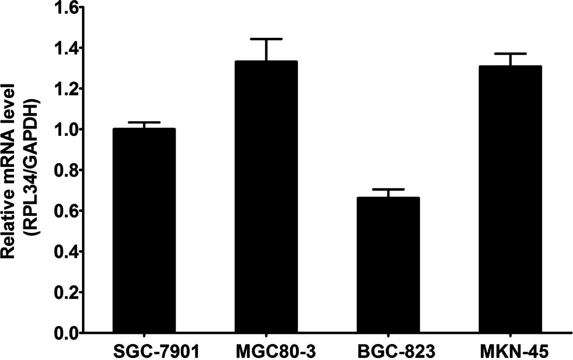
The expression of RPL34 mRNA was assessed in gastric
cancer cell lines SGC-7901, MGC80-3, BGC-823 and MKN-45 by RT-PCR.
The results showed that RPL34 mRNA was expressed in all four cell
lines (Fig. 1).
Knockdown efficiency determined by
western blot analysis
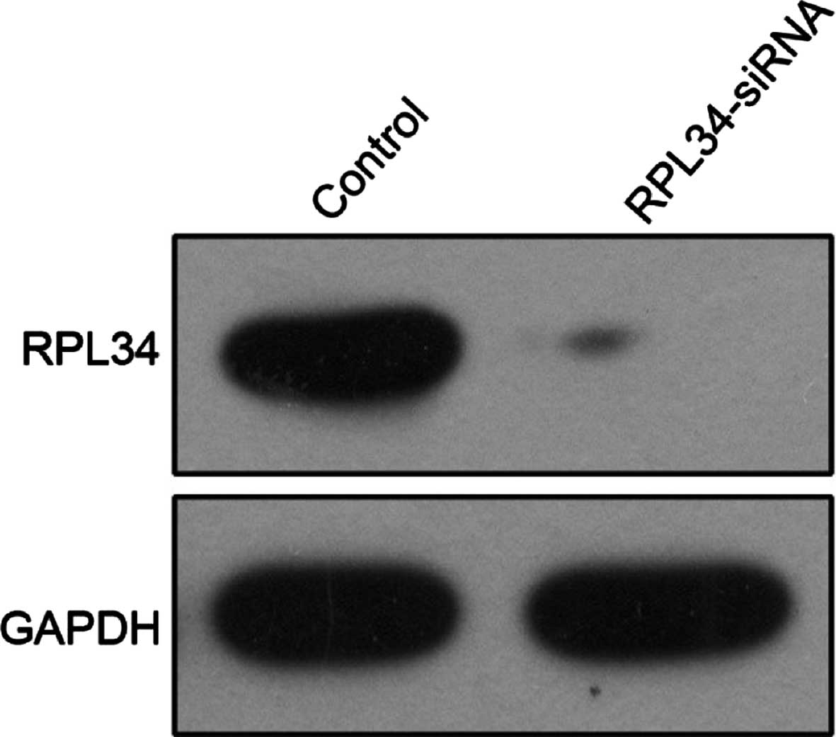
Human embryonic kidney 293T cells were infected with
RPL34-siRNA lentivirus or NC lentivirus. As shown in Fig. 2, RPL34 protein expression was
detected by western blotting in these cells, but was greatly
reduced in the RPL34-siRNA infected cultures, indicating effective
knockdown of the target sequence.
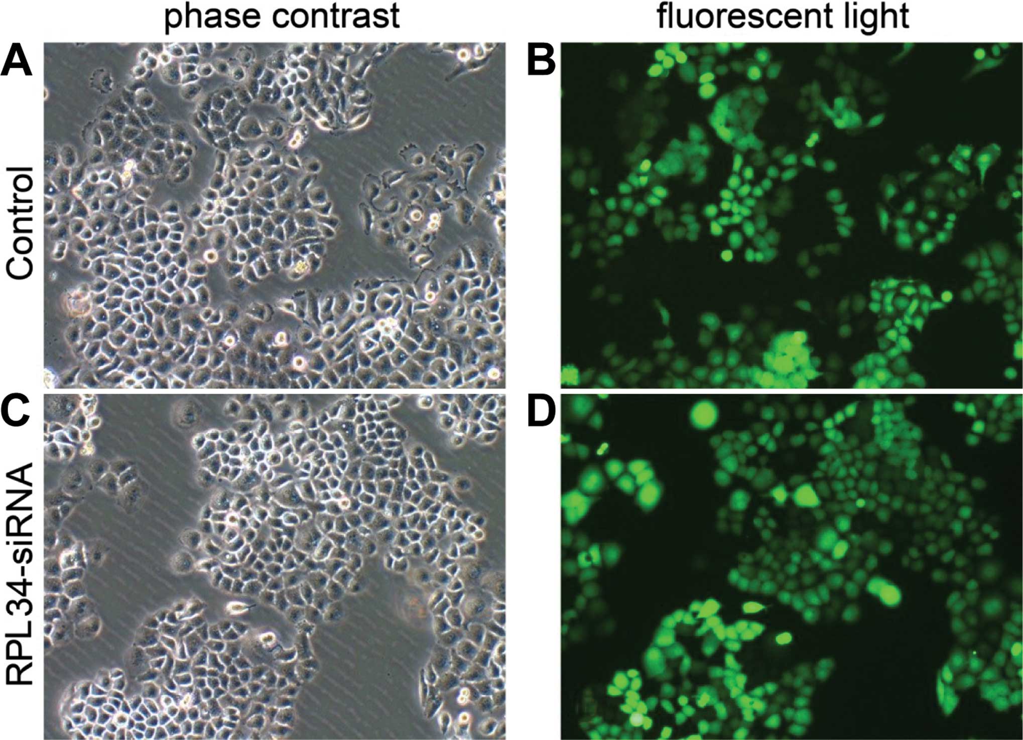
Lentivirus-mediated knockdown of RPL34 in
the human GC cell line SGC-7901
To explore the role of RPL34, we knocked down RPL34
in the SGC-7901 cell line. As shown in Fig. 3, by day 3 post infection, the
proportion of infected cells was >80% for both the RPL34-siRNA
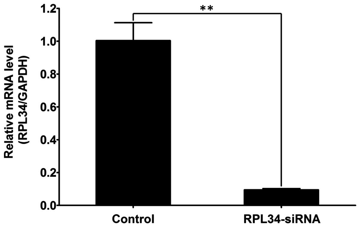
and NC lentivirus. RPL34 mRNA levels were assessed by real-time PCR
at day 5 post infection with either the RPL34-siRNA or NC
lentivirus. RPL34-siRNA lentivirus-infected cultures had
significantly lower levels of RPL34 mRNA compared to levels in the
cultures infected with the NC lentivirus (Fig. 4).
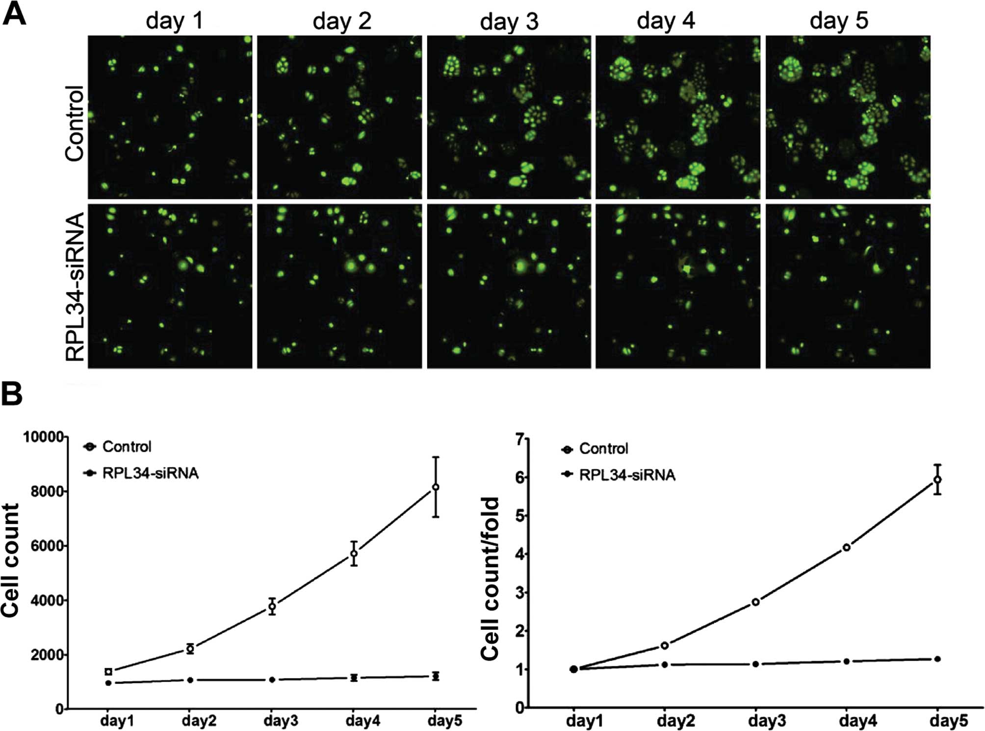
Knockdown of RPL34 in SGC-7901 cells
inhibits cell proliferation
To examine the effect of RPL34 on cell growth,
SGC-7901 cells expressing either the RPL34-siRNA or NC lentivirus
were seeded into 96-well plates and analyzed by Cellomics every day
for 5 days. As illustrated in Fig.
5A and confirmed by quantification in Fig. 5B, control-transfected cells greatly
expanded over the 5 days of the experiment, while the number of
RPL34-siRNA-transfected cells did not change. The cell growth rate
was defined as: Cell count at 9 days/cell count at first day, where
n=2, 3, 4 and 5 (Fig. 5B). The
results of the present study showed that RPL34 knockdown
significantly inhibited proliferation of the SGC-7901 cells.
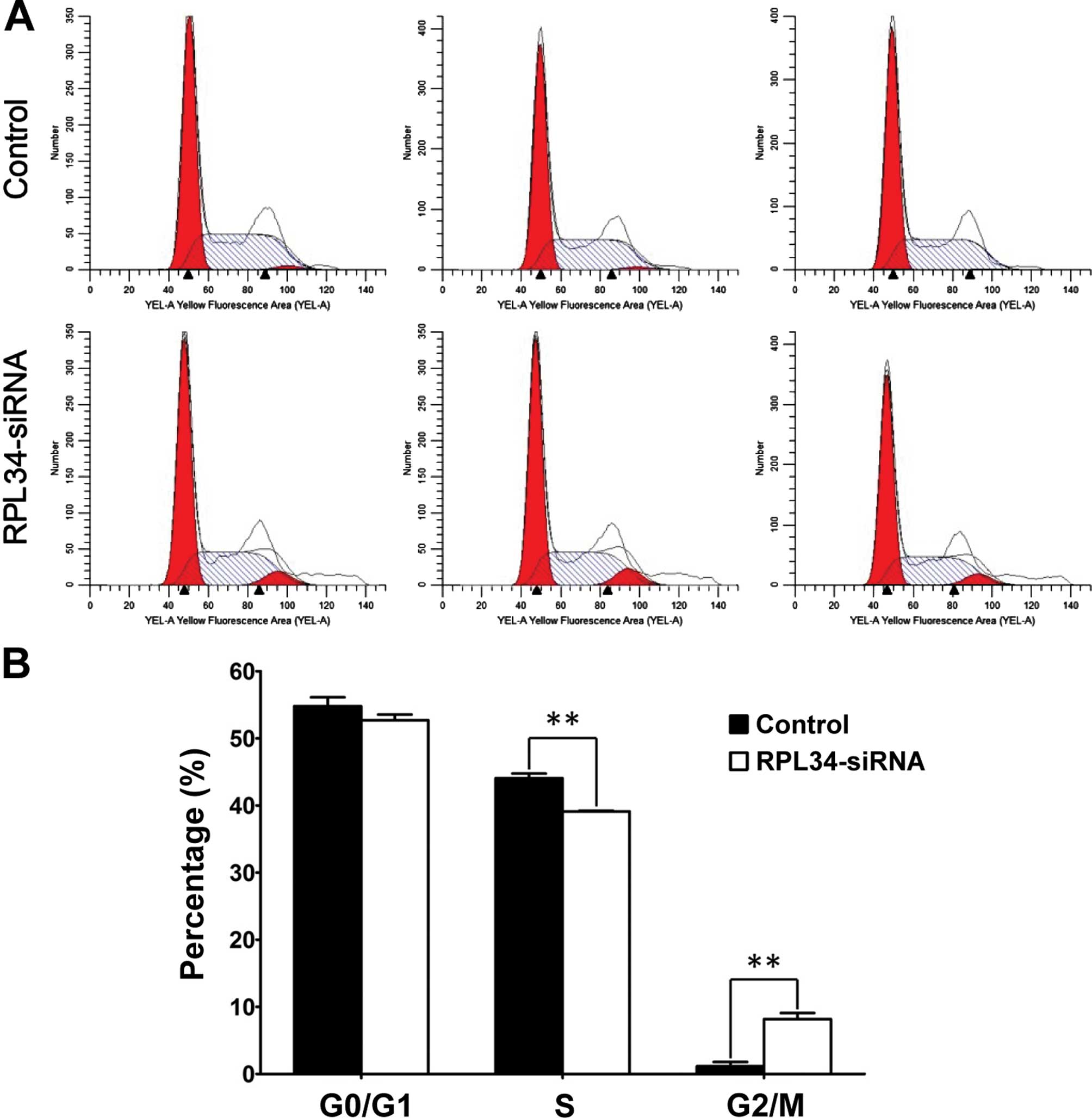
Knockdown of RPL34 in SGC-7901 cells
leads to cell cycle arrest
To determine whether RPL34 is necessary for cell
cycle progression in SGC-7901 cells, we assessed the cell cycle
phases in SGC-7901 cells by flow cytometry (Fig. 6A). The NC group displayed the
following distribution: (G0/G1 phase,
52.02±0.87%; S phase, 41.95±0.98%; and G2/M phase,
6.03±1.40%), and the RPL34-siRNA group displayed the following:
(G0/G1 phase, 67.65±1.00%; S phase,
25.02±0.91%; and G2/M phase, 7.33±0.14%). As shown in
Fig. 6B, compared to the control
cultures, RPL34-siRNA lentivirus cultures displayed a significant
decrease in the percentage of cells in the S phase (p<0.01) and
an increase in the percentage of cells in the G2/M phase
(p<0.01). Taken together, these data suggest that RPL34
regulates cell growth and blocks cell cycle progression in the
G2/M phase.
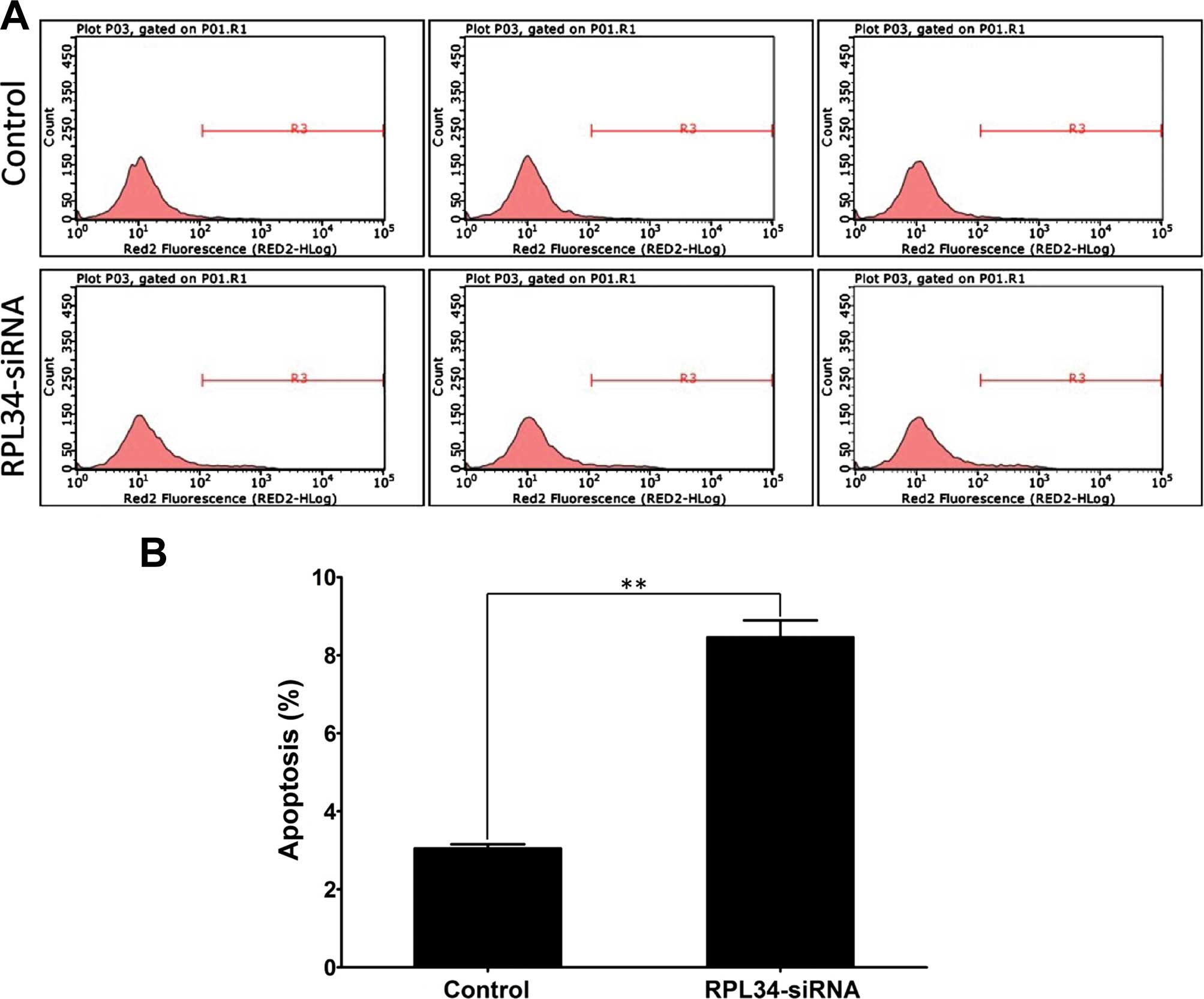
Knockdown of RPL34 in SGC-7901 cells
increases cell apoptosis
To test whether RPL34 expression affects apoptosis
in GC cells, we knocked down RPL34 in SGC-7901 cells. Cell
apoptosis was determined by Annexin V staining followed by flow
cytometry (Fig. 7A). As shown in
Fig. 7B, cell apoptosis was
significantly increased in the RPL34-siRNA group compared to the NC
group (NC 3.05±0.10% vs. RPL34-siRNA 8.46±0.43%, p=0.001). These
results indicate that RPL34 expression is a determinant of cell
apoptosis in SGC-7901 cells.
Discussion
Gastric cancer (GC) is one of the most common
cancers and the third leading cause of cancer-related death in both
genders worldwide (1,2,22).
Gene therapy is being studied as a potential therapeutic modality
for treating cancer (23). However,
the development and progression of GC remain poorly understood.
Therefore, it is particularly important to identify novel factors
associated with gastric malignant transformation and to unravel the
underlying mechanisms (24).
Ribosomal proteins (RPs), encoded by essential
housekeeping RP genes, are constitutively expressed in most
eukaryotic cells. While the interest in identifying human RPs comes
from results indicating their involvement in human cancer (14,25),
most research thus far has focused on the expression and function
of RPL34 in bacteria (26–28), Drosophila (26), mosquito (29) and amphioxus (30). However, RP expression and function
in human GC have not yet been studied.
In the present study, we first determined the
expression levels of RPL34 mRNA in four GC cell lines and found
that it was expressed in all of them. Lentiviral vector is an
efficient gene delivery vehicle due to its unique capability to
deliver target molecules into the host cell DNA and replicate in
non-dividing cells (31). In order
to assess RPL34 function in GC cell lines, we constructed the
RPL34-siRNA lentiviral vector, which efficiently silenced RPL34 in
the SGC-7901 cell line. Compared to the control-infected cells,
RPL34-siRNA-treated cells showed decreased proliferation and a
significant decrease in the proportion of cells in the S phase. A
significantly increased G2/M phase population was also
detected. In addition, we found that knockdown of RPL34 increased
apoptosis in the SGC-7901 cells. Taken together, these results
suggest that RPL34 promotes SGC-7901 cell growth. Further study is
ongoing to validate the anti-apoptotic role of RPL34 in other GC
cell lines.
In conclusion, in the present study we demonstrated
that downregulation of RPL34 expression by RNAi in SCG-7901 cells
inhibited cell proliferation and induced cell apoptosis. Therefore,
knockdown of RPL34 by lentivirus-siRNA may be a candidate approach
for treatment of GCs in which RPL34 is overexpressed.
References
|
1
|
Torre LA, Bray F, Siegel RL, Ferlay J,
Lortet-Tieulent J and Jemal A: Global cancer statistics, 2012. CA
Cancer J Clin. 65:87–108. 2015. View Article : Google Scholar : PubMed/NCBI
|
|
2
|
Sasako M, Inoue M, Lin JT, Khor C, Yang HK
and Ohtsu A: Gastric Cancer Working Group report. Jpn J Clin Oncol.
40(Suppl 1): i28–i37. 2010. View Article : Google Scholar : PubMed/NCBI
|
|
3
|
Catalano V, Labianca R, Beretta GD, Gatta
G, de Braud F and Van Cutsem E: Gastric cancer. Crit Rev Oncol
Hematol. 71:127–164. 2009. View Article : Google Scholar : PubMed/NCBI
|
|
4
|
Danaei G, Vander Hoorn S, Lopez AD, Murray
CJ and Ezzati M; Comparative Risk Assessment collaborating group
(Cancers): Causes of cancer in the world: Comparative risk
assessment of nine behavioural and environmental risk factors.
Lancet. 366:1784–1793. 2005. View Article : Google Scholar : PubMed/NCBI
|
|
5
|
Niccolai E, Taddei A, Prisco D and Amedei
A: Gastric cancer and the epoch of immunotherapy approaches. World
J Gastroenterol. 21:5778–5793. 2015.PubMed/NCBI
|
|
6
|
Quiros RM and Bui CL: Multidisciplinary
approach to esophageal and gastric cancer. Surg Clin North Am.
89:79–96. viii2009. View Article : Google Scholar : PubMed/NCBI
|
|
7
|
Foo M and Leong T: Adjuvant therapy for
gastric cancer: Current and future directions. World J
Gastroenterol. 20:13718–13727. 2014. View Article : Google Scholar : PubMed/NCBI
|
|
8
|
Warner JR, Mitra G, Schwindinger WF,
Studeny M and Fried HM: Saccharomyces cerevisiae coordinates
accumulation of yeast ribosomal proteins by modulating mRNA
splicing, translational initiation, and protein turnover. Mol Cell
Biol. 5:1512–1521. 1985.PubMed/NCBI
|
|
9
|
Wool IG: The structure and function of
eukaryotic ribosomes. Annu Rev Biochem. 48:719–754. 1979.
View Article : Google Scholar : PubMed/NCBI
|
|
10
|
Roberts E, Sethi A, Montoya J, Woese CR
and Luthey-Schulten Z: Molecular signatures of ribosomal evolution.
Proc Natl Acad Sci USA. 105:13953–13958. 2008. View Article : Google Scholar : PubMed/NCBI
|
|
11
|
Ferrari S, Manfredini R, Tagliafico E,
Rossi E, Donelli A, Torelli G and Torelli U: Noncoordinated
expression of S6, S11, and S14 ribosomal protein genes in leukemic
blast cells. Cancer Res. 50:5825–5828. 1990.PubMed/NCBI
|
|
12
|
Fisher EM, Beer-Romero P, Brown LG, Ridley
A, McNeil JA, Lawrence JB, Willard HF, Bieber FR and Page DC:
Homologous ribosomal protein genes on the human X and Y
chromosomes: Escape from X inactivation and possible implications
for Turner syndrome. Cell. 63:1205–1218. 1990. View Article : Google Scholar : PubMed/NCBI
|
|
13
|
Chan YL, Suzuki K, Olvera J and Wool IG:
Zinc finger-like motifs in rat ribosomal proteins S27 and S29.
Nucleic Acids Res. 21:649–655. 1993. View Article : Google Scholar : PubMed/NCBI
|
|
14
|
Frigerio JM, Dagorn JC and Iovanna JL:
Cloning, sequencing and expression of the L5, L21, L27a, L28, S5,
S9, S10 and S29 human ribosomal protein mRNAs. Biochim Biophys
Acta. 1262:64–68. 1995. View Article : Google Scholar : PubMed/NCBI
|
|
15
|
Strausberg RL, Feingold EA, Grouse LH,
Derge JG, Klausner RD, Collins FS, Wagner L, Shenmen CM, Schuler
GD, Altschul SF, et al Mammalian Gene Collection Program Team:
Generation and initial analysis of more than 15,000 full-length
human and mouse cDNA sequences. Proc Natl Acad Sci USA.
99:16899–16903. 2002. View Article : Google Scholar : PubMed/NCBI
|
|
16
|
Aoyama Y, Chan YL and Wool IG: The primary
structure of rat ribosomal protein L34. FEBS Lett. 249:119–122.
1989. View Article : Google Scholar : PubMed/NCBI
|
|
17
|
Tsurugi K, Collatz E, Todokoro K and Wool
IG: Isolation of eukaryotic ribosomal proteins. Isolation of
eukaryotic ribosomal proteins. Purification and characterization of
60 S ribosomal subunit proteins L3, L6, L7′, L8, L10, L15, L17,
L18, L19, L23′, L25, L27′, L28, L29, L31, L32, L34, L35, L36, L36′,
and L37′. J Biol Chem. 252:3961–3969. 1977.PubMed/NCBI
|
|
18
|
Lois C, Hong EJ, Pease S, Brown EJ and
Baltimore D: Germline transmission and tissue-specific expression
of transgenes delivered by lentiviral vectors. Science.
295:868–872. 2002. View Article : Google Scholar : PubMed/NCBI
|
|
19
|
Laemmli UK: Cleavage of structural
proteins during the assembly of the head of bacteriophage T4.
Nature. 227:680–685. 1970. View
Article : Google Scholar : PubMed/NCBI
|
|
20
|
Zhou Y, Su Z, Huang Y, Sun T, Chen S, Wu
T, Chen G, Xie X, Li B and Du Z: The Zfx gene is expressed in human
gliomas and is important in the proliferation and apoptosis of the
human malignant glioma cell line U251. J Exp Clin Cancer Res.
30:1142011. View Article : Google Scholar : PubMed/NCBI
|
|
21
|
Milner AE, Levens JM and Gregory CD: Flow
cytometric methods of analyzing apoptotic cells. Methods Mol Biol.
80:347–354. 1998. View Article : Google Scholar : PubMed/NCBI
|
|
22
|
Hamashima C, Shabana M, Okamoto M, Osaki Y
and Kishimoto T: Survival analysis of patients with interval cancer
undergoing gastric cancer screening by endoscopy. PLoS One.
10:e01267962015. View Article : Google Scholar : PubMed/NCBI
|
|
23
|
Guinn BA and Mulherkar R: International
progress in cancer gene therapy. Cancer Gene Ther. 15:765–775.
2008. View Article : Google Scholar : PubMed/NCBI
|
|
24
|
Ludwig JA and Weinstein JN: Biomarkers in
cancer staging, prognosis and treatment selection. Nat Rev Cancer.
5:845–856. 2005. View
Article : Google Scholar : PubMed/NCBI
|
|
25
|
Pogue-Geile K, Geiser JR, Shu M, Miller C,
Wool IG, Meisler AI and Pipas JM: Ribosomal protein genes are
overexpressed in colorectal cancer: Isolation of a cDNA clone
encoding the human S3 ribosomal protein. Mol Cell Biol.
11:3842–3849. 1991.PubMed/NCBI
|
|
26
|
Akanuma G, Kobayashi A, Suzuki S, Kawamura
F, Shiwa Y, Watanabe S, Yoshikawa H, Hanai R and Ishizuka M: Defect
in the formation of 70S ribosomes caused by lack of ribosomal
protein L34 can be suppressed by magnesium. J Bacteriol.
196:3820–3830. 2014. View Article : Google Scholar : PubMed/NCBI
|
|
27
|
Panagiotidis CA, Huang SC and Canellakis
ES: Relationship of the expression of the S20 and L34 ribosomal
proteins to polyamine biosynthesis in Escherichia coli. Int J
Biochem Cell Biol. 27:157–168. 1995. View Article : Google Scholar : PubMed/NCBI
|
|
28
|
Kruft V, Kapp U and Wittmann-Liebold B:
Characterization and primary structure of proteins L28, L33 and L34
from Bacillus stearothermophilus ribosomes. Biochimie. 73:855–860.
1991. View Article : Google Scholar : PubMed/NCBI
|
|
29
|
Niu LL and Fallon AM: The ribosomal
protein L34 gene from the mosquito, Aedes albopictus: Exon-intron
organization, copy number, and potential regulatory elements.
Insect Biochem Mol Biol. 29:1105–1117. 1999. View Article : Google Scholar : PubMed/NCBI
|
|
30
|
Liu L, Zhang S, Liu Z, Li H, Liu M, Wang Y
and Ma L: Ribosomal proteins L34 and S29 of amphioxus Branchiostoma
belcheri tsingtauense: cDNAs cloning and gene copy number. Acta
Biochim Pol. 52:857–862. 2005.PubMed/NCBI
|
|
31
|
Lever AM, Strappe PM and Zhao J:
Lentiviral vectors. J Biomed Sci. 11:439–449. 2004. View Article : Google Scholar : PubMed/NCBI
|