Introduction
Glioblastoma (GBM) is the most common and most
aggressive primary brain tumor. Despite aggressive surgery,
radiotherapy and chemotherapy, the prognosis of GBM remains poor.
Gene therapy is increasingly being explored as a novel therapeutic
option.
The sodium/iodide symporter (NIS), which facilitates
iodide uptake driven by sodium ion gradients across the plasma
membrane, is a simple and useful reporter. Iodine radioisotopes
were first used as thyroid function tracers and subsequently for
treating hyperthyroidism and benign thyroid diseases (1). NIS can absorb several isotopes, such
as 99mTc, 188Re and 131I, which
are important for nuclear medicine imaging and radionuclide therapy
(2–4). Therefore, as a therapeutic ectopic
gene, NIS has been used in numerous preclinical studies on
the treatment of a variety of cancers (5–7).
Vadysirisack et al showed that the radioiodine uptake is
proportionate to the total NIS protein level and that enhancing NIS
protein expression improves reporter sensitivity and the
therapeutic efficacy of radioiodine therapy (8). The early growth response-1 (Egr1)
promoter is a radio-inducible promoter that can be promoted by
ionizing radiation (9) and induce
the expression of downstream target genes. Our laboratory detected
an 131I radiation positive feedback effect in a
Bac-Egr1-hNIS (a baculovirus-containing Egr1-promoted NIS) system
in U87 glioma cells (10).
Various studies have suggested that antiangiogenic
drugs enhance the tumor response to radiotherapy (11) or radionuclide therapy (5,12).
Kringle 5 (K5), a kringle domain of plasminogen, can induce
apoptosis in dermal microvessel endothelial cells (ECs) (13,14)
and inhibit the proliferation of basic fibroblast growth
factor-stimulated calf pulmonary arterial EC and bovine adrenal
capillary ECs (14,15). In the present study, we constructed
a stable U87 cell line expressing K5 and investigated the effect of
K5 combined with the 131I radiation positive feedback
effect (Egr1-NIS) for treating malignant U87 glioma.
Materials and methods
Cell lines and animals
The U87 human glioma cell line [American Type
Culture Collection (ATCC), Manassas, VA, USA] and HUVECs and the
293T cell line (Cell Bank of the Chinese Academy of Science,
Shanghai, China) were maintained in Dulbecco's modified Eagle's
medium (DMEM) supplemented with 10% fetal bovine serum, 100 U/ml
penicillin and 100 mg/ml streptomycin in 5% CO2 at 37°C.
Twenty-four male BALB/c nude mice (4-weeks old) were purchased from
Shanghai Slaccas Experiment Animal Corporation (Shanghai Institute
for Biological Science, Shanghai, China).
Plasmid construction and lentiviral
preparation
The pcDNA3.1-hNIS vector was kindly provided by Dr
Sissy Jhiang (Ohio University, Athens, OH, USA). The pGL3-Egr1-Luc
plasmid, containing the luciferase gene under the control of the
Egr1 promoter, was a kind gift from Gerald Thiel (University of
Saarland Medical Center, Homburg, Germany). The pET22b-K5 plasmid
(His-tagged) was previously constructed in our laboratory (5). The pLVX-CMV-Puro (puromycin)
expression vector was purchased from Clontech (Takara, Dalian,
China). The NIS, EGR1 and K5 (His-tagged)
genes were PCR-amplified separately from pcDNA3.1-hNIS,
pGL3-Egr1-Luc and pET22b-K5, respectively, and then cloned into the
pLVX-CMV-Puro vector to construct pLVX-CMV-K5-Egr1-NIS,
pLVX-CMV-K5-Egr1-GFP and pLVX-CMV-GFP-Egr1-NIS plasmids,
respectively. The resulting plasmids were recombined with a
destination vector according to the manufacturer's instructions.
The viral particles were produced and amplified in 293T cells.
In vitro infection with the recombinant
lentiviruses
U87 cells were infected at an multiplicity of
infection (MOI) of 20 (2×10⁶ pfu/105 cells in
1 ml complete media) with pLVX-CMV-K5-Egr1-NIS,
pLVX-CMV-K5-Egr1-GFP and pLVX-CMV-GFP-Egr1-NIS, or not transduced.
To generate cell lines harboring the K5-expressing antibiotic
marker puromycin as controlled by the cytomegalovirus
(CMV)-enhancer/promoter (NIS was controlled by the Egr1 promoter),
cells were selected in 0.5 µg/ml puromycin (Sigma, Sydney,
Australia) for the required duration. The stable cell lines
U87-K5-NIS, U87-K5-GFP and U87-GFP-NIS were constructed.
Western blot analyses
U87-K5-NIS, U87-K5-GFP, U87-GFP-NIS and U87 cell
lysates were prepared using standard methods. To detect NIS
expression following 131I irradiation, Na131I
(final concentration, 3.7 MBq/ml) was added into the U87-K5-NIS and
U87-GFP-NIS cell cultures for 24 h, and the cell lysates were
obtained for western blot analysis. To detect K5 expression in the
cell supernatants, western blotting was performed according to a
previous study (16). K5 and NIS
proteins were separately run on 7 and 15% Tris-glycine gels,
respectively. Mouse anti-NIS (Millipore, Boston, MA, USA) and mouse
anti-His-tag (Abgent, Suzhou, China) antibodies were used at a
1:500 dilution at 4°C overnight, followed by incubation with the
secondary anti-mouse antibody and with peroxidase-conjugated goat
anti-mouse immunoglobulin G (1:2,500; Santa Cruz Biotechnology,
Santa Cruz, CA, USA) for 2 h at 4°C. Anti-β-actin (1:5,000; Abgent)
was used as the loading control. Immunodetection was carried out
using an ECL Western Blot Detection kit (Thermo Scientific, Pierce,
Waltman, MA, USA).
In vitro iodide uptake studies
We determined 125I uptake and efflux in
triplicate as previously described (17). The day before the experiment,
U87-K5-NIS, U87-K5-GFP, U87-GFP-NIS and U87 cells were plated
(2×10⁵ cells/well) into 24-well plates. After 24 h, 500
µl HBSS containing 3.7 kBq 125I and 10
µmol/l sodium iodide (NaI) was added. The cells were
incubated at 37°C for 5–120 min, washed twice with ice-cold HBSS
and lysed using 0.5 mol/l NaOH. The radioactivity (counts/min, cpm)
of the cell lysates was measured using an automatic γ-counter
(Shanghai Rihuan Company, Shanghai, China).
In vitro clonogenic assay
U87-K5-NIS, U87-K5-GFP, U87-GFP-NIS and U87 cells
were plated on 10-cm culture dishes (6×10⁶ cells/dish);
4.6 MBq 131I in HBSS was added to each dish. After 8 h,
the cells were washed three times with HBSS, trypsinized and plated
into 6-well culture plates (1,000 cells/well). On day 7, the cells
were stained with 1 ml crystal violet staining solution (Beyotime
Institute of Biotechnology, Shanghai, China) for 10 min, and
colonies containing >50 cells were counted; the results are
expressed as the percentage of surviving cells. The survival rate
was expressed as the percentage of colonies to that of the blank
group without 131I incubation. Data are presented as the
means ± SD.
Cytotoxic effect of K5 on HUVECs in
vitro
HUVECs were plated into 6-well culture plates
(5×10⁵ cells/well). U87-K5-NIS, U87-K5-GFP, U87-GFP-NIS
and U87 cells were plated on 10-cm culture dishes (5×10⁶
cells/dish). After a 24-h incubation, the U87-K5-NIS, U87-K5-GFP,
U87-GFP-NIS and U87 cells were washed three times with
phosphate-buffered saline (PBS) and 3 ml DMEM was added to each
dish. After 12 h, the medium was aspirated and centrifuged at 800 ×
g for 5 min. The HUVEC medium was removed, and then the centrifuged
medium was added to the HUVECs. After 24 h, the HUVECs were
trypsinized and plated into 6-well culture plates (500 cells/well).
On day 7, the cells were stained with 1 ml crystal violet staining
solution (Beyotime Institute of Biotechnology) for 10 min, and
colonies containing >50 cells were counted.
Establishment of xenograft tumors in nude
mice
The right flanks of the BALB/c nude mice were s.c.
injected with U87-K5-NIS, U87-K5-GFP, U87-GFP-NIS or U87 cells in
100 µl PBS. The mice were euthanized by cervical vertebra
dislocation at the end of the experiments. The animal studies were
approved by the local Ethics Committee (Shanghai Jiao Tong
University School of Medicine, Shanghai, China) and performed
according to the principles of ethics related to animal
experimentation.
Micro-single-photon emission computed
tomography/computed tomography (SPECT/CT) imaging
To detect iodide uptake by NIS as promoted by the
Egr1 promoter after iodide irradiation, three U87-K5-NIS
tumor-bearing mice were i.v. injected with 18.5 MBq
125I. Anesthesia was administered and maintained by
isoflurane inhalation, and the mice were positioned spread prone
and scanned using a small-animal micro-SPECT scanner (Bioscan,
Washington, DC, USA) at 1 and 24 h after 125I injection.
Without moving the mice, CT images were acquired (CT dose index;
CTDI = 6.1 cGy) before whole-body nanoSPECT images (10 sec/frame
for systematic scans) were obtained. Two days after the first
125I injection and microSPECT/CT imaging, U87-K5-NIS
tumor-bearing mice were i.v. injected with 18.5 MBq 125I
again and microSPECT/CT imaging was performed at 1 h after the
injection. The images were processed and were reconstructed using
Nuclear v1.02 software; images were acquired using HiSPECT 1.4.2
software and analyzed using InVivoScope 1.44 software (all from
Bioscan). Regions of interest (ROIs) were drawn around the visible
organs, and the radioactivity per volume unit (Conc) in the ROIs
was measured using InVivoScope 1.44.
In vivo 131I therapy
One day before 131I administration, the
animals received a 0.5-ml i.p. injection of 0.9% NaI solution to
block the thyroid uptake of any free radioactive iodide. Treatment
was initiated when the tumors had grown to 3–5 mm in diameter (~70
mm3). U87-K5-NIS, U87-K5-GFP, U87-GFP-NIS or U87
tumor-bearing mice were i.v. injected with 37 MBq 131I
on day 1 and 3. Tumor size was measured on day 7 after the
injection and every three days thereafter up to day 25 using
calipers; tumor volume was calculated as follows: Volume
(mm3) = (L × W2)/2 (18).
Histology and immunohistochemistry
At 25 days after 131I administration, the
animals were sacrificed, and the tumors were cryosectioned (5
µm) and subjected to immunofluorescence and
immunohistochemical analysis using mouse anti-CD31 (1:50; Dako,
Glostrup, Denmark), rabbit anti-human caspase-3 (1:30; Epitomics,
Burlingame, CA, USA) and rabbit anti-human Ki-67 antibodies (1:200;
Thermo Scientific, Fremont, CA, USA). Immunohistochemical analysis
was performed using Image-Pro Plus software (Media Cybernetics,
Rockville, MD, USA). The integral optical density (IOD) of every
visual field was calculated for each section. Data are represented
as means ± SD. Capillary density was defined as the number of
CD31+ ECs/high-power field (hpf; ×200). Five hpfs were
counted per section from six sections/tumor tissue in three
animals/group.
Statistical analysis
Data were analyzed using GraphPad Prism software
(version 5.0; GraphPad Software, Inc., La Jolla, CA, USA); the
means ± SD are presented. Statistical analyses were performed using
two-tailed Student's t-tests for differences between groups and
ANOVA for differences among groups. For all analyses, P<0.05 was
considered to indicate a statistically significant result.
Results
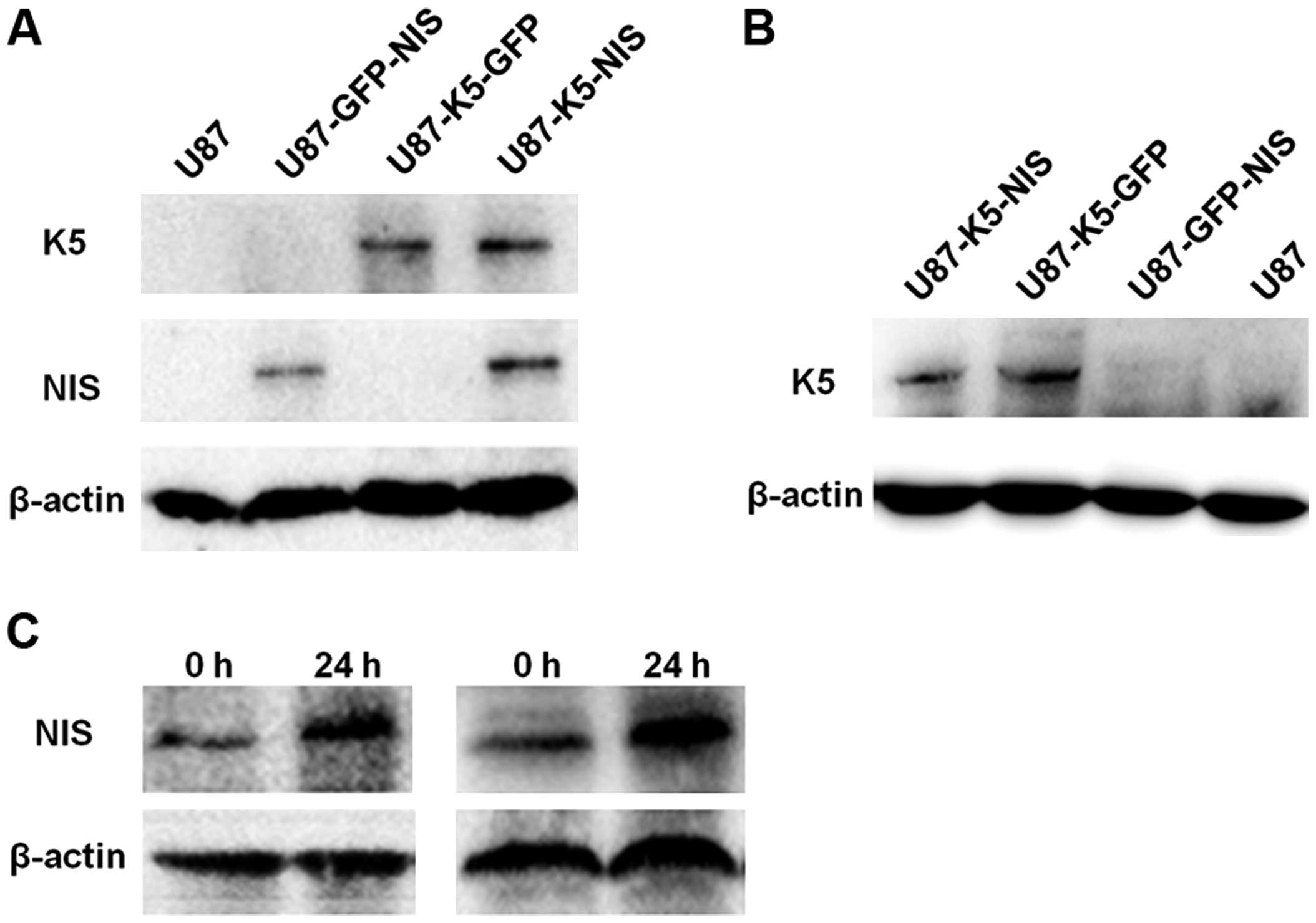
K5 and NIS expression
To investigate K5 and NIS expression in the
U87-K5-NIS, U87-K5-GFP, U87-GFP-NIS and U87 cell lines, we detected
K5 expression in the supernatant of these cell lines using western
blotting. K5 was expressed in the U87-K5-NIS and U87-K5-GFP cells
(Fig. 1A) and their supernatant
(Fig. 1B), but not in the
U87-GFP-NIS and U87 cells (Fig. 1A)
or their supernatant (Fig. 1B). NIS
was expressed in the U87-K5-NIS and U87-GFP-NIS cells but not in
the U87-K5-GFP or U87 cells (Fig.
1A). Western blotting also showed weak expression of NIS
protein in the U87-K5-NIS and U87-GFP-NIS cells without
irradiation, but higher NIS expression after a 24-h 131I
irradiation (Fig. 1C).
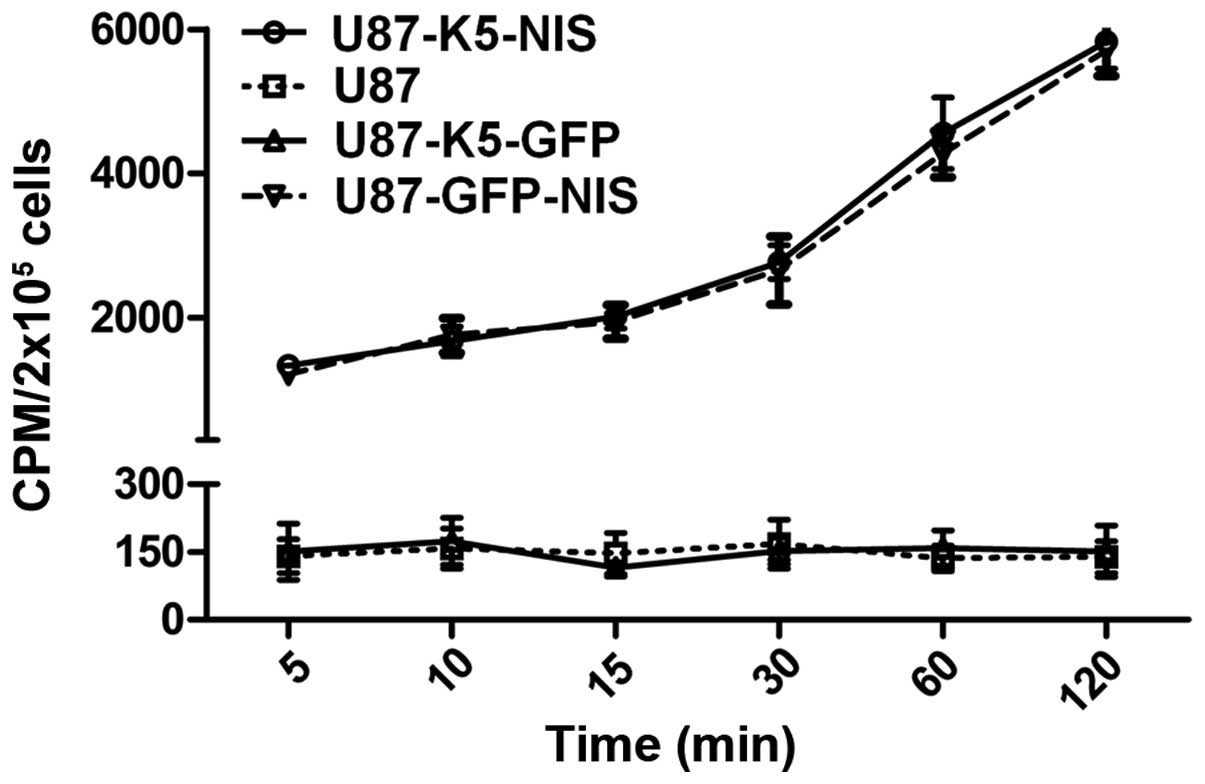
In vivo 125I uptake in cell
lines
The functional activity of NIS protein was clearly
shown by its cellular iodide uptake. 125I uptake by the
U87-K5-NIS and U87-GFP-NIS cells varied with the duration of
incubation. Following its addition to the U87-K5-NIS and
U87-GFP-NIS cells, 125I was gradually absorbed by NIS
protein and was ~6,000 cpm at 120 min; no functional iodide uptake
was observed in the U87-K5-GFP and U87 cells (Fig. 2).
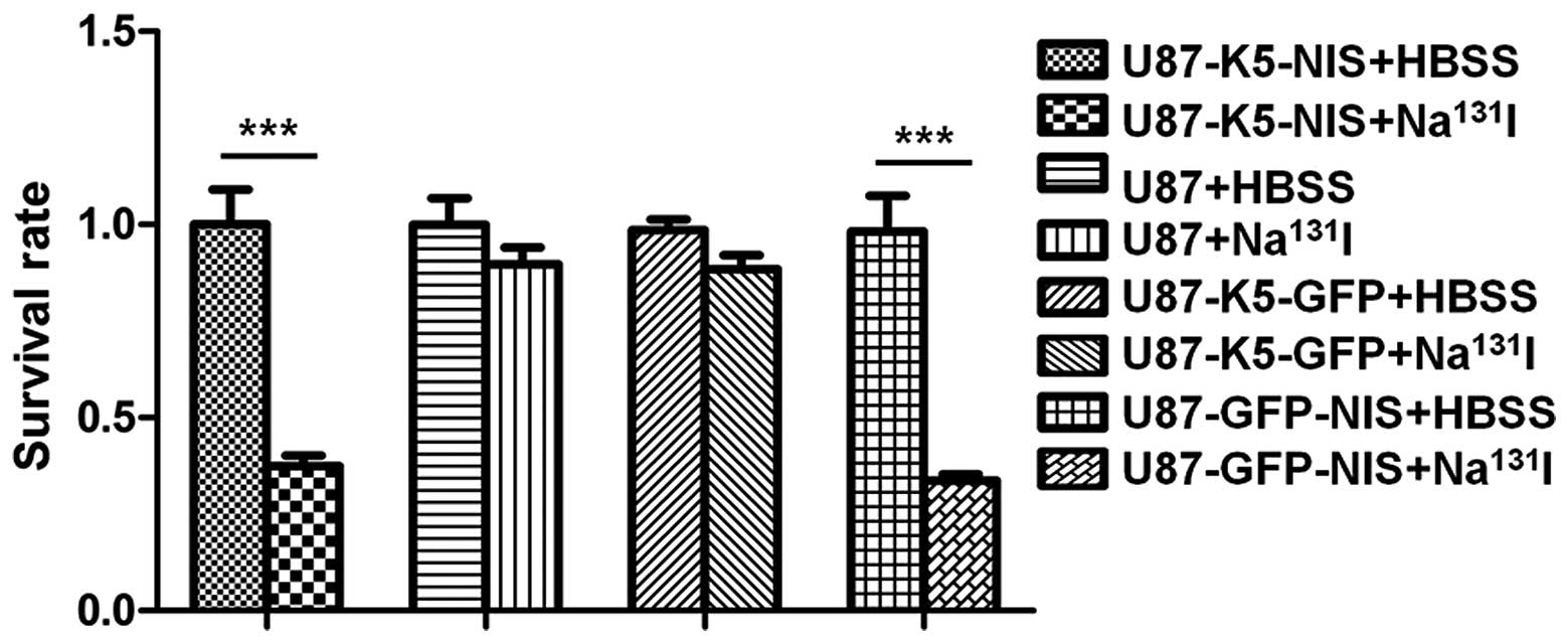
131I reduces the survival of
U87 cells expressing NIS in vitro
In vitro clonogenic assays were performed to
determine the cytotoxic effect of 131I on U87-K5-NIS,
U87-K5-GFP, U87-GFP-NIS and U87 cells. 131I had a
significant cytotoxic effect on the U87-K5-NIS and U87-GFP-NIS
cells when compared to the effect on the U87-K5-GFP and U87 cells
(P<0.001) and the control HBSS-treated U87-K5-NIS, U87-K5-GFP,
U87-GFP-NIS and U87 cells (P<0.001) (Fig. 3).
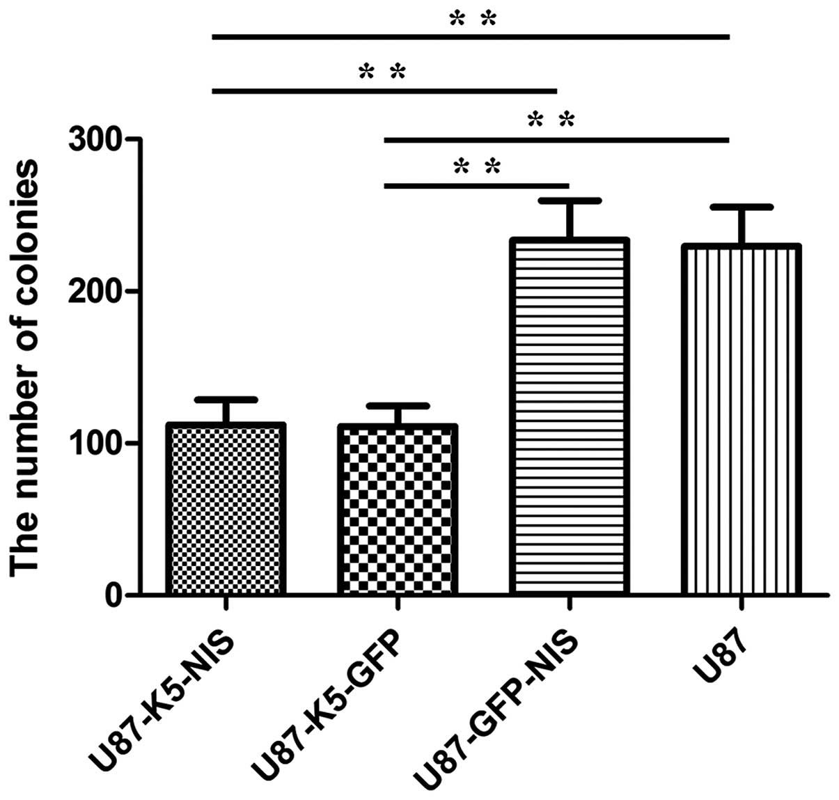
Cytotoxic effect of K5 on HUVECs in
vitro
In vitro clonogenic assays were performed to
determine the effect of K5 on HUVECs. Compared to the U87-GFP-NIS
and U87 cell culture medium, the medium containing K5 secreted by
the U87-K5-NIS and U87-K5-GFP cells had a significant cytotoxic
effect on the HUVECs (P<0.01) (Fig.
4).
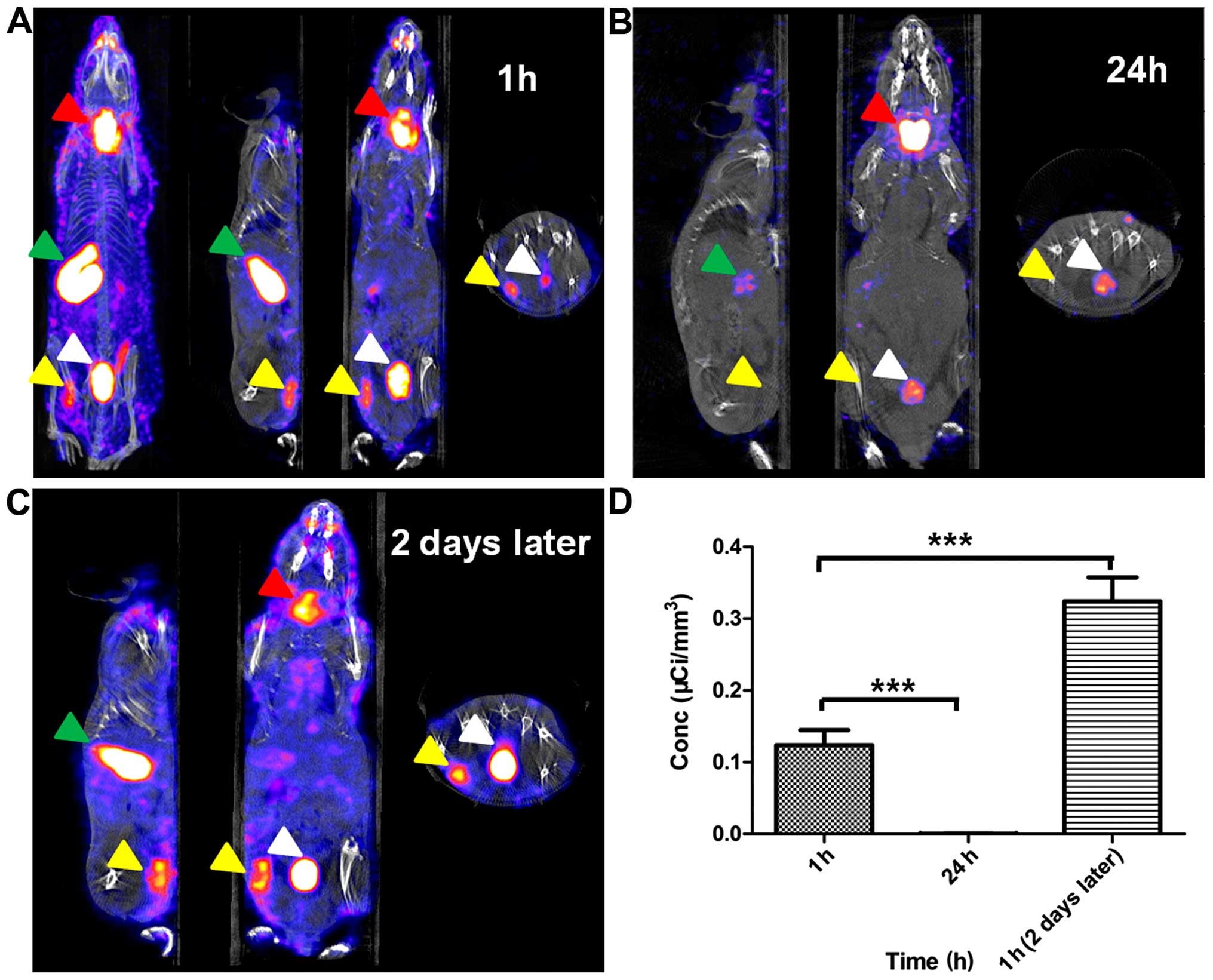
In vivo imaging of 125I
biodistribution in mice bearing U87-K5-NIS xenografts
Significant radioactive uptake was observed in the
U87-K5-NIS tumors at 1 h after 125I injection (Fig. 5A); however, 125I uptake
was not detectable in the U87-K5-NIS tumors 24 h after
125I injection (Fig.
5B). Two days after the first 125I injection, the
U87-K5-NIS tumor-bearing mice were i.v. injected with 18.5 MBq
125I again, and the radioactivity of the U87-K5-NIS
tumors was increased significantly (Fig. 5C). The images were processed and
ROIs were created by CT positioning during SPECT imaging to define
the tissues described above; the obtained Conc value was
0.1240±0.0201 µCi/mm3 at 1 h after the first
125I injection, which was much higher than that at 24 h
after 125I injection (0.0051±0.0002
µCi/mm3) (P<0.001). Two days later at 1 h
after the 125I injection, the Conc value was
0.3243±0.0333 µCi/mm3, which was almost three
times higher than that at 1 h after the first 125I
injection (P<0.001) (Fig. 5D).
Significant radio-iodine accumulation was also observed in tissues
expressing endogenous NIS, including the thyroid and
stomach, and in the urinary bladder due to renal elimination
(Fig. 5A–C).
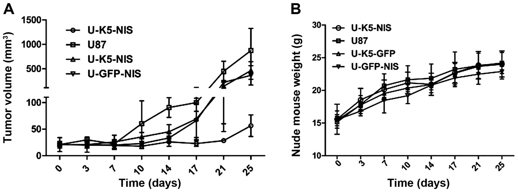
Therapeutic effects of K5 combined with
131I in U87 xenograft tumors expressing K5 and NIS in
vivo
We initiated 131I therapy when the tumors
were 3–5 mm in diameter (~70 mm3). K5 combined with
131I significantly inhibited U87-K5-NIS tumor growth as
compared to the 131I-treated U87-GFP-NIS, U87-K5-GFP and
U87 tumors (P<0.001). There was no significant difference
between U87-GFP-NIS and U87-K5-GFP tumor growth (P>0.05), but
these two tumors grew slowly compared with the U87 tumors
(P<0.05) (Fig. 6A). Therapy did
not affect mouse food intake or physical activity. During
treatment, the weight of the U87-K5-NIS tumor-bearing mice
increased at almost the same rate as the U87-GFP-NIS, U87-K5-GFP
and U87 tumor-bearing mice (P>0.05) (Fig. 6B).
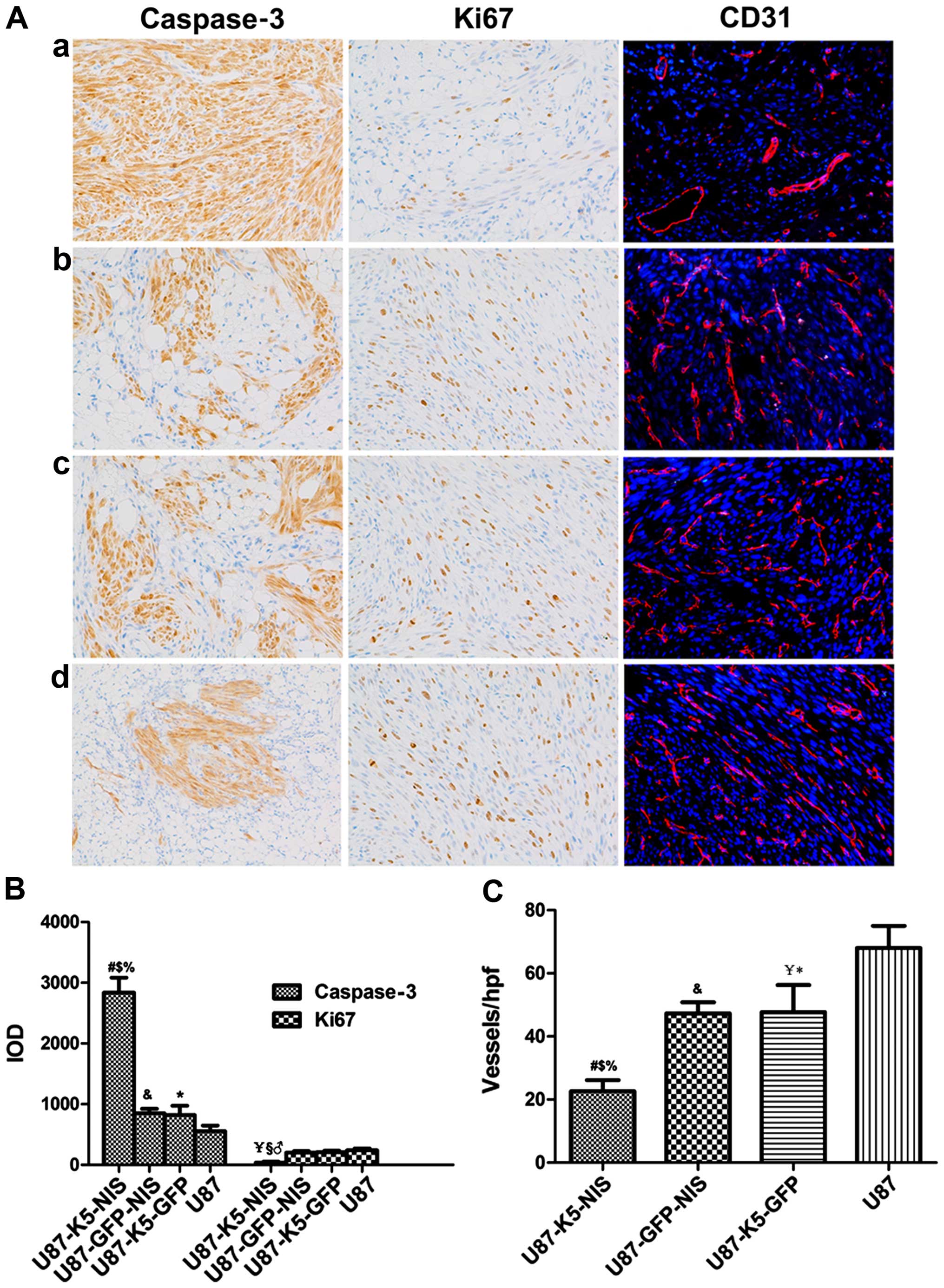
Immunohistochemical and
immunofluorescence analysis
Fig. 7 depicts
representative images of the indirect tumor xenograft
immunohistochemical staining. Immunohistochemical analysis revealed
higher caspase-3 protein expression in the 131I-treated
U87-K5-NIS xenograft cells as compared with the
131I-treated U87-GFP-NIS (P<0.001), U87-K5-GFP
(P<0.001) and U87 (P<0.001) xenograft cells, but lower Ki-67
protein expression in the 131I-treated U87-K5-NIS
xenograft cells as compared with the other three groups (P<0.01)
(Fig. 7A and B). There was higher
caspase-3 protein expression in the U87-GFP-NIS and U87-K5-GFP
xenograft cells compared with the U87 xenograft cells (P<0.05),
but the Ki-67 protein expression levels in the U87-GFP-NIS,
U87-K5-GFP and U87 xenograft cells was nearly identical (Fig. 7A and B). Immunofluorescence revealed
fewer CD31+ capillaries in the U87-K5-NIS group compared
with the U87-GFP-NIS (P<0.01), U87-K5-GFP (P<0.01) and U87
(P<0.001) groups. There were fewer CD31+ capillaries
in the U87-K5-GFP group as compared with the U87-GFP-NIS
(P<0.05) and U87 (P<0.05) groups (Fig. 7A and B).
Discussion
We tested the hypothesis that radioiodide
(131I) therapy mediated by the NIS gene and the
antiangiogenic agent K5 would have a synergistic effect on
therapeutic antitumor efficacy in glioma. To increase the
accumulation and radiotherapeutic effect of 131I, the
radio-inducible Egr1 promoter was used as the NIS gene
promoter, producing an 131I radiation positive feedback
effect. The combined therapy limited glioma growth and progression
more effectively. Our results indicated that the combination
therapy increased tumor cell apoptosis, inhibited tumor cell
proliferation and significantly reduced capillary density in the
U87 glioma tissues.
Gliomas are tumors that arise from glial or glial
progenitor cells in the central nervous system. The median survival
time of patients with malignant glioma is <3 years (19) and is even shorter in the event of
recurrence, usually 3–6 months (20). A combination of chemotherapy or
stereotactic radiosurgery with repeated surgery was previously
found to improve the survival of patients with recurrent GBM
compared to surgery alone, although no patient in the study
survived beyond 44 weeks after treatment (21). However, the use of radiotherapy is
limited in recurrent tumors due to the associated irreversible
brain tissue damage and radiation-induced necrosis of the normal
brain (22) highlighting the need
for new therapeutic strategies. Given the following advantages,
131I therapy, which is mediated by the NIS gene,
is used for treating thyroid cancer in the clinic. 131I
expends 971 KeV decay energy, with γ decay following rapidly after
β decay. The electrons have only 0.6–2 mm tissue penetration
(23) causing a low degree of
injury to the healthy tissues around the tumor. This indicates that
131I, mediated by the ectopic NIS gene, may have
significant potential as an effective low-toxic treatment for
glioma. Previous studies have demonstrated that NIS can be
transferred into a variety of tumors, including nasopharyngeal
carcinoma (17) breast cancer
(24), glioma (6) and prostate carcinoma (25) and is capable of 131I
uptake and has different inhibitory actions on tumor growth.
However, intracellular iodine is rapidly released, resulting in
limited radioiodide retention time within cells; therefore,
131I therapeutic efficacy is limited. To resolve this
issue, we used a radio-inducible Egr1 promoter to promote NIS
expression, creating an 131I radiation positive feedback
effect to increase NIS protein expression levels, increasing the
131I retention time and the amount in the cells. For
neovascularized tumors, neovasculature-targeting therapy is a
promising prospect that is aimed at destroying the blood vessels,
thus depriving tumor cells of oxygen and nutrients. K5 displays its
potent anti-angiogenic effect by inducing EC apoptosis (13,14).
Moreover, inhibition of angiogenesis was observed when colorectal
carcinoma cells stably expressing K5 were propagated subcutaneously
in athymic nude mice (26).
We successfully constructed the lentiviral vectors
pLVX-CMV-K5-Egr1-NIS, pLVX-CMV-K5-Egr1-GFP and
pLVX-CMV-GFP-Egr1-NIS, constructing the stable cell lines
U87-K5-NIS, U87-K5-GFP and U87-GFP-NIS following lentiviral
transduction and puromycin selection. K5 was expressed in
U87-K5-NIS and U87-K5-GFP cells and their supernatant but not in
U87-GFP-NIS and U87 cells or their supernatant; NIS was expressed
in U87-K5-NIS and U87-GFP-NIS cells but not in U87-K5-GFP and U87
cells. There was higher NIS expression following a 24-h
131I irradiation in U87-K5-NIS and U87-GFP-NIS cells.
Guo et al reported more NIS staining by immunofluorescence
testing in vitro following 131I (7.4 MBq/ml)
irradiation in Bac-Egr1-hNIS-transduced glioma cells (10). In vivo microSPECT/CT imaging
showed more significant radioactive uptake (0.3243±0.0333
µCi/mm3) in U87-K5-NIS tumors two days after
irradiation with 18.5 MBq 125I as compared with
U87-K5-NIS tumors without 125I irradiation
(0.1240±0.0201 µCi/mm3) (P<0.001). These
studies showed that the Egr1 promoter may be useful for increasing
NIS protein expression and for increasing NIS radioiodide
accumulation.
The in vitro clonogenic assays showed that
the medium containing K5 secreted by U87-K5-NIS and U87-K5-GFP
cells had a significant cytotoxic effect on HUVECs as compared to
that of U87-GFP-NIS and U87 cells (P<0.05). At 25 days after
131I administration, immunofluorescence showed a lower
density of CD31+ capillaries in the U87-K5-NIS group as
compared with the other three groups. Lower-density
CD31+ capillaries were also observed in the U87-K5-GFP
group compared with the U87-GFP-NIS (P<0.05) and U87 (P<0.05)
groups. Ki-67 and caspase-3 immunohistochemical analysis revealed
significantly fewer proliferating cells and increased apoptosis
following 131I treatment in U87-K5-NIS tumors as
compared to the other three groups. K5 combined with
131I significantly inhibited U87-K5-NIS tumor growth.
These results showed that NIS-mediated 131I irradiation
therapy combined with K5 gene therapy was superior to
single-gene therapy alone. Not only did NIS-mediated
131I irradiation increase EC sensitivity to K5-induced
apoptosis, K5 increased the 131I-mediated glioma cell
apoptosis.
There are various limitations to the present study.
First, although lentiviral vectors can infect both non-dividing and
dividing cells (27), induce
long-term expression of transgenes and lack antigenicity (28,29),
genetic modification with lentiviral vectors in general and stable
integration of the therapeutic gene into the host cell genome raise
concerns regarding personal or environmental safety (30). Second, we constructed stable cell
lines to study the efficacy of the combined gene therapy, which did
not conform to clinical treatment. In the clinic, viruses are
injected directly or i.v. into tumors. Therefore, tumor-specific
promoters in vivo are advantageous due to their lower
sensitivity to promoter inactivation and lower risk of activating
the host cell defense machinery (31). Previously, we showed that NIS
expression under the human telomerase reverse transcriptase (hTERT)
promoter was similar to that under the CMV promoter and that the
hTERT promoter may have not only specificity but also relatively
strong transcriptional activity for targeted gene expression
(5).
In conclusion, the Egr1 promoter induced an
131I radiation positive feedback effect mediated by NIS.
K5 increased 131I-mediated glioma cell apoptosis;
131I irradiation also increased EC sensitivity to
K5-induced apoptosis. This combined gene therapy had a synergistic
effect on therapeutic antitumor efficacy in glioma, inhibiting U87
cell proliferation, arresting angiogenesis and retarding U87 tumor
growth more effectively than single-gene therapy alone.
Acknowledgments
The present study was supported by grants from the
National Natural Science Foundation of China (nos. 81071181,
81271610 and 81471686), the Shanghai Outstanding Academic Leaders
Project (11XD1403700), and the Discipline Leaders Climbing Project
of Ruijin Hospital and Shanghai Health Bureau Youth Foundation
(2010Y021). We are also indebted to the staff of the Department of
Nuclear Medicine, Fudan University Shanghai Cancer Center for their
technological support with micro-SPECT/CT imaging.
References
|
1
|
Chung JK: Sodium iodide symporter: Its
role in nuclear medicine. J Nucl Med. 43:1188–1200. 2002.PubMed/NCBI
|
|
2
|
Verburg FA, de Keizer B, Lips CJ, Zelissen
PM and de Klerk JM: Prognostic significance of successful ablation
with radioiodine of differentiated thyroid cancer patients. Eur J
Endocrinol. 152:33–37. 2005. View Article : Google Scholar : PubMed/NCBI
|
|
3
|
Hackshaw A, Harmer C, Mallick U, Haq M and
Franklyn JA: 131I activity for remnant ablation in
patients with differentiated thyroid cancer: A systematic review. J
Clin Endocrinol Metab. 92:28–38. 2007. View Article : Google Scholar
|
|
4
|
Chung JK, Youn HW, Kang JH, Lee HY and
Kang KW: Sodium iodide symporter and the radioiodine treatment of
thyroid carcinoma. Nucl Med Mol Imaging. 44:4–14. 2010. View Article : Google Scholar : PubMed/NCBI
|
|
5
|
Zhang M, Guo R, Shi S, Miao Y, Zhang Y and
Li B: Baculovirus vector-mediated transfer of sodium iodide
symporter and plasminogen kringle 5 genes for tumor radioiodide
therapy. PLoS One. 9:e923262014. View Article : Google Scholar : PubMed/NCBI
|
|
6
|
Guo R, Zhang M, Xi Y, Ma Y, Liang S, Shi
S, Miao Y and Li B: Theranostic studies of human sodium iodide
symporter imaging and therapy using 188Re: A human
glioma study in mice. PLoS One. 9:e1020112014. View Article : Google Scholar
|
|
7
|
Kim YH, Youn H, Na J, Hong KJ, Kang KW,
Lee DS and Chung JK: Codon-optimized human sodium iodide symporter
(opt-hNIS) as a sensitive reporter and efficient therapeutic gene.
Theranostics. 5:86–96. 2015. View Article : Google Scholar : PubMed/NCBI
|
|
8
|
Vadysirisack DD, Shen DH and Jhiang SM:
Correlation of Na+/I− symporter expression
and activity: Implications of Na+/I−
symporter as an imaging reporter gene. J Nucl Med. 47:182–190.
2006.PubMed/NCBI
|
|
9
|
Wagner M, Schmelz K, Dörken B and Tamm I:
Transcriptional regulation of human survivin by early growth
response (Egr)-1 transcription factor. Int J Cancer. 122:1278–1287.
2008. View Article : Google Scholar
|
|
10
|
Guo R, Tian L, Han B, Xu H, Zhang M and Li
B: Feasibility of a novel positive feedback effect of
131I-promoted Bac-Egr1-hNIS expression in malignant
glioma via baculovirus. Nucl Med Biol. 38:599–604. 2011. View Article : Google Scholar : PubMed/NCBI
|
|
11
|
Gorski DH, Beckett MA, Jaskowiak NT,
Calvin DP, Mauceri HJ, Salloum RM, Seetharam S, Koons A, Hari DM,
Kufe DW, et al: Blockage of the vascular endothelial growth factor
stress response increases the antitumor effects of ionizing
radiation. Cancer Res. 59:3374–3378. 1999.PubMed/NCBI
|
|
12
|
Zhu X, Palmer MR, Makrigiorgos GM and
Kassis AI: Solid-tumor radionuclide therapy dosimetry: New
paradigms in view of tumor microenvironment and angiogenesis. Med
Phys. 37:2974–2984. 2010. View Article : Google Scholar : PubMed/NCBI
|
|
13
|
Davidson DJ, Haskell C, Majest S, Kherzai
A, Egan DA, Walter KA, Schneider A, Gubbins EF, Solomon L, Chen Z,
et al: Kringle 5 of human plasminogen induces apoptosis of
endothelial and tumor cells through surface-expressed
glucose-regulated protein 78. Cancer Res. 65:4663–4672. 2005.
View Article : Google Scholar : PubMed/NCBI
|
|
14
|
Lu H, Dhanabal M, Volk R, Waterman MJ,
Ramchandran R, Knebelmann B, Segal M and Sukhatme VP: Kringle 5
causes cell cycle arrest and apoptosis of endothelial cells.
Biochem Biophys Res Commun. 258:668–673. 1999. View Article : Google Scholar : PubMed/NCBI
|
|
15
|
Cao Y, Chen A, An SS, Ji RW, Davidson D
and Llinás M: Kringle 5 of plasminogen is a novel inhibitor of
endothelial cell growth. J Biol Chem. 272:22924–22928. 1997.
View Article : Google Scholar : PubMed/NCBI
|
|
16
|
Blezinger P, Wang J, Gondo M, Quezada A,
Mehrens D, French M, Singhal A, Sullivan S, Rolland A, Ralston R,
et al: Systemic inhibition of tumor growth and tumor metastases by
intramuscular administration of the endostatin gene. Nat
Biotechnol. 17:343–348. 1999. View
Article : Google Scholar : PubMed/NCBI
|
|
17
|
Shi S, Zhang M, Guo R, Miao Y, Hu J, Xi Y
and Li B: In vivo molecular imaging and radionuclide
(131I) therapy of human nasopharyngeal carcinoma cells
transfected with a lentivirus expressing sodium iodide symporter.
PLoS One. 10:e01165312015. View Article : Google Scholar
|
|
18
|
Sides MD, Sosulski ml, Luo F, Lin Z,
Flemington EK and Lasky JA: Co-treatment with arsenic trioxide and
ganciclovir reduces tumor volume in a murine xenograft model of
nasopharyngeal carcinoma. Virol J. 10(152)2013. View Article : Google Scholar : PubMed/NCBI
|
|
19
|
Chamberlain MC: Emerging clinical
principles on the use of bevacizumab for the treatment of malignant
gliomas. Cancer. 116:3988–3999. 2010. View Article : Google Scholar : PubMed/NCBI
|
|
20
|
Niyazi M, Siefert A, Schwarz SB, Ganswindt
U, Kreth FW, Tonn JC and Belka C: Therapeutic options for recurrent
malignant glioma. Radiother Oncol. 98:1–14. 2011. View Article : Google Scholar
|
|
21
|
Mandl ES, Dirven CM, Buis DR, Postma TJ
and Vandertop WP: Repeated surgery for glioblastoma multiforme:
Only in combination with other salvage therapy. Surg Neurol.
69:506–509. 2008. View Article : Google Scholar : PubMed/NCBI
|
|
22
|
Mayer R and Sminia P: Reirradiation
tolerance of the human brain. Int J Radiat Oncol Biol Phys.
70:1350–1360. 2008. View Article : Google Scholar
|
|
23
|
Skugor M: The Cleveland Clinic Guide to
Thyroid Disorders. Kaplan Publisher; New York: 2006
|
|
24
|
Dwyer RM, Bergert ER, O'connor MK, Gendler
SJ and Morris JC: In vivo radioiodide imaging and treatment of
breast cancer xenografts after MUC1-driven expression of the sodium
iodide symporter. Clin Cancer Res. 11:1483–1489. 2005. View Article : Google Scholar : PubMed/NCBI
|
|
25
|
Dwyer RM, Schatz SM, Bergert ER, Myers RM,
Harvey ME, Classic KL, Blanco MC, Frisk CS, Marler RJ and Davis BJ:
A preclinical large animal model of adenovirus-mediated expression
of the sodium-iodide symporter for radioiodide imaging and therapy
of locally recurrent prostate cancer. Mol Ther. 12:835–841. 2005.
View Article : Google Scholar : PubMed/NCBI
|
|
26
|
Liu XY, Qiu SB, Zou WG, Pei ZF, Gu JF, Luo
CX, Ruan HM, Chen Y, Qi YP and Qian C: Effective gene-virotherapy
for complete eradication of tumor mediated by the combination of
hTRAIL (TNFSF10) and plasminogen k5. Mol Ther. 11:531–541. 2005.
View Article : Google Scholar : PubMed/NCBI
|
|
27
|
Naldini L, Blömer U, Gallay P, Ory D,
Mulligan R, Gage FH, Verma IM and Trono D: In vivo gene delivery
and stable transduction of nondividing cells by a lentiviral
vector. Science. 272:263–267. 1996. View Article : Google Scholar : PubMed/NCBI
|
|
28
|
Sakuma T, Barry MA and Ikeda Y: Lentiviral
vectors: Basic to translational. Biochem J. 443:603–618. 2012.
View Article : Google Scholar : PubMed/NCBI
|
|
29
|
Charrier S, Stockholm D, Seye K, Opolon P,
Taveau M, Gross DA, Bucher-Laurent S, Delenda C, Vainchenker W,
Danos O, et al: A lentiviral vector encoding the human
Wiskott-Aldrich syndrome protein corrects immune and cytoskeletal
defects in WASP knockout mice. Gene Ther. 12:597–606. 2005.
View Article : Google Scholar
|
|
30
|
Rothe M, Modlich U and Schambach A:
Biosafety challenges for use of lentiviral vectors in gene therapy.
Curr Gene Ther. 13:453–468. 2013. View Article : Google Scholar : PubMed/NCBI
|
|
31
|
Liu BH, Wang X, Ma YX and Wang S: CMV
enhancer/human PDGF-beta promoter for neuron-specific transgene
expression. Gene Ther. 11:52–60. 2004. View Article : Google Scholar
|