Introduction
Colorectal cancer (CRC) is one of the most common
malignant types of cancer with the third highest mortality rate
(1). The diagnosis and treatment of
CRC can be quite difficult as the molecular mechanisms are as yet
not fully elucidated. To date, the most effective therapy is
surgery combined with chemotherapy and the 5-year survival rate of
a local tumor is ~93% (2,3). However, nearly half of patients have
metastasis when they are first diagnosed and more than a third
relapse after surgery. Even with advanced surgical techniques and
new molecular therapies, death rates for CRC patients are still
high, and this is largely due to recurrence of cancer due to
metastasis (4).
The molecular mechanism that drive CRC in terms of
progression as well as invasion and metastasis have not been fully
elucidated. It is generally thought that occurrence and progression
of CRC is a multi-factor, multi-phase, multi-step and multi-gene
process (5,6). Most of the mutations that drive CRC
occur in oncogenes driving activation or in tumor suppressor genes
to causing inactivation (7).
Numerous different studies have shown that the occurrence and
progression of CRC, as well as invasion and metastasis, involve
many related genes. For example, mutations in c-myc, Ras, Her-2 and
the cancer suppressor gene p53, which is deleted in colorectal
carcinoma, can drive occurrence. Other genes involved in CRC
progression include PTEN (phosphatase and tensin homologue, deleted
on chromosome 10), a metastasis suppressor gene nm23 and its
encoding genes, such as CD44, MMPs, MUC and dMMR. The loss of DNA
mismatch repair mechanisms also drives progression as well as
mutations in KRAS and BRAF (8,9).
Therefore, the molecular mechanisms are complicated.
The occurrence and progression of a given cancer is
a multi-factor and multi-step process, in which abnormal gene
expression plays a vital role (10). However, with continued research into
pathogenic mechanisms of CRC, molecular markers that play an
essential role in the early diagnosis and drive clinical
progression, have become more apparent and have led to the concept
of individual-based treatment being a realistic goal (9). In addition, this increased knowledge
of biomarkers will undoubtedly assist in earlier diagnosis and more
accurate monitoring of disease progression. To date, nearly 1500
miRNA sequences have been found. Studies of 186 miRNA locus, have
suggested that miRNA may be essential for the occurrence and
development of many different types of cancer (11). In the present study, we correlated
the expression of miR-181a with incidence and survival, in patients
with late liver metastases of CRC.
Materials and methods
Tissue specimens and cell cultures
CRC tissue and non-cancer liver tissue were obtained
from patients with colorectal cancer. All tissues were stored in
liquid nitrogen. Written informed consent was signed by all
participating patients. This study was approved and permitted by
the Ethics Committee of Shandong Cancer Hospital and Institute. All
clinical and histopathological indexes were rendered anonymous. The
human colon NCM460 cells and human colon cancer cells SW620 were
cultured with Dulbecco's modified Eagle's medium (DMEM) and 10%
fetal bovine serum (FBS), 100 U/ml penicillin and 100 mg/ml
streptomycin (Invitrogen, Carlsbad, CA, USA).
Survival analysis
Overall survival (OS) was evaluated in 72 CRC
patients with late liver metastases and 69 normal control patients
from Shandong Cancer Hospital and Institute. The survival curves
were analyzed using the Kaplan-Meier method and statistically
compared using a log-rank test.
RNA extraction and real-time quantitative
RT-PCR
Total RNA was extracted from tissue samples and
NCM460 and SW620 cells using TRIzol reagent (Invitrogen). Total RNA
(2 µg) was used for reverse transcriptase cDNA Moloney
murine leukemia virus reverse transcriptase (Invitrogen) according
to the manufacturer's protocol. Real-time PCR was performed using
SsoFast™ EvaGreen Supermix (Bio-Rad Laboratories) and the
SYBR-Green PCR Master Mix (Toyobo Co., Ltd., Osaka, Japan). PCR
cycling conditions were: 95°C for 45 sec, 40 cycles of 95°C for 45
sec, 60°C for 30 sec and 72°C for 30 sec. 2−ΔCt where
ΔCt = Ct (Target) − Ct (Reference).
Cell viability assay
Cell viability of NCM460 and SW620 cells was
determined using MTT assay (Sigma-Aldrich, St. Louis, MO, USA).
Briefly, NCM460 and SW620 cells were seeded in 96-well plates
(1–2×10³ cells/well) and cultured at 37°C in a
humidified atmosphere of 5% CO2 overnight. For the MTT
assay, 20 µl of MTT was added into each well and incubated
at 37°C for an additional 4 h. The absorbance was detected at 490
nm using a plate microreader (Perkin-Elmer, San Diego, CA,
USA).
Apoptosis
Cell apoptosis was determined using Annexin
V-FITC/propidium iodide assay (Medical and Biological Laboratories
Co., Woburn, MA, USA). Briefly, NCM460 and SW620 cells were seeded
in 96-well plates (1–2×10⁶ cells/well) and cultured at
37°C in a humidified atmosphere of 5% CO2 overnight.
Annexin V-FITC (10 µl) was added to cells and incubated for
30 min in the dark. Propidium iodide was added into stained cells
and immediately analyzed by flow cytometry (Beckman Coulter, Brea,
CA, USA).
Western blotting
Total protein lysates were extracted from tissue and
cell samples using lysed on ice in a buffer (Beyotime Institute of
Biotechnology, Haimen, China), and then quantitated using BCA
protein assay kit (Pierce, Rockford, IL, USA). Equal protein was
resolved on 12% w/v SDS-PAGE gel and transferred onto
polyvinylidene difluoride (PVDF) membrane (GE Healthcare Life
Sciences, Piscataway, NJ, USA). The membrane was blocked with TBST
containing 5% skim milk powder followed by incubation with rabbit
rabbit anti-phosphorylated AKT (1:2,000; Santa Cruz Biotechnology,
Santa Cruz, CA, USA) and rabbit anti-AKT (1:2;000; Santa Cruz
Biotechnology) at 4°C overnight. The membrane was incubated with
secondary horseradish peroxidase-conjugated goat anti-rabbit or
anti-mouse antibody (Beyotime Institute of Biotechnology).
Establishment of stable cell lines
NCM460 and SW620 cells were seeded onto 6-well
plates at ~70–80% confluence. NCM460 and SW620 cells were
transfected with miR-181A, anti-miR-181A or miR-negative control
(NC) using Lipofectamine 2000 (Invitrogen) according to the
manufacturer's instructions. The expression of miR-181A was
confirmed by real-time quantitative RT-PCR, as described above.
Statistical analysis
The experimental data are presented as the mean ±
standard deviation (SD) and performed with the SPSS 17.0 software
using the Kaplan-Meier method and statistically compared using a
log-rank test. Differences were considered statistically
significant at P-values <0.05.
Results
The expression of miR-181A in CRC
patients with late liver metastases
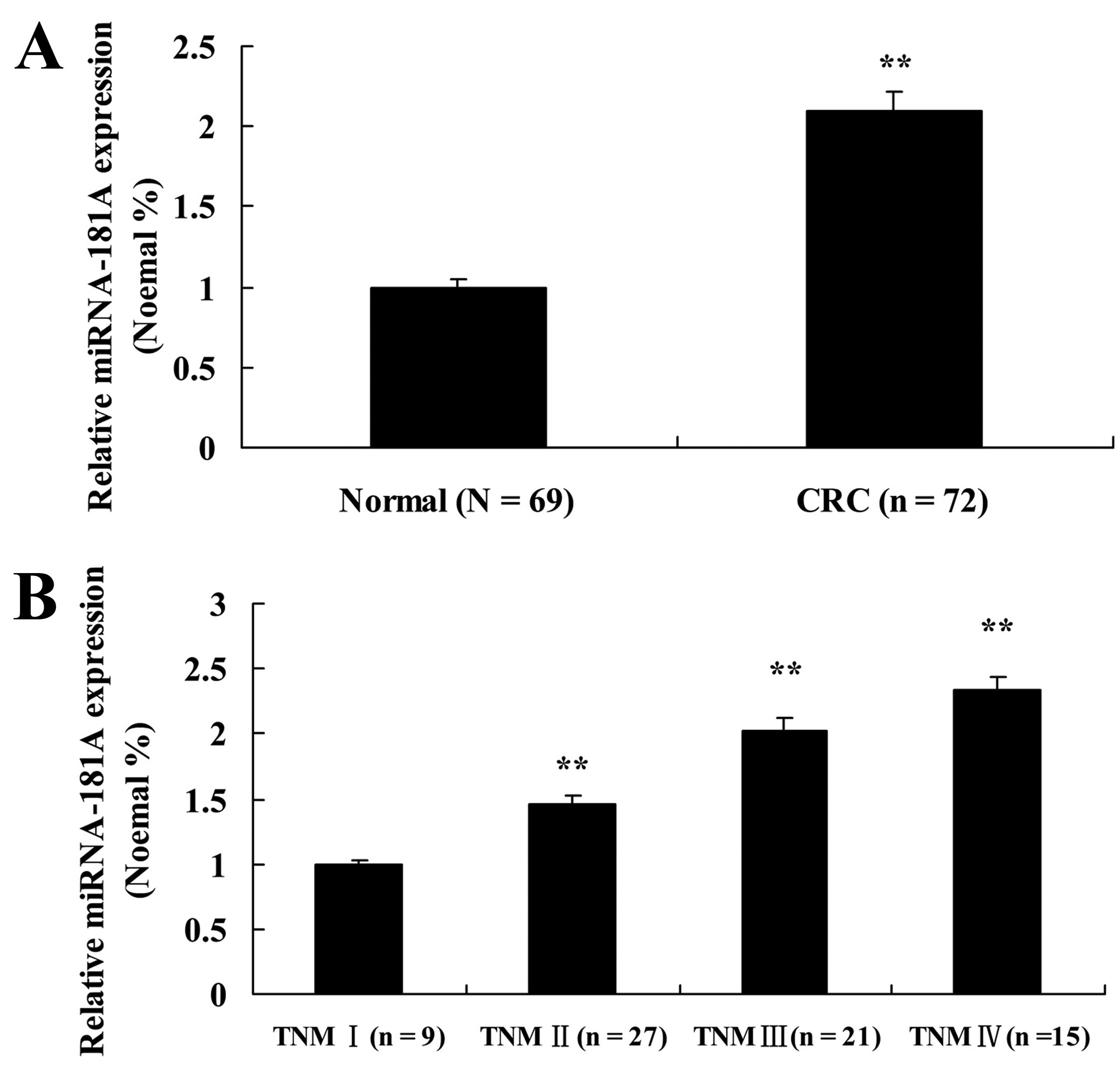
To investigate the expression of miR-181A in CRC
patients with late liver metastases, we used a microarray and
examined expression of miR-181A in 72 CRC patients and 69 normal
(control) patients, from Shandong Cancer Hospital and Institute.
The expression of miR-181A in CRC patients with late liver
metastases was higher than that of the control group (Fig. 1A), whereas the expression of
miR-181A was lower in all the pathological groups (TNM I-TNM IV)
(Fig. 1B).
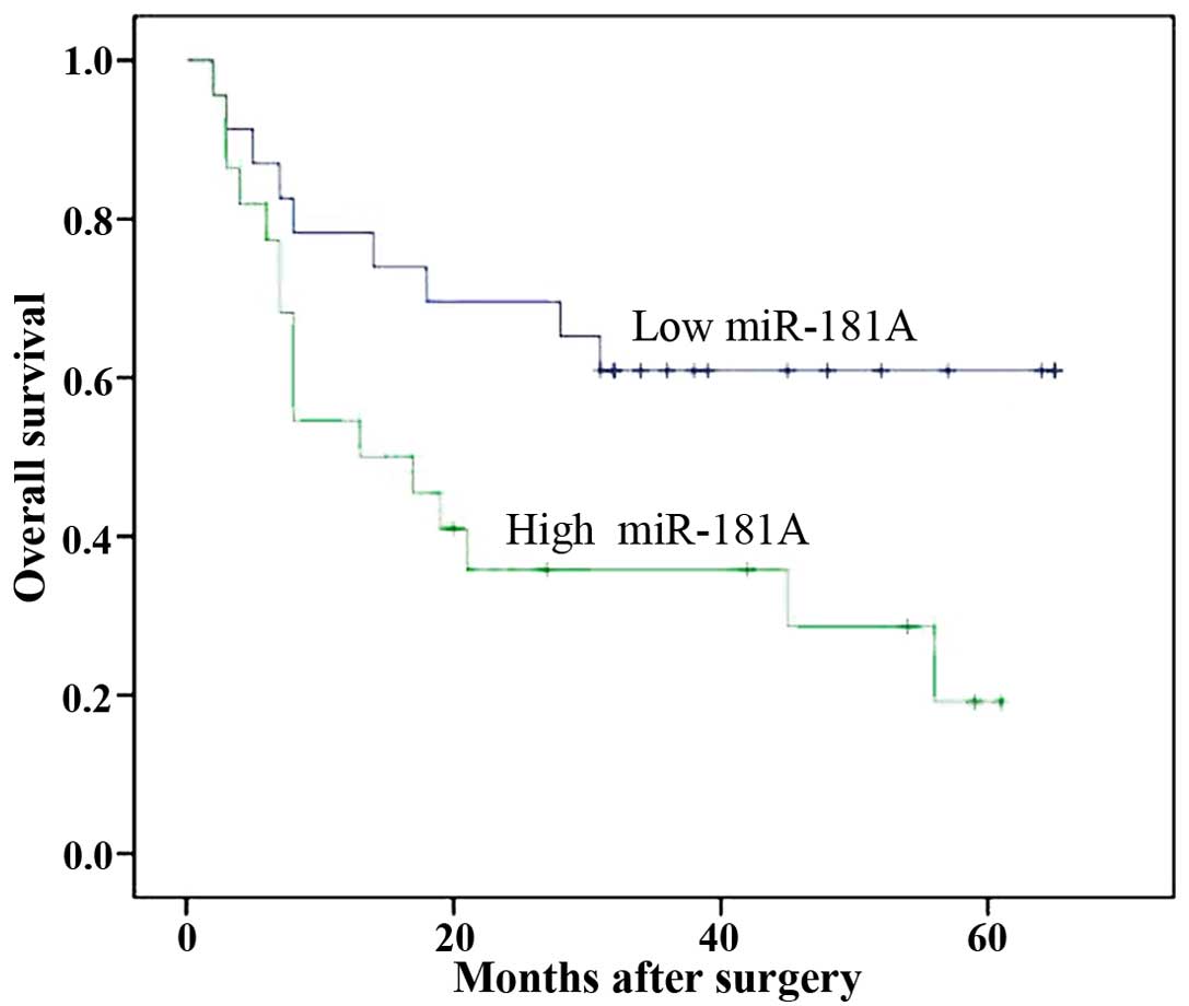
The expression of miR-181A is associated
with survival in late liver metastases
To examine whether the expression of miR-181A is
associated with survival in late liver metastases, we compared the
overall survival (OS) between CRC patients (with late liver
metastases) with lower miR-181A with CRC patients with higher
miR-181A (Fig. 2).
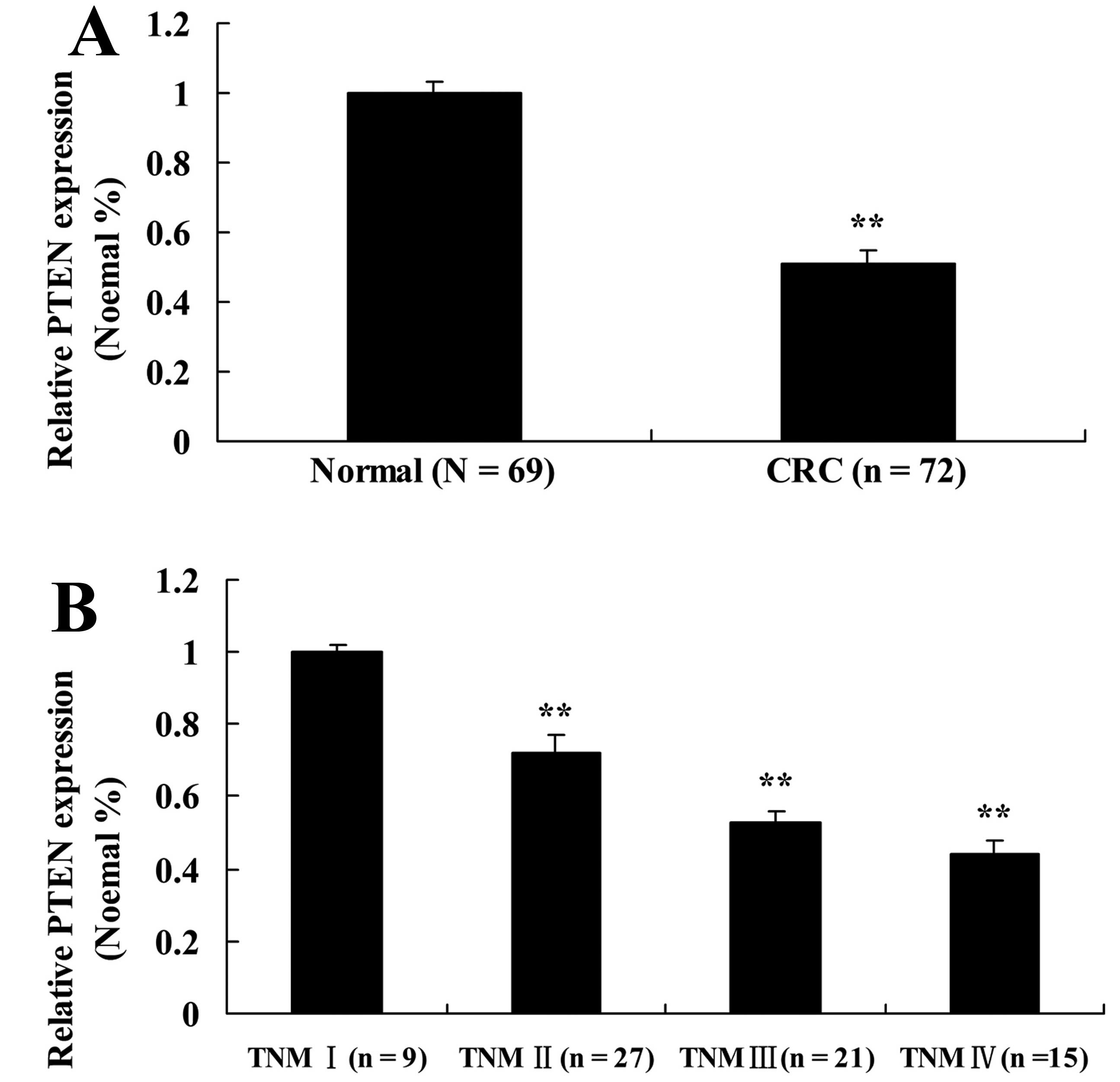
The PTEN in CRC patients with late liver
metastases
To further elucidate the mechanism of miR-181A on
CRC patients with late liver metastases, we examined the expression
of PTEN. Quantitative RT-PCR analyses were carried out in 72 CRC
patients with late liver metastases and 69 normal patients. qRT-PCR
analysis revealed a significant decrease in PTEN expression in CRC
patients with late liver metastases compared to normal patients
(Fig. 3A). In addition, the
expression of PTEN in TNM I CRC patients was markedly enhanced
compared to other pathological groups (Fig. 3B). This result suggests that there
is a specific association between miR-181A, PTEN and CRC
metastases.
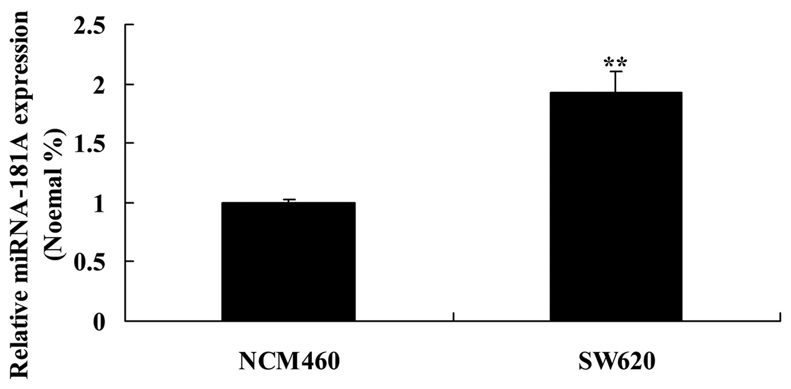
The expression of miR-181A in NCM460 and
SW620 cells
To explore the functional role of miR-181A in
driving late liver metastases, we examined the miR-181A expression
using qRT-PCR analyses in the cell lines NCM460 and SW620. The
miR-181A expression in the colon cancer cells NCM460 was lower than
that of the colon cancer SW620 cells (Fig. 4).
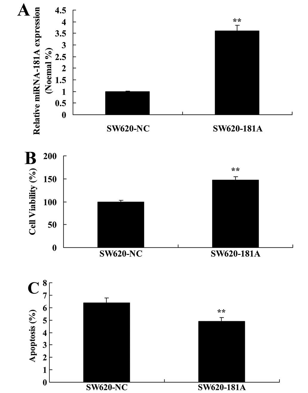
miR-181A induces cell growth and inhibits
apoptosis of SW620 cells
We examined how the upregulation of miR-181A could
affect cell growth and cell apoptosis of SW620 cells, and found
that miR-181A plasmid increased the expression of miR-181A in SW620
cells (Fig. 5A). Moreover,
upregulation of miR-181A enhanced cell viability and inhibited cell
apoptosis of SW620 cells (Fig. 5B and
C).
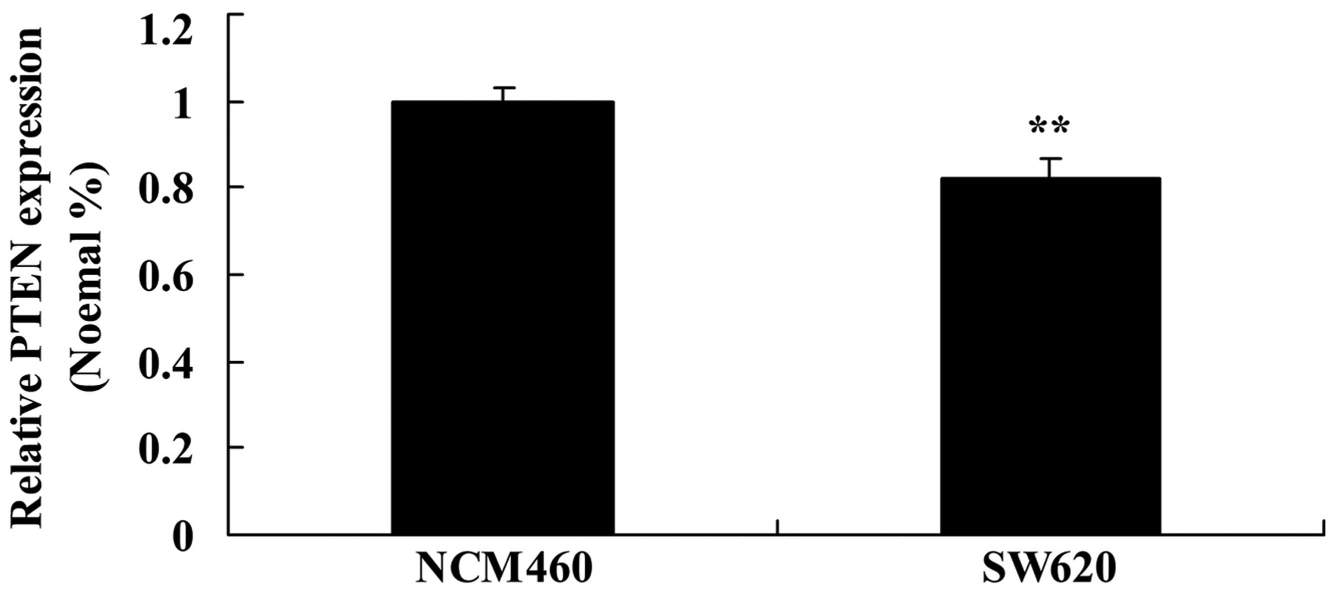
The expression of PTEN in NCM460 and
SW620 cells
To study the role of PTEN in CRC patients with late
liver metastases, we evaluated the PTEN expression using qRT-PCR in
NCM460 and SW620 cells. Consistent with in vitro results,
the expression of PTEN in NCM460 cells was lower than that of PTEN
expression in SW620 cells (Fig.
6).
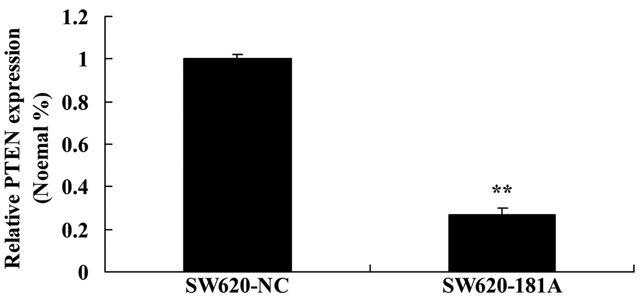
miR-181A inhibits the PTEN expression of
SW620 cells
Notably, miR-181A plasmid increased the expression
of PTEN in SW620 cells (Fig. 7).
These results indicate that PTEN may participate in the effect of
miR-181A on SW620 cells. However, the mechanism of such an
interaction is unknown.
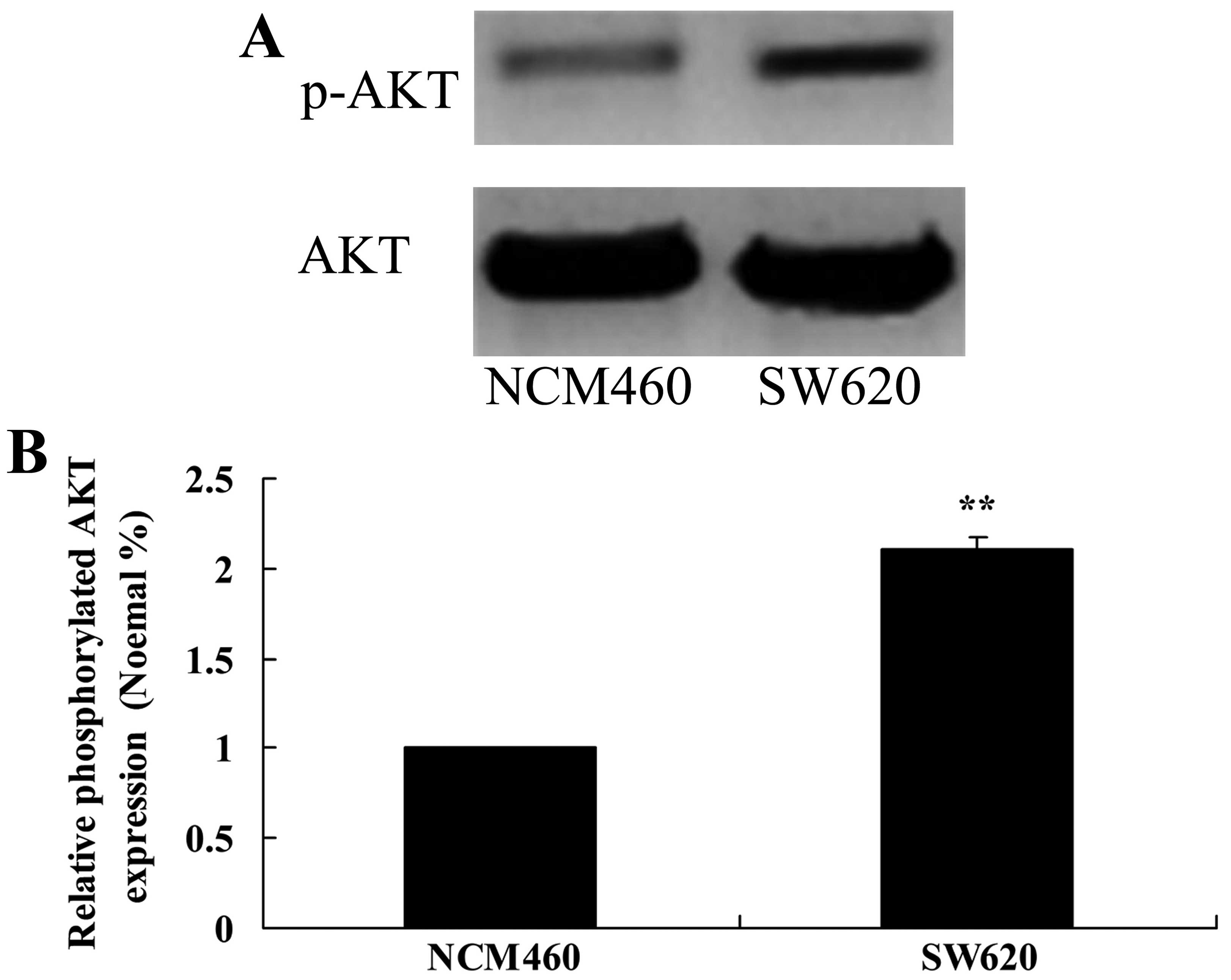
The expression of AKT in NCM460 and SW620
cells
To explore the AKT signaling pathway, western
blotting was performed in NCM460 and SW620 cells. Antibodies were
used against AKT and phosphorylated-AKT to examine protein
expression. The expression of phosphorylated AKT in NCM460 cells
was lower than that of SW620 cells (Fig. 8).
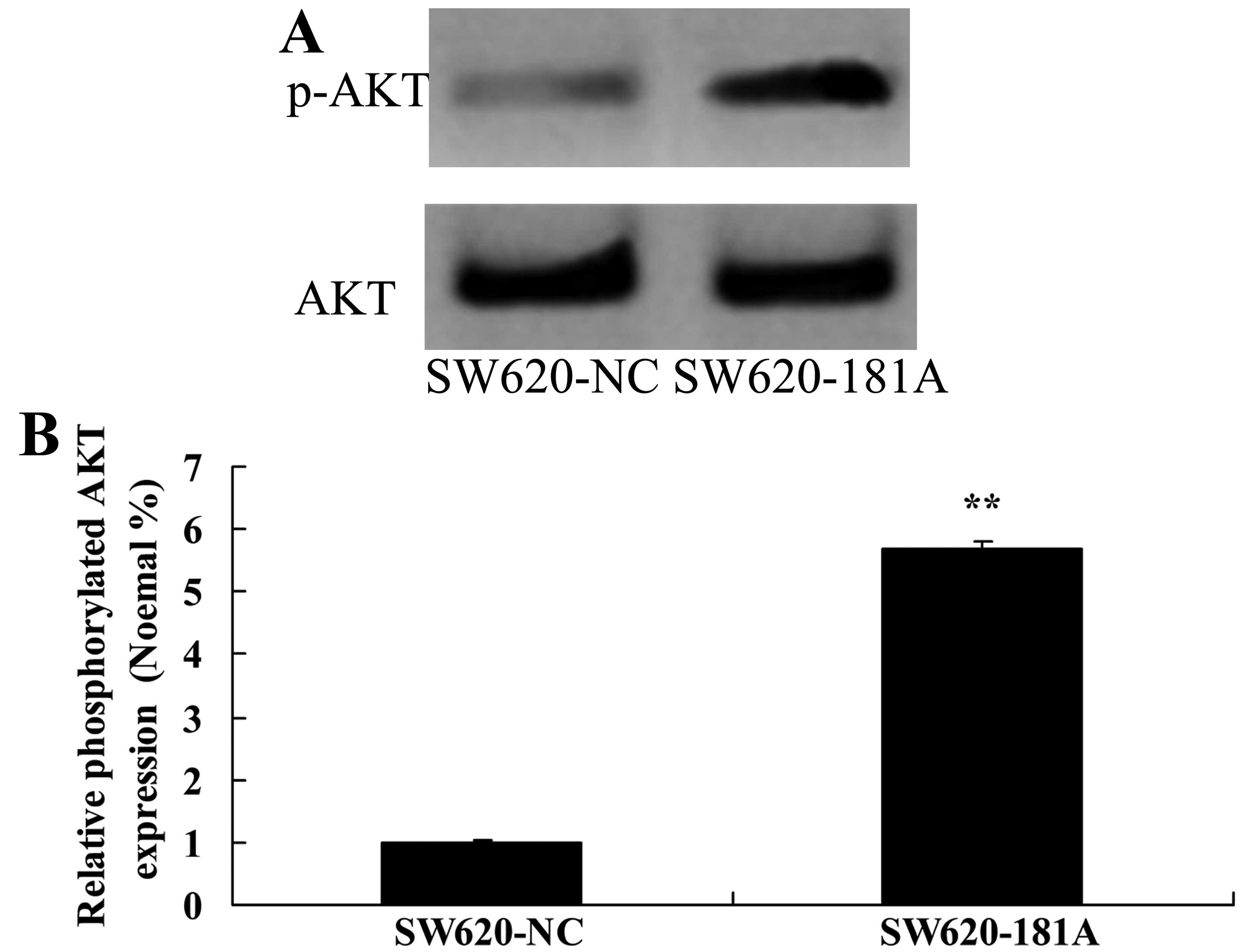
miR-181A induces AKT expression in SW620
cells
Notably, miR-181A plasmid suppressed the expression
of phosphorylated AKT protein in SW620 cells (Fig. 9). This suggests that miR-181 affects
the AKT pathway in human cancer SW620 cells.
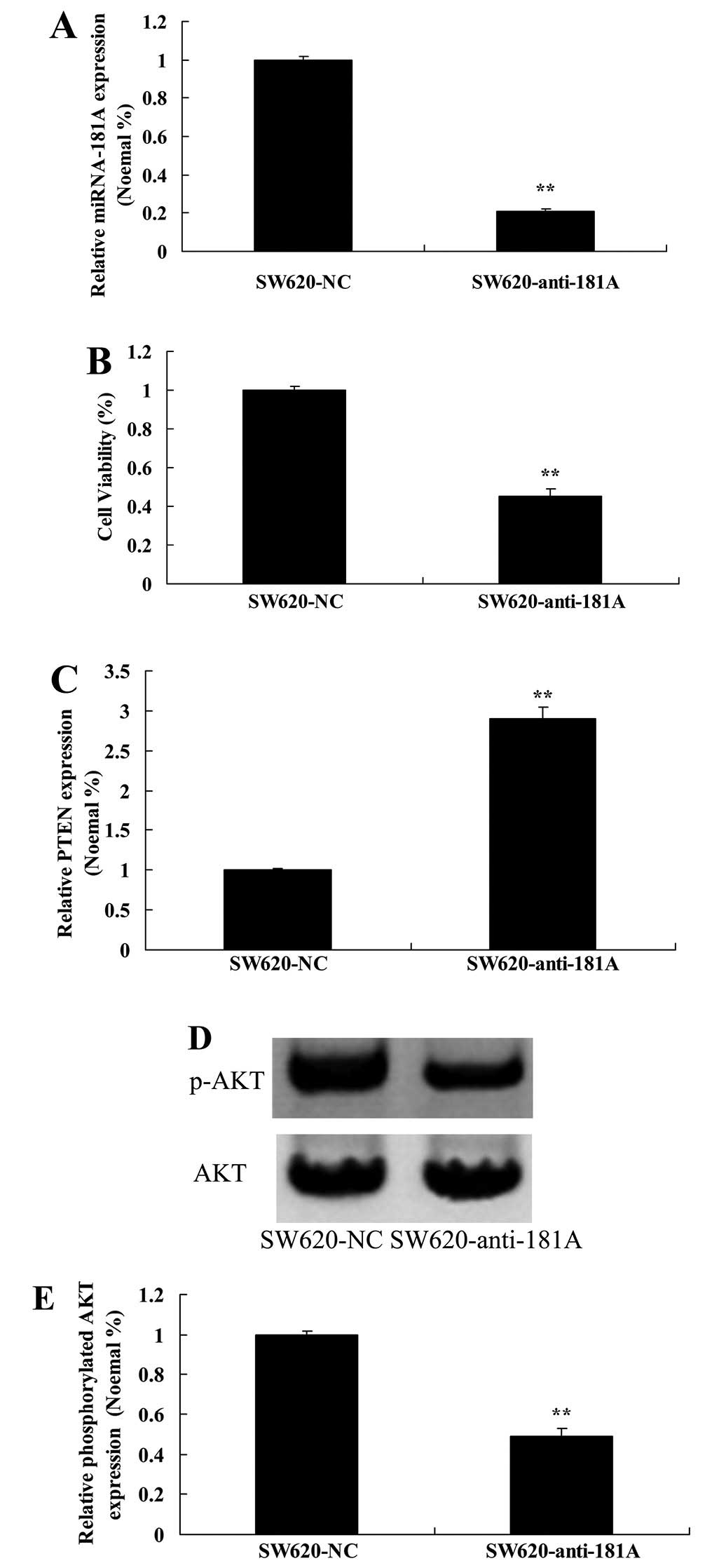
Downregulation of miR-181A inhibits cell
growth of SW620 cells via modulation of the PTEN/AKT signal
pathway
Anti-miR-181A plasmid was able to reduce the
expression of miR-181A in SW620 cells (Fig. 10A). Anti-miR-181A plasmid also
inhibited cell viability of SW620 cells (Fig. 10B). The expression of PTEN was
activated and the expression of phospho-AKT was suppressed by
anti-miR-181A plasmid (Fig.
10C–E).
Discussion
Colorectal cancer is one of the most common
malignant types of cancer, worldwide, with mortality rates of CRC
ranking third. According to data in 2008, there were nearly 1
million new cases of CRC and 500,000 deaths as a result of CRC,
worldwide (12). Despite the rapid
advance of endoscopic techniques, the improvement in material
living standards, and changes in dietary patterns, the morbidity of
CRC in China is increasing (13).
In the 1990s, the morbidity of CRC in urban and rural areas in
China increased by 32. and 8.5%, respectively, compared with that
in 1970s. In China, CRC has become the most common malignant cancer
and a major health issue (13). We
found that the expression of miR-181A in CRC patients with late
liver metastases was higher than that of the normal group and that
the expression of miR-181A was lower in all the pathological groups
(TNM I-TNM IV).
With a length of 19–24 nucleotides, miRNA is a
non-coding small RNA, which can exert a regulatory effect by
combing the untranslated region (3′-UTR) (14). Nearly 1,500 miRNA sequences have
been discovered, to date and >60% of genes reported in the human
genome are miRNA target genes (15). It has been shown that miRNA can
regulate the expression of numerous genes and that miRNAs play an
important role in normal cell processes, such as cell growth and
development, proliferation and apoptosis (14). Increasing evidence reveals that
miRNAs play a crucial role in many types of cancer (16), and indeed many tumour cells have
altered expression of miRNAs (17).
In the present study, we found that there was an
increase of OS in lower expression miR-181A of CRC patients with
late liver metastases compared to CRC patients with higher miR-181A
levels with late liver metastases. Specifically, upregulation of
miR-181A increased the cell viability and inhibited apoptosis of
SW620 cells.
It has been reported that miR-181A has many
different target genes. PTEN is a tumor suppressor gene and has
dual phosphatase activity on lipid and proteins to drive processes
essential in cell processes, such as cell growth, proliferation,
survival, apoptosis, migration and invasion, local adhesion and
angiogenesis (18). Mutations that
drive upregulation or deletion of PTEN can activate the PI3K/Akt
signaling pathway which can trigger occurrence, development,
invasion and metastasis of tumors (19). Studies showing that miR-181A can
target the regulation of PTEN and promote occurrence, development
and metastasis of tumors are common, while studies examining the
related regulatory mechanisms in CRC are rare (18,20).
In the present study, we found that the PTEN expression of the
normal control patients was higher than that of CRC patients with
late liver metastases. Moreover, PTEN expression of TNM I CRC
patients was markedly enhanced compared to other pathological
groups. When we upregulated miR-181A using an expression plasmid,
the expression of PTEN in SW620 cells increased.
Studies have suggested that the PTEN/PI3K/Akt signal
pathway is involved in numerous cell processes, such as growth,
proliferation, survival, apoptosis, metabolism, angiogenesis,
invasion and metastasis, anticancer drug resistance and tumor
immune escape (21). However,
reports examining miR-181A and its regulation of the PTEN/PI3K/Akt
signal pathway are rare. In this study we examined the expression
of related expression of miR-181A, PTEN/PI3K/Akt signaling pathways
in CRC tissues to understand its clinical significance. We would
like to extend this study to verify that miR-21 is a direct target
gene and to examine regulatory mechanisms of the PTEN/PI3K/Akt
signal pathway by miR-181A. Ultimately, we hope to understand how
miR-181A might regulate biological behavior of CRC, such as
proliferation, invasion, metastasis and cell cycle progression, as
this might help provide clues for new therapeutic targets.
Specifically we found that upregulation of miR-181A
induced AKT expression in SW620 cells. Downregulation of miR-181A
could suppress cell growth of SW620 cells, and mediated the
PTEN/AKT signaling pathway.
In conclusion, we report that the expression of
miR-181A in CRC patients with late liver metastases was higher than
that of the normal group. Downregulation of miR-181A was able to
suppress growth of SW620 cells by mediating the PTEN/AKT signal
pathway. Our results support the idea that miR-181A affects
incidence and survival in late liver metastases of colorectal
cancer through the PTEN/AKT signaling pathway.
Acknowledgments
This study was funded by the Science and Technology
Development Plan Project of Shandong Province, China (no.
2013GSF11834), the Shandong Provincial Natural Science Foundation,
China (no. ZR2009CM138) and the Medicine and Health Care in
Shandong Province Science and Technology Development Plan Project
(no. 2011HW06).
References
|
1
|
Li Y, Zeng M, Chen W, Liu C, Wang F, Han
X, Zuo Z and Peng S: Dexmedetomidine reduces isoflurane-induced
neuroapoptosis partly by preserving PI3K/Akt pathway in the
hippocampus of neonatal rats. PLoS One. 9:e936392014. View Article : Google Scholar : PubMed/NCBI
|
|
2
|
Van Schaeybroeck S, Kalimutho M, Dunne PD,
Carson R, Allen W, Jithesh PV, Redmond KL, Sasazuki T, Shirasawa S,
Blayney J, et al: ADAM17-dependent c-MET-STAT3 signaling mediates
resistance to MEK inhibitors in KRAS mutant colorectal cancer. Cell
Rep. 7:1940–1955. 2014. View Article : Google Scholar : PubMed/NCBI
|
|
3
|
Maekawa K, Baba T, Otomo S, Morishita S
and Tamura N: Low pre-existing gray matter volume in the medial
temporal lobe and white matter lesions are associated with
postoperative cognitive dysfunction after cardiac surgery. PLoS
One. 9:e873752014. View Article : Google Scholar : PubMed/NCBI
|
|
4
|
Cai Y, Hu H, Liu P, Feng G, Dong W, Yu B,
Zhu Y, Song J and Zhao M: Association between the apolipoprotein E4
and post-operative cognitive dysfunction in elderly patients
undergoing intravenous anesthesia and inhalation anesthesia.
Anesthesiology. 116:84–93. 2012. View Article : Google Scholar
|
|
5
|
Zhang Q, Li SZ, Feng CS, Qu XD, Wang H,
Zhang XN, Liu Y, Wang Y, Wu AS and Yue Y: Serum proteomics of early
post-operative cognitive dysfunction in elderly patients. Chin Med
J (Engl). 125:2455–2461. 2012.
|
|
6
|
Lin K and Abraham KM: Targets of p56(lck)
activity in immature thymoblasts: Stimulation of the Ras/Raf/MAPK
pathway. Int Immunol. 9:291–306. 1997. View Article : Google Scholar : PubMed/NCBI
|
|
7
|
Kleist B, Kempa M, Novy M, Oberkanins C,
Xu L, Li G, Loland C and Poetsch M: Comparison of neuroendocrine
differentiation and KRAS/NRAS/BRAF/PIK3CA/TP53 mutation status in
primary and metastatic colorectal cancer. Int J Clin Exp Pathol.
7:5927–5939. 2014.PubMed/NCBI
|
|
8
|
Sinha R, Larkin J, Gore M and Fearfield L:
Cutaneous toxicities associated with vemurafenib therapy in 107
patients with BRAF V600E mutation-positive metastatic melanoma,
including recognition and management of rare presentations. Br J
Dermatol. 173:1024–1031. 2015. View Article : Google Scholar : PubMed/NCBI
|
|
9
|
Okamoto K, Okamoto I, Hatashita E, Kuwata
K, Yamaguchi H, Kita A, Yamanaka K, Ono M and Nakagawa K:
Overcoming erlotinib resistance in EGFR mutation-positive non-small
cell lung cancer cells by targeting survivin. Mol Cancer Ther.
11:204–213. 2012. View Article : Google Scholar
|
|
10
|
Cercek A, Shia J, Gollub M, Chou JF,
Capanu M, Raasch P, Reidy-Lagunes D, Proia DA, Vakiani E, Solit DB,
et al: Ganetespib, a novel Hsp90 inhibitor in patients with KRAS
mutated and wild type, refractory metastatic colorectal cancer.
Clin Colorectal Cancer. 13:207–212. 2014. View Article : Google Scholar : PubMed/NCBI
|
|
11
|
Giannoni E, Fiaschi T, Ramponi G and
Chiarugi P: Redox regulation of anoikis resistance of metastatic
prostate cancer cells: Key role for Src and EGFR-mediated
pro-survival signals. Oncogene. 28:2074–2086. 2009. View Article : Google Scholar : PubMed/NCBI
|
|
12
|
Whelan KA, Schwab LP, Karakashev SV,
Franchetti L, Johannes GJ, Seagroves TN and Reginato MJ: The
oncogene HER2/neu (ERBB2) requires the hypoxia-inducible factor
HIF-1 for mammary tumor growth and anoikis resistance. J Biol Chem.
288:15865–15877. 2013. View Article : Google Scholar : PubMed/NCBI
|
|
13
|
Oh HY, Lee EJ, Yoon S, Chung BH, Cho KS
and Hong SJ: Cholesterol level of lipid raft microdomains regulates
apoptotic cell death in prostate cancer cells through EGFR-mediated
Akt and ERK signal transduction. Prostate. 67:1061–1069. 2007.
View Article : Google Scholar : PubMed/NCBI
|
|
14
|
Roomi MW, Kalinovsky T, Rath M and
Niedzwiecki A: In vitro modulation of MMP-2 and MMP-9 in pediatric
human sarcoma cell lines by cytokines, inducers and inhibitors. Int
J Oncol. 44:27–34. 2014.
|
|
15
|
Lawless N, Vegh P, O'Farrelly C and Lynn
DJ: The role of microRNAs in bovine infection and immunity. Front
Immunol. 5:6112014. View Article : Google Scholar : PubMed/NCBI
|
|
16
|
Cortés-Sempere M and Ibáñez de Cáceres I:
microRNAs as novel epigenetic biomarkers for human cancer. Clin
Transl Oncol. 13:357–362. 2011. View Article : Google Scholar : PubMed/NCBI
|
|
17
|
Rotomskis A, Margevičiūtė R, Germanavičius
A, Kaubrys G, Budrys V and Bagdonas A: Differential diagnosis of
depression and Alzheimer's disease with the Addenbrooke's Cognitive
Examination-Revised (ACE-R). BMC Neurol. 15:572015. View Article : Google Scholar : PubMed/NCBI
|
|
18
|
Yen SS: Proteasome degradation of brain
cytosolic tau in Alzheimer's disease. Int J Clin Exp Pathol.
4:385–402. 2011.PubMed/NCBI
|
|
19
|
Zheng J, Wu C, Xu Z, Xia P, Dong P, Chen B
and Yu F: Hepatic stellate cell is activated by microRNA-181b via
PTEN/Akt pathway. Mol Cell Biochem. 398:1–9. 2015. View Article : Google Scholar
|
|
20
|
Jun Z, Li Z, Fang W, Fengzhen Y, Puyuan W,
Wenwen L, Zhi S and Bondy SC: Melatonin decreases levels of S100β
and NFKB, increases levels of synaptophysin in a rat model of
Alzheimer's disease. Curr Aging Sci. 6:142–149. 2013. View Article : Google Scholar
|
|
21
|
Granic I, Dolga AM, Nijholt IM, van Dijk G
and Eisel UL: Inflammation and NF-kappaB in Alzheimer's disease and
diabetes. J Alzheimers Dis. 16:809–821. 2009.PubMed/NCBI
|