Introduction
Prostate cancer is the most common cancer in males
in economically developed countries. In 2014, there were an
estimated 233,000 new cases of prostate cancer and 29,480 related
deaths in the US (1). Metastatic
castration-resistant prostate cancer is the primary cause of death
for most patients. Unlike the majority of solid cancers, prostate
cancer usually shows a poor response to chemotherapy. Therefore,
more effective strategies for the treatment of castration-resistant
prostate cancer are urgently required.
Therapeutics based on RNA interference (RNAi) have
become powerful and ideal methods for the treatment of many
diseases including cancer which are mainly caused by overactive
oncogenes due to the high specificity, high efficacy and low
toxicity of the RNAi trigger - small double-stranded RNAs (dsRNAs)
(2–4). However, there are many types of cancer
that are mainly caused by complete inactivation or reduced
expression of tumor-suppressor genes (TSGs). Notably, new evidence
has emerged that synthetic small dsRNAs induce sequence-specific
transcriptional gene activation of E-cadherin,
p21WAF1/CIP1 and VEGF by targeting specific regions in
their gene promoters (5). This
phenomenon has been termed RNA-induced gene activation (RNAa) and
such dsRNAs as small activating RNAs (saRNAs) (5). Their observation was supported by
subsequent studies which suggest that RNAa may be a general and
conserved phenomenon of gene regulation (6–14).
Moreover, several studies have demonstrated that restoration of p21
expression by saRNAs in different cancer cells could inhibit cell
proliferation and tumor growth (15–20).
Thus, RNAa holds great promise as an alternative to traditional
vector-based systems to activate target genes and would supplement
RNA-mediated gene silencing to broaden the gene pool susceptible to
regulation by small RNAs.
The human PAWR (PRKC apoptosis WT1 regulator) gene,
whose other aliases include PAR4 and Par-4, is located on
chromosome 12q21 and encodes a leucine zipper domain protein first
identified in prostate cancer cells undergoing apoptosis induced by
an exogenous insult (21,22). Mutations in the PAWR gene have not
been reported in cancer cells, and endogenous PAWR expressed in
normal and cancer cells does not, by itself, cause apoptosis.
However, inhibition of endogenous PAWR with antisense
oligodeoxynucleotides, a dominant-negative leucine zipper domain or
RNAi, precludes apoptosis by exogenously applied agents, thus
indicating that functional PAWR protein is essential for apoptosis
via diverse cell death pathways (23–25).
More importantly, ectopic PAWR overexpression is sufficient to
induce apoptosis in most cancer cells in vitro and growth
inhibition of prostate cancer xenografts in nude mice, but not in
normal or immortalized cells (23,26).
Therefore, PAWR is an ideal target and a candidate TSG for
RNAa.
Our previous study demonstrated that several small
dsRNAs targeting the PAWR promoter can upregulate PAWR gene
expression effectively in human cancer cells (14). In the present study, we investigated
the antitumor effects of dsPAWR-433 on prostate cancer cell lines
and found that upregulation of PAWR by saRNA inhibited the growth
of prostate cancer cells by inducing cell apoptosis.
Materials and methods
dsRNA design and synthesis
The sequence of dsPAWR-433 [S, 5′-AAU ACG GUC UUG
UAC UUA A (dT)(dT)-3′; AS, 5′-UUA AGU ACA AGA CCG UAU U
(dT)(dT)-3′] was designed as previously described (17); and the control dsRNA [dsCon: S,
5′-ACU ACU GAG UGA CAG UAG A (dT)(dT)-3′; AS, 5′-UCU ACU GUC ACU
CAG UAG U (dT)(dT)-3′] is the same as the dsCon-2 which was
specifically designed by Li et al to lack homology to all
known human sequences (5). All
dsRNAs were chemically synthesized by GenePharma (Shanghai, China)
with dTdT-3′ overhangs.
Cell culture and transfection
The human prostate cancer cell lines DU145 and PC3
were obtained from the Shanghai Institute of Cell Biology, Chinese
Academy of Science. The cells were cultured in RPMI-1640 medium
supplemented with 10% heat-inactivated fetal bovine serum,
penicillin (100 U/ml) and streptomycin (100 mg/l) in a humidified
atmosphere containing 5% CO2 maintained at 37°C. The day
before transfection, the cells were plated in growth medium without
antibiotics at a density of 30–40%. Transfections of dsRNAs were
carried out using Lipofectamine 2000 (Invitrogen, Carlsbad, CA,
USA) according to the manufacturer's protocol and lasted for 24, 48
or 72 h. Cell images were captured using a phase-contrast
microscope at a magnification of x100 (Olympus, Japan).
Cell growth/viability assay
Proliferation of cells was determined by the
3-(4,5-dimethylthiazol-2-yl)-2,5-diphe-nyltetrazolium bromide (MTT)
assay (16). Approximately
5,000–10,000 cells were plated in each well of a 96-well plate.
After overnight incubation, the cells were treated with the
appropriate dsRNAs for 24, 48 or 72 h. At the various times after
treatment, 20 µl MTT (5 mg/ml) was added to each well and
the plates were incubated at 37°C for 4 h. After that, the crystals
were dissolved in 150 µl of dimethyl sulfoxide at room
temperature. Absorbance was measured at 490 nm in an absor-bance
reader (MRX II; Dynex Technologies, Chantilly, VA, USA). The
reduction in viability of each group was expressed as a percentage
of the mock group, which was considered to be 100% viable.
Real-time quantitative RT-PCR
Total RNA was extracted from cells using TRIzol
(Invitrogen) and reverse transcribed using oligo(dT) primers. The
resulting cDNA was amplified in a real-time PCR system (ABI Prism
7500; Applied Biosystems, Foster City, CA, USA) using the
DNA-binding dye SYBR-Green I (Invitrogen) for detection of PCR
products. Values are expressed as fold-difference compared with the
mock group. Primer sequences for PAWR were:
5′-GCCGCAGAGTGCTTAGATGAG-3′ (forward) and
5′-GCAGATAGGAACTGCCTGGATC-3′ (reverse) and; for GAPDH were:
5′-AAGAAGGTGGTGAAGCAGGC-3′ (forward) and 5′-TCCACCACCC
TGTTGCTGTA-3′ (reverse).
Western blot analysis
Protein extraction and western blot analysis were
carried out according to a previously described method (16). The primary and secondary antibodies
were all purchased from the Cell Signaling Technology (Beverly, MA,
USA).
To determine NF-κB cellular localization, nuclear
and cytoplasmic proteins were isolated from the cells using a cell
fractionation kit (KeyGen, Wuhan, China). NF-κB expression in the
nuclear and cytoplasmic compartments was determined by immunoblot
analysis as described above.
Detection of apoptotic cells by flow
cytometry
A quantitative assessment of apoptosis was carried
out by determining the percentage of cells with highly condensed or
fragmented nuclei. Cells were plated in 6-well plates and incubated
overnight before treatment. Then, cells were harvested at 72 h
after dsRNA treatment, washed twice with pre-chilled
phosphate-buffered saline (PBS), and resuspended in 100 µl
1X binding buffer at a concentration of 1×10⁶ cells/ml.
Double staining with fluorescein isothiocyanate (FITC)-conjugated
Annexin V and propidium iodide (PI) (Annexin V-FITC apop-tosis
detection kit; BD Biosciences, San Jose, CA, USA) was performed in
accordance with the manufacturer's protocol. Cell apoptosis
analysis was performed within 1 h using the Beckman Coulter FC500
Flow Cytometry system with CXP Software (Beckman Coulter,
Fullerton, CA, USA).
Statistical analysis
All values are expressed as means ± SD. Statistical
significance was compared between treatment groups and controls
using the Student's t-test. P<0.05 was considered to indicate a
statistically significant result.
Results
dsPAWR-433 induces PAWR gene activation
in prostate cancer cells
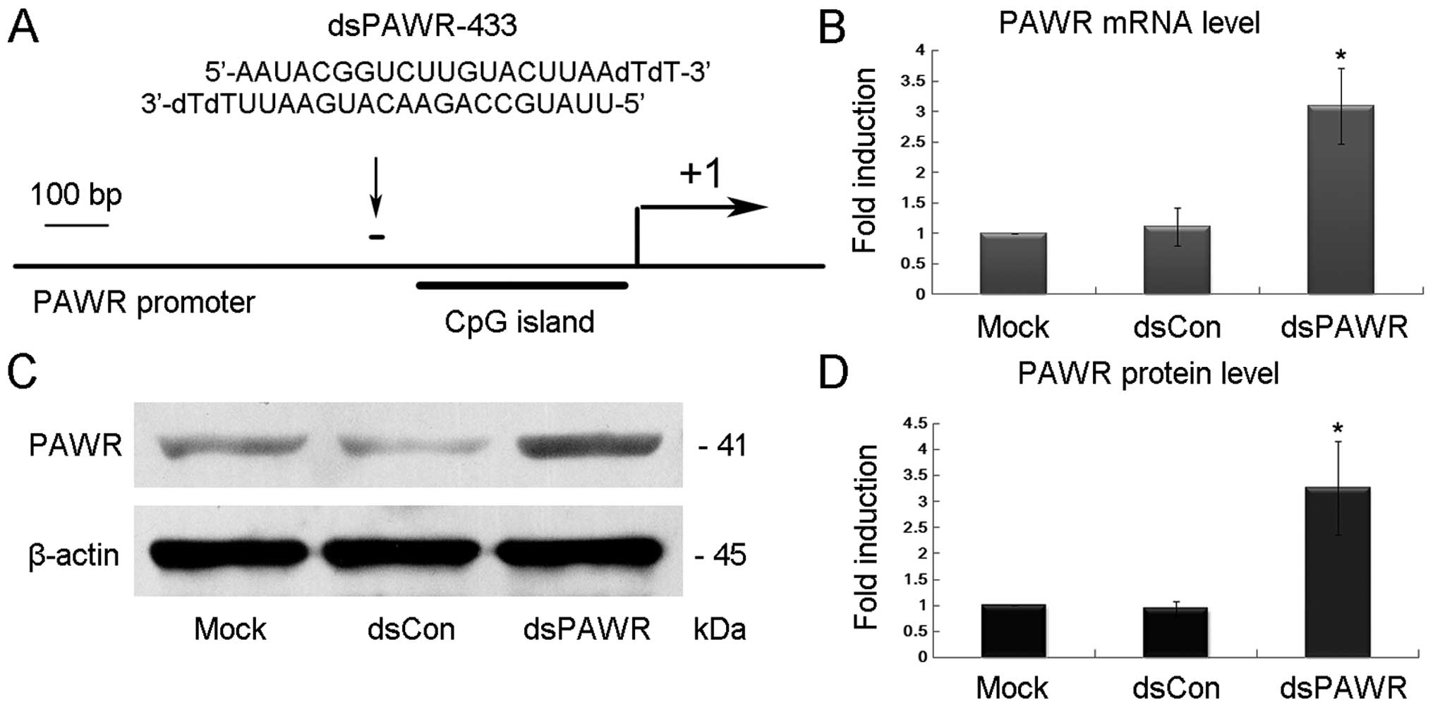
We previously reported that several dsRNAs targeting
the PAWR gene promoter at position -433-435 relative to the
transcription start site (dsPAWR-433-435, Fig. 1A) had the ability to activate PAWR
expression in T24 bladder cancer cells (14). In the present study, we investigated
whether dsPAWR-433 could induce PAWR gene expression in prostate
cancer cells. Fifty nmol/l (nM) dsPAWR-433 and a non-specific
control dsRNA (dsCon) were transfected into DU145 human prostate
cancer cells and PAWR expression levels were evaluated 48 and 72 h
later. Compared with the mock and dsCon groups, dsPAWR-433 caused a
>3-fold induction in the PAWR mRNA level in the DU145 cells
(Fig. 1B). Induction of PAWR was
also confirmed by western blot analysis and the elevated levels of
PAWR protein were strongly correlated to the increase in PAWR mRNA
expression (Fig. 1C and D).
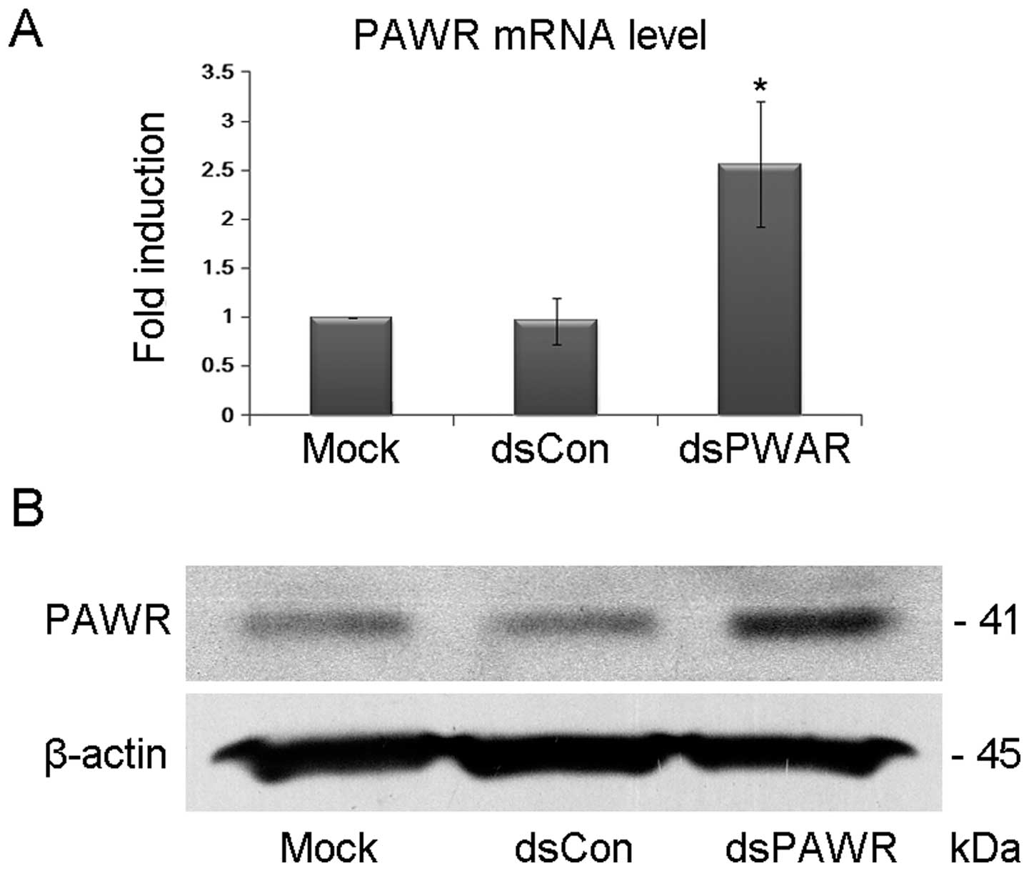
Transfection of dsPAWR-433 was also performed in
another human prostate cancer cell line PC3. As shown in Fig. 2, dsPAWR-433 transfection resulted in
a >2-fold induction of PAWR gene expression in the PC3
cells.
dsPAWR-433 inhibits prostate cancer cell
growth and viability
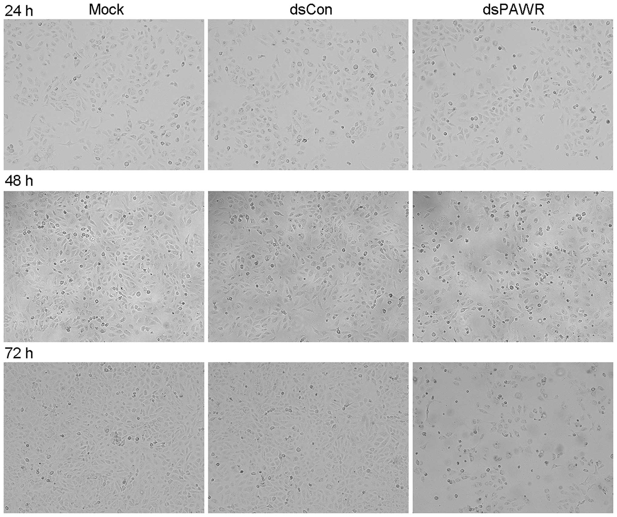
Ectopic PAWR overexpression has been shown to induce
growth inhibition in most cancer cells in vitro (26). In the present study, we investigated
whether the upregulation of PAWR by saRNA has similar effects on
prostate cancer cells. DU145 prostate cancer cells were transfected
with 50 nM dsPAWR-433 and dsCon for 48 or 72 h, and the
dsPAWR-433-transfected cells gradually displayed growth inhibition
and cell shrinkage (Fig. 3).
Moreover, evidently decreased cell density and more floating dead
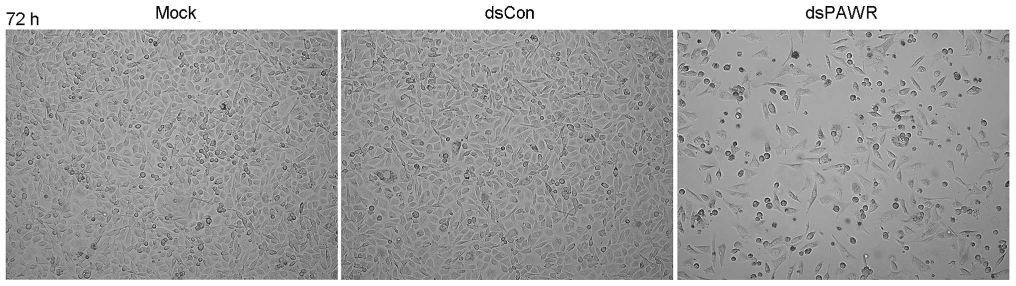
cells were observed in the dsPAWR-433-treated group (Fig. 3). These morphological changes were
also observed in the prostate cancer cell line PC3 (Fig. 4).
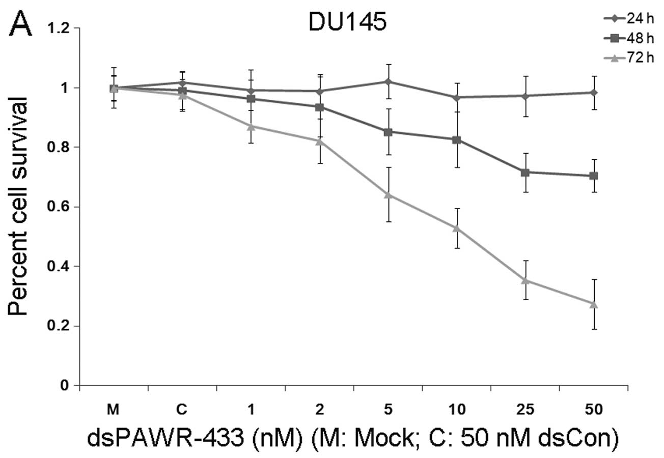
Then, the effects of dsPAWR-433 on the proliferation
and viability of human prostate cancer DU145 cells were determined
at varying concentrations and times (24–72 h) by MTT assay. As
shown in Fig. 5A, the effects of
dsPAWR-433 on cell viability, which were dose- and time-dependent,
occurred within 48 h and at dsRNA concentrations as low as 5 nM.
Compared with the mock and dsCon transfections, reduction in
viability of the DU145 cells following dsRNA treatment at
concentrations of 1–50 nM after 48 h ranged from 3.6 to 29.5%,
whereas after 72 h this ranged from 13.0 to 72.6% (Fig. 5A). Accordingly, lower concentrations
of dsPAWR-433 (5–25 nM) could also elevate the PAWR expression and
its effects also appeared to be dose-dependent (Fig. 5B).
dsPAWR-433 induces cell apoptosis in
prostate cancer cells
The antitumor ability by ectopic PAWR overexpression
is related to its essential role in inducing apoptosis via diverse
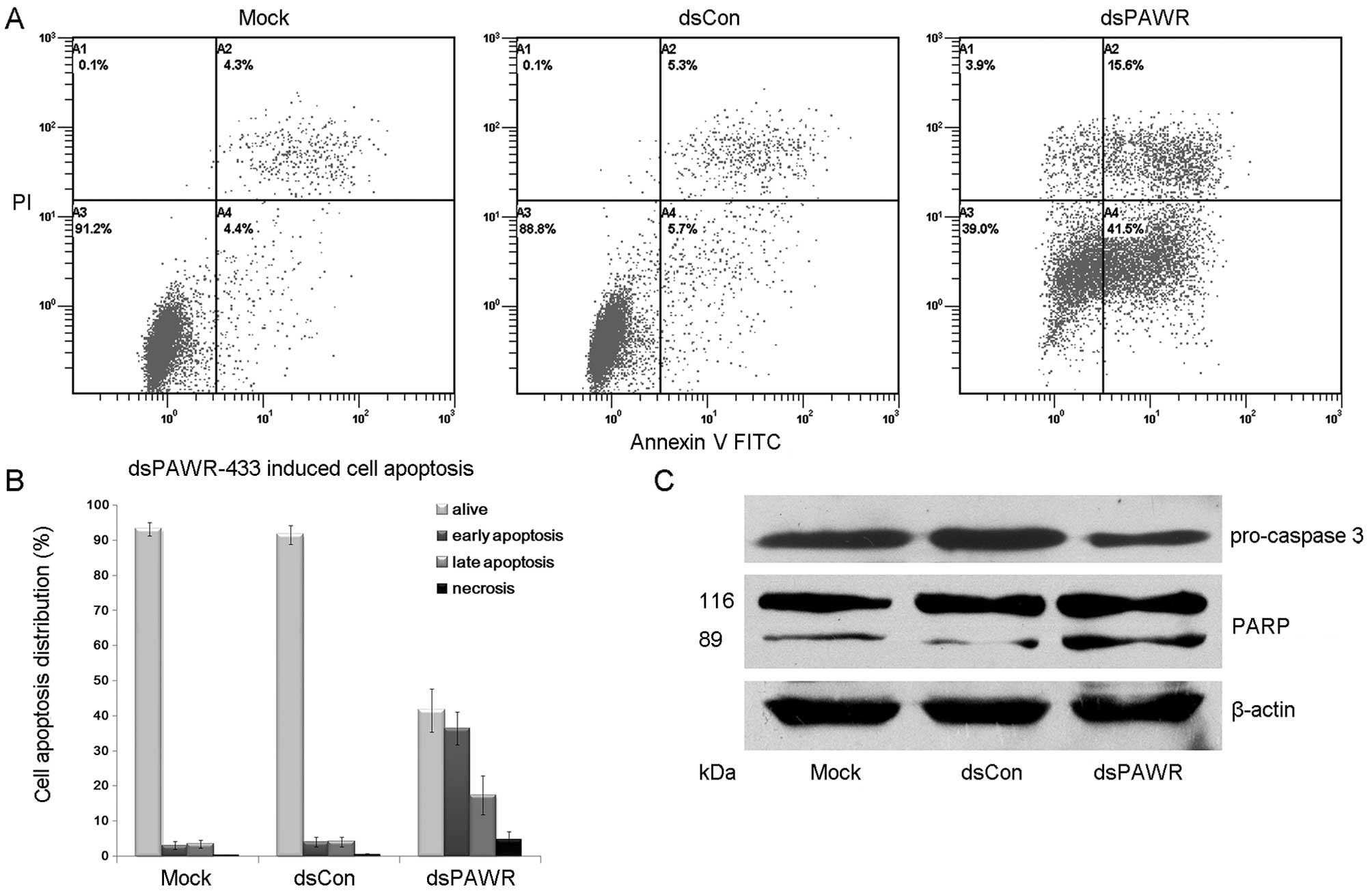
cell death pathways (23–25). Thus, we investigated the
relationship between dsPAWR-433-mediated loss of cell viability and
apoptosis by flow cytometric analysis of DU145 cells labeled with
PI and Annexin V. We found that dsPAWR-433 caused evident apoptosis
in the DU145 cells at 72 h following treatment. The number of early
apoptotic cells (LR quadrant) increased to ~40% and the number of
late apoptotic cells (UR quadrant) increased to nearly 20%
(Fig. 6A and B). These data also
showed that dsPAWR-433 treatment resulted in not only apoptosis but
also tiny cell necrosis, which may be a secondary event in the
apoptotic process.
Caspase-3 and poly(ADP-ribose) polymerase (PARP)
play central roles in apoptosis. Accordingly, we observed that the
level of pro-caspase-3 was markedly decreased in the 50 nM
dsPAWR-433-treated DU145 cells at 72 h following treatment
(Fig. 6C). Moreover, the 89 kDa
cleaved PARP fragment was detected in the dsPAWR-433-treated
samples. Thus, the significant changes in apoptosis-related
proteins caused by dsPAWR-433 confirmed the ongoing apoptosis above
and the anti-carcinogenic effects on the DU145 human prostate
cancer cells.
The molecular mechanism related to
dsPAWR-433-induced cell apoptosis
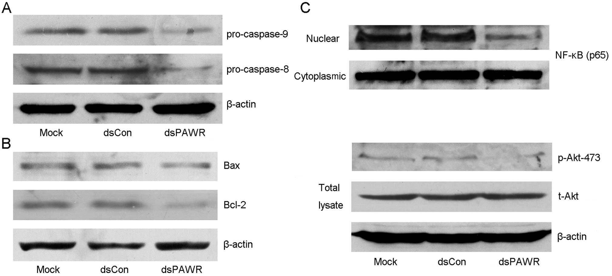
To examine which pathway plays a role in the
dsPAWR-433-induced cell apoptosis, the cleavage of caspase-8 and -9
was examined. As shown in Fig. 7A,
the levels of pro-caspase-8 and -9 were markedly decreased in the
50 nM dsPAWR-433-treated DU145 cells at 72 h following treatment,
indicating that both extrinsic and intrinsic pathways were active
in the dsPAWR-433-treated cells.
PAWR-mediated apoptosis requires downregulation of
Bcl-2 levels and PAWR regulates Bcl-2 gene expression through a
WT1-binding site in its promoter leading to a decrease in
transcription (27,28). Consistently, the expression of Bcl-2
was found to decrease in the dsPAWR-433-treated cells compared with
the controls (Fig. 7B). However,
the level of Bax, the pro-apoptotic member of the Bcl-2 family, was
not altered after the treatment of dsPAWR-433.
Previous reseach has shown that PAWR is an important
intersection in the network of tumor suppressors that involves the
NF-κB and Akt pathways (29), which
are both deregulated during prostate tumorigenesis. Thus, we
detected these proteins and found that nuclear translocation of
NF-κB and phosphorylation of Akt were inhibited in the
dsPAWR-433-treated cells compared with the controls (Fig. 7C), which implied inactivation of
these signaling pathways.
Discussion
RNA activation (RNAa) is an interesting and
promising discovery of small RNA-mediated gene upregulation
originally identified in several human cancer cell lines (5,6). It
may offer an alternative to manipulate gene expression potently and
specifically if this phenomenon exists in most genes as RNAi and
its rules could be deciphered. RNAa thus holds great promise as a
therapeutic for reactivation of functionally silenced or low
expressed TSGs in cancer patients. Our group and other
investigators have obtained exciting results whereby upregulation
of p21 by saRNA induced cell cycle arrest and apoptosis in human
bladder cancer cells (15,16) and renal cell carcinoma cells
(17) in vitro, and
inhibited the growth of xenograft prostate tumors (19) and orthotopic bladder tumors
(20) in vivo.
Functional PAWR is a TSG essential for apoptosis and
a cancer-selective target for cancer therapeutics (30), implicating its potential as a
candidate for RNAa. Our attempts with dsRNAs targeting the PAWR
promoter have successfully induced transcriptional activation of
the PAWR gene in human cancer cells (14). Moreover, dsPAWR-435 actually induced
growth inhibition of bladder and prostate cancer cells, suggesting
that the increased PAWR protein by saRNA is physiologically
functional and has great potential for the application in cancer
therapy (14). In the present
study, we demonstrated that another PAWR promoter targeted dsRNA,
dsPAWR-433, could potently induce activation of PAWR gene
expression in prostate cancer cells. Promisingly, MTT assay and
flow cytometric analysis showed that dsPAWR-433 inhibited cell
viability in a dose- and time-dependent manner and it was related
to apoptotic cell death after treatment.
Apoptosis by ectopic PAWR involves activation of the
Fas death receptor signaling pathway and concurrent nuclear
factor-κB (NF-κB) inhibition, which withdraws the anti-apoptotic
roadblocks and allows the caspase cascade to proceed uninterrupted
(23). PAWR induces apoptosis in
hormone-independent cancer cells by enabling the translocation of
Fas and Fas ligand (Fas/FasL) to the plasma membrane, which
recruits the adapter protein Fas-dependent death domain (FADD),
induces the formation of the death-inducing signaling complex
(DISC), and thereby initiates the caspase-8 dependent cascade
(23). In parallel, PAWR
translocates to the nucleus and inhibits NF-κB-mediated cell
survival mechanism, which constitutes one of the mechanisms of
PAWR-induced apoptosis (31).
Moreover, PAWR has been shown to function in the cytoplasm, wherein
it represses the tumor necrosis factor-α-induced nuclear
translocation of the p65 (Rel A) subunit by blocking the atypical
protein kinase C (aPKC) or IκB kinase (IKKβ)-mediated
phosphorylation of the NF-κB inhibitory protein IκB (32). Activation of the Akt pathway is a
frequent molecular event in human cancer and one of the major
signaling pathways implicated in advanced prostate cancer (33,34).
Akt is also a direct substrate of aPKC, which places PAWR as a
common step in the regulation of the Akt and NF-κB pathways
(35). These pathways regulate a
number of pro-survival genes, including, but not limited to,
anti-apoptotic genes such as those of the Bcl-2 family (33,36).
PAWR-mediated apoptosis requires downregulation of Bcl-2 levels and
PAWR regulates Bcl-2 gene expression through a WT1-binding site in
its promoter leading to a decrease in transcription (27,28).
Therefore, these interacting factors regulated by overexpressive
PAWR promote the intracellular apoptotic cascade.
In the present study, two androgen-independent
prostate cancer cell lines, DU145 and PC3, were chosen to test the
antitumor effects of dsPAWR-433. Activation of PAWR gene expression
by dsPAWR-433 not only activated the caspase-8-dependent
extracellular apoptotic pathway but also induced the
caspase-9-dependent intracellular apoptosis by inhibition of Akt
and NF-κB pathways and downregulation of Bcl-2 protein. Then, the
activation of caspase-3 plays a central role in apoptosis by
cleaving intracellular proteins vital for cell survival and growth,
such as PARP (37,38), leading to the completion of
apoptosis in the dsPAWR-433-treated prostate cancer cells.
To date, it appears difficult to discern the
definite mechanisms of RNAa due to only a few genes activated and
the diversity of the results from different genes. In contrast, we
still have to screen multiple targets in order to activate a
particular promoter. Regardless, RNAa offers a new approach to
enhance endogenous gene expression and holds great promise as a
therapeutic for reactivation of functionally silenced or lowly
expressed TSGs in cancer patients. Despite the promise, further
studies are needed to delineate the exact mechanism of RNAa and
develop safe and effective in vivo saRNA delivery methods
for clinical use.
Acknowledgments
The present study was supported by grants from the
National Natural Science Foundation of China (grant no. 81101718)
and the Natural Science Foundation of Zhejiang Province (grant nos.
LY16H160012 and LY13H160009).
Abbreviations:
|
RNAa
|
RNA activation
|
|
saRNA
|
small activating RNA
|
|
RNAi
|
RNA interference
|
|
dsRNA
|
double-stranded RNA
|
|
PAWR
|
PRKC apoptosis WT1 regulator
|
|
TSG
|
tumo-suppressor gene
|
|
MTT
|
3-(4,5-dimethylthiazol-2-yl)-2,5-diphenyltetrazolium bromide
|
|
nM
|
nmol/l
|
References
|
1
|
Siegel R, Ma J, Zou Z and Jemal A: Cancer
statistics, 2014. CA Cancer J Clin. 64:9–29. 2014. View Article : Google Scholar : PubMed/NCBI
|
|
2
|
Lares MR, Rossi JJ and Ouellet DL: RNAi
and small interfering RNAs in human disease therapeutic
applications. Trends Biotechnol. 28:570–579. 2010. View Article : Google Scholar : PubMed/NCBI
|
|
3
|
Castanotto D and Rossi JJ: The promises
and pitfalls of RNA-interference-based therapeutics. Nature.
457:426–433. 2009. View Article : Google Scholar : PubMed/NCBI
|
|
4
|
Tiemann K and Rossi JJ: RNAi-based
therapeutics-current status, challenges and prospects. EMBO Mol
Med. 1:142–151. 2009. View Article : Google Scholar
|
|
5
|
Li LC, Okino ST, Zhao H, Pookot D, Place
RF, Urakami S, Enokida H and Dahiya R: Small dsRNAs induce
transcriptional activation in human cells. Proc Natl Acad Sci USA.
103:17337–17342. 2006. View Article : Google Scholar : PubMed/NCBI
|
|
6
|
Janowski BA, Younger ST, Hardy DB, Ram R,
Huffman KE and Corey DR: Activating gene expression in mammalian
cells with promoter-targeted duplex RNAs. Nat Chem Biol. 3:166–173.
2007. View Article : Google Scholar : PubMed/NCBI
|
|
7
|
Place RF, Li LC, Pookot D, Noonan EJ and
Dahiya R: MicroRNA-373 induces expression of genes with
complementary promoter sequences. Proc Natl Acad Sci USA.
105:1608–1613. 2008. View Article : Google Scholar : PubMed/NCBI
|
|
8
|
Turunen MP, Lehtola T, Heinonen SE, Assefa
GS, Korpisalo P, Girnary R, Glass CK, Väisänen S and Ylä-Herttuala
S: Efficient regulation of VEGF expression by promoter-targeted
lentiviral shRNAs based on epigenetic mechanism: A novel example of
epigenetherapy. Circ Res. 105:604–609. 2009. View Article : Google Scholar : PubMed/NCBI
|
|
9
|
Huang V, Qin Y, Wang J, Wang X, Place RF,
Lin G, Lue TF and Li LC: RNAa is conserved in mammalian cells. PLoS
One. 5:e88482010. View Article : Google Scholar : PubMed/NCBI
|
|
10
|
Wang J, Place RF, Huang V, Wang X, Noonan
EJ, Magyar CE, Huang J and Li LC: Prognostic value and function of
KLF4 in prostate cancer: RNAa and vector-mediated overexpression
identify KLF4 as an inhibitor of tumor cell growth and migration.
Cancer Res. 70:10182–10191. 2010. View Article : Google Scholar : PubMed/NCBI
|
|
11
|
Matsui M, Sakurai F, Elbashir S, Foster
DJ, Manoharan M and Corey DR: Activation of LDL receptor expression
by small RNAs complementary to a noncoding transcript that overlaps
the LDLR promoter. Chem Biol. 17:1344–1355. 2010. View Article : Google Scholar : PubMed/NCBI
|
|
12
|
Wang X, Wang J, Huang V, Place RF and Li
LC: Induction of NANOG expression by targeting promoter sequence
with small activating RNA antagonizes retinoic acid-induced
differentiation. Biochem J. 443:821–828. 2012. View Article : Google Scholar : PubMed/NCBI
|
|
13
|
Wang T, Li M, Yuan H, Zhan Y, Xu H, Wang
S, Yang W, Liu J, Ye Z and Li LC: saRNA guided iNOS up-regulation
improves erectile function of diabetic rats. J Urol. 190:790–798.
2013. View Article : Google Scholar : PubMed/NCBI
|
|
14
|
Yang K, Shen J, Xie YQ, Lin YW, Qin J, Mao
QQ, Zheng XY and Xie LP: Promoter-targeted double-stranded small
RNAs activate PAWR gene expression in human cancer cells. Int J
Biochem Cell Biol. 45:1338–1346. 2013. View Article : Google Scholar : PubMed/NCBI
|
|
15
|
Chen Z, Place RF, Jia ZJ, Pookot D, Dahiya
R and Li LC: Antitumor effect of dsRNA-induced
p21WAF1/CIP1 gene activation in human bladder cancer
cells. Mol Cancer Ther. 7:698–703. 2008. View Article : Google Scholar : PubMed/NCBI
|
|
16
|
Yang K, Zheng XY, Qin J, Wang YB, Bai Y,
Mao QQ, Wan Q, Wu ZM and Xie LP: Up-regulation of
p21WAF1/Cip1 by saRNA induces G1-phase arrest and
apoptosis in T24 human bladder cancer cells. Cancer Lett.
265:206–214. 2008. View Article : Google Scholar : PubMed/NCBI
|
|
17
|
Whitson JM, Noonan EJ, Pookot D, Place RF
and Dahiya R: Double stranded-RNA-mediated activation of P21 gene
induced apoptosis and cell cycle arrest in renal cell carcinoma.
Int J Cancer. 125:446–452. 2009. View Article : Google Scholar : PubMed/NCBI
|
|
18
|
Wei J, Zhao J, Long M, Han Y, Wang X, Lin
F, Ren J, He T and Zhang H: p21WAF1/CIP1 gene transcriptional
activation exerts cell growth inhibition and enhances
chemosensitivity to cisplatin in lung carcinoma cell. BMC Cancer.
10:6322010. View Article : Google Scholar : PubMed/NCBI
|
|
19
|
Place RF, Wang J, Noonan EJ, Meyers R,
Manoharan M, Charisse K, Duncan R, Huang V, Wang X and Li LC:
Formulation of small activating RNA into lipidoid nanoparticles
inhibits xenograft prostate tumor growth by inducing p21
expression. Mol Ther Nucleic Acids. 1:e152012. View Article : Google Scholar :
|
|
20
|
Kang MR, Yang G, Place RF, Charisse K,
Epstein-Barash H, Manoharan M and Li LC: Intravesical delivery of
small activating RNA formulated into lipid nanoparticles inhibits
orthotopic bladder tumor growth. Cancer Res. 72:5069–5079. 2012.
View Article : Google Scholar : PubMed/NCBI
|
|
21
|
Johnstone RW, Tommerup N, Hansen C,
Vissing H and Shi Y: Mapping of the human PAWR (par-4) gene to
chromosome 12q21. Genomics. 53:241–243. 1998. View Article : Google Scholar : PubMed/NCBI
|
|
22
|
Sells SF, Wood DP Jr, Joshi-Barve SS,
Muthukumar S, Jacob RJ, Crist SA, Humphreys S and Rangnekar VM:
Commonality of the gene programs induced by effectors of apoptosis
in androgen-dependent and -independent prostate cells. Cell Growth
Differ. 5:457–466. 1994.PubMed/NCBI
|
|
23
|
Chakraborty M, Qiu SG, Vasudevan KM and
Rangnekar VM: Par-4 drives trafficking and activation of Fas and
Fasl to induce prostate cancer cell apoptosis and tumor regression.
Cancer Res. 61:7255–7263. 2001.PubMed/NCBI
|
|
24
|
Gurumurthy S, Goswami A, Vasudevan KM and
Rangnekar VM: Phosphorylation of Par-4 by protein kinase A is
critical for apoptosis. Mol Cell Biol. 25:1146–1161. 2005.
View Article : Google Scholar : PubMed/NCBI
|
|
25
|
Goswami A, Burikhanov R, de Thonel A,
Fujita N, Goswami M, Zhao Y, Eriksson JE, Tsuruo T and Rangnekar
VM: Binding and phosphorylation of par-4 by akt is essential for
cancer cell survival. Mol Cell. 20:33–44. 2005. View Article : Google Scholar : PubMed/NCBI
|
|
26
|
El-Guendy N, Zhao Y, Gurumurthy S,
Burikhanov R and Rangnekar VM: Identification of a unique core
domain of par-4 sufficient for selective apoptosis induction in
cancer cells. Mol Cell Biol. 23:5516–5525. 2003. View Article : Google Scholar : PubMed/NCBI
|
|
27
|
Qiu G, Ahmed M, Sells SF, Mohiuddin M,
Weinstein MH and Rangnekar VM: Mutually exclusive expression
patterns of Bcl-2 and Par-4 in human prostate tumors consistent
with down-regulation of Bcl-2 by Par-4. Oncogene. 18:623–631. 1999.
View Article : Google Scholar : PubMed/NCBI
|
|
28
|
Cheema SK, Mishra SK, Rangnekar VM, Tari
AM, Kumar R and Lopez-Berestein G: Par-4 transcriptionally
regulates Bcl-2 through a WT1-binding site on the bcl-2 promoter. J
Biol Chem. 278:19995–20005. 2003. View Article : Google Scholar : PubMed/NCBI
|
|
29
|
Diaz-Meco MT and Abu-Baker S: The
Par-4/PTEN connection in tumor suppression. Cell Cycle.
8:2518–2522. 2009. View Article : Google Scholar : PubMed/NCBI
|
|
30
|
Goswami A, Ranganathan P and Rangnekar VM:
The phos-phoinositide 3-kinase/Akt1/Par-4 axis: A cancer-selective
therapeutic target. Cancer Res. 66:2889–2892. 2006. View Article : Google Scholar : PubMed/NCBI
|
|
31
|
Nalca A, Qiu SG, El-Guendy N, Krishnan S
and Rangnekar VM: Oncogenic Ras sensitizes cells to apoptosis by
Par-4. J Biol Chem. 274:29976–29983. 1999. View Article : Google Scholar : PubMed/NCBI
|
|
32
|
Diaz-Meco MT, Lallena MJ, Monjas A, Frutos
S and Moscat J: Inactivation of the inhibitory kappaB protein
kinase/nuclear factor kappaB pathway by Par-4 expression
potentiates tumor necrosis factor alpha-induced apoptosis. J Biol
Chem. 274:19606–19612. 1999. View Article : Google Scholar : PubMed/NCBI
|
|
33
|
Manning BD and Cantley LC: AKT/PKB
signaling: Navigating downstream. Cell. 129:1261–1274. 2007.
View Article : Google Scholar : PubMed/NCBI
|
|
34
|
Malik SN, Brattain M, Ghosh PM, Troyer DA,
Prihoda T, Bedolla R and Kreisberg JI: Immunohistochemical
demonstration of phospho-Akt in high Gleason grade prostate cancer.
Clin Cancer Res. 8:1168–1171. 2002.PubMed/NCBI
|
|
35
|
Joshi J, Fernandez-Marcos PJ, Galvez A,
Amanchy R, Linares JF, Duran A, Pathrose P, Leitges M, Cañamero M,
Collado M, et al: Par-4 inhibits Akt and suppresses Ras-induced
lung tumorigenesis. EMBO J. 27:2181–2193. 2008. View Article : Google Scholar : PubMed/NCBI
|
|
36
|
Barkett M and Gilmore TD: Control of
apoptosis by Rel/NF-kappaB transcription factors. Oncogene.
18:6910–6924. 1999. View Article : Google Scholar : PubMed/NCBI
|
|
37
|
Salvesen GS and Dixit VM: Caspase
activation: The induced-proximity model. Proc Natl Acad Sci USA.
96:10964–10967. 1999. View Article : Google Scholar : PubMed/NCBI
|
|
38
|
Ivana Scovassi A and Diederich M:
Modulation of poly(ADP-ribo-sylation) in apoptotic cells. Biochem
Pharmacol. 68:1041–1047. 2004. View Article : Google Scholar : PubMed/NCBI
|