Introduction
Lymphoma is a common hematopoietic malignancy
originating from lymphoid system. Based on their immunophenotypes,
most lymphomas are B cell type (1).
Although chemotherapy remains the treatment of choice for B cell
lymphoma, the main barrier to its success is the development of
multidrug resistance (MDR), by which tumors become insensitive to
multiple chemotherapeutic agents after exposure to one (2). Tumor development of MDR is thought to
be due to their overexpression of MDR-related genes. Several novel
factors, including a defective apoptosis pathway, the pump effect
of MDR-related proteins on drugs, and enhanced DNA repair activity,
have been found to play critical roles in the development of MDR.
Since MDR is a primary cause of the clinical failure of
chemotherapy, there is an urgent need to identify novel and more
effective strategies to prevent drug resistance. Investigation of
the mechanisms underlying MDR may identify chemosensitizers for
clinical application in patients with B cell lymphoma.
Interleukin-24 (IL-24), a novel member of the IL-10
family of cytokines also called melanoma differentiation associated
gene-7 (Mda-7), was first identified in human melanoma cells
(3–5). IL-24 expression was found to be lost
in a broad spectrum of malignant tumors, including hematopoietic
malignancies, whereas overexpression of this gene resulted in
ubiquitous growth inhibition, induction of apoptosis, reversal of
malignant phenotypes and terminal differentiation in a variety of
tumors (5–7). Recently, overexpression of Mda-7/IL-24
was reported to result in the apoptosis of various cancer cells,
including lung, hepatoma, pancreatic, breast, melanoma and
prostatic carcinoma, but to have no harmful effects on normal cells
(7–9). Mda-7/IL-24 was found to reverse MDR
and enhance sensitivity to chemotherapy drugs of human colorectal
and hepatocellular carcinomas. This ability of Mda-7/IL-24 to
reverse MDR in solid tumors led to our hypothesis, that
overexpression of Mda-7/IL24 may sensitize human B lymphoma cells
to chemotherapy.
The present study reports, for the first time, that
Mda-7/IL-24 sensitized B lymphoma cells lines to treatment with the
chemotherapy agents cis-diamminedichloroplatinum (CDDP),
epirubicin and vinblastine (VCR) resulting in significantly higher
percentages of apoptotic cells. The 50% inhibitory concentrations
(IC50) of these chemotherapy drugs were significantly
lower in lymphoma cells overexpressing Mda-7/IL-24 than in those
that were not. Moreover, the expression of multidrug resistance
protein 1 (MDR1), multi-drug resistance-related protein 1 (MRP1),
and B-cell-specific moloney murine leukemia virus insertion site 1
(BMI1) was lower, whereas topoisomerase II (Topo II) expression was
higher, in lymphoma cells overexpressing Mda-7/IL-24. Furthermore,
the activities of ERK signaling pathway in Raji and Daudi cells
overpressing Mda-7/IL-24 were suppressed. These results suggest
that the combination of chemotherapy drugs and Mda-7/IL-24 may be a
potential clinical strategy to avoid MDR in patients with B cell
lymphoma.
Materials and methods
Cell lines and regents
The human B lymphoma cells Raji and Daudi were
obtained from the Research Center of the Fourth Hospital of Hebei
Medical University (Hebei, China) and cultured in RPMI-1640 medium
(Sigma, St. Louis, MA, USA) supplemented with 10% fetal calf serum
(FCS; Gibco, Grand Island, NY, USA) in a 5% CO2
humidified incubator at 37°C. MTS and Rhodamine-123 were obtained
from Sigma. Annexin V-FITC and 7-AAD double stain kit was purchased
from BD Pharmingen (San Diego, CA, USA). Antibodies to total p44/42
MAPK (ERK 1/2), pho-p44/42 MAPK (p-ERK 1/2) and GTP-RhoA were all
purchased from Cell Signaling Technology, Inc. (Beverly, MA, USA).
Antibodies against Mda-7/IL-24, P-gp, MRP1, BMI1, Topo II and
β-actin were purchased from Abcam (Cambridge, MA, USA). TRIzol
reagent was purchased from Invitrogen (Carlsbad, CA, USA).
GoTaq® qPCR Master Mix was purchased from Promega
(Madison, WI, USA). RevertAid™ First Strand cDNA Synthesis kit was
purchased from MBI Fermentas (Hanover, MD, USA). CDDP was purchased
from Qilu Pharmaceutical Co., Ltd. Shandong, China (lot no.
1WA2A1501008). Epirubicin was purchased from Hisun Pharmaceutical
Inc., Zhejiang, China (lot no. 1140102A). VCR was purchased from
Shenzhen Main Luck Pharmaceuticals Inc., Guangdong, China (lot no.
1406V3).
Transfection of lymphoma cells
Raji and Daudi cells were transfected with the
Mda-7/IL-24 gene linked to a lentiviral vector. Briefly, Raji and
Daudi cells were cultured with supernatants of 293T cells producing
lentiviral vector encoding the human Mda-7/IL-24 gene and selected
in the presence of G418 (500 µg/ml). As a control, Raji and
Daudi cells were transfected with vector alone.
MTS assay
Raji and Daudi cells (1×10⁵/200
µl/well) were seeded into 96-well plates (Gibco). After 24
h, 10 µl MTS solution [5 mg/ml in phosphate-buffered saline
(PBS)] were added to each well and the plates incubated for 4 h.
The absorbance value (OD value) of each well was measured at 492 nm
on an ELISA microplate reader set. The inhibition rate (%) of
chemotherapy drugs was calculated as: [(OD value control group − OD
value experimental group)/OD value control group] × 100%.
Flow cytometry
To investigate apoptosis, single-cell suspensions of
Raji and Daudi cells (1×10⁶ cells/sample) were
resuspended in ice-cold PBS, with the volume adjusted to 1 ml with
PBS. The cells were incubated with Annexin V-FITC and 7-AAD double
stain according to the manufacturer's instructions and analyzed
using a flow cytometer (FACSCalibur™; Becton-Dickinson, USA). Data
were analyzed with CellQuest Pro software (FACSCalibur™; BD) and
expressed as the mean ± standard error of the mean of three
independent experiments.
Western blot analysis
Raji and Daudi cells were lysed with 250 µl
of lysis buffer (1% Triton X-100, 150 mM NaCl, 10 mM Tris-HCl, pH
7.4, 1 mM EDTA, 1 mM EGTA, pH 8.0, 0.2 mM
Na3VO4, 0.2 mM phenylmethylsulfonylfluride
and 0.5% NP-40). The lysates were subjected to sodium dodecyl
sulfate-polyacrylamide gel electrophoresis (SDS-PAGE) and were
electrotransferred onto a polyvinylidene difluoride membrane. The
membranes were incubated in PBS containing 5% bovine serum albumin
for 2 h at room temperature, followed by overnight incubation at
4°C with 1:1,000 dilutions of the primary antibodies, including
antibodies to Mda-7/IL-24, P-gp, MRP1, BMI1, Topo II, GTP-RhoA,
Total-ERK1/2, p-ERK1/2 and β-actin Ab. The membranes were developed
with the Odyssey infrared imaging system according to the
manufacturer's instructions. The levels of protein in each sample
were normalized relative to those of β-actin. Each experiment
contained triplicate wells of each sample and all experiments were
repeated at least three times.
RNA preparation and reverse transcription
quantitative polymerase chain reaction (RT-qPCR) analysis
Total RNA was extracted from Raji and Daudi cells
using TRIzol reagent according to the manufacturer's directions.
RNA concentration was routinely measured spectrophotometrically and
its quality was evaluated by visualization after agarose gel
electrophoresis and ethidium bromide staining. First strand cDNAs
were generated from one microgram aliquots of total RNA using
RevertAid™ First Strand cDNA Synthesis kit and incubation at 42°C
for 60 min. The resultant cDNAs were amplified by RT-qPCR using
GoTaq® qPCR Master Mix and specific primers for MDR1,
MRP1, BMI1, Topo II and β-actin (Shanghai Generay Biotech; Table I), with the latter used as an
internal control. PCR products were separated on 2% agarose gels
and visualized by ethidium bromide staining. The relative level of
expression of each target gene was assessed by the
2−ΔΔCt method, where ΔΔCt = (Ct target gene of
experimental group − Ct β-actin of experimental group) − (Ct target
gene of control group − Ct β-actin of control group).
 | Table IPrimer sequences for the reverse
transcription quantitative polymerase chain reaction. |
Table I
Primer sequences for the reverse
transcription quantitative polymerase chain reaction.
| Gene | Primer sequence | (°C)a |
|---|
| da-7/IL-24 | F
5′-CGACAGCCTCTCAAATGCAG-3′ | 60 |
| R
5′-GCTCTCCGGAATAGCAGAAACC-3′ | |
| β-actin | F
5′-GTTGTGATGGGTTCTGA-3′ | 60 |
| R
5′-GAGCAATAGCGTCTGTG-3′ | |
| MRP1 | F
5′-CGCTGAGTTCCTGCGTACC-3′ | 60 |
| R
5′-TCTGCGGTGCTGTTGTGG-3′ | |
| MDR1 | F
5′-CAGAGGGGATGGTCAGTGTT-3′ | 60 |
| R
5′-CGTGGTGGCAAACAATACAG-3′ | |
| BMI1 | F
5′-AAATGCTGGAGAACTGGAAAG-3′ | 60 |
| R
5′-AACTGTGGATGAGGAGACTG-3′ | |
| Topo II | F
5′-GGCTCGATTGTTATTTCCAC-3′ | 60 |
| R
5′-GGTTGTAGAATTAAGAATAGC-3′ | |
Intracellular accumulation of
Rhodamine-123 in lymphoma cells
To assess the efflux of MDR-related proteins,
Rhodamine-123 exclusion assays were performed. Single-cell
suspensions of Raji and Daudi cells were resuspended in ice-cold
PBS, washed twice with PBS and co-cultured with Rhodamine-123 (5
mg/ml) for 35 min. The cells were again washed and analyzed using a
FACScan flow cytometer to determine the intensity of fluorescence.
Data were analyzed by CellQuest Pro software.
Statistical analysis
All statistical analyses were performed using SPSS
13.0 software (SPSS, Inc., Chicago, IL, USA). Differences were
assessed by one-way analysis of variance, with Student's t-tests
used to compare two independent samples. P<0.05 was considered
statistically significant. The IC50 values of Raji and
Daudi cells were calculated by origin 5 software (OriginLab,
Northampton, MA, USA). All data are expressed as the mean ±
standard deviation. Results shown in the images are representative
of at least three independent experiments.
Results
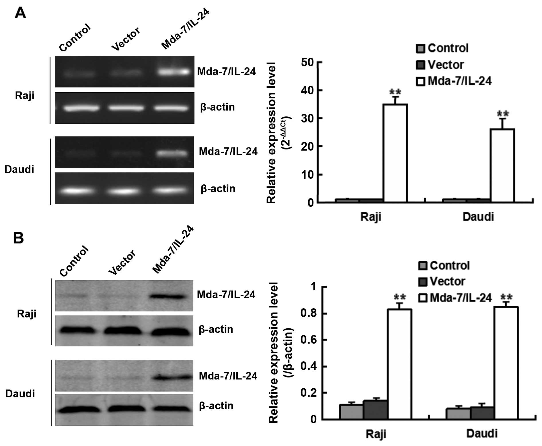
Expression of Mda-7/IL-24 in human Raji
and Daudi lymphoma cell lines
Mda-7/IL-24 mRNA and protein were assayed in parent
Raji and Daudi cells (control), in cells transfected with vector
alone, and in cells overexpressing Mda-7/IL-24 by RT-qPCR and
western blotting, respectively. The expression of Mda-7/IL-24 mRNA
and protein was weakly detectable in control cells and cells
transfected with empty vector. In contrast, Mda-7/IL-24
overexpressing cell lines showed significantly increased expression
of both Mda-7/IL-24 mRNA and protein (Fig. 1).
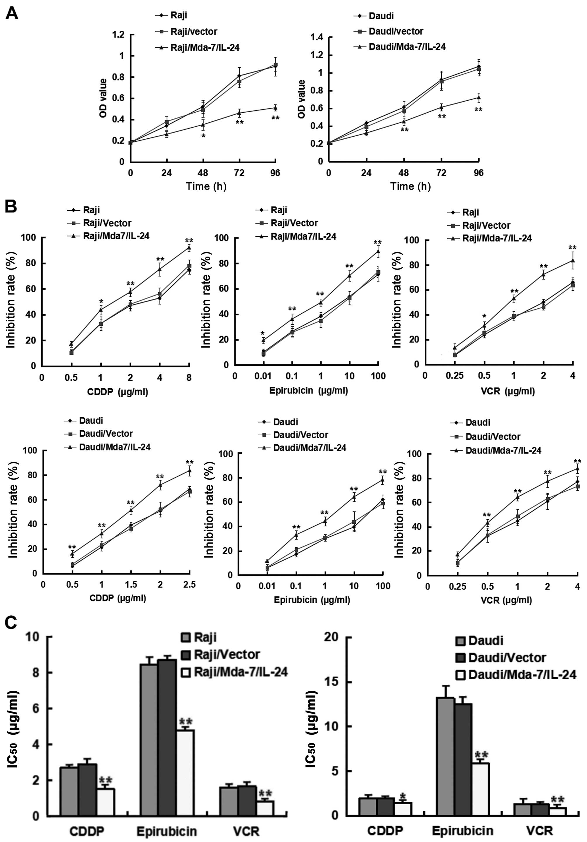
Mda-7/IL-24 retards proliferation and
enhances the sensitivity of B lymphoma cells to chemotherapeutic
agents
To investigate the effect of Mda-7/IL-24 on
proliferation and chemotherapy sensitivity of B lymphoma cells, MTS
assays were performed. As shown in Fig.
2A, transfection with Mda-7/IL-24 significantly induced growth
suppression in Raji and Daudi cells overexpressing Mda-7/IL-24,
compared with non-transfected cells and cells transfected with
vector alone. We further explored whether Mda-7/IL-24 had
chemosensitizing effects on B lymphoma cells. As shown in Fig. 2, the proliferation of the two
lymphoma cell lines over 24 h was dose-dependently inhibited by
CDDP, epirubicin and VCR, with significantly higher inhibition
observed in Raji and Daudi cells overexpressing Mda-7/IL-24 than in
non-transfected cells and cells transfected with vector alone
(Fig. 2A and B). The
IC50 values of CDDP, epirubicin and VCR were lower in
cells overexpressing Mda-7/IL-24 than in non-transfected cells and
cells transfected with empty vector (Fig. 2C). These results suggested that
Mda-7/IL-24 can retard proliferation of B lymphoma cells, and
enhance the sensitivity of B lymphoma cells to chemotherapy
drugs.
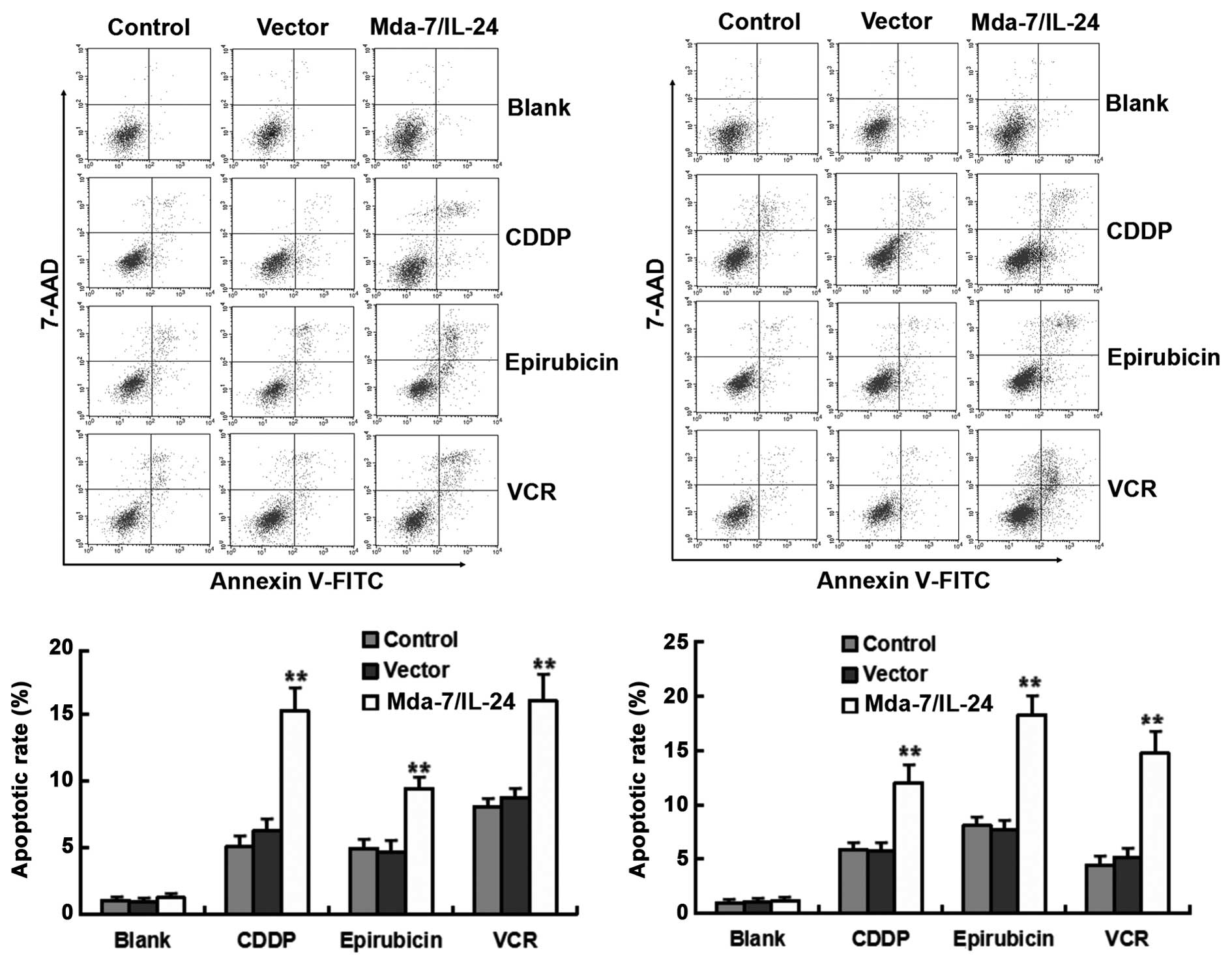
To further determine the potential chemosensitizing
effects of Mda-7/IL-24, flow cytometry was performed to investigate
whether overexpressing Mda-7/IL-24 induced Raji and Daudi cell
apoptosis. As shown in Fig. 3, no
difference in the percentage of Annexin V-FITC-positive cells was
observed between the cell lines of Raji and Daudi with and without
Mda-7/IL-24 expression using stable transfection methods, compared
with non-transfected cells and cells transfected with vector alone,
this finding suggested that the growth suppression observed in
these cells was not due to an increase in cell death. However, when
Raji and Daudi cells were treated with CDDP, epirubicin and VCR,
the percentage of Annexin V-FITC-positive cells was significantly
higher in cells overexpressing Mda-7/IL-24 than in non-transfected
and vector-transfected cells, and there was no significant
difference between the latter two cultures. Those results indicate
that apoptosis may not be involved in Mda-7/IL-24-induced growth
inhibition of Raji and Daudi cells, but Mda-7/IL-24 promotes the
apoptosis of Raji and Daudi cells treated with chemotherapeutic
agents, so that Mda-7/IL-24 is a strong chemosensitizer for B cell
lymphoma.
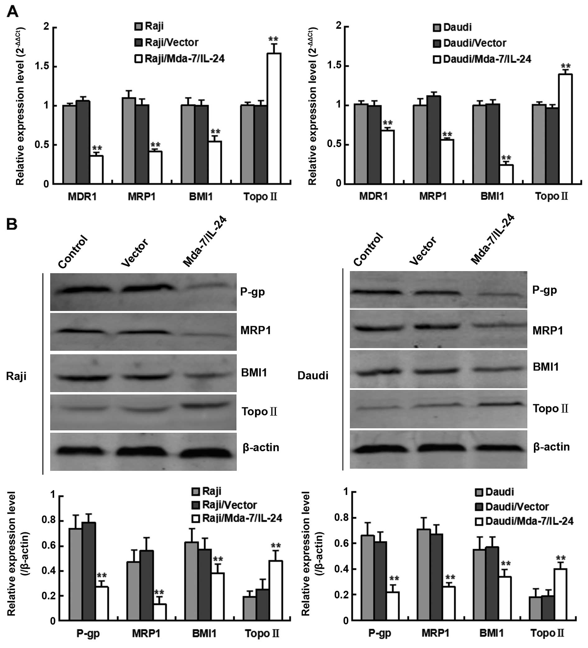
The multidrug-resistance genes MDR1,
MRP1, BMI1 and Topo II may be involved in sensitivity of
Mda-7/IL-24 overpressing cells to chemotherapy drugs
To address the underlying mechanism that may be
responsible for Mda-7/IL-24-mediated chemosensitizing effect, we
analyzed the changes of multidrug-resistance genes expression.
Since MDR1, MRP1, BMI and Topo II are genes associated with the
induction of multi-drug resistance, their expression was assessed
in Raji and Daudi overexpressing Mda-7/IL-24. Our results showed
that the levels of expression of MDR1, MRP1 and BMI1 mRNA and
protein were reduced, whereas the levels of Topo II were increased,
in lymphoma cells overexpressing Mda-7/IL-24 compared with both
non-transfected and vector-transfected cells (Fig. 4A and B). Those results indicate that
the multidrug-resistance genes MDR1, MRP1, BMI1 and Topo II may be
involved in sensitivity of overpressing cells to chemotherapy
drugs.
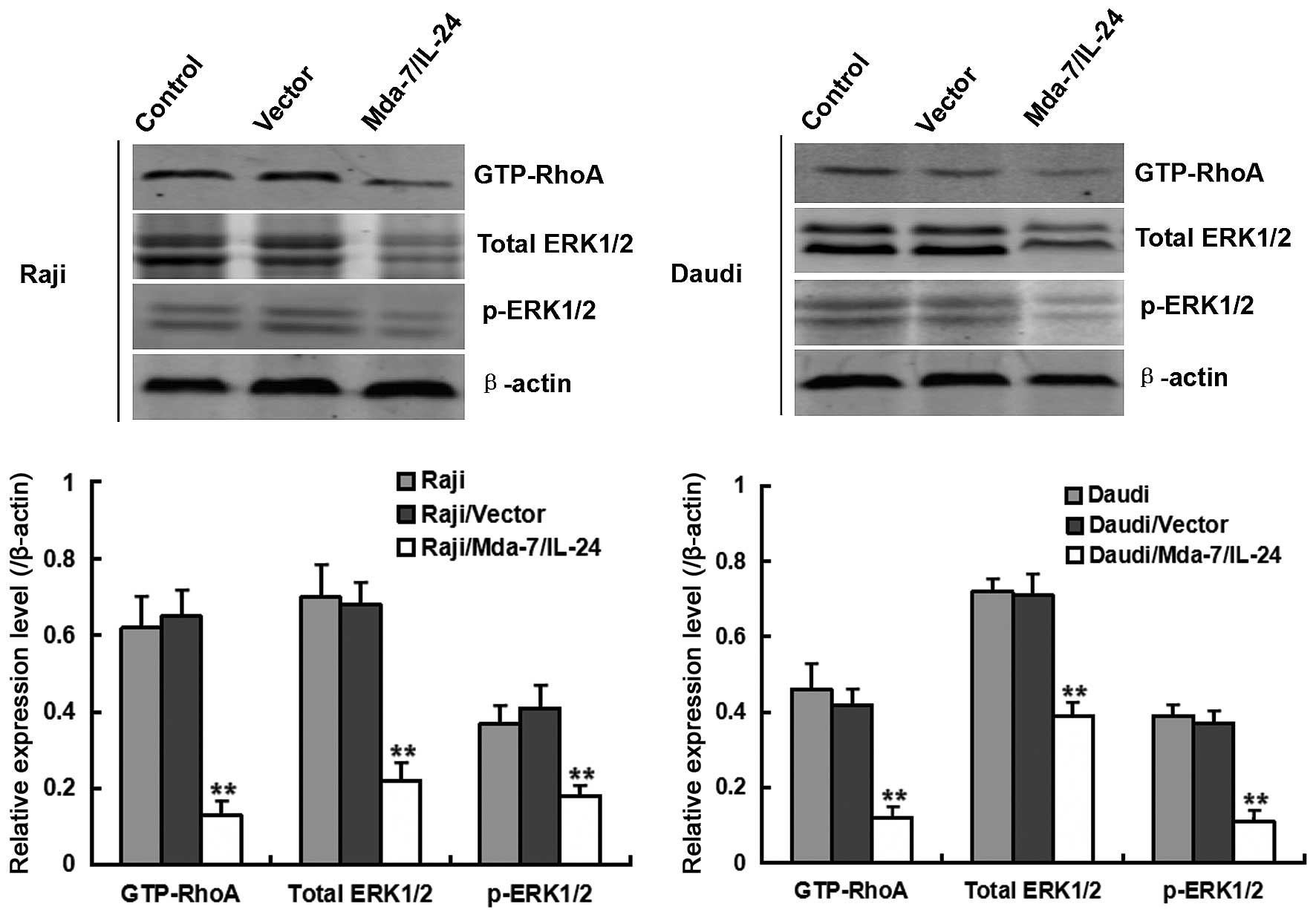
To further determine the signaling pathway
influenced by Mda-7/IL-24, the effects of Mda-7/IL-24 on the
activity of ERK signal that is involved in regulating expression of
MDR-related genes was evaluated. As shown in Fig. 5, the expression levels of total
ERK1/2 and p-ERK1/2 were significantly decreased in Raji and Daudi
overexpressing Mda-7/IL-24. It suggested that Mda-7/IL-24 can
suppress the activity of ERK signaling pathway. Several reports
have shown that MDR-related proteins including P-gp, MRP1 and BMI1
are positively regulated by the ERK pathway and blockade of the ERK
pathway can suppress their expression (10–13).
Thus, ERK signaling pathway may be a potential drug target for
circumventing MDR. Furthermore, it is well known that GTP-RhoA
signaling pathway is upstream of the ERK signaling pathway
(14,15). Western blot assay showed that
expression of GTP-RhoA was also influenced by Mda-7/IL-24 (Fig. 5). Taken together, the above data
indicated that there is an axis of GTP-RhoA-ERK regulating
expression of MDR-related genes MDR1, MRP1 and BMI1, but the
mechanism that upregulates the expression of Topo II is still
unknown.
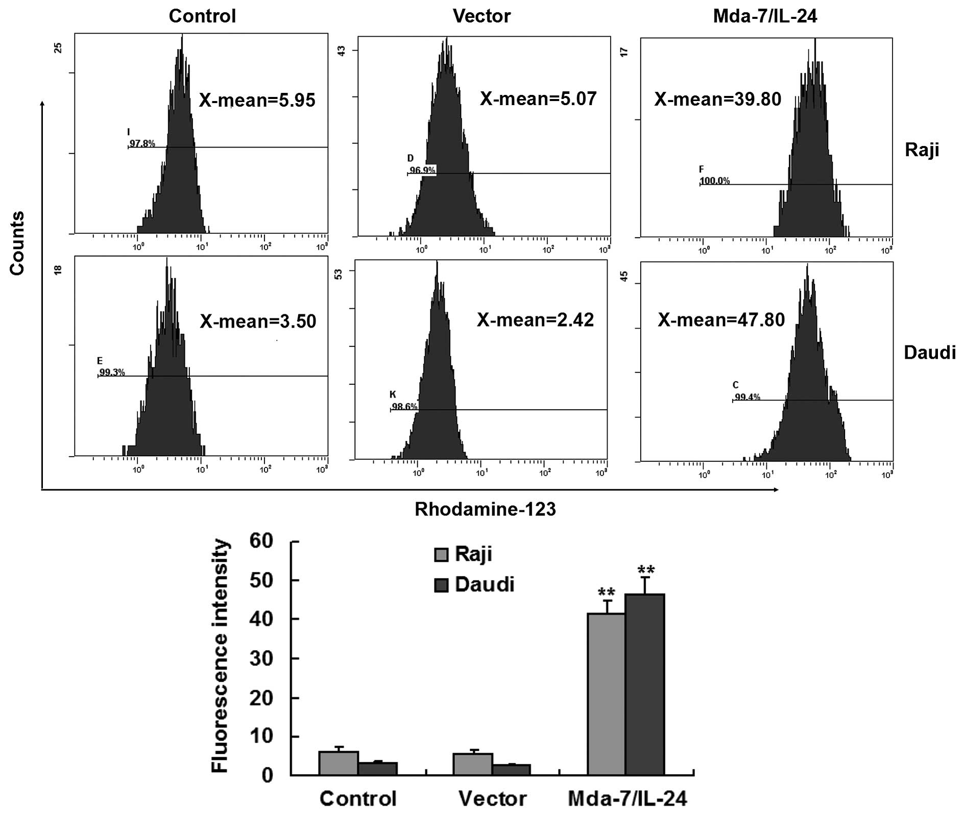
Mda-7/IL-24 enhances the intracellular
accumulation of Rhodamine-123 in lymphoma cells
The ability of Mda-7/IL-24 to inhibit the efflux of
MDR-related proteins such as P-glycoprotein (P-gp) and MRP1 was
assessed by Rhodamine-123 exclusion assays. The intracellular
accumulation of Rhodamine-123 was significantly higher in
Mda-7/IL-24 overexpressing cells than in non-transfected and
vector-transfected cells (Fig. 6).
These results suggested that Mda-7/IL-24 may reduce the pump effect
of MDR-related proteins in Raji and Daudi cells, increasing the
intracellular accumulation and reducing the efflux of chemotherapy
drugs.
Discussion
B cell lymphoma is one of the most common types of
hematopoietic tumors (16).
Chemotherapy is the first-line choice for lymphoma treatment.
However, tumors often become resistant to chemotherapy drugs during
treatment. MDR can result in the failure of chemotherapy and the
death of patients. Studies are required to elucidate the mechanisms
of drug resistance, and new treatment strategies are needed to
improve cure rates and survival in patients with lymphoma. Changes
in molecular regulatory networks that increase anti-apoptotic
activity and reduce the apoptosis of tumor cells are the primary
mechanisms associated with resistance to chemotherapy drugs
(2,17). Methods of enhancing sensitivity to
chemotherapy and avoiding MDR remain among the most difficult
problems in tumor treatment.
Combinations of chemotherapy with gene therapy are
promising in the treatment of B cell lymphoma (18). Although Mda-7/IL-24 has been widely
regarded as an antitumor molecule and has been shown to inhibit
hematopoietic tumor proliferation (5,6),
little is known about its effects on lymphoma cell sensitivity to
chemotherapy. Recent studies have shown that Mda-7/IL-24 could
reverse MDR in human colorectal and hepatocellular cancers
(2,19), but whether it plays a similar role
in B cell lymphoma has not been determined. The present study is
the first to show that transfection of endogenous Mda-7/IL-24
sensitized Raji and Daudi lymphoma cells to chemotherapy drugs by
affecting factors related to chemotherapy drug resistance.
In the present study, we first assessed the
chemosensitizing effects of Mda-7/IL-24 gene in B lymphoma cells
in vitro. MTS assay results showed that Mda-7/IL-24
significantly retarded proliferation of Raji and Daudi cells, and
enhanced the sensitivity of these lymphoma cells to various
chemotherapy drugs, including CDDP, epirubicin and VCR, and that
IC50 was lower in cells transfected with Mda-7/IL-24
than in non-transfected and vector-transfected cells. These results
were in good agreement with the effects of Mda-7/IL-24 on these
types of tumor cells (2,5,6). In
addition, although the apoptosis-inducing capability of Mda-7/IL-24
has been widely documented in various solid tumors, we did not
observe significant induction of apoptosis in the two lymphoma cell
lines. However, flow cytometry results showed that transfecting
Mda-7/IL-24 enhanced the apoptotic rates in cells treated with
these chemotherapeutic agents. The effect that Mda-7/IL-24 plus
chemotherapy drugs enhanced the induction of apoptosis in Raji and
Daudi cells indicating that Mda-7/IL-24 could be a potent model of
adjuvant chemotherapy for B cell lymphoma.
To study the mechanisms that may be responsible for
Mda-7/IL-24-mediated chemosensitizing effect, we analyzed the
changes of expression in various multidrug-resistance genes.
Abnormal levels of expression of MDR1, MRP1, BMI1 and Topo II were
found to be important in impairing tumor chemosensitivity,
resulting in MDR (20–23). P-gp is the best-known and principal
mediator of MDR. P-gp, which is encoded by MDR1, is involved in
pumping chemotherapy drugs from the inside to the outside of tumor
cells, and in preventing intracellular accumulation of these drugs
(20,24). MRP1 functions similarly to MDR1 in
reducing tumor sensitivity to chemotherapy, and it can recognize
and pump out chemotherapy drugs bound to glutathione in tumor
cells. High expression of MDR1 and MRP1 in different types of B
cell lymphoma tissues has been associated with poor response to
chemotherapy (25). Thus, our
finding, that Mda-7/IL-24 over-expression downregulated MDR1 and
MRP1 expression in Raji and Daudi cells, suggests that Mda-7/IL-24
may enhance the intracellular accumulation of anticancer drugs.
BMI1, which is essential for the self-renewal of
normal and malignant stem cells, is another important gene that
induces MDR. The mechanism of action of BMI1 may be related to
anti-oxidation and the upregulation of bcl-2, resulting in
resistance to chemotherapy-induced apoptosis (26,27).
Downregulation of BMI1 may increase drug-induced tumor cell
apoptosis, thus enhancing sensitivity to chemotherapy (28). Topo II, which can bind to
chemotherapeutic agents, with binding of these drugs to the α chain
of Topo II inhibiting the proliferation and duplication of DNA in
tumor cells (17). Upregulating the
expression of Topo II by transfection has been reported to enhance
the cytotoxicity of chemotherapy drugs (23,29).
Thus, the ability of Mda-7/IL-24 to enhance Topo II expression in
Raji and Daudi suggests that it may enhance sensitivity to
chemotherapy in vivo. Collectively, all of these findings
suggest that Mda-7/IL-24 enhances sensitivity to chemotherapy via
various targets, with the changes in MDR related protein
expression, enhancing the cytotoxic effects of chemotherapy agents
on lymphoma cells.
To further investigate the molecular mechanism of
Mda-7/IL-24-induced chemosensitizing effect, we analyzed the
activities of ERK and GTP-RhoA signaling pathways. It is known that
ERK plays an important role in positively regulating expression of
various MDR-related genes including MDR1, MRP1 and BMI1 (10–13). A
previous report showed that decreased activities of ERK1/2 was
detected in Human leukemia U937 and HL60 cells overpressing
Mda-7/IL-24 (4). In the present
study, overpressing Mda-7/IL-24 also resulted in a similar result
in Raji and Daudi cells. Moreover, GTP-RhoA is known as upstream
activator of cell ERK pathway, and it was also reported to be
related to induction of MDR (30).
In the present study, we found that decreased GTP-RhoA in
Mda-7/IL-24 overpressing lymphoma cells indicated that GTP-RhoA may
be a key factor affected by Mda-7/IL-24 regulating the expression
of MDR-related genes.
In addition, Mda-7/IL-24 overexpression also
significantly increased the intracellular accumulation of
Rhodamine-123 in Raji and Daudi cells, suggesting that Mda-7/IL-24
impaired the pump function of MDR-related proteins.
In summary, we found that Mda-7/IL-24 can enhance
the sensitivity of B lymphoma cells to chemotherapy by decreasing
the expression of MDR1, MRP1 and BMI1 via GTP-RhoA-ERK signaling
pathway, and by increasing the expression of Topo II and the
intracellular accumulation of anticancer drugs. In addition,
Mda-7/IL-24 could increase the apoptosis rate of B lymphoma cells
treated with chemotherapy drugs. Mda-7/IL-24 may therefore be a
potential candidate for treatment of MDR in B cell lymphomas.
Acknowledgments
The present study was supported by a grant from the
National Natural Science Foundation of China (no. 81173611).
Abbreviations:
|
Mda-7/IL-24
|
melanoma differentiation associated
gene-7/interleukin-24
|
|
CDDP
|
cis-diamminedichloroplatinum
|
|
VCR
|
vinblastine
|
|
IC50
|
50% inhibiting concentration
|
|
MDR
|
multidrug resistance
|
References
|
1
|
Morin RD, Mendez-Lago M, Mungall AJ, Goya
R, Mungall KL, Corbett RD, Johnson NA, Severson TM, Chiu R, Field
M, et al: Frequent mutation of histone-modifying genes in
non-Hodgkin lymphoma. Nature. 476:298–303. 2011. View Article : Google Scholar : PubMed/NCBI
|
|
2
|
Fang P, Zhang X, Gao Y, Ding CR, Cui F and
Jiao SC: Reversal effect of melanoma differentiation associated
gene-7/interleukin-24 on multidrug resistance in human
hepatocellular carcinoma cells. Anat Rec. 295:1639–1646. 2012.
View Article : Google Scholar
|
|
3
|
Huo W, Li ZM, Zhu XM, Bao YM and An LJ:
MDA-7/IL-24 suppresses tumor adhesion and invasive potential in
hepatocellular carcinoma cell lines. Oncol Rep. 30:986–992.
2013.PubMed/NCBI
|
|
4
|
Rahmani M, Mayo M, Dash R, Sokhi UK,
Dmitriev IP, Sarkar D, Dent P, Curiel DT, Fisher PB and Grant S:
Melanoma differentiation associated gene-7/interleukin-24 potently
induces apoptosis in human myeloid leukemia cells through a process
regulated by endoplasmic reticulum stress. Mol Pharmacol.
78:1096–1104. 2010. View Article : Google Scholar : PubMed/NCBI
|
|
5
|
Dong CY, Zhang F, Duan YJ, Yang BX, Lin YM
and Ma XT: mda-7/IL-24 inhibits the proliferation of hematopoietic
malignancies in vitro and in vivo. Exp Hematol. 36:938–946. 2008.
View Article : Google Scholar : PubMed/NCBI
|
|
6
|
Qian W, Liu J, Tong Y, Yan S, Yang C, Yang
M and Liu X: Enhanced antitumor activity by a selective
conditionally replicating adenovirus combining with
MDA-7/interleukin-24 for B-lymphoblastic leukemia via induction of
apoptosis. Leukemia. 22:361–369. 2008. View Article : Google Scholar
|
|
7
|
Sahoo A and Im SH: Molecular mechanisms
governing IL-24 gene expression. Immune Netw. 12:1–7. 2012.
View Article : Google Scholar : PubMed/NCBI
|
|
8
|
Gopalan B, Shanker M, Chada S and Ramesh
R: MDA-7/IL-24 suppresses human ovarian carcinoma growth in vitro
and in vivo. Mol Cancer. 6:112007. View Article : Google Scholar : PubMed/NCBI
|
|
9
|
Pataer A, Vorburger SA, Barber GN, Chada
S, Mhashilkar AM, Zou-Yang H, Stewart AL, Balachandran S, Roth JA,
Hunt KK, et al: Adenoviral transfer of the melanoma
differentiation-associated gene 7 (mda7) induces apoptosis of lung
cancer cells via up-regulation of the double-stranded RNA-dependent
protein kinase (PKR). Cancer Res. 62:2239–2243. 2002.PubMed/NCBI
|
|
10
|
Yan F, Bai LP, Gao H, Zhu CM, Lin L and
Kang XP: EGF reverses multi-drug resistance via the p-ERK pathway
in HepG2/ADM and SMMC7721/ADM hepatocellular carcinoma models.
Asian Pac J Cancer Prev. 15:2619–2623. 2014. View Article : Google Scholar : PubMed/NCBI
|
|
11
|
Wang NN, Zhao LJ, Wu LN, He MF, Qu JW,
Zhao YB, Zhao WZ, Li JS and Wang JH: Mechanistic analysis of
taxol-induced multidrug resistance in an ovarian cancer cell line.
Asian Pac J Cancer Prev. 14:4983–4988. 2013. View Article : Google Scholar : PubMed/NCBI
|
|
12
|
Yan F, Wang XM, Pan C and Ma QM:
Down-regulation of extracellular signal-regulated kinase 1/2
activity in P-glycoprotein-mediated multidrug resistant
hepatocellular carcinoma cells. World J Gastroenterol.
15:1443–1451. 2009. View Article : Google Scholar : PubMed/NCBI
|
|
13
|
Suh HN and Han HJ: Collagen I regulates
the self-renewal of mouse embryonic stem cells through α2β1
integrin- and DDR1-dependent Bmi-1. J Cell Physiol. 226:3422–3432.
2011. View Article : Google Scholar : PubMed/NCBI
|
|
14
|
Jayakumar T, Chiu CC, Wang SH, Chou DS,
Huang YK and Sheu JR: Anti-cancer effects of CME-1, a novel
polysaccharide, purified from the mycelia of Cordyceps sinensis
against B16-F10 melanoma cells. J Cancer Res Ther. 10:43–49. 2014.
View Article : Google Scholar : PubMed/NCBI
|
|
15
|
Sánchez-Mir L, Soto T, Franco A, Madrid M,
Viana RA, Vicente J, Gacto M, Pérez P and Cansado J: Rho1 GTPase
and PKC ortholog Pck1 are upstream activators of the cell integrity
MAPK pathway in fission yeast. PLoS One. 9:e880202014. View Article : Google Scholar : PubMed/NCBI
|
|
16
|
Chen S, Wang Z, Dai X, Pan J, Ge J, Han X,
Wu Z, Zhou X and Zhao T: Re-expression of microRNA-150 induces
EBV-positive Burkitt lymphoma differentiation by modulating c-Myb
in vitro. Cancer Sci. 104:826–834. 2013. View Article : Google Scholar : PubMed/NCBI
|
|
17
|
Xu Y, Zheng H, Kang JS, Zhang L, Su J, Li
HY and Sun LK: 5-Nitro-2-(3-phenylpropylamino) benzoic acid induced
drug resistance to cisplatin in human erythroleukemia cell lines.
Anat Rec. 294:945–952. 2011. View
Article : Google Scholar
|
|
18
|
Zhu X, Ma Y and Liu D: Novel agents and
regimens for acute myeloid leukemia: 2009 ASH annual meeting
highlights. J Hematol Oncol. 3:172010. View Article : Google Scholar : PubMed/NCBI
|
|
19
|
Emdad L, Lebedeva IV, Su ZZ, Sarkar D,
Dent P, Curiel DT and Fisher PB: Melanoma differentiation
associated gene-7/interleukin-24 reverses multidrug resistance in
human colorectal cancer cells. Mol Cancer Ther. 6:2985–2994. 2007.
View Article : Google Scholar : PubMed/NCBI
|
|
20
|
Loo TW, Bartlett MC, Shi L and Clarke DM:
Corrector-mediated rescue of misprocessed CFTR mutants can be
reduced by the P-glycoprotein drug pump. Biochem Pharmacol.
83:345–354. 2012. View Article : Google Scholar
|
|
21
|
Wang E, Bhattacharyya S, Szabolcs A,
Rodriguez-Aguayo C, Jennings NB, Lopez-Berestein G, Mukherjee P,
Sood AK and Bhattacharya R: Enhancing chemotherapy response with
Bmi-1 silencing in ovarian cancer. PLoS One. 6:e179182011.
View Article : Google Scholar : PubMed/NCBI
|
|
22
|
Baird NJ, Fang XW, Srividya N, Pan T and
Sosnick TR: Folding of a universal ribozyme: The ribonuclease P
RNA. Q Rev Biophys. 40:113–161. 2007. View Article : Google Scholar : PubMed/NCBI
|
|
23
|
Tokiniwa H, Horiguchi J, Takata D, Kikuchi
M, Rokutanda N, Nagaoka R, Sato A, Odawara H, Tozuka K, Oyama T, et
al: Topoisomerase II alpha expression and the Ki-67 labeling index
correlate with prognostic factors in estrogen receptor-positive and
human epidermal growth factor type-2-negative breast cancer. Breast
Cancer. 19:309–314. 2012. View Article : Google Scholar
|
|
24
|
Lu D, Shi HC, Wang ZX, Gu XW and Zeng YJ:
Multidrug resistance-associated biomarkers PGP, GST-pi, Topo-II and
LRP as prognostic factors in primary ovarian carcinoma. Br J Biomed
Sci. 68:69–74. 2011.PubMed/NCBI
|
|
25
|
Ohsawa M, Ikura Y, Fukushima H, Shirai N,
Sugama Y, Suekane T, Hirayama M, Hino M and Ueda M:
Immunohistochemical expression of multidrug resistance proteins as
a predictor of poor response to chemotherapy and prognosis in
patients with nodal diffuse large B-cell lymphoma. Oncology.
68:422–431. 2005. View Article : Google Scholar : PubMed/NCBI
|
|
26
|
Crea F, Duhagon Serrat MA, Hurt EM, Thomas
SB, Danesi R and Farrar WL: BMI1 silencing enhances docetaxel
activity and impairs antioxidant response in prostate cancer. Int J
Cancer. 128:1946–1954. 2011. View Article : Google Scholar
|
|
27
|
Siddique HR, Parray A, Tarapore RS, Wang
L, Mukhtar H, Karnes RJ, Deng Y, Konety BR and Saleem M: BMI1
polycomb group protein acts as a master switch for growth and death
of tumor cells: Regulates TCF4-transcriptional factor-induced BCL2
signaling. PLoS One. 8:e606642013. View Article : Google Scholar : PubMed/NCBI
|
|
28
|
Zhu D, Wan X, Huang H, Chen X, Liang W,
Zhao F, Lin T, Han J and Xie W: Knockdown of Bmi1 inhibits the
stemness properties and tumorigenicity of human bladder cancer stem
cell-like side population cells. Oncol Rep. 31:727–736. 2014.
|
|
29
|
Zhu WY, Hunag YY, Liu XG, He JY, Chen DD,
Zeng F, Zhou JH and Zhang YK: Prognostic evaluation of CapG,
gelsolin, P-gp, GSTP1, and Topo-II proteins in non-small cell lung
cancer. Anat Rec. 295:208–214. 2012. View
Article : Google Scholar
|
|
30
|
Rigoni M, Riganti C, Vitale C, Griggio V,
Campia I, Robino M, Foglietta M, Castella B, Sciancalepore P,
Buondonno I, et al: Simvastatin and downstream inhibitors
circumvent constitutive and stromal cell-induced resistance to
doxorubicin in IGHV unmutated CLL cells. Oncotarget. 6:29833–29846.
2015.PubMed/NCBI
|