Introduction
The COP9 signalosome (CSN) is an evolutionarily
conserved complex of 8 to 9 protein subunits that regulates
numerous cellular and biological processes such as embryonic
development, cell cycle, DNA damage repair, checkpoint repair
control, signal transduction, circadian rhythm, T-cell development
and autophagy (1). CSN accomplishes
these various functions by controlling cullin-RING-E3 ligases
(CRLs), the most prominent class of E3-enzymes, which promote
ubiquitination of a variety of regulatory proteins subsequently
targeted to proteasomal degradation (2). All CRLs are composed of a constitutive
cullin RING-based catalytic core and of exchangeable substrate
recognition modules (3). The human
genome encodes at least seven cullins (Cul1, 2, 3, 4A, 4B, 5 and
7), which share some sequence homology and a similar elongated
arc-shaped structure (4,5). The substrate recognition module is
specific to each cullin, and consists of a substrate recognition
subunit connected to the cullin by an adaptor subunit, such as DDB1
in CRL4 ligases. Despite their considerable diversity, all CRLs are
activated by conjugation of the ubiquitin-like protein Nedd8 to the
cullin subunit (neddylation) (3,6). CSN
inactivates cullin proteins both by hydrolyzing the cullin-Nedd8
conjugates (7) and by inhibiting
CRL activity as a direct Cullin-Roc1 binding partner, thereby
preventing substrate recruitment (8,9). The
association of CSN with CRL is highly dynamic, and context- as well
as substrate-dependent (10,11).
Previously, we discovered that two Cullin
4-Roc1-DDB1 ligases (CRL4s) are involved in the repair of
ultraviolet (UV)-induced DNA lesions by the nucleotide excision
repair (NER) pathway.
NER repairs helix-distorting forms of single-strand
DNA damage such as UV-created cyclobutane pyrimidine dimers (CPDs)
and 6-4 pyrimidone phosphoproducts (6-4PPs) or chemotherapy
generated bulky adducts. NER consists of two main sub-pathways,
global genomic NER (GG-NER) that repairs the whole genome on one
hand and transcription-coupled NER (TC-NER) that is restricted to
actively transcribed genes. Mutations in the NER pathway cause two
well-known genetic diseases: the developmental disorder Cockayne
syndrome is caused by mutations in TC-NER; the skin cancer
predisposition syndrome Xeroderma pigmentosum is the consequence of
an impairment of GG-NER. TC-NER and GG-NER are similar, and the
main differences are in the mechanism of DNA lesion recognition, a
step at which NER-associated CRL4 ligases are involved. DDB2 (DNA
damage binding protein 2) and CSA (Cockayne Syndrome protein A) are
the substrate recognition receptors of these CLR4 ligases
(CRL4DDB2 and CRL4CSA). DDB2 is the smaller
subunit of DDB, a heterodimeric protein implicated in the ethiology
of Xeroderma pigmentosum group E (XPE), while CSA is a TC-NER
component and a complementation factor for the Cockayne syndrome
disease (12). CRL4DDB2
and CRL4CSA are regulated by CSN, albeit in a different
manner (10). Immediately after
UV-irradiation, CRL4DDB2 is released from CSN and
activated by neddylation (10,13).
By contrast, CRL4CSA is silenced by CSN at the beginning
of the repair process and is activated at later stages, upon
dissociation of CSN (10,14).
DNA damages trigger the DNA damage response (DDR)
pathway, a signaling network leading to a coordinated cell response
that includes cell cycle arrest, apoptosis, senescence, and DNA
repair. Two related protein kinases, ATM (ataxia telangiectasia
mutated) and ATR (ATM and Rad3-related), are essential in DDR. ATM,
and its regulator the MRN (Mre11-Rad50-NBS1) complex, is the
general sensor of DNA double-strand breaks (DSBs). ATR and its
regulator ATRIP (ATR-interacting protein) detect the single-strand
DNA (ssDNA) species generated by NER, as well as ssDNA present at
stalled replication forks (15,16).
UV irradiation and the NER pathway also activate ATM (17,18).
The second wave of signaling involves phosphorylation of common
downstream targets of ATM and ATR, e.g., H2AX, Chk1 and Chk2. The
Elledge laboratory has identified over 700 new ATM and ATR targets
phosphorylated at SQ/TQ sites in response to DSB-inducing ionized
radiation (IR) and/or UV irradiation. Three subunits of CSN, CSN1,
CSN3 and CSN7a, are phosphorylated in response to IR. CSN1 is also
phosphorylated in response to UV (19).
Here, we examined phosphorylation of CSN in response
to UV irradiation. We detected several new ATM-dependent
phosphorylation sites in CSN1 and CSN7a. We provide evidence, based
on gain- and loss-of-function experiments, that CSN1
phosphorylation on S474 is not required for DNA repair, but rather
appears to be involved in DDR and participates in inducing
apoptosis in response to UV. Thus, in UV-irradiated cells, CSN
appears to be involved both in the DNA repair process and in cell
apoptosis, a downstream cell response.
Materials and methods
Plasmid construction
CSN1 phosphomutant (S474A) was generated using the
QuickChange Site-Directed Mutagenesis kit (Stratagene). Full length
human CSN1 wild-type and CSN1 phosphomutant (S474A) were inserted
in frame into the pREV-HTF retroviral vector as previously
described (20).
Cell culture and cell lines
HeLa S3 cells were grown in Dulbecco's modified
Eagle's medium (DMEM) supplemented with 10% fetal bovine serum.
HeLa S3 cell lines stably expressing FLAG and HA-tagged CSN1 or its
phosphomutant were generated by retroviral transduction according
to a previously established protocol (10). A HeLa cell line transduced with the
empty pREV vehicle vector was used as a negative control. For UV
irradiation, cells were grown on 15-cm tissue culture dishes,
washed with PBS, irradiated with UV at 25 J/m2 (unless
noted otherwise) and incubated in fresh medium for the indicated
periods of time. Where indicated, 5 µM of the ATM-specific
inhibitor, 2-morpholin-4-yl-6-thianthren-1-yl-pyran-4-one
(KU-55933) was added.
Antibodies
We used the following antibodies: CSA (sc-10997;
Santa Cruz Biotechnology; GTX100145; GeneTex), ATM protein kinase
pS1981 (200-301-400; Rockland), CSN1 (PW8285; Enzo/Affinity),
β-actin (A5441; Sigma), cyclobutane pyrimidine dimers (TDM2,
D194-1, MBL) and (6-4) photoproducts (6-4M2, D195-1, MBL).
Purification of protein complexes
A solubilized chromatin fraction was prepared as
described previously (10).
Briefly, 1×10⁸ cells were suspended in
hypotonic Micrococcal Nuclease (MNase) buffer (20 mM Tris, pH 8.0,
5 mM NaCl, 2.5 mM CaCl2) and disrupted by
freezing/thawing cycles. Nuclei were collected by centrifugation at
1,000 × g for 5 min, washed once with 1 ml of MNase buffer and
resus-pended in 400 µl of MNase buffer. MNase (N3755; Sigma)
was added at 10 U/ml and the samples were incubated for 10 min at
room temperature. MNase reaction was terminated with 10 mM EGTA.
The samples were centrifuged at 5,000 × g for 5 min and the
supernatants were collected (solubilized chromatin fraction).
Protein complexes were immunoprecipitated with anti-FLAG M2 agarose
(Sigma) and eluted with FLAG peptide. They were either immediately
analyzed, or further affinity purified with anti-HA-conjugated
agarose and eluted with the HA peptide. Complexes were resolved by
SDS-PAGE and stained using the Silver Quest kit (Invitrogen).
LC-MS/MS based shotgun sequencing
SDS-PAGE-resolved protein bands were subjected to
trypsin-based in-gel digestion and LC-MS/MS based shotgun
sequencing using an LTQ Orbitrap mass spectrometer essentially as
described previously (21–25). As was carried out previously, the
resulting tandem mass spectra were data-searched using the SEQUEST
algorithm allowing for variable modifications on methionine
(+15.9949 Da) and serine, threonine and tyrosine (+79.9663 Da) with
full trypsin specificity, 2 missed cleavages and a precursor
tolerance of ±1.1 Da against the UniProt human database (www.uniprot.org). Results were filtered with a ±3 ppm
mass measurement accuracy cutoff (from theoretical) and a
delta-Xcorr of 0.08, and all CSN phosphopeptide MS/MS spectra were
manually inspected for consistency.
Analysis of UV-induced photolesions
UV-induced photolesions were quantified on a
slot-blot, using TD-2 and 6-4M2 antibodies specifically recognizing
CPDs and 6-4 photoproducts respectively. Irradiated cells were
lysed in MNase buffer and centrifuged to obtain nuclei pellets that
were subsequently treated with MNase as described above. DNA for
analysis was purified by phenol-chlorophorm and measured on a
NanoDrop (ND-1000; NanoDrop Technologies, Inc.). One milligram of
nucleosome-associated DNA from each sample was transferred onto
nitrocellulose membranes (Hybond™-N+ nylon transfer membrane 0.45
µm RPN2020B; Amersham) using the MINIFOLD I slot-blot system
(Schleicher & Schuell). Immunoblot analysis was performed as
previously described (14).
Results
CSN1 is phosphorylated by ATM in response
to UV irradiation
To test whether CSN subunits are regulated by
phosphorylation in response to UV, HeLa S3 cells expressing a Flag
tag on the CSN5 subunit (10) were
UV irradiated (or not for the controls). Chromatin-associated CSN
complexes were affinity purified with anti-Flag antibodies 30 min
or 4 h following UV exposure. Analysis of the complexes by
mass-spectrometry revealed five phosphorylation sites on the CSN1
subunit and one phosphorylation site on the CSN7α subunit (Table I). Except for the CSN1 pS468 and
pT479 sites, the amount of phosphorylated peptides increased at 4 h
after UV exposure but not at 30 min.
 | Table IIdentification of phosphorylated
subunits by mass spectrometry. |
Table I
Identification of phosphorylated
subunits by mass spectrometry.
| Subunit | Phospho-site | Total peptide matches
after exposure to UV (min)
|
|---|
| 0 | 30 | 240 |
|---|
| CSN1 | pS468 | 2 | 0 | 1 |
| pS474 | 6 | 5 | 9 |
| pT479 | 4 | 4 | 4 |
| pS483 | 0 | 0 | 2 |
| pS485 | 0 | 0 | 2 |
| CSN7 | pS232 | 1 | 0 | 2 |
The sequences adjacent to the S474 and S483
phosphorylation sites on CSN1 and the S232 site on CSN7a match the
phosphorylation site by ATM or/and ATR kinases (SQ sites). These
sites were also identified as ATM/ATR substrates in a proteomic
screen for the DDR associated ATM/ATR targets performed by the
Elledge laboratory (19). To
confirm that the CSN1 S474Q site was phosphorylated by ATM, we used
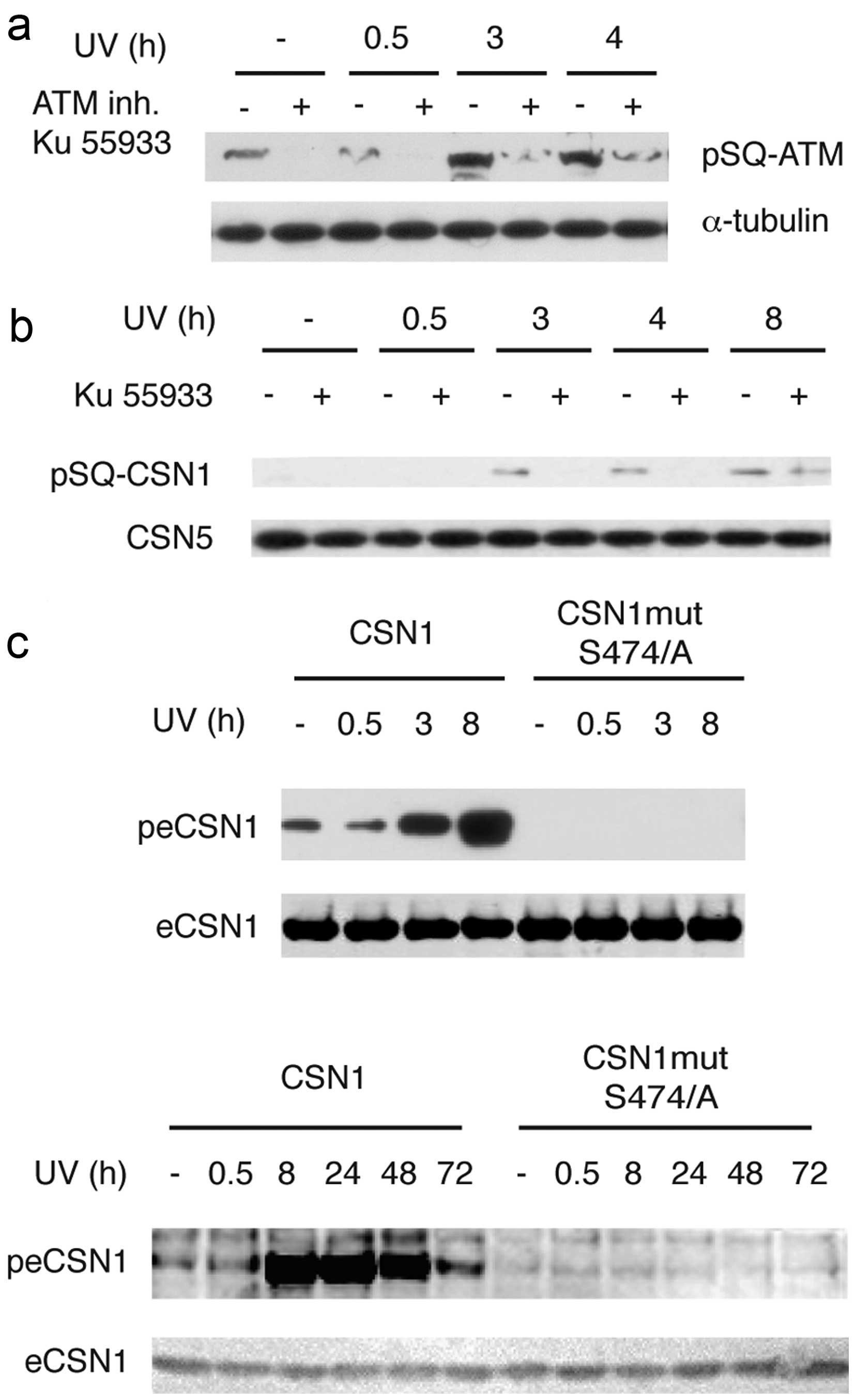
pSQ antibodies generated against ATM pS1981-Q (19): these antibodies detected ATM
phosphorylation at 3 h but not at 30 min after UV irradiation
(Fig. 1a). The ATM specific
inhibitor Ku55933 dramatically reduced ATM phosphorylation,
confirming ATM autophosphorylation. The same antibody detected a
band in CSN complexes purified from chromatin. The size of the band
matched the size of pCSN1. The band was detected at 3 h but not at
30 min post-UV irradiation (Fig.
1b), in good concordance with the kinetics of phosphorylation
observed using mass-spectrometry. Moreover, cell pre-treatment with
Ku55933 abolished the appearance of this band, supporting the idea
that phosphorylation was carried out by ATM (Fig. 1b). Taken together, our results
suggest that CSN1 is phosphorylated on S474 by ATM in response to
UV irradiation. To confirm this hypothesis, we mutated S474 in
CSN1; the resulting S474/A mutant was not phosphorylated in
response to UV (Fig. 1c). The
phosphorylation of ectopic CSN1 was detected 3 h post irradiation,
peaked at 8 h and persisted up to 48 h. Taken together, these data
indicate that CSN1 is phosphorylated in response to UV, and that
CSN1 phosphorylation is a late event.
UV-dependent CSN1 phosphorylation is
important for UV-induced apoptosis
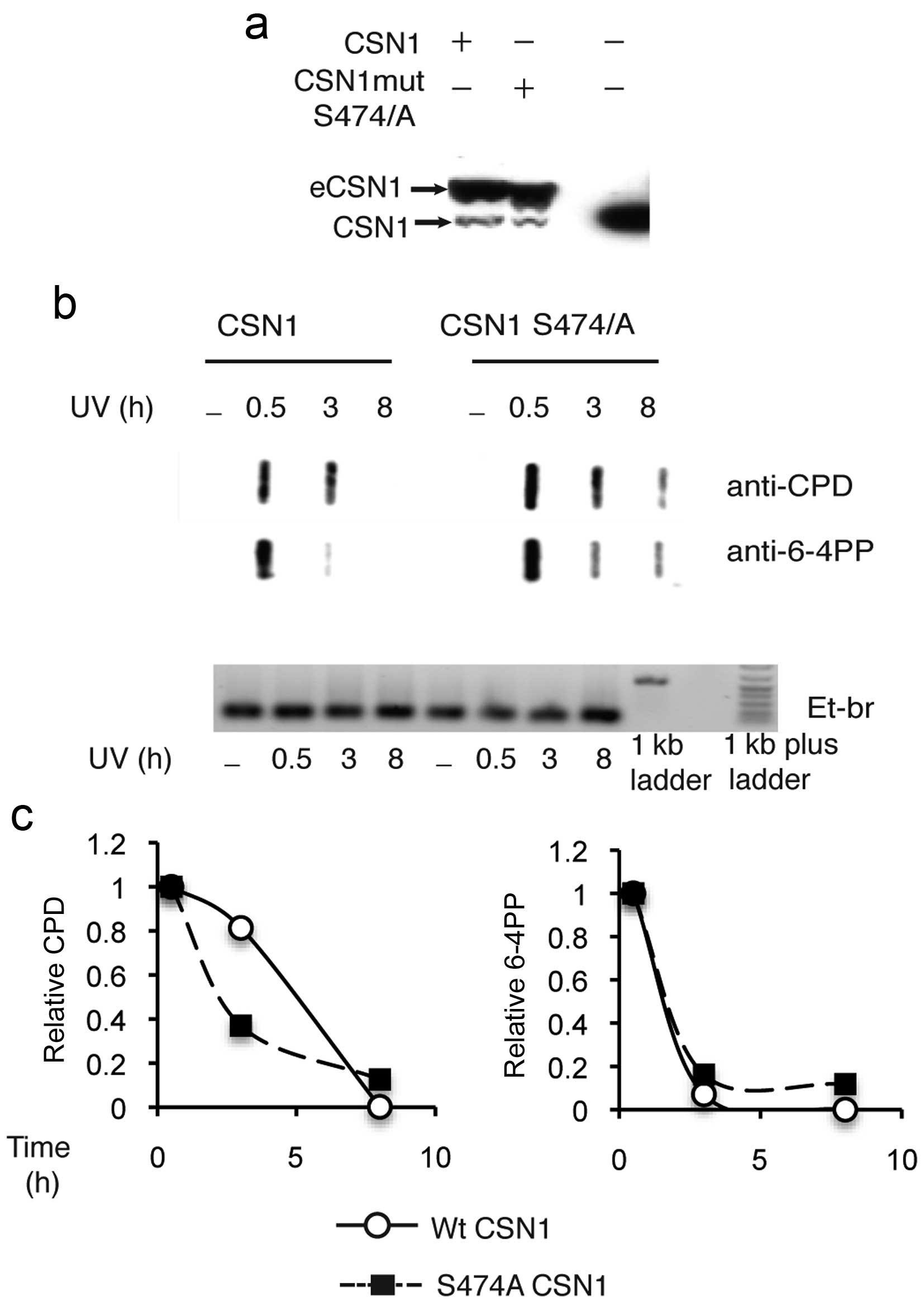
The ectopic expression of a large excess of mutant
CSN1 (Fig. 2a), potentially acting
as a negative transdominant mutant, did not decrease the repair of
6-4PP or CPD damages. Repair of CPD damages appeared even to occur
at slightly faster rates (Fig. 2b and
c). Consistently, the timing of phosphorylation, which peaked
at 8 h, a time at which DNA repair was completed (Fig. 2c) and its persistance for 48 h
post-irradiation (Fig. 1c),
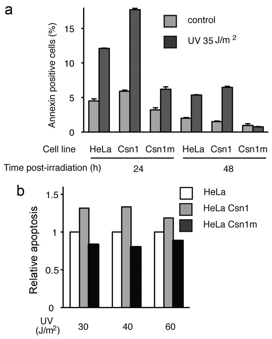
suggests an involvement in downstream events. We thus tested the
hypothesis that CSN1 phosphorylation could be involved in apoptosis
induced by high doses of UV. Apoptosis was monitored using an
Annexin V assay. Apoptosis was inhibited in HeLa cells
overexpressing the S474/A mutant CSN1 (Fig. 3a); whereas 12% of HeLa cells were
scored as apoptotic 24 h post-irradiation, only 6% were apoptotic
in cells expressing excess amounts of the mutant. At 48 h
post-irradiation, apoptotic cells could not be detected in cells
expressing the mutant, whereas 6% of the cells were scored as
apoptotic in the HeLa cells. Conversely, an excess of wild-type
CSN1 resulted in increased apoptosis (from 12% in HeLa cells to 17%
in HeLa cells expressing wild-type CSN1). This tendency was
observed at various energies of irradiation (Fig. 3b). Thus, the phosphorylation of CSN1
appears to be involved in UV-induced apoptosis.
Discussion
Here, we demonstrated that CSN1 is phosphorylated on
S474 by ATM in response to UV irradiation. CSN1 binds to
CRL4DDB2 and CRL4CSA ligases, and a tempting
hypothesis was that CSN1 phosphorylation by ATM regulates the
interaction between CSN1 and these ligases. However, our
experiments (data not shown) indicated that this was not the case,
as mutating the CSN phosphorylation site did not affect the
co-immunoprecipitation of the CSN complex with CRL4DDB2
or CRL4CSA ligases. This result was consistent with the
timing of phosphorylation, which occurred later than the
dissociation of the CSN1/CLR4 complex. This result is also
consistent with the X-ray structure of CSN, recently solved by the
Thomä laboratory (28). This
structure demonstrates that Ser 474 is located at the surface of
the CSN C-terminal helical bundle, separated by structured linkers
from the CRL4 interacting PCI ring structure. Three other COP9/CSN
subunits, CSN3, CSN7a and CSN8, were phosphorylated in response to
UV irradiation and these phosphosites are also located in the same
C-terminal helical bundle according to CSN X-ray structure.
Moreover, CSN1 itself is phosphorylated on multiple serines
(19,27). Thus, eliminating only one of these
phosphorylation events may not be sufficient to impact CSN/CLR4
interaction, and to achieve maximal inhibition of apoptosis.
The observation that ATM-dependent CSN1
phosphorylation is a late event correlates with a role in apoptosis
rather then in DNA lesion repair. Conversely, CSN3 is
phosphorylated at earlier times (from 0.5–8 h) after γ-irradiation
and this phosphorylation event delayed double strands break repair
in irradiated fibroblasts (27). We
did not observe any effect of phosphorylation on DNA repair for
S474 phosphorylation, but rather our data showed that CSN1
phosphorylation was important for cell progression toward
programmed cell death (apoptosis) in response to high dose UV
irradiation, as ectopic expression of the S474/A mutant reduced
apoptosis. Two recent high throughput studies of cell
cycle-dependent phosphorylation identified specific phosphorylation
of CSN1 and CSN3 by ATM/ATR during the S phase of the cell cycle
(29,30). The activation of DDR phosphorylation
network mediated by ATM and ATR during the S phase of the cell
cycle serves as a checkpoint and promotes the elimination of cells
with unrepaired damages via apoptosis. Here, we demonstrated a role
for CSN1 S474 phosphorylation in UV-induced apoptosis, as ectopic
expression of the non-phosphorylated mutant reduced cell apoptosis.
A similar case has been described for Ku70 in chronic lymphocytic
leukemia (CLL). Upon genotoxic stress, phosphorylation of Ku70 is
increased in an ATM-dependent manner in CLL cells, and mutation of
this site reduced CLL cell apoptosis. By enhancing double-strand
break (DSB) repair, and interfering with apoptosis, phospho-Ku70
likely contributes to the CLL drug resistant phenotype and to
cancer progression. Similarly, phospho-CSN could also play a
specific role in different steps of tumorigenesis. CSN has been
implicated in the development of many cancers, partially via the
proteasome-mediated degradation of oncogenes or tumor suppressors
p53 (31), Mdm2 (32), p27 (33), Id1 and Id3 (34).
References
|
1
|
Wei N, Serino G and Deng XW: The COP9
signalosome: More than a protease. Trends Biochem Sci. 33:592–600.
2008. View Article : Google Scholar : PubMed/NCBI
|
|
2
|
Petroski MD and Deshaies RJ: Function and
regulation of cullin-RING ubiquitin ligases. Nat Rev Mol Cell Biol.
6:9–20. 2005. View
Article : Google Scholar : PubMed/NCBI
|
|
3
|
Merlet J, Burger J, Gomes JE and Pintard
L: Regulation of cullin-RING E3 ubiquitin-ligases by neddylation
and dimerization. Cell Mol Life Sci. 66:1924–1938. 2009. View Article : Google Scholar : PubMed/NCBI
|
|
4
|
Zheng N, Schulman BA, Song L, Miller JJ,
Jeffrey PD, Wang P, Chu C, Koepp DM, Elledge SJ, Pagano M, et al:
Structure of the Cul1-Rbx1-Skp1-F boxSkp2 SCF ubiquitin
ligase complex. Nature. 416:703–709. 2002. View Article : Google Scholar : PubMed/NCBI
|
|
5
|
Angers S, Li T, Yi X, MacCoss MJ, Moon RT
and Zheng N: Molecular architecture and assembly of the DDB1-CUL4A
ubiquitin ligase machinery. Nature. 443:590–593. 2006.PubMed/NCBI
|
|
6
|
Rabut G and Peter M: Function and
regulation of protein neddylation. 'Protein modifications: Beyond
the usual suspects' review series. EMBO Rep. 9:969–976. 2008.
View Article : Google Scholar : PubMed/NCBI
|
|
7
|
Wee S, Geyer RK, Toda T and Wolf DA: CSN
facilitates Cullin-RING ubiquitin ligase function by counteracting
auto-catalytic adapter instability. Nat Cell Biol. 7:387–391. 2005.
View Article : Google Scholar : PubMed/NCBI
|
|
8
|
Emberley ED, Mosadeghi R and Deshaies RJ:
Deconjugation of Nedd8 from Cul1 is directly regulated by
Skp1-F-box and substrate, and the COP9 signalosome inhibits
deneddylated SCF by a noncatalytic mechanism. J Biol Chem.
287:29679–29689. 2012. View Article : Google Scholar : PubMed/NCBI
|
|
9
|
Lingaraju GM, Bunker RD, Cavadini S, Hess
D, Hassiepen U, Renatus M, Fischer ES and Thomä NH: Crystal
structure of the human COP9 signalosome. Nature. 512:161–165. 2014.
View Article : Google Scholar : PubMed/NCBI
|
|
10
|
Groisman R, Polanowska J, Kuraoka I,
Sawada J, Saijo M, Drapkin R, Kisselev AF, Tanaka K and Nakatani Y:
The ubiquitin ligase activity in the DDB2 and CSA complexes is
differentially regulated by the COP9 signalosome in response to DNA
damage. Cell. 113:357–367. 2003. View Article : Google Scholar : PubMed/NCBI
|
|
11
|
Hannss R and Dubiel W: COP9 signalosome
function in the DDR. FEBS Lett. 585:2845–2852. 2011. View Article : Google Scholar : PubMed/NCBI
|
|
12
|
Reardon JT and Sancar A: Nucleotide
excision repair. Prog Nucleic Acid Res Mol Biol. 79:183–235. 2005.
View Article : Google Scholar : PubMed/NCBI
|
|
13
|
Takedachi A, Saijo M and Tanaka K: DDB2
complex-mediated ubiquitylation around DNA damage is oppositely
regulated by XPC and Ku and contributes to the recruitment of XPA.
Mol Cell Biol. 30:2708–2723. 2010. View Article : Google Scholar : PubMed/NCBI
|
|
14
|
Fousteri M, Vermeulen W, van Zeeland AA
and Mullenders LH: Cockayne syndrome A and B proteins
differentially regulate recruitment of chromatin remodeling and
repair factors to stalled RNA polymerase II in vivo. Mol Cell.
23:471–482. 2006. View Article : Google Scholar : PubMed/NCBI
|
|
15
|
Shiloh Y: ATM: Expanding roles as a chief
guardian of genome stability. Exp Cell Res. 329:154–161. 2014.
View Article : Google Scholar : PubMed/NCBI
|
|
16
|
Weber AM and Ryan AJ: ATM and ATR as
therapeutic targets in cancer. Pharmacol Ther. 149:124–138. 2015.
View Article : Google Scholar
|
|
17
|
Marti TM, Hefner E, Feeney L, Natale V and
Cleaver JE: H2AX phosphorylation within the G1 phase after UV
irradiation depends on nucleotide excision repair and not DNA
double-strand breaks. Proc Natl Acad Sci USA. 103:9891–9896. 2006.
View Article : Google Scholar : PubMed/NCBI
|
|
18
|
Wakasugi M, Sasaki T, Matsumoto M, Nagaoka
M, Inoue K, Inobe M, Horibata K, Tanaka K and Matsunaga T:
Nucleotide excision repair-dependent DNA double-strand break
formation and ATM signaling activation in mammalian quiescent
cells. J Biol Chem. 289:28730–28737. 2014. View Article : Google Scholar : PubMed/NCBI
|
|
19
|
Matsuoka S, Ballif BA, Smogorzewska A,
McDonald ER III, Hurov KE, Luo J, Bakalarski CE, Zhao Z, Solimini
N, Lerenthal Y, et al: ATM and ATR substrate analysis reveals
extensive protein networks responsive to DNA damage. Science.
316:1160–1166. 2007. View Article : Google Scholar : PubMed/NCBI
|
|
20
|
Ouararhni K, Hadj-Slimane R, Ait-Si-Ali S,
Robin P, Mietton F, Harel-Bellan A, Dimitrov S and Hamiche A: The
histone variant mH2A1.1 interferes with transcription by
down-regulating PARP-1 enzymatic activity. Genes Dev. 20:3324–3336.
2006. View Article : Google Scholar : PubMed/NCBI
|
|
21
|
Zhang A, Petrov KO, Hyun ER, Liu Z, Gerber
SA and Myers LC: The Tlo proteins are stoichiometric components of
Candida albicans mediator anchored via the Med3 subunit. Eukaryot
Cell. 11:874–884. 2012. View Article : Google Scholar : PubMed/NCBI
|
|
22
|
Hood EA, Kettenbach AN, Gerber SA and
Compton DA: Plk1 regulates the kinesin-13 protein Kif2b to promote
faithful chromosome segregation. Mol Biol Cell. 23:2264–2274. 2012.
View Article : Google Scholar : PubMed/NCBI
|
|
23
|
Liu M, Kang S, Ray S, Jackson J, Zaitsev
AD, Gerber SA, Cuny GD and Glicksman MA: Kinetic, mechanistic, and
structural modeling studies of truncated wild-type leucine-rich
repeat kinase 2 and the G2019S mutant. Biochemistry. 50:9399–9408.
2011. View Article : Google Scholar : PubMed/NCBI
|
|
24
|
Sano H, Peck GR, Kettenbach AN, Gerber SA
and Lienhard GE: Insulin-stimulated GLUT4 protein translocation in
adipocytes requires the Rab10 guanine nucleotide exchange factor
Dennd4C. J Biol Chem. 286:16541–16545. 2011. View Article : Google Scholar : PubMed/NCBI
|
|
25
|
Choi SH, Wright JB, Gerber SA and Cole MD:
Myc protein is stabilized by suppression of a novel E3 ligase
complex in cancer cells. Genes Dev. 24:1236–1241. 2010. View Article : Google Scholar : PubMed/NCBI
|
|
26
|
Balaganur V, Pathak NN, Lingaraju MC, More
AS, Latief N, Kumari RR, Kumar D and Tandan SK: Effect of
S-methylisothiourea, an inducible nitric oxide synthase inhibitor,
in joint pain and pathology in surgically induced model of
osteoarthritis. Connect Tissue Res. 55:367–377. 2014. View Article : Google Scholar : PubMed/NCBI
|
|
27
|
Füzesi-Levi MG, Ben-Nissan G, Bianchi E,
Zhou H, Deery MJ, Lilley KS, Levin Y and Sharon M: Dynamic
regulation of the COP9 signalosome in response to DNA damage. Mol
Cell Biol. 34:1066–1076. 2014. View Article : Google Scholar : PubMed/NCBI
|
|
28
|
Meir M, Galanty Y, Kashani L, Blank M,
Khosravi R, Fernández-Ávila MJ, Cruz-García A, Star A, Shochot L,
Thomas Y, et al: The COP9 signalosome is vital for timely repair of
DNA double-strand breaks. Nucleic Acids Res. 43:4517–4530. 2015.
View Article : Google Scholar : PubMed/NCBI
|
|
29
|
Olsen JV, Vermeulen M, Santamaria A, Kumar
C, Miller ML, Jensen LJ, Gnad F, Cox J, Jensen TS, Nigg EA, et al:
Quantitative phosphoproteomics reveals widespread full
phosphorylation site occupancy during mitosis. Sci Signal.
3:ra32010. View Article : Google Scholar : PubMed/NCBI
|
|
30
|
Dephoure N, Zhou C, Villén J, Beausoleil
SA, Bakalarski CE, Elledge SJ and Gygi SP: A quantitative atlas of
mitotic phosphorylation. Proc Natl Acad Sci USA. 105:10762–10767.
2008. View Article : Google Scholar : PubMed/NCBI
|
|
31
|
Bech-Otschir D, Kraft R, Huang X, Henklein
P, Kapelari B, Pollmann C and Dubiel W: COP9 signalosome-specific
phosphorylation targets p53 to degradation by the ubiquitin system.
EMBO J. 20:1630–1639. 2001. View Article : Google Scholar : PubMed/NCBI
|
|
32
|
Mani SA, Guo W, Liao MJ, Eaton EN, Ayyanan
A, Zhou AY, Brooks M, Reinhard F, Zhang CC, Shipitsin M, et al: The
epithelial-mesenchymal transition generates cells with properties
of stem cells. Cell. 133:704–715. 2008. View Article : Google Scholar : PubMed/NCBI
|
|
33
|
Yang X, Menon S, Lykke-Andersen K, Tsuge
T, Di Xiao, Wang X, Rodriguez-Suarez RJ, Zhang H and Wei N: The
COP9 signalosome inhibits p27(kip1) degradation and impedes G1-S
phase progression via deneddylation of SCF Cul1. Curr Biol.
12:667–672. 2002. View Article : Google Scholar : PubMed/NCBI
|
|
34
|
Berse M, Bounpheng M, Huang X, Christy B,
Pollmann C and Dubiel W: Ubiquitin-dependent degradation of Id1 and
Id3 is mediated by the COP9 signalosome. J Mol Biol. 343:361–370.
2004. View Article : Google Scholar : PubMed/NCBI
|