Introduction
Lung cancer is the leading cause of cancer-related
deaths in China for both men and women, and ~85% of these cases are
non-small cell lung cancer (NSCLC) (1). Research indicates that disorders of
signaling pathways such as epidermal growth factor receptor (EGFR)
may lead to NSCLC progression. EGFR is overexpressed or mutated in
60–70% of NSCLC, but less in normal lung tissues (2–4). The
most common EGFR mutation is exon 19 deletion (EGFR Del 19),
accounting for 45% of EGFR mutations (5), and EGFR Del 19 may upregulate the
downstream signaling pathway and promote new signaling molecular
expression, leading to tumor proliferation and metastasis. Although
NSCLC patients with EGFR Del 19 often markedly respond to EGFR
tyrosine kinase inhibitors (TKIs) (6), high cost and emergence of acquired
resistance are virtually inevitable, thus limiting the therapeutic
effects in these patients. In order to solve this issue and
identify a new therapeutic target for the treatment of NSCLC with
EGFR Del 19, it is necessary to research downstream molecular
mechanisms when EGFR is mutated.
Fibroblast growth factor-inducible 14 (Fn14; gene
TNFRSF12A) is the smallest member of the tumor necrosis
factor (TNF) receptor family and is induced in tissue-injured
situations (7,8–12). Low
expression of Fn14 has been reported in fibroblasts, endothelial
and epithelial cells of healthy homeostatic tissue, but is
overexpressed in damaged tissues, such as tumors (13–15).
Whitsett et al (16) showed
that Fn14 is frequently overexpressed in NSCLC cell lines, and it
is correlated with p-EGFR expression. In addition, it may promote
tumor proliferation, invasion and migration through the JAK/STAT
pathway (17). Given all these
elements, we speculated that EGFR Del 19 may promote NSCLC cell
survival and proliferation through the Fn14/JAK/STAT pathway, but
the activation mechanism is unclear. Thus, the present study
focused on the correlation of EGFR Del 19 and Fn14 signaling
pathway expression in NSCLC in order to provide a basis for further
study on the developmental mechanism of EGFR Del 19 in regards to
NSCLC.
Materials and methods
Patients and tissue samples
Paraffin-embedded tissue specimens from 343 patients
with confirmed NSCLC (including 125 EGFR Del 19, 100 L858R
mutations as well as 118 wild-type EGFR selected by ARMS, and the
latter two as comparisons) and 30 corresponding normal lung
tissues, collected from 2010 to 2013, were analyzed from an
archived thoracic oncology tissue repository at the Department of
Thoracic Surgery of Tangdu Hospital affiliated with The Fourth
Military Medical University. Patients who received preoperative
chemotherapy, radiotherapy or EGFR-targeted therapy were excluded
from the present study. Detailed information was collected from the
medical records of the enrolled patients in a computerized registry
database including patient age, gender, smoking history, clinical
manifestation, surgical method, tumor status, histological
differentiation, nodal status and follow-up information. The
follow-up lasted until September 30, 2014, with a median follow-up
period of 21.65 months for living patients (range, 7.43–48.17
months). The day of surgery was considered as the starting day for
estimating postoperative survival time. Histological classification
of tumors was reviewed by pathologists and based on the World
Health Organization Criteria. All tumors were staged according to
the pathological tumor/node/metastasis (pTNM) classification (7th
edition) of The World Health Organization. The study protocol was
approved by the Regional Ethics Committee for Clinical Research of
the Fourth Military Medical University. All patients provided
written informed consent for use of their medical records and
tissue specimens for research purposes.
Immunohistochemistry
The tumor samples were fixed with 10% formaldehyde
and embedded with paraffin. Sections were sliced to 4-µm
thickness, deparaffinized with a series of xylene and rehydrated
through a graded series of alcohol. Microwave antigen retrieval was
performed at 750 W for 5 min and 450 W for 15 min in citrate buffer
(pH 6.0) to enhance the immunoreactivity. After blocking the
endogenous peroxidase activity with 3% hydrogen peroxidase for 30
min, the sections were incubated with 5% normal goat serum for 30
min at room temperature to block non-specific antibody reaction.
After washing the tissue samples with phosphate-buffered saline
(PBS) three times for 5 min, the sections were incubated with the
primary antibodies [EGFR Del 19 at 1:100 (Cell Signaling
Technology, Beverly, CA, USA); Fn14 at 1:100 (Abcam, Cambridge,
UK); p-JAK1 at 1:100; p-STAT1 at 1:100 (both from Abcam)] overnight
at 4°C, and incubated with an EnVision™ Detection kit (Dako,
Glostrup, Denmark) following the manufacturer's instructions. The
sections were then reacted with 0.003% 3,3′-diaminobenzidine and
counterstained with hematoxylin. To confirm the specificity of the
immunostaining, negative controls were obtained by replacing the
primary antibody with PBS.
Evaluation of the immunohistochemical
(IHC) staining
Five random fields from each section were viewed
under a light microscope (Leica DM4000B; Leica, Wetzlar, Germany)
in a high-power field (magnification, x400). We scored all sections
in accordance with previous studies on IHC staining (18,19).
Briefly, a total immunostaining score was calculated as the product
of a proportion score and an intensity score. The proportion score
represented the estimated fraction of positively stained tumor
cells (0, 0–5; 1, 6–25; 2, 26–50; 3, 51–75 and 4, 76–100%). The
intensity score represented the estimated staining intensity (0,
negative; 1, weak; 2, moderate; 3, strong). The score of these were
measured according to the result of the degree multiplied by the
score of the staining intensity: 0, 0; 1+, 1–4; 2+, 5–8 and 3+,
9–12. Score 0 was considered negative, whereas scores (1+ to 3+)
were considered positive. Thus, the total score ranged from 0 to
12. All slides were assessed by two independent investigators who
were blinded to the clinical features and outcomes. The final IHC
staining score reported is the average of the scores from the two
investigators.
RT-qPCR
RT-qPCR was performed for gene expression detection
in specimens of NSCLC with EGFR Del 19 and corresponding normal
lung tissues. Total RNA was isolated using the E.Z.N.A. Total RNA
Kit I Bio-Tek (R6834; omega Bio-Tek, Norcross, GA, USA) and
reverse-transcribed to cDNA using RevertAid First Strand cDNA
Synthesis kit (K1622; Thermo Fisher Scientific, Vilnius,
Lithuania). The primers were as follows: forward primer,
5′-CCAAGCTCCTCCAACCACAA-3′ and reverse primer,
5′-TGGGGCCTAGTGTCAAGTCT-3′ for Fn14 gene; forward primer,
5′-GGGAAATCTGCTACAATGGC-3′ and reverse primer,
5′-TGATGGCTCGGAAGAAAGGC-3′ for JAK1 gene; forward primer,
5′-GTTGAACCCTACACGAA-3′ and reverse primer, 5′-TACAGAGCCCACTATCC-3′
for STAT1 gene; forward primer, 5′-GAGCTACGAGCTGCCTGACG-3′ and
reverse primer, 5′-CCTAGAAGCATTTGCGGTGG-3′ for β-actin gene.
β-actin served as the internal control.
Western blotting
Cytoplasmic and nuclear protein were extracted from
the tissues using RIPA lysate (P0013B; Beyotime Institute of
Biotechnology, Jiangsu, China) and western blotting was performed
using the EGFR Del 19 monoclonal antibody (6B6; Cell Signaling
Technology), Fn14-specific monoclonal antibody (EPR3179), p-JAK1
monoclonal antibody (EPR1899), p-STAT1 monoclonal antibody
(EPR3146) (all from Abcam) and the β-actin-specific polyclonal
antibody (CW0097; CW Biotech Co., Ltd., Beijing, China).
Protein concentrations were determined using the BCA
assay kit (Pierce Biotechnology, Inc., Rockford, IL, USA).
Equivalent amounts of each protein sample were mixed with loading
buffer (CW0027A; CWBio, Beijing, China), heated at 65°C for 30 min,
and subjected to SDS-PAGE using 12% separation and 5% spacer gels.
Then the protein was transferred to a polyvinylidene fluoride
(PVDF) membrane (Solarbio, Beijing, China) by electroblotting
(Bio-Rad, Philadelphia, PA, USA). The membrane was blocked for 3 h
at room temperature in Tris-buffered saline and Tween-20 (TBST) (25
mM Tris/HCl, pH 7.5, 150 mM NaCl, 0.1% Tween-20) containing 5%
non-fat dry milk. The membranes were incubated overnight at 4°C in
1:2,000 dilution of anti-EGFR Del 19 monoclonal antibody, 1:5,000
dilution of anti-Fn14 monoclonal antibody, 1:5,000 dilution of
p-JAK1 monoclonal antibody, 1:7,500 dilution of p-STAT1 monoclonal
antibody and 1:2,500 dilution of β-actin polyclonal antibody with
WB Antibody Diluent (P0023A; Beyotime Institute of Biotechnology),
washed six times in TBST, and incubated for 35 min in a 1:5,000
dilution of goat anti-rabbit Ig-HRP (EK020; Zhuangzhi Bio, Xi'an,
China) with WB Secondary Antibody Diluent (P0023D; Beyotime
Institute of Biotechnology). Immunoreactive bands were revealed by
the enhanced chemiluminescence system (Santa Cruz Biotechnology,
Santa Cruz, CA, USA). Images were captured and analyzed by GelDox
XR system (Bio-Rad).
Immunohistofluorescence
The tumor samples were fixed with 10% formaldehyde
and embedded with paraffin. Sections were sliced to 4-µm
thickness, deparaffinized with a series of xylene and rehydrated
through a graded series of alcohol. Microwave antigen retrieval was
performed at 750 W for 5 min and 450 W for 15 min in citrate buffer
(pH 6.0) to enhance the immunoreactivity. After blocking the
endogenous peroxidase activity with 3% hydrogen peroxidase for 30
min, the sections were incubated with 5% normal goat serum for 30
min at room temperature to block non-specific antibody reaction.
After washing the tissue samples with PBS three times for 5 min,
the sections were incubated with the primary antibodies [EGFR Del
19 at 1:100 (Cell Signaling Technology); Fn14 at 1:100; p-JAK1 at
1:100; p-STAT1 at 1:100 (all from Abcam)] overnight at 4°C. After
an additional series of washes, the slides were stained with goat
anti-rabbit (Cy3; Zhuangzhi Bio, Xi'an, China) at room temperature
for 50 min, and then the tissue samples were washed with PBS three
times for 5 min. The sections were then incubated with the other
primary antibodies overnight at 4°C. After an additional series of
washes, the slides were stained with goat anti-mouse (Alexa Fluor
488; Zhuangzhi Bio) at room temperature for 50 min. After the final
washing, the slides were mounted in 50% glycerol (in PBS) and
examined by a fluorescence microscope (Leica DM4000B).
Statistical analysis
As our data were not normally distributed, we used
non-parametric tests. The degree of protein expression in the
primary tumor was compared by cross-table analysis. The expression
differences among more than three groups were analyzed by
Kruskal-Wallis H test, otherwise Mann-Whitney U analysis was used.
Survival curve was examined using the Kaplan-Meier method, and
correlation analyses of the survival time and various
clinicopathological variables were performed by univariate and
multivariate analyses using the Cox regression model. All analyses
were performed using SPSS 15.0 software (SPSS, Inc., Chicago, IL,
USA). P-values were adjusted for multiple testing and a P<0.05
was considered to indicate a statistically significant result.
Spearman's rank correlation coefficients were calculated for the
assessment of overall concordance, and rs was used to
express the relevant coefficient, and when P<0.05,
rs>0.3 was considered positive relevance.
Results
Patient characteristics
The clinicopathological characteristics of the NSCLC
patients with EGFR Del 19 are summarized in Table I. There were 62 female and 63 male
patients with a median age of 58 years (range, 36–79 years). The
patients were diagnosed with squamous cell carcinoma (SCC) (n=30,
24%) and adenocarcinoma (ADC) (n=95, 76%). Histopathologic
diagnosis included: well differentiated (n=23, 18.4%), moderately
differentiated (n=83, 66.4%) and poorly differentiated (n=19,
15.2%) tumors. Postoperative staging evaluation demonstrated stage
I disease in 43 patients, stage II disease in 19 patients, stage
III disease in 57 patients and stage IV disease in 6 patients.
 | Table IAssociation of EGFR Del 19, Fn14, JAK1
and STAT1 expression with clinicopathological features in NSCLC
specimens. |
Table I
Association of EGFR Del 19, Fn14, JAK1
and STAT1 expression with clinicopathological features in NSCLC
specimens.
| Variables | n | EGFR Del 19
expression
| Fn14 expression
| p-JAK1 expression
| p-STAT1 expression
|
|---|
| − | + | ++ | +++ | P-value | + | ++ | +++ | P-value | − | + | ++ | +++ | P-value | + | ++ | +++ | P-value |
|---|
| Gender | | | | | | | | | | | | | | | | | | | |
| Male | 63 | 1 | 23 | 32 | 7 | 0.563 | 19 | 35 | 9 | 0.282 | 10 | 25 | 27 | 1 | 0.340 | 8 | 37 | 18 | 0.777 |
| Female | 62 | 0 | 28 | 27 | 7 | | 21 | 38 | 3 | | 11 | 29 | 22 | 0 | | 8 | 38 | 16 | |
| Age (years) | | | | | | | | | | | | | | | | | | | |
| <58 | 60 | 0 | 25 | 28 | 7 | 0.911 | 15 | 42 | 3 | 0.498 | 8 | 28 | 24 | 0 | 0.724 | 9 | 33 | 18 | 0.858 |
| ≥58 | 65 | 1 | 26 | 31 | 7 | | 25 | 31 | 9 | | 13 | 26 | 25 | 1 | | 7 | 42 | 16 | |
| Smoking
history | | | | | | | | | | | | | | | | | | | |
| Smoker | 65 | 1 | 24 | 33 | 7 | 0.626 | 19 | 37 | 9 | 0.217 | 10 | 26 | 28 | 1 | 0.283 | 8 | 39 | 18 | 0.856 |
| Non-smoker | 60 | 0 | 27 | 26 | 7 | | 21 | 36 | 3 | | 11 | 28 | 21 | 0 | | 8 | 36 | 16 | |
| Histological
type | | | | | | | | | | | | | | | | | | | |
| ADC | 95 | 1 | 40 | 44 | 10 | 0.480 | 35 | 51 | 9 | 0.080 | 16 | 43 | 36 | 0 | 0.440 | 14 | 53 | 28 | 0.822 |
| SCC | 30 | 0 | 11 | 15 | 4 | | 5 | 22 | 3 | | 5 | 11 | 13 | 1 | | 2 | 22 | 6 | |
|
Differentiation | | | | | | | | | | | | | | | | | | | |
| Well | 23 | 0 | 18 | 5 | 0 | <0.001 | 15 | 8 | 0 | <0.001 | 10 | 11 | 2 | 0 | <0.001 | 7 | 14 | 2 | 0.001 |
| Moderate | 83 | 1 | 30 | 44 | 8 | | 23 | 51 | 9 | | 10 | 37 | 36 | 0 | | 8 | 53 | 22 | |
| Poor | 19 | 0 | 3 | 10 | 6 | | 2 | 14 | 3 | | 1 | 6 | 11 | 1 | | 1 | 8 | 10 | |
| pTNM stages | | | | | | | | | | | | | | | | | | | |
| I–II | 62 | 1 | 32 | 27 | 2 | 0.001 | 32 | 30 | 0 | <0.001 | 15 | 33 | 14 | 0 | <0.001 | 11 | 44 | 7 | <0.001 |
| III–IV | 63 | 0 | 19 | 32 | 12 | | 8 | 43 | 12 | | 6 | 21 | 35 | 1 | | 5 | 31 | 27 | |
| Primary tumor size
(cm) | | | | | | | | | | | | | | | | | | | |
| <4 | 46 | 1 | 25 | 17 | 3 | 0.008 | 21 | 21 | 4 | 0.029 | 9 | 22 | 15 | 0 | 0.199 | 10 | 26 | 10 | 0.057 |
| ≥4 | 79 | 0 | 26 | 42 | 11 | | 19 | 52 | 8 | | 12 | 32 | 34 | 1 | | 6 | 49 | 24 | |
| Lymph node
metastasis | | | | | | | | | | | | | | | | | | | |
| Yes | 74 | 0 | 25 | 35 | 14 | 0.003 | 11 | 51 | 12 | <0.001 | 8 | 24 | 41 | 1 | <0.001 | 6 | 37 | 31 | <0.001 |
| No | 51 | 1 | 26 | 24 | 0 | | 29 | 22 | 0 | | 13 | 30 | 8 | 0 | | 10 | 38 | 3 | |
| Tumor location
(type) | | | | | | | | | | | | | | | | | | | |
| Central | 43 | 0 | 14 | 20 | 9 | 0.032 | 8 | 30 | 5 | 0.031 | 7 | 17 | 18 | 1 | 0.509 | 7 | 22 | 14 | 0.732 |
| Peripheral | 82 | 1 | 37 | 39 | 5 | | 32 | 43 | 7 | | 14 | 37 | 31 | 0 | | 9 | 53 | 20 | |
Protein expression in NSCLC with EGFR Del
19 and the correlation with clinicopathological parameters
In the present study, 99.2% (124/125) of the tumor
sections were classified as EGFR Del 19-positive as detected by IHC
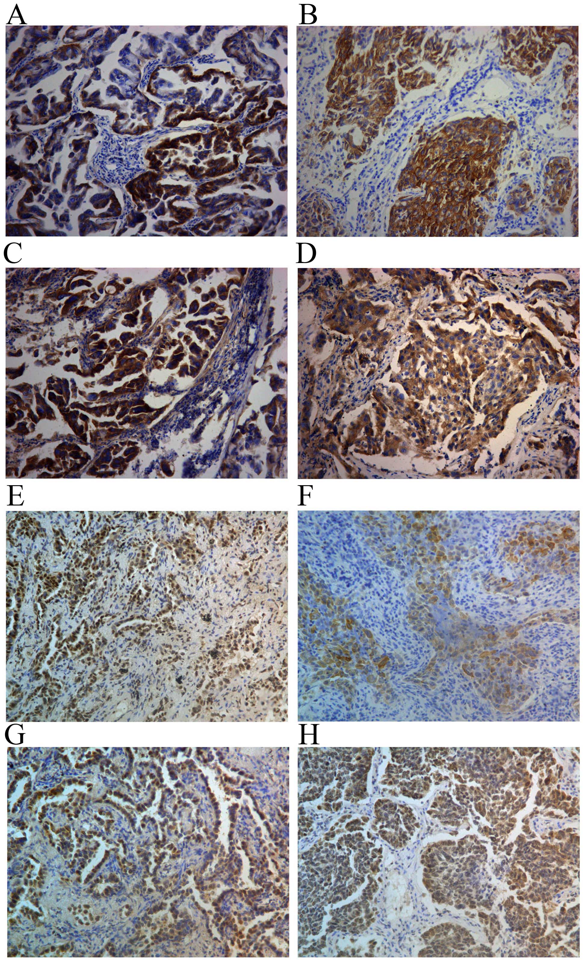
and positive staining was mainly located in the cytoplasm (Fig. 1A and B). The correlation of EGFR Del
19 expression with clinicopathological parameters was then
investigated. EGFR Del 19 expression was significantly associated
with differentiation (P<0.001), pTNM stage (P=0.001), primary
tumor size (P=0.008), lymph node metastasis (P=0.003) and tumor
location (P=0.032). No significant relationship was noted between
EGFR Del 19 expression and gender (P=0.563), age (P=0.911), smoking
history (P=0.626) and histological type (P=0.480) (Table I).
Differences in the expression of Fn14 were detected
in NSCLC with EGFR Del 19 (n=125) and its corresponding normal lung
tissues (n=30), EGFR L858R samples (n=100) and EGFR wild-type NSCLC
samples (n=118). A total of 100% (125/125) of tumor sections were
classified as Fn14-positive while 100% (30/30) corresponding normal
lung tissue sections were classified as Fn14-negative. Seventy-two
percent (72/100) and 48.3% (57/118) of the tumor sections were
classified as Fn14-positive in L858R EGFR and wild-type EGFR NSCLC
samples, nevertheless the rate of high expression of Fn14 (++−+++)
in Del 19 EGFR, L858R EGFR and wild-type EGFR NSCLC samples was
65.6, 17 and 12.7%, and there was significant difference in Fn14
expression among EGFR Del 19, L858R EGFR, and wild-type EGFR
samples (P<0.001). Thus, Fn14 was highly expressed in NSCLC with
EGFR Del 19 (Table II).
| Table IIFn14 is highly expressed in NSCLC
cases with EGFR Del 19. |
Table II
Fn14 is highly expressed in NSCLC
cases with EGFR Del 19.
| Group | n | Fn14 expression
| P-value |
|---|
| − | + | ++ | +++ | Positive rate
(%) |
|---|
| EGFR Del 19
samples | 125 | 0 | 43 | 73 | 9 | 100.0 | |
| EGFR wild-type
samples | 118 | 61 | 42 | 15 | 0 | 48.3 | |
| EGFR L858R
samples | 100 | 28 | 55 | 17 | 0 | 72.0 | <0.001 |
| Normal lung
samples | 30 | 30 | 0 | 0 | 0 | 0.0 | |
Fn14-positive staining was mainly located on the
cell membrane and in the cytoplasm (Fig. 1C and D). In order to evaluate the
role of Fn14 in NSCLC, the correlation of Fn14 expression with
clinicopathological parameters was investigated (Table I). The results showed that Fn14
expression was significantly associated with differentiation
(P<0.001), pTNM stage (P<0.001), primary tumor size
(P=0.029), lymph node metastasis (P<0.001) and tumor location
(P= 0.031). No significant relationship was noted between Fn14
expression and gender (P=0.282), age (P=0.498), smoking history
(P=0.217) and histological type (P=0.080).
p-JAK1 (83.2%, 104/125) (Fig. 1E and F) and p-STAT1 (100%, 125/125)
(Fig. 1G and H) were mainly located
in the cytoplasm and cell nucleus. Correlation of JAK1 and STAT1
expression with clinicopathological parameters was then
investigated. p-JAK1 and p-STAT1 expression was significantly
associated with differentiation (P<0.01), pTNM stage
(P<0.001) and lymph node metastasis (P<0.001). No significant
relationship was noted with gender, age, smoking history,
histological type, primary tumor size and tumor location (Table I).
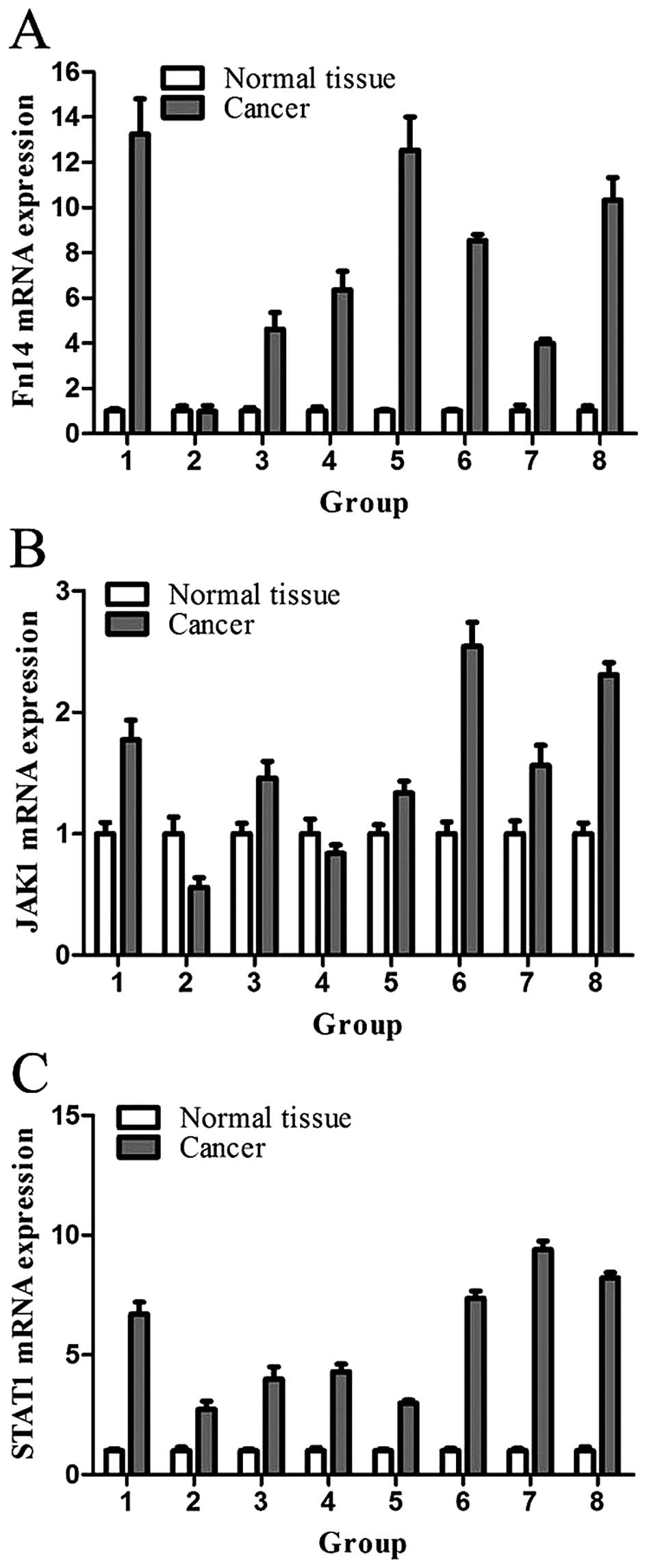
The mRNA expression levels of Fn14, JAK1 and STAT1
were detected by RT-qPCR. Compared to corresponding normal lung
tissues, they were increased in the NSCLC with EGFR Del 19 tissues
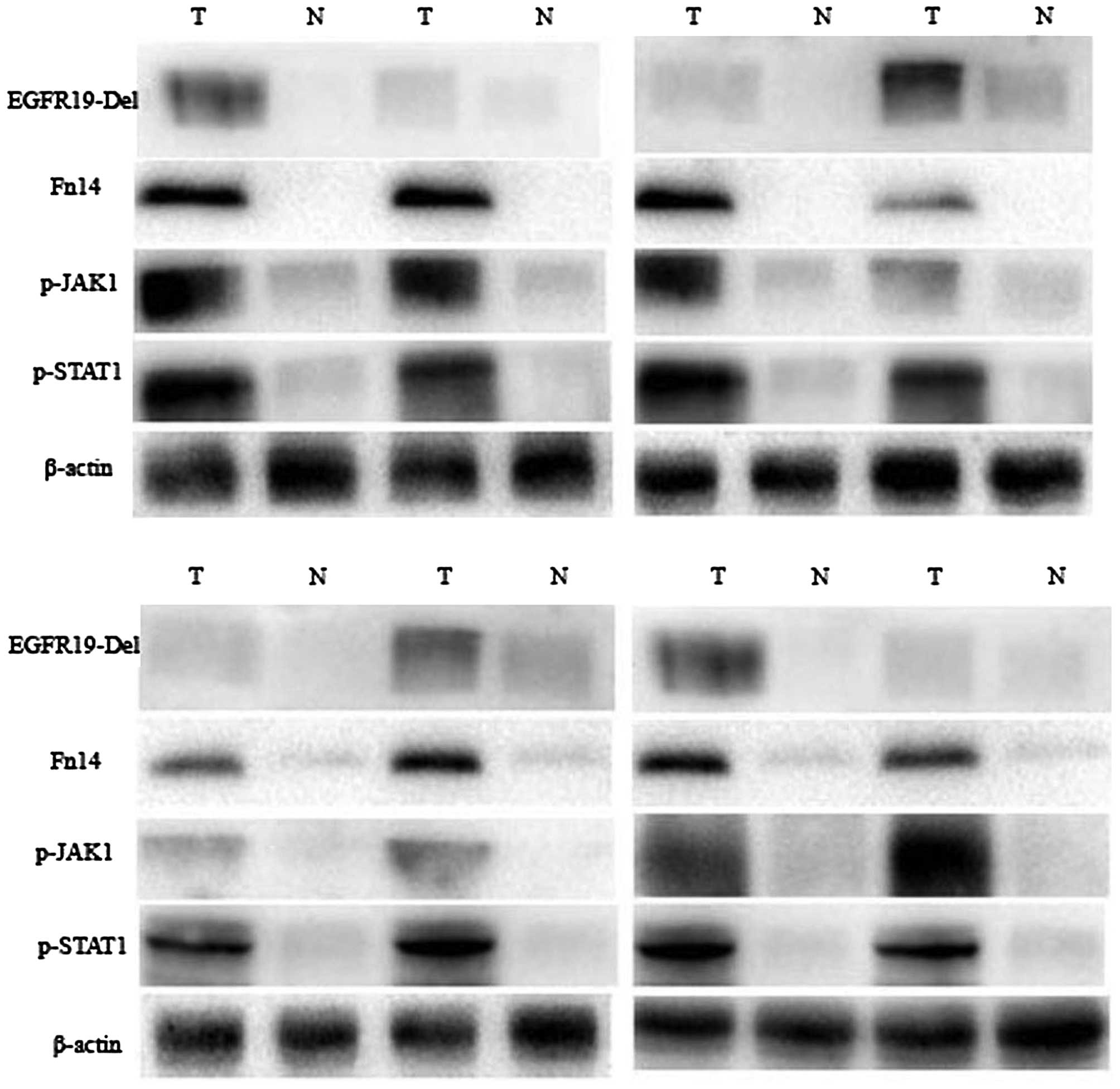
(Fig. 2). In addition, the protein
expression levels of EGFR Del 19, Fn14, p-JAK1 and p-STAT1 were
detected by western blotting in NSCLC with EGFR Del 19 and the
corresponding normal lung tissues. The results showed that when
compared to the normal lung tissues, Fn14 and p-STAT1 were
increased in NSCLC, but this trend was less evident for p-JAK1
protein expression (Fig. 3).
Correlation of EGFR Del 19 with Fn14,
p-JAK1, p-STAT1 expression in NSCLC specimens
In order to investigate the correlation of EGFR Del
19 with Fn14, p-JAK1, p-STAT1 expression, the expression rate and
intensity of these proteins were analyzed (Table III). The correlation of EGFR Del
19 with Fn14, p-JAK1 and p-STAT1 expression was obvious (P<0.05;
rs>0.3) in NSCLC, ADC and SCC. The correlation of
EGFR Del 19 with Fn14, p-JAK1 and p-STAT1 was high in moderate
pathologic stage tissues (P<0.01; rs>0.423). In
addition, there were good correlations for TNM stage in NSCLC
(P<0.05; rs>0.376).
| Table IIIThe correlation of EGFR Del 19 with
Fn14, p-JAK1 and p-STAT1 expression in NSCLC. |
Table III
The correlation of EGFR Del 19 with
Fn14, p-JAK1 and p-STAT1 expression in NSCLC.
| Group | EGFR Del 19 | Fn14
| p-JAK1
| p-STAT1
|
|---|
| + | ++ | +++ | Statistics | − | + | ++ | +++ | Statistics | + | ++ | +++ | Statistics |
|---|
| Histological
type | | | | | | | | | | | | | | |
| NSCLC | − | 1 | 0 | 0 |
χ2=42.814 | 0 | 1 | 0 | 0 |
χ2=42.874 | 1 | 0 | 0 |
χ2=63.251 |
| + | 28 | 20 | 3 | | 16 | 29 | 6 | 0 |
rs=0.523 | 14 | 35 | 2 |
rs=0.620 |
| ++ | 11 | 45 | 3 |
rs=0.468 | 4 | 21 | 34 | 0 | P<0.001 | 1 | 39 | 19 | P<0.001 |
| +++ | 0 | 8 | 6 | P<0.001 | 0 | 4 | 9 | 1 | | 0 | 1 | 13 | |
| ADC | − | 1 | 0 | 0 |
χ2=33.152 | 1 | 0 | 0 | 0 |
χ2=36.752 | 1 | 0 | 0 |
χ2=52.873 |
| + | 25 | 13 | 2 | | 12 | 25 | 3 | 0 |
rs=0.586 | 12 | 27 | 1 |
rs=0.665 |
| ++ | 9 | 32 | 3 |
rs=0.509 | 3 | 16 | 25 | 0 | P<0.001 | 1 | 26 | 17 | P<0.001 |
| +++ | 0 | 6 | 4 | P<0.001 | 0 | 2 | 8 | 0 | | 0 | 0 | 10 | |
| SCC | + | 3 | 7 | 1 |
χ2=10.220 | 4 | 4 | 3 | 0 |
χ2=12.794 | 2 | 8 | 1 |
χ2=12.131 |
| ++ | 2 | 13 | 0 | | 1 | 5 | 9 | 0 |
rs=0.377 | 0 | 13 | 2 |
rs=0.468 |
| +++ | 0 | 2 | 2 |
rs=0.314 | 0 | 2 | 1 | 1 | P=0.046 | 0 | 1 | 3 | P=0.016 |
| | | | | P=0.037 | | | | | | | | | |
|
Differentiation | | | | | | | | | | | | | | |
| Well | + | 12 | 6 | 0 |
χ2=0.077 | 8 | 10 | 0 | 0 |
χ2=8.252 | 6 | 12 | 0 |
χ2=7.886 |
| ++ | 3 | 2 | 0 |
rs=0.058 | 2 | 1 | 2 | 0 |
rs=0.212 | 1 | 2 | 2 |
rs=0.331 |
| | | | | P=0.782 | | | | | P=0.016 | | | | P=0.019 |
| Moderate | − | 1 | 0 | 0 |
χ2=30.119 | 1 | 0 | 0 | 0 |
χ2=24.740 | 1 | 0 | 0 |
χ2=39.843 |
| + | 15 | 12 | 3 | | 7 | 18 | 5 | 0 |
rs=0.481 | 7 | 21 | 2 |
rs=0.558 |
| ++ | 7 | 35 | 2 |
rs=0.423 | 2 | 17 | 25 | 0 | P<0.001 | 0 | 31 | 13 | P<0.001 |
| +++ | 0 | 4 | 4 | P<0.001 | 0 | 2 | 6 | 0 | | 0 | 1 | 7 | |
| Poor | + | 1 | 2 | 0 |
χ2=4.087 | 1 | 1 | 1 | 0 |
χ2=8.147 | 1 | 2 | 0 |
χ2=13.490 |
| ++ | 1 | 8 | 1 |
rs=0.436 | 0 | 3 | 7 | 0 |
rs=0.287 | 0 | 6 | 4 |
rs=0.729 |
| +++ | 0 | 4 | 2 | P=0.394 | 0 | 2 | 3 | 1 | P=0.228 | 0 | 0 | 6 | P=0.009 |
| pTNM stages | | | | | | | | | | | | | | |
| I–II | − | 1 | 0 | 0 |
χ2=10.446 | 1 | 0 | 0 | 0 |
χ2=18.557 | 1 | 0 | 0 |
χ2=30.337 |
| + | 22 | 10 | 0 | | 10 | 20 | 2 | 0 |
rs=0.440 | 9 | 23 | 0 |
rs=0.528 |
| ++ | 9 | 18 | 0 |
rs=0.406 | 4 | 13 | 10 | 0 | P=0.005 | 1 | 21 | 5 | P<0.001 |
| +++ | 0 | 2 | 0 | P=0.015 | 0 | 0 | 2 | 0 | | 0 | 0 | 2 | |
| III–IV | + | 6 | 10 | 3 |
χ2=18.102 | 6 | 9 | 4 | 0 |
χ2=26.200 | 5 | 12 | 2 |
χ2=28.038 |
| ++ | 2 | 27 | 3 | | 0 | 8 | 24 | 0 |
rs=0.463 | 0 | 18 | 14 |
rs=0.602 |
| +++ | 0 | 6 | 6 |
rs=0.376 | 0 | 4 | 7 | 1 | P=0.001 | 0 | 1 | 11 | P<0.001 |
| | | | | P<0.001 | | | | | | | | | |
Correlation of Fn14 expression with the
JAK1/STAT1 pathway in NSCLC specimens with EGFR Del 19
In order to understand the role of Fn14 in NSCLC
with EGFR Del 19 and the influence of Fn14 on downstream signaling
pathway, correlations between Fn14 expression with JAK1/STAT1
signaling molecules were analyzed (Table IV). There were obvious correlations
in ADC and SCC (P<0.05; rs>0.3). Correlations
between Fn14 and JAK1 and STAT1 were strong in moderately
differentiated tissues (P<0.01; rs>0.366). In
addition, there was a good correlation with pTNM stage in NSCLC
(P<0.01; rs>0.336).
| Table IVCorrelation of Fn14 with p-JAK1 and
p-STAT1 expression in NSCLC. |
Table IV
Correlation of Fn14 with p-JAK1 and
p-STAT1 expression in NSCLC.
| Group | Fn14 | p-JAK1
| p-STAT1
|
|---|
| − | + | ++ | +++ | Statistics | + | ++ | +++ | Statistics |
|---|
| Histological
type | | | | | | | | | | |
| ADC | + | 12 | 22 | 1 | 0 |
χ2=33.472 | 13 | 19 | 3 |
χ2=30.414 |
| ++ | 4 | 19 | 28 | 0 |
rs=0.583 | 1 | 31 | 19 |
rs=0.518 |
| +++ | 0 | 2 | 7 | 0 | P<0.001 | 0 | 3 | 6 | P<0.001 |
| + | 2 | 3 | 0 | 0 |
χ2=16.608 | 1 | 4 | 0 |
χ2=12.749 |
| SCC | ++ | 2 | 8 | 12 | 0 |
rs=0.394 | 0 | 18 | 4 |
rs=0.313 |
| +++ | 1 | 0 | 1 | 1 | P=0.011 | 1 | 0 | 2 | P=0.013 |
|
Differentiation | | | | | | | | | | |
| Well | + | 8 | 6 | 1 | 0 |
χ2=1.720 | 7 | 7 | 1 |
χ2=5.367 |
| ++ | 2 | 5 | 1 | 0 |
rs=0.268 | 0 | 7 | 1 |
rs=0.446 |
| | | | | | P=0.423 | | | | P=0.068 |
| Moderate | + | 6 | 17 | 0 | 0 |
χ2=25.722 | 6 | 15 | 2 |
χ2=16.877 |
| ++ | 3 | 18 | 30 | 0 |
rs=0.498 | 1 | 35 | 15 |
rs=0.366 |
| +++ | 1 | 2 | 6 | 0 | P<0.001 | 1 | 3 | 5 | P=0.002 |
| Poor | + | 0 | 2 | 0 | 0 |
χ2=10.939 | 1 | 1 | 0 |
χ2=12.350 |
| ++ | 1 | 4 | 9 | 0 |
rs=0.556 | 0 | 7 | 7 |
rs=0.572 |
| +++ | 0 | 0 | 2 | 1 | P=0.090 | 0 | 0 | 3 | P=0.015 |
| pTNM stages | | | | | | | | | | |
| I–II | + | 12 | 19 | 1 | 0 |
χ2=16.396 | 11 | 19 | 2 |
χ2=13.053 |
| ++ | 3 | 14 | 13 | 0 |
rs=0.494 | 0 | 25 | 5 |
rs=0.424 |
| | | | | | P<0.001 | | | | P=0.001 |
| III–IV | + | 2 | 6 | 0 | 0 |
χ2=16.980 | 3 | 4 | 1 |
χ2=15.647 |
| ++ | 3 | 13 | 27 | 0 |
rs=0.385 | 1 | 24 | 18 |
rs=0.336 |
| +++ | 1 | 2 | 8 | 1 | P=0.009 | 1 | 3 | 8 | P=0.004 |
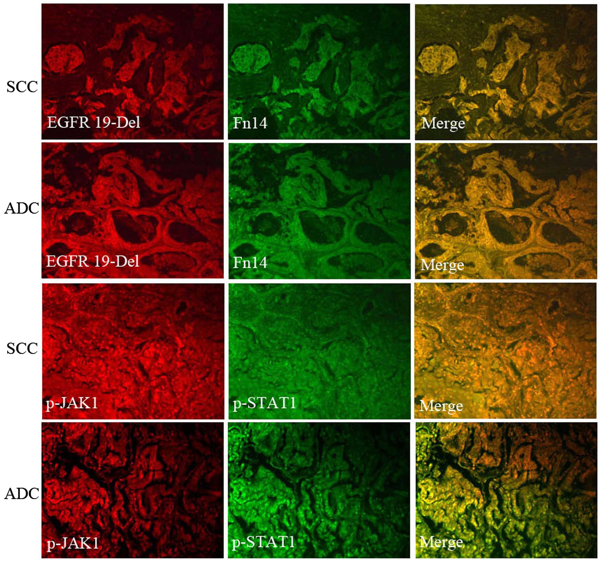
Co-localization of EGFR Del 19 and Fn14
protein expression as well as p-JAK1 and p-STAT1 protein
expression
EGFR Del 19 and Fn14 were mainly expressed on the
cell membrane. In order to further verify whether there is a
co-localization phenomenon when they are expressed,
immunofluorescence experiments were conducted. Preliminary
experimental results showed that the expression of localization
phenomenon existed. p-JAK1 and p-STAT1 were mainly located in the
cytoplasm and cell nucleus. In addition, the same method was used
for co-localization of p-JAK1 and p-STAT1 protein expression.
Co-localization phenomenon also existed (Fig. 4).
Correlation of EGFR Del 19 and Fn14
expression with survival in NSCLC with EGFR Del 19 cases
Among factors such as patient age, gender, smoking
history, tumor histological type, lymph node metastasis,
differentiation and pTNM stages, only the pTNM stage,
differentiation and lymph node metastasis were significantly
associated with patient survival. The median survival time of
patients with pTNM I/II tumors (n=61) was not reached [95%
confidence interval (CI) was not reached], whereas the median
survival time of those with TNM III/IV tumors (n=64) was 24 months
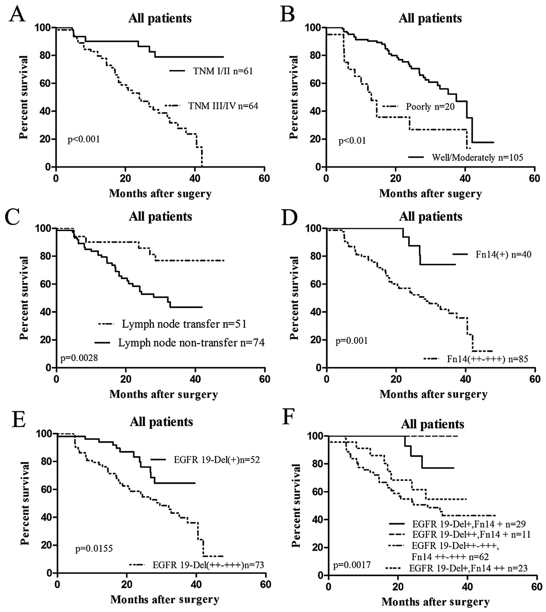
(95% CI, 17.96–30.04 months; P<0.001; Fig. 5A). Patients with well or moderately
differentiated tumors (n=140, median survival time, 37.47 months,
95% CI, 31.73–43.20 months) had a longer survival time than those
with poorly differentiated tumors (n=51, median survival time, 13
months; 95% CI, 8.67–17.34 months; P<0.001; Fig. 5B). The median survival time of
patients with no lymph node metastasis (n=74) was not reached (95%
CI was not reached), whereas the median survival time of those with
lymph node metastasis (n=51) was 32.07 months (95% CI of ratio,
1.382–4.733; P<0.01; Fig.
5C).
To investigate the relationship between EGFR Del 19
and Fn14 expression and the clinical outcome of NSCLC patients, the
correlation of EGFR Del 19 and Fn14 expression with patient
survival and EGFR Del 19, Fn14 expression status were analyzed.
Patients with Fn14 high positive expression had a significantly
worse prognosis than those with Fn14 low positive expression
(P=0.001; Fig. 5D). The median
survival time of patients with Fn14 high positive NSCLC (n=85) was
28 months (95% CI, 17.99–38.01 months) and the mean survival time
was 27.07 months (95% CI, 23.49–30.66 months), whereas the median
survival time of patients with Fn14 low positive NSCLC (n=40) was
not reached and the mean survival time was 33.86 months (95% CI,
31.17–36.55 months). Patients with high positive expression of EGFR
Del 19 had a significantly worse prognosis than those with low
positive expression of EGFR Del 19 (P=0.0155; Fig. 5E). The median survival time of
patients with EGFR Del 19 low positive NSCLC (n=52) was 32.8 months
(95% CI of ratio, 0.2551–0.8663), whereas the median survival time
of patients with EGFR Del 19 high positive NSCLC (n=73) was not
reached. Comprehensive statistics were analyzed concerning the
influence of EGFR Del 19 and Fn14 on patient prognosis. We found
that with increased expression of EGFR Del 19 and Fn14, the shorter
the patient survival (P<0.01; Fig.
5F).
To further assess whether EGFR Del 19 and Fn14
expression represents a common prognostic parameter in NSCLC
patients with EGFR Del 19, regression analysis using the Cox's
proportional hazards model was performed. The covariate parameters
included several clinicopathological factors in addition to EGFR
Del 19 and Fn14, as shown in Table
V. In the univariate analysis, factors including EGFR Del 19
expression, Fn14 expression, pTNM stage and differentiation showed
a significantly higher hazard ratio for poor prognosis.
| Table VCox proportional hazards model
analysis of variables affecting survival in NSCLC patients. |
Table V
Cox proportional hazards model
analysis of variables affecting survival in NSCLC patients.
| Variables | Categories | Univariate analysis
| Multivariate
analysis
|
|---|
| HR (95% CI) | P-value | HR (95% CI) | P-value |
|---|
| Age (years) | ≥58/<58 | 0.751
(0.428–1.318) | 0.319 | | |
| Gender | Male/female | 1.031
(0.592–1.796) | 0.914 | | |
| Smoking
history |
Smoking/non-smoking | 1.001
(0.573–1.747) | 0.998 | | |
| Histological
type | SCC/ADC | 0.712
(0.383–1.323) | 0.282 | | |
|
Differentiation |
Poor/well+moderate | 0.291
(0.154–0.549) | <0.001 | 0.424
(0.218–0.824) | 0.011 |
| pTNM stage | I/II/III/IV | 0.222
(0.108–0.457) | <0.001 | 0.350
(0.160–0.768) | 0.009 |
| Fn14
expression | High positive/low
positive | 0.183
(0.066–0.512) | 0.001 | 0.316
(0.102–0.983) | 0.047 |
| EGFR Del 19 | High positive/low
positive | 0.430
(0.223–0.829) | 0.012 | 1.065
(0.508–2.233) | 0.867 |
Moreover, multivariate analysis was carried out
using the significant factors observed in the univariate analysis.
The results showed that, in addition to TNM stage and
differentiation, Fn14 expression was an independent prognostic
factor (P=0.047; Table V). These
results strongly indicate that Fn14 expression in NSCLC patients is
closely related to poor prognosis.
Discussion
Normally, most NSCLC patients carry EGFR mutations,
and EGFR Del 19 is one of the most common forms. Although NSCLC
patients with EGFR Del 19 are sensitive to EGFR TKI, acquired
resistance is inevitable to limiting the therapeutic effects in
patients. Researchers have conjectured whether NSCLC carcinogenesis
is related with EGFR Del 19 and whether the EGFR Del 19 mutation
may upregulate new downstream signaling pathways or promote a novel
molecular expression, and this issue is unclear. Thus, we aimed to
ascertain whether the pathogenesis of NSCLC is associated with EGFR
Del 19.
Increasing evidence shows that Fn14 is weakly
expressed in healthy tissues (7–9), but
is highly expressed in contexts of tissue damage caused by hypoxia,
oxidative stress and tumor growth (10–14).
Thus, researchers have increasingly given more attention to the
function of Fn14 in cancers and found that Fn14 expression may be a
valuable potential biomarker (20–22).
In NSCLC cell lines, Fn14 expression levels may be correlated with
EGFR mutations (16), and it may
function in cancer through the JAK/STAT1 pathway. Although STAT1 is
well known as a master transcription factor for IFN-related
intracellular signaling, leading to antiviral activity (23–26),
emerging data has revealed that in certain cellular contexts the
IFN/STAT1 pathway may facilitate tumor cell growth (27,28).
There is limited research concerning the functions among EGFR Del
19, Fn14 and the JAK/STAT pathway. Thus, in the present study, we
assessed the correlation of EGFR Del 19 with Fn14 and the JAK/STAT
pathway.
In the present study, we performed IHC, RT-qPCR and
western blot analysis on surgically resected NSCLC specimens to
investigate EGFR Del 19, Fn14, JAK1 and STAT1 expression. IHC and
western blotting results showed that compared to EGFR L858R, EGFR
wild-type NSCLC tissues and corresponding normal lung tissues, Fn14
protein expression was significantly higher in NSCLC with EGFR Del
19. Fn14, JAK1 and STAT1 were increased at the mRNA levels and were
associated with clinicopathological characteristics, such as pTNM
stage, differentiation, lymph node metastasis, but not with age,
gender, smoking history and histological subtypes.
The present study was the first to investigate the
correlation of EGFR Del 19 with Fn14 and JAK1/STAT1 pathway. The
correlations of EGFR Del 19 with Fn14, p-JAK1, p-STAT1 and Fn14
with p-JAK1, p-STAT1 were significant in regards to histological
type, differentiation and pTNM stage (rs>0.3;
P<0.05). From the data, we found that both EGFR Del 19 and Fn14
were associated with p-JAK1 and p-STAT1. This indicates that there
is better relationship among these four factors. In addition, EGFR
mutations in NSCLC can selectively activate signaling pathways
which promote cell survival and induce proliferation such as STAT
(29). Thus, this is a substantial
proof which can testify the relationship between EGFR Del 19 and
p-JAK1 and p-STAT1. Furthermore, one study showed that Fn14
expression was higher in EGFR Del 19 cell lines, and erlotinib (one
of TKIs) inhibited Fn14 expression (16). Thus, we may infer that EGFR Del 19
may upregulate Fn14, JAK1 and STAT1 protein expression, but the
detailed mechanism of action is not clear. In order to further
verify whether there is a co-localization phenomenon when EGFR Del
19 and Fn14 are expressed, immunofluorescence experiments were
conducted. Preliminary experimental results showed that a
co-localization phenomenon existed. All these signs indicated that
EGFR Del 19 may play an important role in migration, invasion and
metastasis of NSCLC and EGFR Del 19 may upregulate Fn14 and
JAK1/STAT1 pathway to promote NSCLC progression.
In addition, to the best of our knowledge, the
present study was the first to indicate an association between EGFR
Del 19 and Fn14 positivity in NSCLC patients and poor prognosis,
and these factors are pivotal to affecting patient prognosis. The
results of survival analysis suggested that Fn14 may be an
independent prognostic factor of NSCLC patients. Due to the
limitations of the sample size and follow-up, the median survival
time in many groups could not be obtained. To compensate for these
shortcomings, we intend to carry out further multicenter clinical
studies, expand the sample size and enrich the means of
detection.
Taken together with the results of the present
study, we believe that EGFR Del 19 may promote NSCLC progression
through upregulation of Fn14 and the JAK1/STAT1 pathway, and Fn14
may be a potential biomarker for NSCLC with EGFR Del 19 patients.
However, the mechanism of NSCLC progression by EGFR Del 19
upregulating Fn14 and JAK/STAT pathway needs to be further
examined.
In conclusion, we showed that a high proportion of
NSCLC with EGFR Del 19 expressed Fn14, JAK1 and STAT1 and the
expression of these proteins was significantly associated with
differentiation, pTNM stage and lymphatic metastasis. Moreover,
levels of Fn14, JAK1 and STAT1 expression were related with EGFR
Del 19, and there was a co-localization phenomenon when EGFR Del 19
and Fn14 were expressed. Thus, our results indicate that EGFR Del
19 may promote Fn14 and JAK1/STAT1 expression in NSCLC, and Fn14
has a significant role in NSCLC with EGFR Del 19, and may serve as
a prognostic biomarker in NSCLC with EGFR Del 19.
Acknowledgments
We thank Liangbo Fan, Peng Chen and Guang Yang
(Thoracic Surgery Department Laboratory) for their help in
providing experimental technical support. Finally, we would like to
thank all members of our research team, who collaborated to ensure
that the study proceeded smoothly.
References
|
1
|
Devesa SS, Bray F, Vizcaino AP and Parkin
DM: International lung cancer trends by histologic type:
Male:female differences diminishing and adenocarcinoma rates
rising. Int J Cancer. 117:294–299. 2005. View Article : Google Scholar : PubMed/NCBI
|
|
2
|
Nicholson RI, Gee JM and Harper ME: EGFR
and cancer prognosis. Eur J Cancer. 37(Suppl 4): S9–S15. 2001.
View Article : Google Scholar : PubMed/NCBI
|
|
3
|
Krause DS and Van Etten RA: Tyrosine
kinases as targets for cancer therapy. N Engl J Med. 353:172–187.
2005. View Article : Google Scholar : PubMed/NCBI
|
|
4
|
Hynes NE and Lane HA: ERBB receptors and
cancer: The complexity of targeted inhibitors. Nat Rev Cancer.
5:341–354. 2005. View
Article : Google Scholar : PubMed/NCBI
|
|
5
|
Paez JG, Jänne PA, Lee JC, Tracy S,
Greulich H, Gabriel S, Herman P, Kaye FJ, Lindeman N, Boggon TJ, et
al: EGFR mutations in lung cancer: Correlation with clinical
response to gefitinib therapy. Science. 304:1497–1500. 2004.
View Article : Google Scholar : PubMed/NCBI
|
|
6
|
Kaneda T, Hata A, Tomioka H, Tanaka K,
Kaji R, Fujita S, Tomii K and Katakami N: Possible differential
EGFR-TKI efficacy among exon 19 deletional locations in EGFR-mutant
non-small cell lung cancer. Lung Cancer. 86:213–218. 2014.
View Article : Google Scholar : PubMed/NCBI
|
|
7
|
Girgenrath M, Weng S, Kostek CA, Browning
B, Wang M, Brown SA, Winkles JA, Michaelson JS, Allaire N,
Schneider P, et al: TWEAK, via its receptor Fn14, is a novel
regulator of mesenchymal progenitor cells and skeletal muscle
regeneration. EMBO J. 25:5826–5839. 2006. View Article : Google Scholar : PubMed/NCBI
|
|
8
|
Baxter FO, Came PJ, Abell K, Kedjouar B,
Huth M, Rajewsky K, Pasparakis M and Watson CJ: IKKbeta/2 induces
TWEAK and apoptosis in mammary epithelial cells. Development.
133:3485–3494. 2006. View Article : Google Scholar : PubMed/NCBI
|
|
9
|
Winkles JA: The TWEAK-Fn14
cytokine-receptor axis: Discovery, biology and therapeutic
targeting. Nat Rev Drug Discov. 7:411–425. 2008. View Article : Google Scholar : PubMed/NCBI
|
|
10
|
Burkly LC, Michaelson JS and Zheng TS:
TWEAK/Fn14 pathway: An immunological switch for shaping tissue
responses. Immunol Rev. 244:99–114. 2011. View Article : Google Scholar : PubMed/NCBI
|
|
11
|
Kim SH, Kang YJ, Kim WJ, Woo DK, Lee Y,
Kim DI, Park YB, Kwon BS, Park JE and Lee WH: TWEAK can induce
pro-inflammatory cytokines and matrix metalloproteinase-9 in
macrophages. Circ J. 68:396–399. 2004. View Article : Google Scholar : PubMed/NCBI
|
|
12
|
Pettersen I, Baryawno N, Abel F, Bakkelund
WH, Zykova SN, Winberg JO, Moens U, Rasmuson A, Kogner P, Johnsen
JI, et al: Expression of TWEAK/Fn14 in neuroblastoma: implications
in tumorigenesis. Int J Oncol. 42:1239–1248. 2013.PubMed/NCBI
|
|
13
|
Lynch CN, Wang YC, Lund JK, Chen YW, Leal
JA and Wiley SR: TWEAK induces angiogenesis and proliferation of
endothelial cells. J Biol Chem. 274:8455–8459. 1999. View Article : Google Scholar : PubMed/NCBI
|
|
14
|
Harada N, Nakayama M, Nakano H, Fukuchi Y,
Yagita H and Okumura K: Pro-inflammatory effect of TWEAK/Fn14
interaction on human umbilical vein endothelial cells. Biochem
Biophys Res Commun. 299:488–493. 2002. View Article : Google Scholar : PubMed/NCBI
|
|
15
|
Chicheportiche Y, Chicheportiche R, Sizing
I, Thompson J, Benjamin CB, Ambrose C and Dayer JM: Proinflammatory
activity of TWEAK on human dermal fibroblasts and synoviocytes:
Blocking and enhancing effects of anti-TWEAK monoclonal antibodies.
Arthritis Res. 4:126–133. 2002. View
Article : Google Scholar : PubMed/NCBI
|
|
16
|
Whitsett TG, Cheng E, Inge L, Asrani K,
Jameson NM, Hostetter G, Weiss GJ, Kingsley CB, Loftus JC, Bremner
R, et al: Elevated expression of Fn14 in non-small cell lung cancer
correlates with activated EGFR and promotes tumor cell migration
and invasion. Am J Pathol. 181:111–120. 2012. View Article : Google Scholar : PubMed/NCBI
|
|
17
|
Chapman MS, Wu L, Amatucci A, Ho SN and
Michaelson JS: TWEAK signals through JAK-STAT to induce tumor cell
apoptosis. Cytokine. 61:210–217. 2013. View Article : Google Scholar
|
|
18
|
Gastl G, Spizzo G, Obrist P, Dünser M and
Mikuz G: Ep-CAM overexpression in breast cancer as a predictor of
survival. Lancet. 356:1981–1982. 2000. View Article : Google Scholar : PubMed/NCBI
|
|
19
|
Zhao J, Zhou Y, Zhang Z, Tian F, Ma N, Liu
T, Gu Z and Wang Y: Upregulated fascin1 in non-small cell lung
cancer promotes the migration and invasiveness, but not
proliferation. Cancer Lett. 290:238–247. 2010. View Article : Google Scholar
|
|
20
|
Kwon OH, Park SJ, Kang TW, Kim M, Kim JH,
Noh SM, Song KS, Yoo HS, Wang Y, Pocalyko D, et al: Elevated
fibroblast growth factor-inducible 14 expression promotes gastric
cancer growth via nuclear factor-κB and is associated with poor
patient outcome. Cancer Lett. 314:73–81. 2012. View Article : Google Scholar
|
|
21
|
Zhou H, Ekmekcioglu S, Marks JW,
Mohamedali KA, Asrani K, Phillips KK, Brown SA, Cheng E, Weiss MB,
Hittelman WN, et al: The TWEAK receptor Fn14 is a therapeutic
target in melanoma: Immunotoxins targeting Fn14 receptor for
malignant melanoma treatment. J Invest Dermatol. 133:1052–1062.
2013. View Article : Google Scholar
|
|
22
|
Huang M, Narita S, Tsuchiya N, Ma Z,
Numakura K, Obara T, Tsuruta H, Saito M, Inoue T, Horikawa Y, et
al: Overexpression of Fn14 promotes androgen-independent prostate
cancer progression through MMP-9 and correlates with poor treatment
outcome. Carcinogenesis. 32:1589–1596. 2011. View Article : Google Scholar : PubMed/NCBI
|
|
23
|
Townsend PA, Scarabelli TM, Davidson SM,
Knight RA, Latchman DS and Stephanou A: STAT-1 interacts with p53
to enhance DNA damage-induced apoptosis. J Biol Chem.
279:5811–5820. 2004. View Article : Google Scholar
|
|
24
|
Stephanou A, Brar BK, Knight RA and
Latchman DS: Opposing actions of STAT-1 and STAT-3 on the Bcl-2 and
Bcl-x promoters. Cell Death Differ. 7:329–330. 2000. View Article : Google Scholar : PubMed/NCBI
|
|
25
|
Chin YE, Kitagawa M, Kuida K, Flavell RA
and Fu XY: Activation of the STAT signaling pathway can cause
expression of caspase 1 and apoptosis. Mol Cell Biol. 17:5328–5337.
1997. View Article : Google Scholar : PubMed/NCBI
|
|
26
|
Kumar A, Commane M, Flickinger TW, Horvath
CM and Stark GR: Defective TNF-alpha-induced apoptosis in
STAT1-null cells due to low constitutive levels of caspases.
Science. 278:1630–1632. 1997. View Article : Google Scholar : PubMed/NCBI
|
|
27
|
Fryknäs M, Dhar S, Oberg F, Rickardson L,
Rydåker M, Göransson H, Gustafsson M, Pettersson U, Nygren P,
Larsson R, et al: STAT1 signaling is associated with acquired
crossresistance to doxorubicin and radiation in myeloma cell lines.
Int J Cancer. 120:189–195. 2007. View Article : Google Scholar
|
|
28
|
Roberts D, Schick J, Conway S, Biade S,
Laub PB, Stevenson JP, Hamilton TC, O'Dwyer PJ and Johnson SW:
Identification of genes associated with platinum drug sensitivity
and resistance in human ovarian cancer cells. Br J Cancer.
92:1149–1158. 2005. View Article : Google Scholar : PubMed/NCBI
|
|
29
|
Zimmer S, Kahl P, Buhl TM, Steiner S,
Wardelmann E, Merkelbach-Bruse S, Buettner R and Heukamp LC:
Epidermal growth factor receptor mutations in non-small cell lung
cancer influence downstream Akt, MAPK and Stat3 signaling. J Cancer
Res Clin Oncol. 135:723–730. 2009. View Article : Google Scholar
|