Introduction
CRC is one of the leading causes of death worldwide
(1). Aggressive local invasion and
metastasis make CRC difficult to treat (2). Therefore clarifying the mechanism of
invasion of CRC to find new treatment targets is urgent.
Metastasis of cancer cells is a complex processes
during which the degradation of the extracellular matrix (ECM)
plays an important role (3).
MMP-2/−9 play important roles in degrading ECM and its involvement
is complex in the process of cancer metastasis (3,4).
Overactivity of ERK signaling pathway has been found in almost half
of known human tumor cell lines and in many human primary tumors
derived from different origins (5).
ERK signaling pathway plays a vital role in the synthesis of
MMP-2/−9 (3,6). Overactivity of ERK pathway is observed
in CRC (7,8) and is correlated with the metastasis of
CRC (9,10).
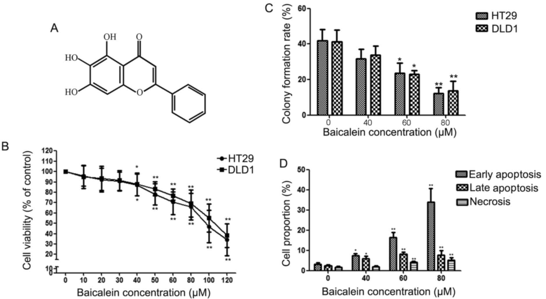
Baicalein is extracted from the roots of
Scutellaria baicalensis or Scutellaria radix. The
chemical structure of baicalein is shown in Fig. 1A. The antitumor biological effect of
baicalein has been found in many tumors (11–13).
Baicalein has been found to possess antitumor effect in
hepatocellular carcinoma, glioma and bladder cancer (3,14–16),
but the anti-metastatic effect and related mechanism(s) in CRC are
still unclear. Thus, the present study investigated the effects of
baicalein in CRC invasion and metastasis and related
mechanisms.
Materials and methods
Cell lines and reagents
CRC HT29 and DLD1 cell lines were purchased from the
American Type Culture Collection (ATCC; Manassas, VA, USA) and
cultured in RPMI-1640 (HyClone Laboratories, Inc., Logan, UT, USA)
medium with 10% fetal bovine serum (FBS; HyClone Laboratories). The
cells were cultured at 37°C with 5% CO2. Anti-MMP-2,
ERK, MMP-9 and p-ERK were purchased from Cell Signaling Technology
(Beverly, MA, USA). Unless otherwise indicated, all the other
chemicals were purchased from Sigma-Aldrich (St. Louis, MO,
USA).
Apoptosis assays
Apoptotic and/or necrotic cells were analysed by
propidium iodide (PI) uptake using Annexin V binding an Annexin
V-FITC/PI kit as previously described (15). Briefly, CRC cells were seeded into
6-well plates at a density of 1×10⁵ cells/well for 12 h
and treated with 0, 40, 60 and 80 µM of baicalein for 48 h. After
washing with cold phosphate-buffered saline (PBS), cells were
resuspended in Annexin V binding buffer. Cells were incubated in
Annexin V-FITC for 15 min, washed and then incubated with PI.
Samples were detected by flow cytometer with CellQuest
software.
Cell growth assay and focus formation
assay
Cell growth rate was detected using the MTT assay.
Briefly, cells were seeded in a 96-well plate at a density of
1.5×10³ cells. Cells were treated with baicalein (0, 10,
20, 30, 40, 50, 60, 80, 100 and 120 µM) (R&D Systems, Inc.,
Minneapolis, MN, USA) for 24 h. MTT assay was done in accordance
with the manufacturers instructions. Each experiment was done in
triplicate and data were expressed as mean ± SD. In focus formation
assay, 100 cells were firstly seeded onto a plate and then cultured
with (0, 40, 60 and 80 µM) baicalein for 2 weeks. The plates were
fixed and then stained with 1% crystal violet. All experiments were
performed in triplicate.
Construction of expression plasmids
and transfection
The construction of the expression plasmids and
their transfection was performed in accordance with the
manufacturers instructions (3).
First, the full-length pcDNA3.1 (Invitrogen, Carlsbad, CA, USA)
MEK1 vector was made by cloning the full-length PCR product of MEK1
with KOD® DNA polymerase (Toyobo Co., Ltd., Osaka,
Japan). Then we measured the plasmid sequences to confirm the
sequences of the plasmid. In transient transfection experiments,
cells were plated at a density of 2×10⁵ cells/well in a
6-well plate, 24 h later they were transfected with 4.0 µg
pcDNA3.1(+)-MEK1 vector via Lipofectamine 2000 (Invitrogen) in
accordance with the manufacturers protocol. The 4.0 µg pcDNA3.1(+)
empty vector was used as a negative control.
Cell invasion and migration assay
The migration and invasion ability of cells were
detected by Transwell assays. Migration and invasion assay was
performed with 24-well Transwell chambers coated with Matrigel or
not (Becton-Dicknson, Billerica, MA, USA) as previously described.
For the migration assay, cells were seeded into inner well and
cultured in medium with 0, 10, 20 and 30 µM baicalein. After 16 h,
cells on the bottom side of the inner well were fixed in alcohol,
fixed in crystal violet and counted. Invasion assay was performed
with 24-well Transwell chamber coated with Matrigel as previously
described (3).
Western blotting assay
After treated with U0126 or baicalein,
2×10⁶ cells were added into 200 µl of lysis buffer
(Fermentas, Waltham, MA, USA). By 10% SDS-polyacrylamide gel
electrophoresis, the proteins (60 µg) were separated and bloted
onto PVDF membranes. To block non-specific binding, the PVDF
membranes were subsequently blocked in Tris-buffered saline with
Tween-20 (TBST) containing 5% defatted milk buffer for 1 h at 37°C
and were then incubated with antibodies against ERK, p-ERK, MMP-2,
MMP-9 or β-actin overnight in defatted milk 5% in TBST at 4°C. The
membranes were incubated with a horseradish peroxidase goat
anti-rabbit or anti-mouse IgG antibody at room temperature for 1 h.
The pictures were examined with an enhanced chemiluminescence kit
(ECL Plus; Amersham, Freiburg, Germany) according to the
manufacturers instructions and captured by autoradiography. The
relative photographic density was quantified with ImageJ software
(GE Healthcare, Buckinghamshire, UK) and expressed as arbitrary
units (a.u.).
Tumor xenograft experiments and
animals
To assess the anti-proliferation of baicalein in
vivo, Balb/c athymic nude mice (4- to 6-week-old) were obtained
from Shanghai SLAC Laboratory Animal, Co., Ltd., (Shanghai, China).
Each mouse was subcutenously implanted with 1×10⁶ viable
DLD1 cells in the right flank area. One group was treated with
vehicle dimethyl sulfoxide (DMSO), the other group was treated with
baicalein (20 mg/kg/day) for 3 weeks. At day 21, all the mice were
sacrificed and the tumor tissues were removed and weighed. The
experimental protocols used herein were evaluated and approved by
the Animal Care and Use Committee of the Medical School of Xian
Jiaotong University.
Results
Baicalein inhibits the proliferation
of HT29 and DLD1 cells
The inhibition effects of baicalein on the
proliferation of CRC cells at different concentrations (0 to 120
µM) for 24 h are shown in Fig. 1B.
At concentrations >40 µM, the anti-proliferation effect of
baicalein on HT29 and DLD1 cells was significant so we chose
concentrations <40 µM for all the following experiments.
To determine the effect of baicalein on apoptosis in
detail, HT29 cells were incubated for 48 h with baicalein (40, 60
or 80 µM) and then analysed by flow cytometry. As shown in Fig. 1C, the effect of baicalein on
apoptosis of HT29 cells was concentration-dependent. After
treatment for 48 h, both early and late apoptotic cells increased
significantly in the HT29 cells treated with baicalein. Baicalein
reduced the number of colony HT29 cells in vitro
significantly (Fig. 1D).
Baicalein inhibits the metastasis of
HT29 and DLD1 cells
Fig. 2 shows the
inhibition effect of baicalein (0, 10, 20 and 30 µM) on cell
migration (16 h) and invasion (24 h) in HT29 and DLD1 cells,
respectively. We found that inhibition effect of baicalein on the
migration (Fig. 2A and B) and
invasion (Fig. 2C and D) of HT29
and DLD1 cells was concentration-dependent.
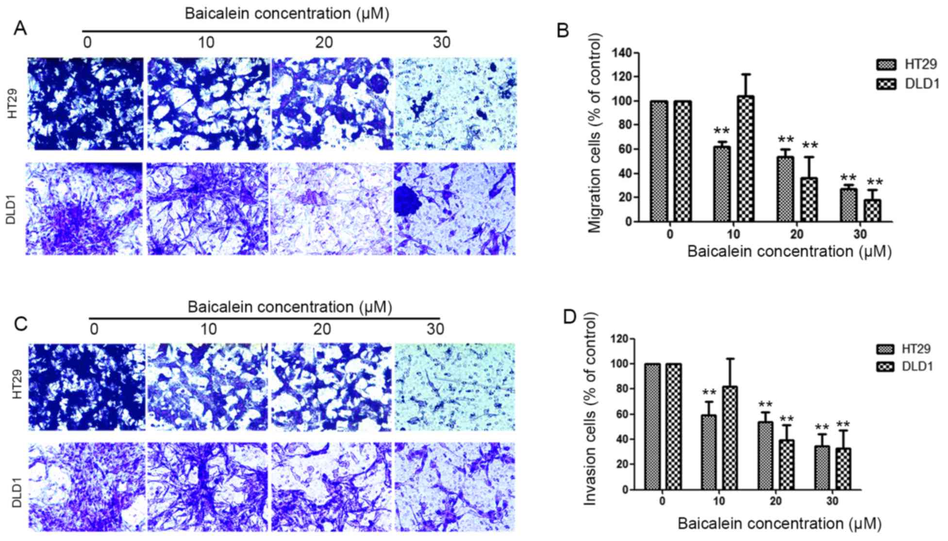 | Figure 2.Baicalein inhibits the migration and
invasion of CRC cells. (A) HT29 and DLD1 cells were pretreated with
0, 10, 20 and 30 µM baicalein for 24 h. Then seeded onto the upper
wells, FBS (10%) was added to the bottom chambers for 16 h to
induce cell migration. After 16 h, cells on the bottom side of the
filter were fixed, stained and counted. (B) The percentage of
migration rate was expressed as a percentage of control (0 µM). (C)
HT29 and DLD1 cells were pretreated with 0, 10, 20 and 30 µM
baicalein for 24 h. Then seeded onto the upper wells, FBS (10%) was
added to the bottom chambers for 24 h to induce cell invasion.
After 24 h, cells on the bottom side of the filter were fixed,
stained and counted. (D) The percentage of invasive rate was
expressed as a percentage of control (0 µM). Values are presented
as means ± SD of three independent experiments performed in
triplicate. **P<0.01 compared with the control group,
respectively. |
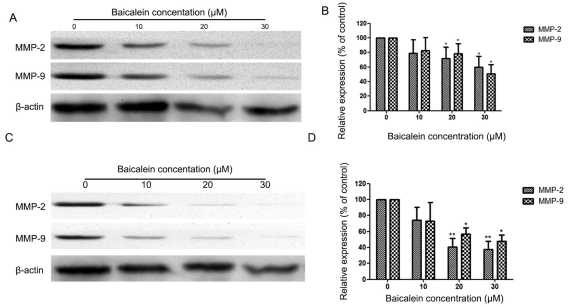
Baicalein inhibits the expression of
MMP-2/−9
We analysed the effect of baicalein on the
expression of MMP-2/−9 in HT29 and DLD1 at various concentrations.
Cells were treated with baicalein (0, 10, 20 and 30 µM) for 24 h
and then analysed by western blotting. Fig. 3A and B show the inhibited effect of
baicalein on the expression of MMP-2/−9 in HT-29 cells. The effect
is concentration-dependent. Fig. 3C and
D show the effect of baicalein on the expression of MMP-2/−9 in
DLD1 cells.
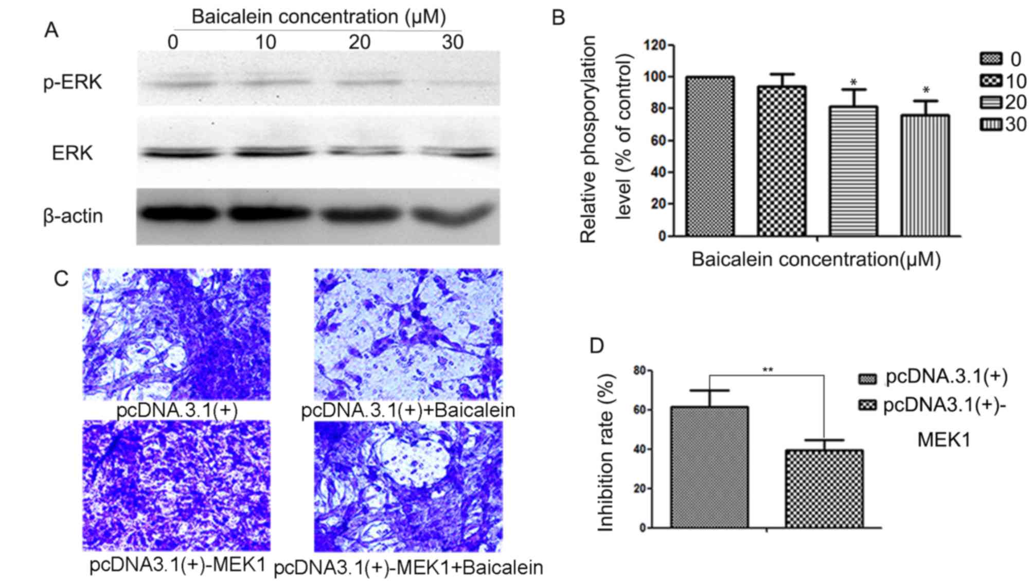
The ERK signaling pathway is involved
in the anti-metastatic mechanism of baicalein in CRC
ERK signaling pathway plays an vital role in the
metastasis of cancer cells by regulating MMP-2/−9 (3); hence, we detected the impact of
baicalein on the activity of ERK signaling pathway in CRC cells.
The results of the western blotting showed that baicalein reduced
the phosphorylation level of ERK1/2 in DLD1 cells treated with
baicalein (Fig. 4A and B). To
further check the role of ERK signaling pathway in the
anti-metastatic effect of baicalein, we upregulated the activity of
ERK singnaling pathway in DLD1 cells via transfecting the plasmid
[pcDNA3.1 (+)-MEK1] expressing human MEK1 and found that the
anti-invasion effect of baicalein was reversed by the high
expression of MEK1 (Fig. 4C). The
positive clones were selected by G418. DLD1 cells were separated
into 3 groups: no treatment as the control group ‘C’, transfected
with an empty vector pcDNA3.1(+) as the negative control group ‘N’,
transfected with a pcDNA3.1(+)-MEK1 as the positive group ‘M’.
The inhibition rate was ~61.3 and 39.3% after 24 h
of treatment with 0 or 30 µM of baicalein (Fig. 4D). Baicalein could also inhibit the
phosphorylation level of ERK in the HT29 cells (data not shown).
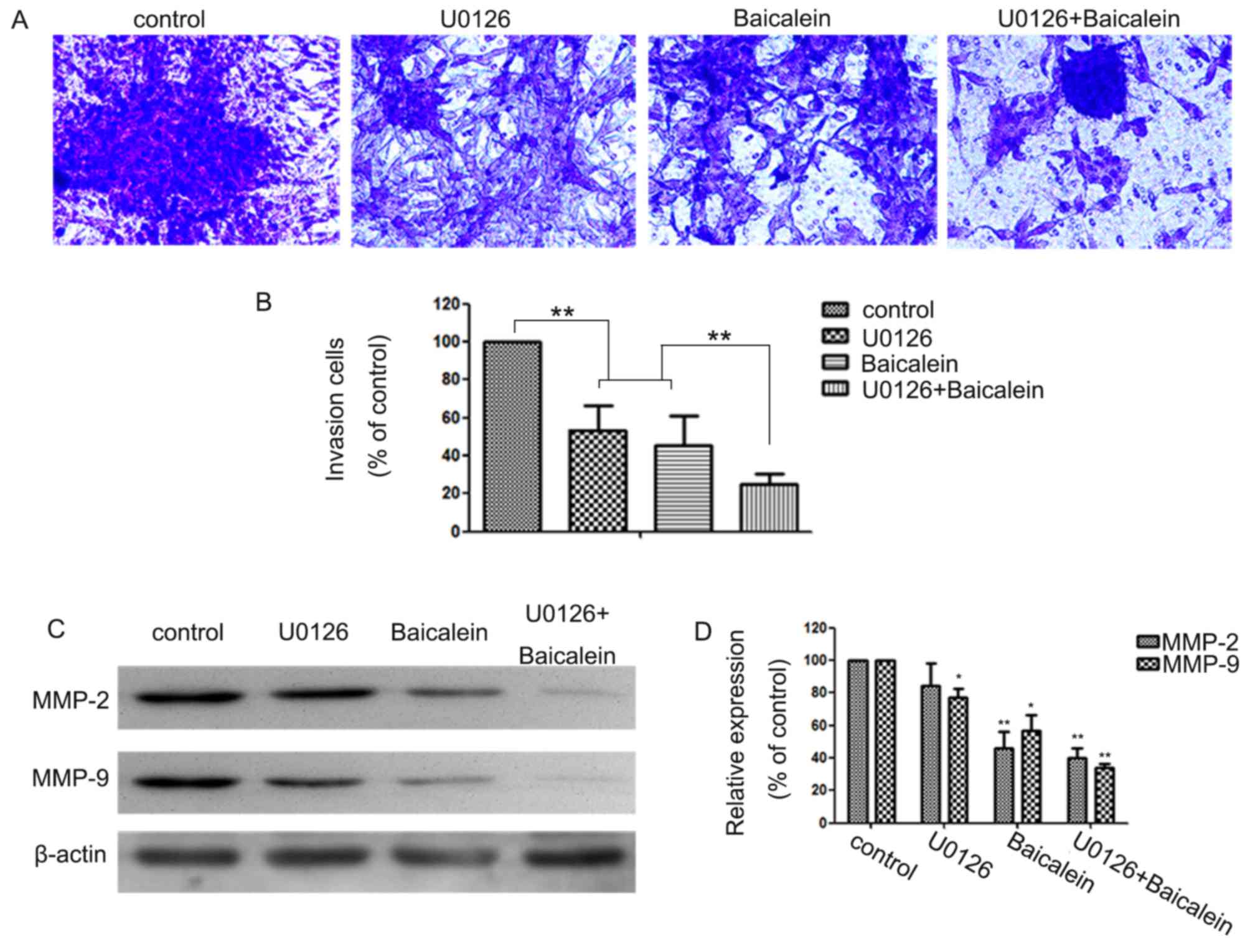
Our results also show that the combined treatment with the
baicalein and U0126 (an ERK inhibitor) reduced both the MMP-2/−9
protein expression (Fig. 5A and B)
and the cell invasion significantly (Fig. 5C and D).
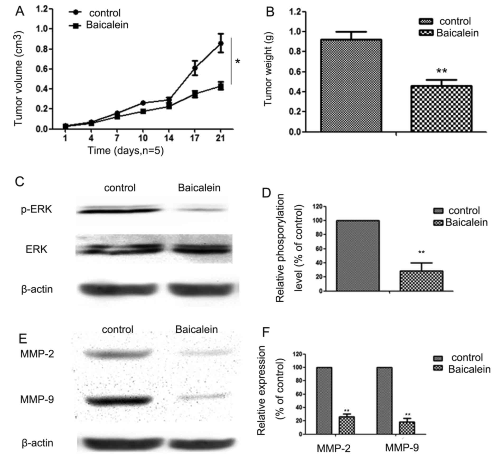
Baicalein inhibits the growth of CRC
tumors in vivo
The inhibition effect of baicalein on DLD1 xenograft
growth is shown in Fig. 6A. We
found that baicalein could inhibit CRC growth significantly. At day
21, all the mice were sacrificed and the tumor tissues were removed
and weighed. Compared to the control group, baicalein reduced the
volume of solid tumors significantly (Fig. 6B). We also found that baicalein
significantly inhibited phosphorylation level of ERK and the
expression of MMP-2/−9 in vivo. The inhibition effect of
baicalein on the phosphorylation level of ERK is shown in Fig. 6C and D, and on expression of
MMP-2/−9 is shown Fig. 6E and F
in vivo.
Discussion
Baicalein possesses antitumor effect in many tumors
(17–19), but the reports on baicalein effect
are rare in CRC. We found that baicalein significantly inhibited
the migration and invasion of CRC cells by inhibiting MMP-2/−9
expression via inhibiting the ERK signaling pathway.
Our results showed that baicalein inhibited the
proliferation and colony formation ability of CRC cells and the
mechanism is correlated with apoptosis. Transwell chamber is
generally used as an assay to detect the metastasis ability of
cancer cells. In the study, we chose the concentration below
cytotoxicity to detect the effect of baicalein on the migration of
CRC cells. We found that baicalein could inhibit the migration and
invasion ability of CRC cells. Our finding is similar to previous
research in other cancers (3,11,15,16),
in which baicalein could inhibit the proliferation of hepatocellar
carcinoma cells, glioma and breast cancer cells. As concentrations
are below the cytotoxicity concentration, the inhibition effect is
not correlated with the cytotoxicity of baicalein. To the best of
our knowledge, we first show the effect of baicalein on DLD1 CRC
cells whicn was used for research on proliferation, apoptosis and
invasion of CRC (20–22). The inhibitory properties are
probably related to the specific structural features of baicalein
(as shown in Fig. 1A). It was
reported that the hydroxylation of C5 and C7 in A-ring
significantly improved the anticancer activity of flavonoids over
that of 5-fluorouracil (23).
Moreover, it has been demonstrated that the hydroxyl substitutions
in the A-ring (C7) of baicalein are crucial for its anti-metastatic
effects against human hematoma cells (24).
To date, metastasis of CRC causes thousands of
deaths every year worldwide (25).
Degradation of the ECM of lymph or blood vessels excert important
roles in the metastasis of cancers via allowing the invasion of
cancer cells into the circulation system and invade distant organs
or tissues (26). MMPs, especially
MMP-2/−9, play important roles in degrading ECM (27). Baicalein shows inhibition effect on
the expression of MMP-2/−9 in hepatocellular carcinoma and glioma
(3,16). Our results showed the inhibition
effect of baicalein on MMP-2/−9 expression in CRC cells. The
results suggest that MMP-2/−9 play important roles in the
anti-metastatic effect of baicalein in CRC cells.
Multiple signaling cascades play important roles in
the synthesis of MMPs, especially the ERK signaling pathway
(19,28,29).
ERK signaling pathway plays an important role in tumor invasion via
promoting the degradation of ECM proteins (30,31).
Studies have shown that the inhibition of ERK phosphorylation
reduces the expression of MMP-2/−9 in the CRC cells (32,33).
To investigate the related mechanism(s) of the inhibition effect of
baicalein on CRC metastasis, we detected the activity of ERK
signaling pathway in CRC cells. The results proved that baicalein
reduced the activity of ERK signaling pathway significantly.
Upregulation of ERK signaling pathway activity abolished the
baicaleins antimetastatic effect. The results was similar to a
previous study, in which baicalein inhibited the expression and
activity of MMP-2 and MMP-9 via inhibiting the activity of ERK
signaling pathway (3).
Our results also showed that combined treatment with
baicalein and U0126 (an ERK inhibitor) reduced both the MMP-2/−9
protein expression (Fig. 5A and B)
and the cell invasion significantly (Fig. 5C and D) suggesting that baicalein
directly downregulated the ERK signaling pathway, which were
similar to previous studies conducted in other types of carcinomas
(3). Finally, our results
demonstrated that baicalein can inhibit the growth of CRC
xenografts in vivo.
In conclusion, the present study demonstrated the
anti-metastatic effect of baicalein on CRC. Furthermore, ERK
signaling pathway plays a vitally important role in the
anti-metastatic effect of baicalein on CRC cells by regulating
MMP-2/−9. These findings revealed that baicalein may represent a
new potential anti-metastatic therapy for CRC.
References
|
1
|
Ma YS, Hsiao YP, Lin JH, Hsu SC, Chueh FS,
Weng SW, Lai KC, Lin JG and Chung JG: Crude extract of Rheum
palmatum L inhibits migration and invasion of LS1034 human
colon cancer cells acts through the inhibition of matrix
metalloproteinase-2/−9 by MAPK signaling. Environ Toxicol.
30:852–863. 2014. View Article : Google Scholar : PubMed/NCBI
|
|
2
|
Patiutko IuI, Pylev AL, Sagaĭdak IV,
Poliakov AN, Chuchuev ES, Abgarian MG and Shishkina NA: Surgical
and combined treatment of patients with metastatic liver and lymph
nodes invasion by colorectal cancer. Khirurgiia (Mosk). 7:49–54.
2010.(In Russian).
|
|
3
|
Chen K, Zhang S, Ji Y, Li J, An P, Ren H,
Liang R, Yang J and Li Z: Baicalein inhibits the invasion and
metastatic capabilities of hepatocellular carcinoma cells via
down-regulation of the ERK pathway. PLoS One. 8:e729272013.
View Article : Google Scholar : PubMed/NCBI
|
|
4
|
Gialeli C, Theocharis AD and Karamanos NK:
Roles of matrix metalloproteinases in cancer progression and their
pharmacological targeting. FEBS J. 278:16–27. 2011. View Article : Google Scholar : PubMed/NCBI
|
|
5
|
Hoshino R, Chatani Y, Yamori T, Tsuruo T,
Oka H, Yoshida O, Shimada Y, Ari-i S, Wada H, Fujimoto J, et al:
Constitutive activation of the 41-/43-kDa mitogen-activated protein
kinase signaling pathway in human tumors. Oncogene. 18:813–822.
1999. View Article : Google Scholar : PubMed/NCBI
|
|
6
|
Zhang H, Shen B, Swinarska JT, Li W, Xiao
K and He P: 9-Hydroxypheophorbide α-mediated photodynamic therapy
induces matrix metalloproteinase-2 (MMP-2) and MMP-9
down-regulation in Hep-2 cells via ROS-mediated suppression of the
ERK pathway. Photodiagn Photodyn Ther. 11:55–62. 2014. View Article : Google Scholar
|
|
7
|
Kress TR, Raabe T and Feller SM: High Erk
activity suppresses expression of the cell cycle inhibitor p27Kip1
in colorectal cancer cells. Cell Commun Signal. 8:12010. View Article : Google Scholar : PubMed/NCBI
|
|
8
|
Park KS, Lee NG, Lee KH, Seo JT and Choi
KY: The ERK pathway involves positive and negative regulations of
HT-29 colorectal cancer cell growth by extracellular zinc. Am J
Physiol Gastrointest Liver Physiol. 285:G1181–G1188. 2003.
View Article : Google Scholar : PubMed/NCBI
|
|
9
|
Kim HC, Kim YS, Oh HW, Kim K, Oh SS, Kim
JT, Kim BY, Lee SJ, Choe YK, Kim DH, et al: Collagen triple helix
repeat containing 1 (CTHRC1) acts via ERK-dependent induction of
MMP9 to promote invasion of colorectal cancer cells. Oncotarget.
5:519–529. 2014. View Article : Google Scholar : PubMed/NCBI
|
|
10
|
Ai X, Wu Y, Zhang W, Zhang Z, Jin G, Zhao
J, Yu J, Lin Y, Zhang W, Liang H, et al: Targeting the ERK pathway
reduces liver metastasis of Smad4-inactivated colorectal cancer.
Cancer Biol Ther. 14:1059–1067. 2013. View Article : Google Scholar : PubMed/NCBI
|
|
11
|
Wang L, Ling Y, Chen Y, Li CL, Feng F, You
QD, Lu N and Guo QL: Flavonoid baicalein suppresses adhesion,
migration and invasion of MDA-MB-231 human breast cancer cells.
Cancer Lett. 297:42–48. 2010. View Article : Google Scholar : PubMed/NCBI
|
|
12
|
Kim DH, Hossain MA, Kang YJ, Jang JY, Lee
YJ, Im E, Yoon JH, Kim HS, Chung HY and Kim ND: Baicalein, an
active component of Scutellaria baicalensis Georgi, induces
apoptosis in human colon cancer cells and prevents AOM/DSS-induced
colon cancer in mice. Int J Oncol. 43:1652–1658. 2013.PubMed/NCBI
|
|
13
|
Chen J, Li Z, Chen AY, Ye X, Luo H, Rankin
GO and Chen YC: Inhibitory effect of baicalin and baicalein on
ovarian cancer cells. Int J Mol Sci. 14:6012–6025. 2013. View Article : Google Scholar : PubMed/NCBI
|
|
14
|
Li HL, Zhang S, Wang Y, Liang RR, Li J, An
P, Wang ZM, Yang J and Li ZF: Baicalein induces apoptosis via a
mitochondrial-dependent caspase activation pathway in T24 bladder
cancer cells. Mol Med Rep. 7:266–270. 2013.PubMed/NCBI
|
|
15
|
Liang RR, Zhang S, Qi JA, Wang ZD, Li J,
Liu PJ, Huang C, Le XF, Yang J and Li ZF: Preferential inhibition
of hepatocellular carcinoma by the flavonoid Baicalein through
blocking MEK-ERK signaling. Int J Oncol. 41:969–978.
2012.PubMed/NCBI
|
|
16
|
Zhang Z, Lv J, Lei X, Li S, Zhang Y, Meng
L, Xue R and Li Z: Baicalein reduces the invasion of glioma cells
via reducing the activity of p38 signaling pathway. PLoS One.
9:e903182014. View Article : Google Scholar : PubMed/NCBI
|
|
17
|
Chandrashekar N, Selvamani A, Subramanian
R, Pandi A and Thiruvengadam D: Baicalein inhibits pulmonary
carcinogenesis-associated inflammation and interferes with COX-2,
MMP-2 and MMP-9 expressions in-vivo. Toxicol Appl Pharmacol.
261:10–21. 2012. View Article : Google Scholar : PubMed/NCBI
|
|
18
|
Wu B, Li J, Huang D, Wang W, Chen Y, Liao
Y, Tang X, Xie H and Tang F: Baicalein mediates inhibition of
migration and invasiveness of skin carcinoma through Ezrin in A431
cells. BMC Cancer. 11:5272011. View Article : Google Scholar : PubMed/NCBI
|
|
19
|
Wang ZD, Huang C, Li ZF, Yang J, Li BH,
Liang RR, Dai ZJ and Liu ZW: Chrysanthemum indicum ethanolic
extract inhibits invasion of hepatocellular carcinoma via
regulation of MMP/TIMP balance as therapeutic target. Oncol Rep.
23:413–421. 2010.PubMed/NCBI
|
|
20
|
Organ SL, Hai J, Radulovich N, Marshall
CB, Leung L, Sasazuki T, Shirasawa S, Zhu CQ, Navab R, Ikura M, et
al: p120RasGAP is a mediator of rho pathway activation and
tumorigenicity in the DLD1 colorectal cancer cell line. PLoS One.
9:e861032014. View Article : Google Scholar : PubMed/NCBI
|
|
21
|
Laezza C, Caruso MG, Gentile T,
Notarnicola M, Malfitano AM, Di Matola T, Messa C, Gazzerro P and
Bifulco M: N6-isopentenyladenosine inhibits cell proliferation and
induces apoptosis in a human colon cancer cell line DLD1. Int J
Cancer. 124:1322–1329. 2009. View Article : Google Scholar : PubMed/NCBI
|
|
22
|
Shida D, Kitayama J, Yamaguchi H, Okaji Y,
Tsuno NH, Watanabe T, Takuwa Y and Nagawa H: Lysophosphatidic acid
(LPA) enhances the metastatic potential of human colon carcinoma
DLD1 cells through LPA1. Cancer Res. 63:1706–1711. 2003.PubMed/NCBI
|
|
23
|
Ibrahim A, Sobeh M, Ismail A, Alaa A,
Sheashaa H, Sobh M and Badria F: Free-B-Ring flavonoids as
potential lead compounds for colon cancer therapy. Mol Clin Oncol.
2:581–585. 2014.PubMed/NCBI
|
|
24
|
Chiu YW, Lin TH, Huang WS, Teng CY, Liou
YS, Kuo WH, Lin WL, Huang HI, Tung JN, Huang CY, et al: Baicalein
inhibits the migration and invasive properties of human hepatoma
cells. Toxicol Appl Pharmacol. 255:316–326. 2011. View Article : Google Scholar : PubMed/NCBI
|
|
25
|
Dziki L, Przybyłowska K, Majsterek I,
Trzciński R, Mik M and Sygut A: A/G polymorphism of the MMP-7 gene
promoter region in colorectal cancer. Pol Przegl Chir. 83:622–626.
2011.PubMed/NCBI
|
|
26
|
Artym VV, Yamada KM and Mueller SC: ECM
degradation assays for analyzing local cell invasion. Methods Mol
Biol. 522:211–219. 2009. View Article : Google Scholar : PubMed/NCBI
|
|
27
|
Zhang Y, Liu H, Jin J, Zhu X, Lu L and
Jiang H: The role of endogenous reactive oxygen species in
oxymatrine-induced caspase-3-dependent apoptosis in human melanoma
A375 cells. Anticancer Drugs. 21:494–501. 2010. View Article : Google Scholar : PubMed/NCBI
|
|
28
|
Cohen M, Meisser A, Haenggeli L and
Bischof P: Involvement of MAPK pathway in TNF-alpha-induced MMP-9
expression in human trophoblastic cells. Mol Hum Reprod.
12:225–232. 2006. View Article : Google Scholar : PubMed/NCBI
|
|
29
|
Liu SQ, Huang JA, Qin MB, Su YJ, Lai MY,
Jiang HX and Tang GD: Sphingosine kinase 1 enhances colon cancer
cell proliferation and invasion by upregulating the production of
MMP-2/9 and uPA via MAPK pathways. Int J Colorectal Dis.
27:1569–1578. 2012. View Article : Google Scholar : PubMed/NCBI
|
|
30
|
Chen YY, Liu FC, Chou PY, Chien YC, Chang
WS, Huang GJ, Wu CH and Sheu MJ: Ethanol extracts of fruiting
bodies of Antrodia cinnamomea suppress CL1-5 human lung
adenocarcinoma cells migration by inhibiting matrix
metalloproteinase-2/9 through ERK, JNK, p38, and PI3K/Akt signaling
pathways. Evid Based Complement Alternat Med.
2012:3784152012.PubMed/NCBI
|
|
31
|
Lin F, Chengyao X, Qingchang L, Qianze D,
Enhua W and Yan W: CRKL promotes lung cancer cell invasion through
ERK-MMP9 pathway. Mol Carcinog. 54:(Suppl 1). E35–E44. 2014.
View Article : Google Scholar : PubMed/NCBI
|
|
32
|
Deng W, Sui H, Wang Q, He N, Duan C, Han
L, Li Q, Lu M and Lv S: A Chinese herbal formula, Yi-Qi-Fu-Sheng,
inhibits migration/invasion of colorectal cancer by down-regulating
MMP-2/9 via inhibiting the activation of ERK/MAPK signaling
pathways. BMC Complement Altern Med. 13:652013. View Article : Google Scholar : PubMed/NCBI
|
|
33
|
Babykutty S, Suboj P, Srinivas P, Nair AS,
Chandramohan K and Gopala S: Insidious role of nitric oxide in
migration/invasion of colon cancer cells by upregulating MMP-2/9
via activation of cGMP-PKG-ERK signaling pathways. Clin Exp
Metastasis. 29:471–492. 2012. View Article : Google Scholar : PubMed/NCBI
|