Introduction
Endometrial cancer is the fourth most common
malignancy among women in developed countries (1). Abnormal bleeding is one of the useful
signs for early diagnosis of endometrial cancer, but 15–20% of
patients recur after primary surgery (2) and their 10-year survival rate is still
poor at 18% (3). Hence, novel
therapeutic options based on better understanding of endometrial
cancer, particularly of immune microenvironment, should be
developed to improve the poor prognosis.
In the process of antitumor cytotoxic T-cell
response, the first event is the capture of cancer-specific
antigens by antigen presenting cells (APCs) such as dendritic cells
and macrophages (4). APCs process
these antigen proteins to peptides, present the antigens on human
leukocyte antigen (HLA) class I molecules to CD8+ T
cells and activate T-cell responses against specific antigens
(4). Then, the activated effector
CD8+ T cells infiltrate into tumor tissues and recognize
cancer cells through the interaction between T cell receptor (TCR)
and its cognate antigen bound to HLA class I molecule (4). Finally, the activated cytotoxic T
cells kill cancer cells by the releasing cytotoxic agents such as
perforin and granzyme, and then cancer cells killed by T cells
further releasing cancer-specific antigens (4). These sequential events are known as
‘cancer immunity cycle’ (5), and
many molecules are involved in the present cycle. To escape from
the host immune attack, cancer cells frequently downregulate the
expression of HLA molecules (6) or
overexpress immune checkpoint molecules such as programmed
death-ligand 1 (PD-L1) and 2 (PD-L2) (7). Expression levels of PD-L1/L2 are
enhanced proportionally according to the amount of
tumor-infiltrating T lymphocytes (TILs), and are associated with
favorable prognosis in several types of cancer, including melanoma,
breast and ovarian cancer (8–11).
Significance of immune microenvironment in prognosis
of endometrial cancer patients has been investigated, since the
patients with hypermutations by polymerase ε (POLE) gene
mutations or microsatellite instability (MSI) were indicated to
have better progression-free survival (PFS) (12). These high MSI cases were suggested
to possess a higher number of somatic mutations, and considered to
generate a higher number of antigenic neo-epitopes and contain
significantly higher tumor-infiltrating CD8+ T cells
(13,14). High T-cell infiltration into tumor
was shown to be associated with favorable prognosis of patients
with endometrial cancer (15–17).
These results indicate the clinical importance of immune
microenvironment in endometrial cancer, but no previous study
investigated the possible significance of T cell clonality and
expression of genes related to cancer immune responses in
endometrial cancer.
TCRs are expressed on the surface of T cells, and
95% of T cells possess TCRs consisting of a heterodimer of α and β
chains. Diversity of TCRs is generated by a somatic recombination
process of variable (V), diversity (D) (only for β chain), and
joining (J) exons, termed the V(D)J recombination (18,19).
Rearrangement of these segments generates the highly variable
complementarity determining region 3 (CDR3), which is important for
the recognition of an antigen on the HLA molecule (20). We recently developed a method to
characterize TCR repertoire from mRNA isolated from cancer tissues
using a next-generation DNA sequencer (21). In this study, we aimed to
investigate the clinical significance of the clonality of TILs and
intratumor expression levels of immune-related genes in prognosis
of endometrial cancer.
Materials and methods
Patient samples
Surgical samples from 32 patients with endometrioid
endometrial carcinoma were obtained at Saitama Medical University
International Medical Center. Details of the patient
characteristics are summarized in Table
I. No patient received any chemotherapy before surgery. The
study protocol was approved by the Institutional Review Boards of
Saitama Medical University International Medical Center and The
University of Chicago. Written informed consent was obtained from
each of the study participants.
 | Table I.Patient characteristics of 32
endometrial cancer samples. |
Table I.
Patient characteristics of 32
endometrial cancer samples.
|
Characteristics | Cases | Frequency |
|---|
| Total | 32 |
|
| Histology |
|
|
|
Endometrioid | 32 | (100%) |
| Stage |
|
|
|
Early | 15 | (47%) |
|
Advanced | 17 | (53%) |
| Grade |
|
|
|
1/2 | 22 | (69%) |
| 3 | 10 | (31%) |
| Menopause |
|
|
|
Pre | 6 | (19%) |
|
Post | 22 | (69%) |
|
Unknown | 4 | (12%) |
| Lymph node
metastasis | 8 | (25%) |
| Lymphvascular
invasion | 16 | (50%) |
| Muscle
invasion |
|
|
|
<1/2 | 27 | (84%) |
|
≥1/2 | 5 | (16%) |
| Recurrence |
|
|
|
Yes | 7 | (22%) |
| No | 25 | (78%) |
| Age (years) |
|
|
|
Median | 61 | (range, 35–78) |
| BMI |
|
|
|
Median | 24 | (range,
17.6–34.7) |
| Tumor size
(mm) |
|
|
|
Median | 44.6 | (range,
23.3–108) |
TaqMan gene expression analysis
From the tumor tissues, total RNAs were obtained
using an RNeasy Mini kit (Qiagen, Valencia, CA, USA). cDNA with
5-rapid amplification of cDNA ends (5-RACE) adapter was synthesized
using SMART cDNA library construction kit (Clontech Laboratories,
Inc., Mountain View, CA, USA). Quantitative real-time PCR was
conducted using TaqMan gene expression assays (Thermo Fisher
Scientific, Carlsbad, CA, USA) on the Applied Biosystems ViiA 7
Real-Time PCR system (Thermo Fisher Scientific) according to the
manufacturers instructions. The following TaqMan gene expression
assays were used; TCRβ (TRB) (forward,
GAGCCATCAGAAGCAGAGATCTC and reverse; GGCCAGGCACACCAGTGT, MGB probe;
ACACC AAAAGGC), CD8A (Hs00233520_m1), granzyme A
(GZMA) (Hs00989184_m1), HLA-A (Hs01058806_g1), CD11c
(ITGAX) (Hs00174217_m1) and PD-L1 (Hs01125301_m1).
mRNA expression levels were normalized to GAPDH expression
(Hs02758991_g1).
TCGA dataset analysis
We obtained the mRNA expression dataset in
endometrial cancer from The Cancer Genome Atlas (TCGA) database
(12). The files with
IlluminaHiSeq_RNASeqV2 and IlluminaGA_RNASeqV2 platform code were
downloaded from the TCGA website (see https://tcga-data.nci.nih.gov/tcga/). Total of 546
cases were obtained from the TCGA website, and we excluded 6 cases
from this analysis because of the lack of clinical information we
needed.
TCR sequencing
The detailed method of library preparation for TCR
sequencing was previously described (21). In brief, we used 957 to 1878 ng of
total RNA. cDNAs were synthesized as described above, and performed
two step PCRs to obtain sequence libraries. The first PCR was
performed to amplify TCRβ cDNA using a forward primer corresponding
to the SMART adapter sequence and a reverse primer corresponding to
the constant region of TCRβ. The second PCR was done to add the
index sequences for Illumina sequencer with barcode sequence using
the Nextera Index kit (Illumina, San Diego, CA, USA). The prepared
libraries were subjected to sequencing using the MiSeq Reagent v3
600-cycles kit on the MiSeq (Illumina).
TCR repertoire was analyzed using
Tcrip software (22)
Briefly, sequencing reads in fastq files were mapped
to the TCR reference sequences derived from IMGT/GENE-DB
(http://www.imgt.org) using Bowtie2 aligner
(version 2.1.0) (23). The V, D and
J genes were designated according to the nomenclature provided by
the international ImMunoGeneTics information system (IMGT)
(24,25). A CDR3 region was defined by
identifying the second conserved cysteine encoded in the 3 portion
of the V segment and the conserved phenylalanine or tryptophan
encoded in the 5 portion of the J segment. TCRβ clonality was
defined as the number of abundant TCRβ clonotypes adjusted by
TRB mRNA expression, as shown below:
TCRβclonality=NumberofTCRβclonotypes<0.1%frequencyTRBmRNAexpression
Statistical analysis
Survival analysis was performed using the
Kaplan-Meier method and the log-rank test. Differences between two
groups were evaluated by the Mann-Whitney U test. All statistical
analyses including multivariate analysis were performed using JMP
v11 (SAS, Inc., Cary, NC, USA) and GraphPad Prism 6 (GraphPad
Software, La Jolla, CA, USA). In all statistical tests, differences
were considered to be statistically significant at P<0.05.
Results
Association of expression levels of
cancer immune-related genes with patient prognosis in endometrial
cancer
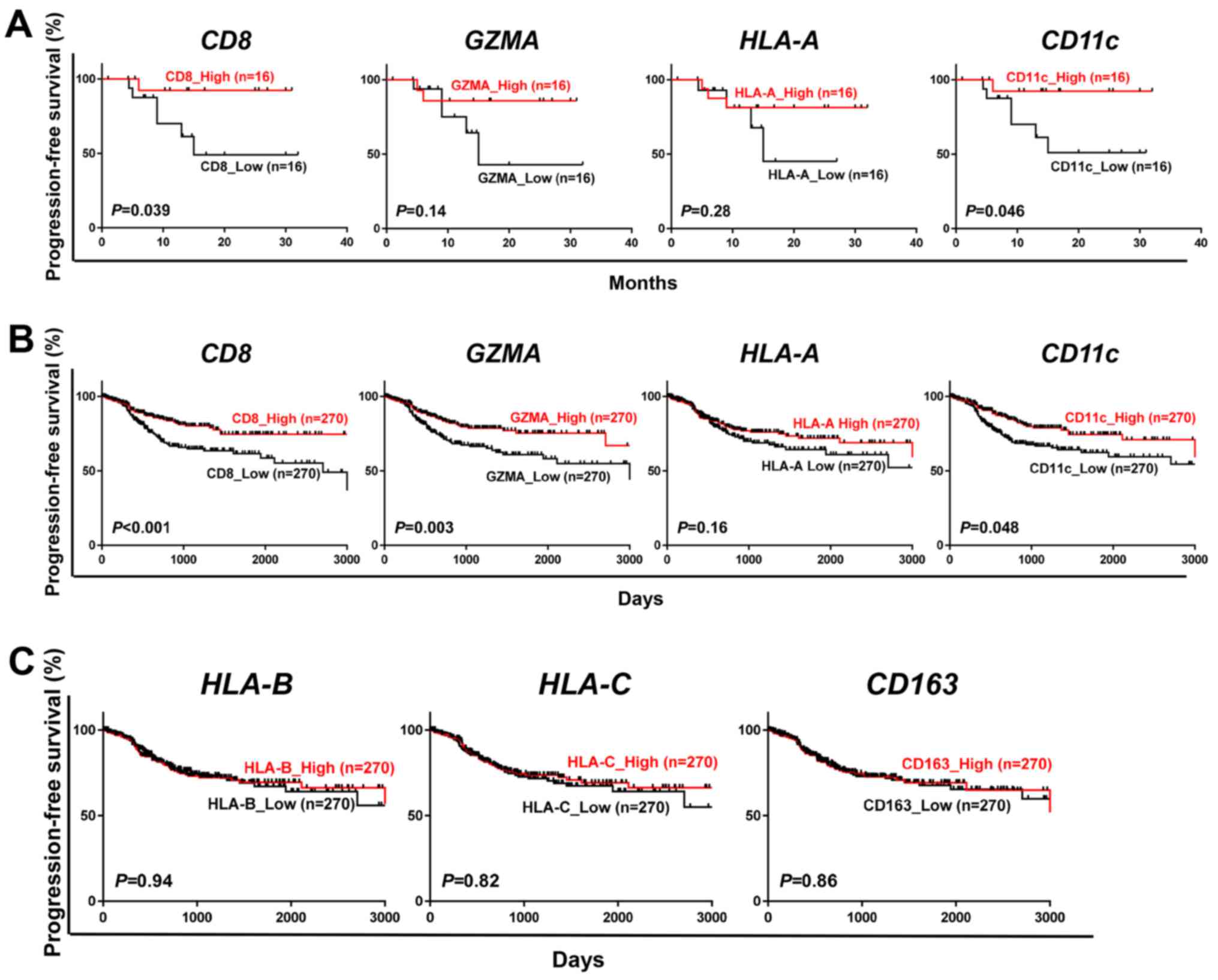
To investigate any effects of expression levels of
cancer immune-related genes on the prognosis of endometrial cancer
patients, we measured mRNA expression levels of CD8,
GZMA (one of molecular markers for cytolytic activity),
HLA-A (one of major HLA class I molecules) and CD11c
(one of markers for dendritic cells/macrophages) in the
surgically-resected cancer tissues from 32 endometrial cancer
patients. We classified the cases into two groups by the median
expression level of each gene and then compared the
progression-free survival (PFS). Higher mRNA expression of
CD8 (P=0.039) and CD11c (P=0.046) was significantly
associated with longer PFS (Fig.
1A). Expression levels of GZMA (P=0.14) and HLA-A
(P=0.28) showed some tendencies although statistically not
significant. In 540 cases from the TCGA dataset, we identified
significant associations of higher expression level of CD8
(P<0.001), GZMA (P=0.003) and CD11c (P=0.048) with
longer PFS (Fig. 1B). In addition,
we explored other cancer immune-related genes such as HLA-B
(HLA class I molecule), HLA-C (HLA class I molecule) and
CD163 (a macrophage marker) using the 540 TCGA cases, but no
significant association with prognosis was observed (Fig. 1C). These results imply the
importance of the infiltration of CD8+ T cells and APCs
into tumor tissues for prognosis in endometrial cancer
patients.
Association of TCRβ clonality in TILs
with prognosis of endometrial cancer patients
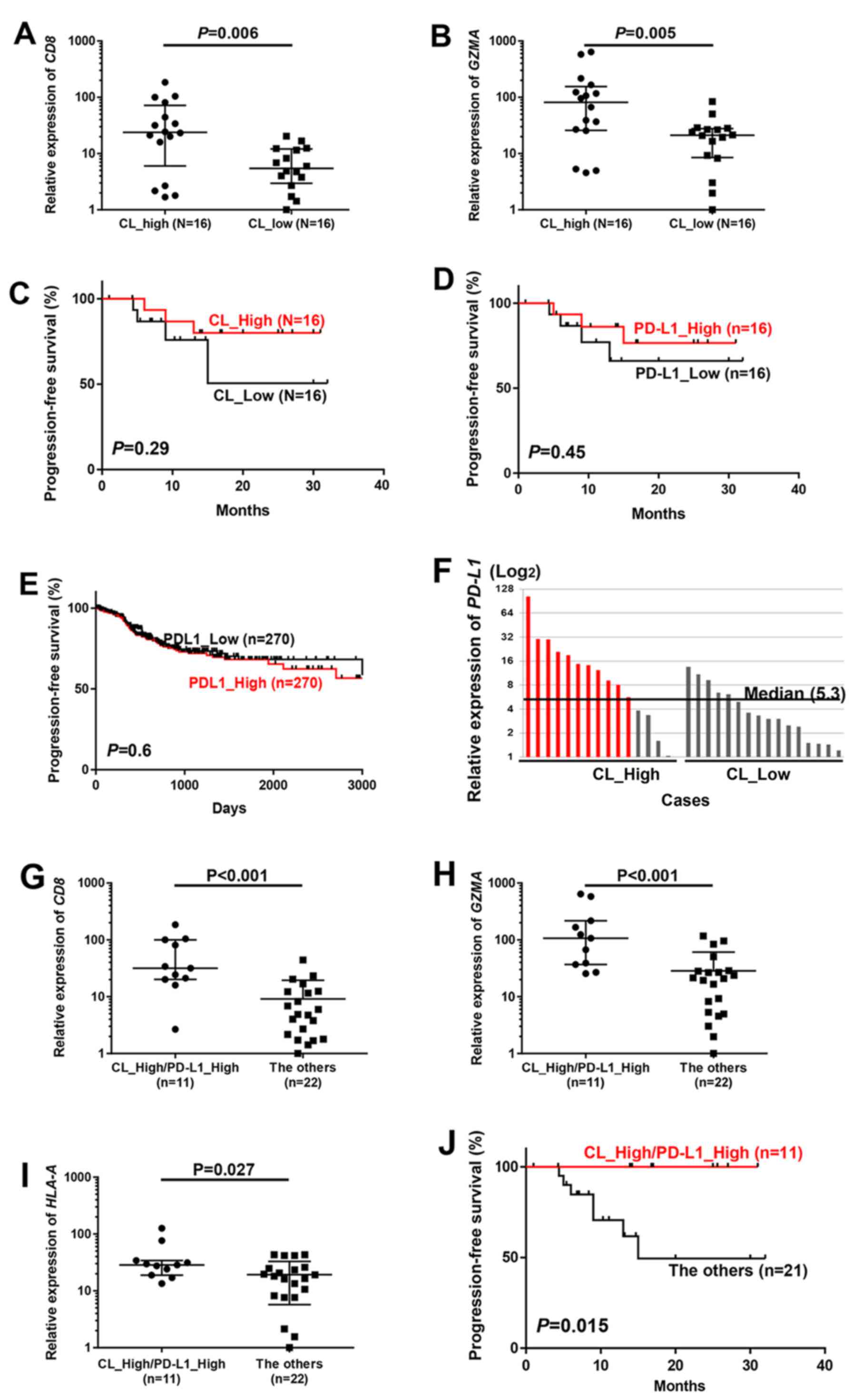
To examine whether clonal expansion of TILs is
related to prognosis of patients with endometrial cancer, we
explored the TCR repertoire in the 32 endometrial cancer tissues.
Through the cDNA sequencing of TCRβ, we obtained total sequence
reads of 270,255 to 4,692,690 (average, 1,834,576), and identified
3,765 to 80,739 (average, 23,598) unique TCRβ CDR3 clonotypes
(Table II). To evaluate the TCRβ
clonality, the numbers of TCRβ clonotypes of >0.1% frequency
were adjusted by TRB mRNA expression levels; this value is lower
when certain T cells are enriched (defined as ‘CL_High’), and is
higher when the number of enriched clones is limited (defined as
‘CL_Low’). In this classification, mRNA expression levels of
CD8 (P=0.006) and GZMA (P=0.005) were significantly
higher in patients with CL_High, and tend to have longer PFS
(Fig. 2A-C).
| Table II.TCRβ sequencing of 32 endometrial
cancer samples. |
Table II.
TCRβ sequencing of 32 endometrial
cancer samples.
| Samples | RNA (µg) | Total reads | Observed
clonotypes | Unique
clonotypes |
|---|
| 1 | 1256 | 2,799,659 | 2,000,034 | 47,474 |
| 2 | 1515 | 1,706,242 | 1,172,922 | 31,040 |
| 3 | 1248 | 3,483,543 | 1,528,768 | 37,262 |
| 4 | 1032 | 2,014,597 | 1,318,455 | 31,655 |
| 5 | 1356 | 1,259,422 | 887,850 | 39,312 |
| 6 | 1389 | 530,160 | 111,787 | 2,631 |
| 7 | 1203 | 1,244,915 | 791,062 | 9,131 |
| 8 | 1704 | 270,255 | 135,501 | 1,376 |
| 9 | 1383 | 2,088,016 | 1,654,058 | 19,699 |
| 10 | 1095 | 749,670 | 553,644 | 8,055 |
| 11 | 1272 | 349,401 | 206,553 | 3,899 |
| 12 | 1084 | 1,561,372 | 957,506 | 8,705 |
| 13 | 1212 | 4,692,690 | 3,286,445 | 80,739 |
| 14 | 1878 | 2,785,592 | 2,229,815 | 34,177 |
| 15 | 1080 | 841,590 | 710,246 | 8,985 |
| 16 | 957 | 1,884,946 | 1,392,866 | 15,713 |
| 17 | 1026 | 1,661,339 | 858,528 | 5,567 |
| 25 | 1011 | 1,505,156 | 811,544 | 27,736 |
| 26 | 1734 | 760,442 | 313,160 | 6,543 |
| 27 | 1072 | 1,368,263 | 695,285 | 27,130 |
| 28 | 1006 | 2,174,686 | 1,258,224 | 16,901 |
| 29 | 1120 | 800,381 | 223,986 | 4,352 |
| 31 | 1156 | 2,683,768 | 1,483,747 | 38,150 |
| 32 | 1353 | 1,640,097 | 1,047,746 | 23,619 |
| 33 | 1349 | 1,005,383 | 381,802 | 8,887 |
| 34 | 1730 | 1,964,567 | 1,448,467 | 32,797 |
| 35 | 1470 | 1,362,842 | 1,047,478 | 34,492 |
| 36 | 1171 | 3,347,286 | 2,430,597 | 55,515 |
| 37 | 1169 | 3,034,346 | 1,655,574 | 43,294 |
| 38 | 980 | 1,715,642 | 930,467 | 19,235 |
| 39 | 1122 | 4,078,715 | 83,682 | 3,765 |
| 40 | 1267 | 1,341,472 | 992,980 | 27,295 |
Several recent reports indicated that expression of
PD-L1 in tumor cells reflect the presence of antigen-induced
antitumor immune pressure mediated by TILs (8–11).
However, PD-L1 expression itself did not show any prognostic value
in our 32 cases (P=0.45) or in 540 cases in TCGA database (P=0.60)
(Fig. 2D and E). Among our 16 cases
with CL_High, 11 cases showed higher PD-L1 expression than the
median PD-L1 expression level of 32 cases, and we defined
them as CL_High/PD-L1_High which is considered to have strong
immune pressure in their tumor tissues (Fig. 2F). In these 11 CL_High/PD-L1_High
cases, we observed significantly higher levels of CD8
(P<0.001), GZMA (P<0.001) and HLA-A (P=0.027)
than in the remaining cases (Fig.
2G-I). In prognostic analysis, none of 11 cases in the
CL_High/PD-L1_High group had recurrence while the remaining 21
cases showed the significantly higher relapse rate (P=0.015;
Fig. 2J).
Association of immune-related gene
expression and TCRβ clonality in TILs with patient
characteristics
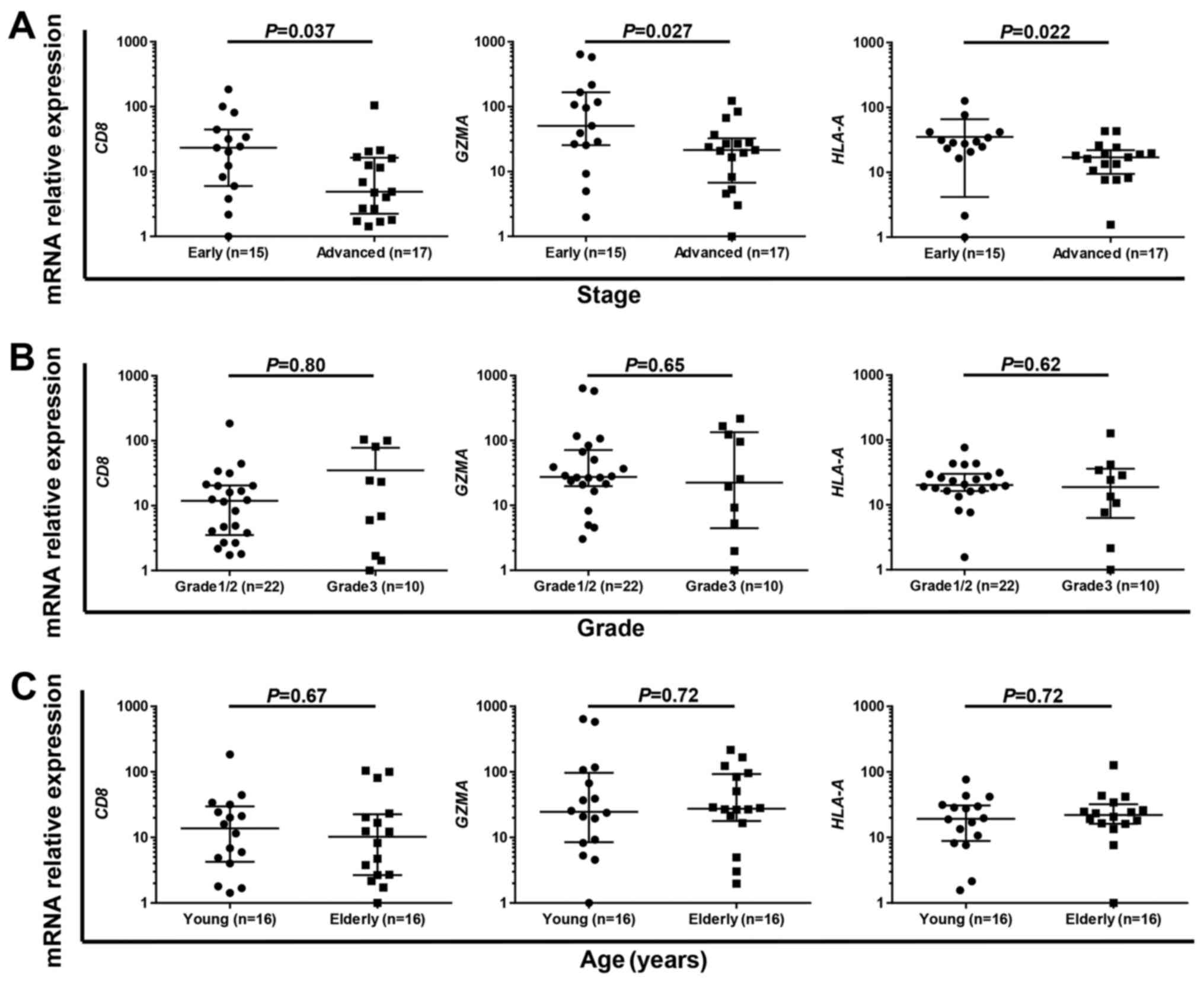
Finally, we examined the association of
immune-related gene expression and TCRβ clonality with
clinicopathological characteristics such as clinical stage, grade
and age. Multivariable analysis revealed that CL_High/PD-L1_High
was the only independent favorable prognostic factor in our results
(Table III). Therefore, we
examined the association between expression level of immune-related
genes such as CD8, GZMA and HLA-A with clinicopathological
characteristics. We found that the expression levels of CD8
(P=0.037), GZMA (P=0.027) and HLA-A (P=0.022) were
significantly higher in early-stage cases than advanced-stage cases
(Fig. 3A), while no significant
difference was observed in grade (Fig.
3B) or age (young vs. elderly, classified by the median of 61
years; Fig. 3C). These results
suggest that early-stage endometrial tumors are immunologically
more active compared to advanced-stage tumors.
| Table III.Multivariate analysis of expression
levels of immune-related genes. |
Table III.
Multivariate analysis of expression
levels of immune-related genes.
|
| Multivariate
analysis |
|---|
|
|
|
|---|
|
Characteristics | Hazard ratio | 95% CI | P-value |
|---|
|
CL_High/PD-L1_High |
|
|
|
|
Yes | 1.06E-09 | 0-0.43 | 0.01 |
| No
(ref) |
|
|
|
| Stage |
|
|
|
| I and
II | 0.14 | 0.01–1.04 | 0.06 |
| III and
IV (ref) |
|
|
|
| Grade |
|
|
|
| 1 and
2 | 0.3 | 0.03–1.88 | 0.2 |
| 3
(ref) |
|
|
|
| Age (years) |
|
|
|
|
<60 | 0.58 | 0.05–6.12 | 0.64 |
| ≥60
(ref) |
|
|
|
| CD8 |
|
|
|
|
>Median | 0.25 | 0.01–1.9 | 0.19 |
| ≤Median
(ref) |
|
|
|
| Stage |
|
|
|
| I and
II | 0.19 | 0.01–1.33 | 0.1 |
| III and
IV (ref) |
|
|
|
| Grade |
|
|
|
| 1 and
2 | 0.22 | 0.06–1.85 | 0.22 |
| 3
(ref) |
|
|
|
| Age (years) |
|
|
|
| 60 | 1.15 | 0.15–9.4 | 0.89 |
| ≥60
(ref) |
|
|
|
| CD11c |
|
|
|
|
>Median | 0.21 | 0.01–2.34 | 0.21 |
| ≤Median
(ref) |
|
|
|
| Stage |
|
|
|
| I and
II | 0.16 | 0.01–1.28 | 0.09 |
| III and
IV (ref) |
|
|
|
| Grade |
|
|
|
| 1 and
2 | 0.18 | 0.61–15.4 | 0.18 |
| 3
(ref) |
|
|
|
| Age (years) |
|
|
|
|
<60 | 0.8 | 0.08–7.79 | 0.84 |
| ≥60
(ref) |
|
|
|
Discussion
In the present study, we analyzed mRNA expression
levels of immune-related genes and TCR repertoire of T lymphocytes
in tumor tissues of 32 endometrial cancer patients, and
demonstrated that: i) the association of higher mRNA expression
levels of CD8, GZMA and CD11c with better
prognosis; ii) the association of coexistence of higher clonal
enrichment of certain T cells and higher PD-L1 expression
(CL_High/PD-L1_High) with higher expression levels of CD8,
GZMA and HLA-A as well as favorable prognosis; and
ii) higher levels of CD8, GZMA and HLA-A
expression in early-stage endometrial cancer.
We first explored clinical significance of cancer
immunity-related molecules in our 32 endometrial cancer samples as
well as the 540 TCGA cases, and identified that CD8
expression level could be an important factor to predict patient
prognosis, as reported based on several previous investigations
(26,27). In addition, we identified
CD11c expression level, which represents the number of
infiltrated dendritic cells/macrophages into tumors, was
significantly associated with prognosis of endometrial cancer
patients. These cells play a crucial role as APCs in defining
immune microenvironment, and association of higher CD11c
expression level with favorable prognosis was also suggested in
previous studies for gastric, colon and cervical cancers (28). This is the first study showing
prognostic significance of CD11c expression levels in endometrial
cancer.
Since CD11c is a maker of dendritic
cells/macrophages, which play an important role for antigen
presentation, we also focused on the TCRβ repertoire in endometrial
cancer. We previously demonstrated that TCRβ clonality was
associated with prognosis in bladder cancer (29). Since TCRβ clonality itself was not
significantly associated with prognosis of endometrial cancer
patients, we combined T cell clonality and PD-L1 expression for
further analysis. PD-L1 expression is associated with prognosis in
several types of cancer including breast, ovarian cancer and
melanoma (8–11), although our result in endometrial
cancer did not clearly show the prognostic significance of PD-L1
expression. However, 11 CL_High/PD-L1_High cases, in which we
observed higher T cell clonal expansion along with higher PD-L1
expression in tumor tissues, showed significantly higher expression
levels of immune-related genes such as CD8, GZMA and
HLA-A than the remaining cases. In addition, none of these
11 cases with CL_High/PD-L1_High experienced recurrence, indicating
a prognostic importance of immune microenvironment characterized by
expression levels of immune-related genes and the clonality of
infiltrated T cells in endometrial cancer.
Finally, we performed subgroup analysis based on the
patient clinicopathological characteristics, and identified that
CL_high/PD-L1_high was an independent prognostic factor in
endometrial cancer. In addition, the anti-immune status of
early-stage patients was considered to be more active, although two
previous reports were controversial for the association between
immune status and clinical stage (26,30).
Our results imply that the non-inflamed phenotype is one of the
characteristics of tumor progression process in endometrial
cancer.
In conclusion, we identified clinical significance
of expression levels of cancer immune response-related factors such
as CD8 and dendritic cells/macrophages in endometrial tumor
tissues. In addition, the clonality of TILs in combination with
PD-L1 expression in tumor tissues could be a good prognostic maker
in endometrial cancer.
Acknowledgements
The super-computing resource was provided by the
Human Genome Center, the Institute of Medical Science, the
University of Tokyo (http://sc.hgc.jp/shirokane.html).
References
|
1
|
Siegel RL, Miller KD and Jemal A: Cancer
statistics, 2016. CA Cancer J Clin. 66:7–30. 2016. View Article : Google Scholar : PubMed/NCBI
|
|
2
|
Salvesen HB, Haldorsen IS and Trovik J:
Markers for individualised therapy in endometrial carcinoma. Lancet
Oncol. 13:e353–e361. 2012. View Article : Google Scholar : PubMed/NCBI
|
|
3
|
Creutzberg CL, Nout RA, Lybeert ML,
Wárlám-Rodenhuis CC, Jobsen JJ, Mens JW, Lutgens LC, Pras E, van de
Poll-Franse LV and van Putten WL: PORTEC Study Group: Fifteen-year
radiotherapy outcomes of the randomized PORTEC-1 trial for
endometrial carcinoma. Int J Radiat Oncol Biol Phys. 81:e631–e638.
2011. View Article : Google Scholar : PubMed/NCBI
|
|
4
|
Melero I, Gaudernack G, Gerritsen W, Huber
C, Parmiani G, Scholl S, Thatcher N, Wagstaff J, Zielinski C,
Faulkner I, et al: Therapeutic vaccines for cancer: An overview of
clinical trials. Nat Rev Clin Oncol. 11:509–524. 2014. View Article : Google Scholar : PubMed/NCBI
|
|
5
|
Chen DS and Mellman I: Oncology meets
immunology: The cancer-immunity cycle. Immunity. 39:1–10. 2013.
View Article : Google Scholar : PubMed/NCBI
|
|
6
|
Hicklin DJ, Marincola FM and Ferrone S:
HLA class I antigen downregulation in human cancers: T-cell
immunotherapy revives an old story. Mol Med Today. 5:178–186. 1999.
View Article : Google Scholar : PubMed/NCBI
|
|
7
|
Ohaegbulam KC, Assal A, Lazar-Molnar E,
Yao Y and Zang X: Human cancer immunotherapy with antibodies to the
PD-1 and PD-L1 pathway. Trends Mol Med. 21:24–33. 2015. View Article : Google Scholar : PubMed/NCBI
|
|
8
|
Schalper KA, Velcheti V, Carvajal D,
Wimberly H, Brown J, Pusztai L and Rimm DL: In situ tumor PD-L1
mRNA expression is associated with increased TILs and better
outcome in breast carcinomas. Clin Cancer Res. 20:2773–2782. 2014.
View Article : Google Scholar : PubMed/NCBI
|
|
9
|
Harter PN, Bernatz S, Scholz A, Zeiner PS,
Zinke J, Kiyose M, Blasel S, Beschorner R, Senft C, Bender B, et
al: Distribution and prognostic relevance of tumor-infiltrating
lymphocytes (TILs) and PD-1/PD-L1 immune checkpoints in human brain
metastases. Oncotarget. 6:40836–40849. 2015.PubMed/NCBI
|
|
10
|
Webb JR, Milne K, Kroeger DR and Nelson
BH: PD-L1 expression is associated with tumor-infiltrating T cells
and favorable prognosis in high-grade serous ovarian cancer.
Gynecol Oncol. 141:293–302. 2016. View Article : Google Scholar : PubMed/NCBI
|
|
11
|
Inozume T, Hanada K, Wang QJ, Ahmadzadeh
M, Wunderlich JR, Rosenberg SA and Yang JC: Selection of
CD8+PD-1+ lymphocytes in fresh human
melanomas enriches for tumor-reactive T cells. J Immunother.
33:956–964. 2010. View Article : Google Scholar : PubMed/NCBI
|
|
12
|
Kandoth C, Schultz N, Cherniack AD, Akbani
R, Liu Y, Shen H, Robertson AG, Pashtan I, Shen R, Benz CC, et al:
Cancer Genome Atlas Research Network: Integrated genomic
characterization of endometrial carcinoma. Nature. 497:67–73. 2013.
View Article : Google Scholar : PubMed/NCBI
|
|
13
|
van Gool IC, Eggink FA, Freeman-Mills L,
Stelloo E, Marchi E, de Bruyn M, Palles C, Nout RA, de Kroon CD,
Osse EM, et al: POLE proofreading mutations elicit an antitumor
immune response in endometrial cancer. Clin Cancer Res.
21:3347–3355. 2015. View Article : Google Scholar : PubMed/NCBI
|
|
14
|
Howitt BE, Shukla SA, Sholl LM,
Ritterhouse LL, Watkins JC, Rodig S, Stover E, Strickland KC,
DAndrea AD, Wu CJ, et al: Association of polymerase e-mutated and
microsatellite-instable endometrial cancers with neoantigen load,
number of tumor-infiltrating lymphocytes, and expression of PD-1
and PD-L1. JAMA Oncol. 1:1319–1323. 2015. View Article : Google Scholar : PubMed/NCBI
|
|
15
|
Čermáková P, Melichar B, Tomšová M, Zoul
Z, Kalábová H, Spaček J and Doležel M: Prognostic significance of
CD3+ tumor-infiltrating lymphocytes in patients with
endometrial carcinoma. Anticancer Res. 34:5555–5561.
2014.PubMed/NCBI
|
|
16
|
de Jong RA, Leffers N, Boezen HM, ten Hoor
KA, van der Zee AG, Hollema H and Nijman HW: Presence of
tumor-infiltrating lymphocytes is an independent prognostic factor
in type I and II endometrial cancer. Gynecol Oncol. 114:105–110.
2009. View Article : Google Scholar : PubMed/NCBI
|
|
17
|
Suemori T, Susumu N, Iwata T, Banno K,
Yamagami W, Hirasawa A, Sugano K, Matsumoto E and Aoki D:
Intratumoral CD8 lymphocyte infiltration as a prognostic factor and
its relationship with cyclooxygenase 2 expression and
microsatellite instability in endometrial cancer. Int J Gynecol
Cancer. 25:1165–1172. 2015. View Article : Google Scholar : PubMed/NCBI
|
|
18
|
Scaviner D and Lefranc MP: The human T
cell receptor alpha variable (TRAV) genes. Exp Clin Immunogenet.
17:83–96. 2000. View Article : Google Scholar : PubMed/NCBI
|
|
19
|
Folch G and Lefranc MP: The human T cell
receptor beta variable (TRBV) genes. Exp Clin Immunogenet.
17:42–54. 2000. View Article : Google Scholar : PubMed/NCBI
|
|
20
|
Danska JS, Livingstone AM, Paragas V,
Ishihara T and Fathman CG: The presumptive CDR3 regions of both T
cell receptor alpha and beta chains determine T cell specificity
for myoglobin peptides. J Exp Med. 172:27–33. 1990. View Article : Google Scholar : PubMed/NCBI
|
|
21
|
Jang M and Yew P: Deep sequencing of
T-cell and B-cell receptors with next-generation DNA
sequencersImmunopharmacogenomics. Nakamura Y: Springer; pp. 3–26.
2015, View Article : Google Scholar
|
|
22
|
Yamaguchi R, Inoto S and Miyano S: A TCR
sequence data analysis pipeline: TcripImmunopharmacogenomics.
Nakamura Y: Springer; pp. 27–43. 2015, View Article : Google Scholar
|
|
23
|
Langmead B and Salzberg SL: Fast
gapped-read alignment with Bowtie 2. Nat Methods. 9:357–359. 2012.
View Article : Google Scholar : PubMed/NCBI
|
|
24
|
Lefranc MP, Giudicelli V, Ginestoux C,
Jabado-Michaloud J, Folch G, Bellahcene F, Wu Y, Gemrot E, Brochet
X, Lane J, et al: IMGT, the international ImMunoGeneTics
information system. Nucleic Acids Res. 37:(Database). D1006–D1012.
2009. View Article : Google Scholar : PubMed/NCBI
|
|
25
|
Lefranc MP: IMGT, the International
ImMunoGeneTics Information System. Cold Spring Harb Protoc.
2011:595–603. 2011. View Article : Google Scholar : PubMed/NCBI
|
|
26
|
Yamagami W, Susumu N, Tanaka H, Hirasawa
A, Banno K, Suzuki N, Tsuda H, Tsukazaki K and Aoki D:
Immunofluorescence-detected infiltration of
CD4+FOXP3+ regulatory T cells is relevant to
the prognosis of patients with endometrial cancer. Int J Gynecol
Cancer. 21:1628–1634. 2011. View Article : Google Scholar : PubMed/NCBI
|
|
27
|
Kondratiev S, Sabo E, Yakirevich E, Lavie
O and Resnick MB: Intratumoral CD8+ T lymphocytes as a
prognostic factor of survival in endometrial carcinoma. Clin Cancer
Res. 10:4450–4456. 2004. View Article : Google Scholar : PubMed/NCBI
|
|
28
|
Origoni M, Parma M, DellAntonio G, Gelardi
C, Stefani C, Salvatore S and Candiani M: Prognostic significance
of immunohistochemical phenotypes in patients treated for
high-grade cervical intraepithelial neoplasia. BioMed Res Int.
2013:8319072013. View Article : Google Scholar : PubMed/NCBI
|
|
29
|
Choudhury NJ, Kiyotani K, Yap KL,
Campanile A, Antic T, Yew PH, Steinberg G, Park JH, Nakamura Y and
ODonnel PH: Low T-cell receptor diversity, high somatic mutation
burden, and high neoantigen load as predictors of clinical outcome
in muscle-invasive bladder cancer. Eur Urol Focus. 2:445–452. 2015.
View Article : Google Scholar
|
|
30
|
Jung IK, Kim SS, Suh DS, Kim KH, Lee CH
and Yoon MS: Tumor-infiltration of T-lymphocytes is inversely
correlated with clinicopathologic factors in endometrial
adenocarcinoma. Obstet Gynecol Sci. 57:266–273. 2014. View Article : Google Scholar : PubMed/NCBI
|