|
1
|
Snover D, Ahnen DJ, Burt RW and Odze RD:
Serrated polyps of the colon and rectum and serrated polyposis. WHO
Classification of Tumours of the Digestive System. 3. 4th. Bosman
FT, Carneiro F, Hruban RH and Theise ND: Lyon, France: IARC; pp.
160–165. 2010
|
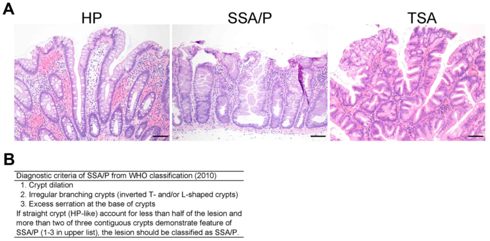
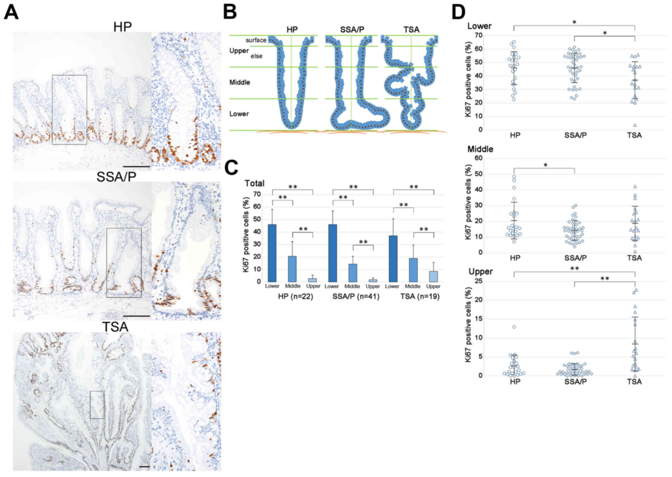
|
2
|
Snover DC: Update on the serrated pathway
to colorectal carcinoma. Hum Pathol. 42:1–10. 2011. View Article : Google Scholar : PubMed/NCBI
|
|
3
|
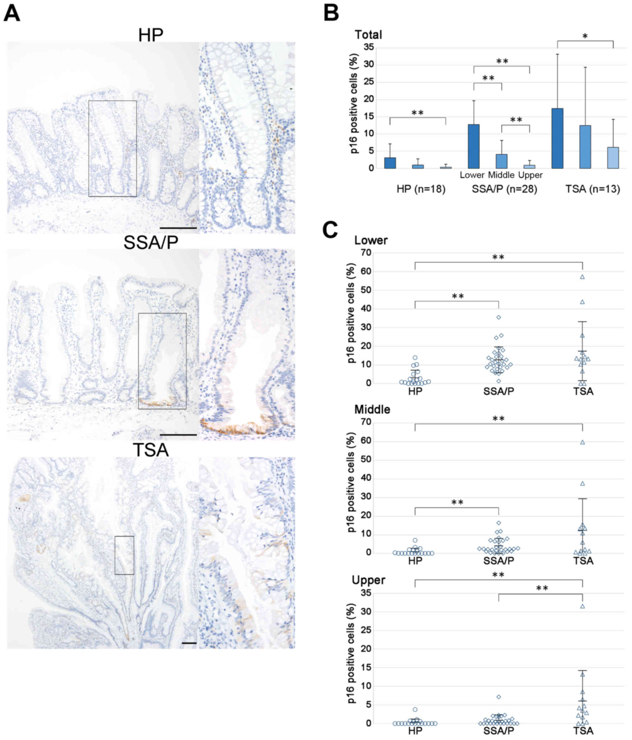
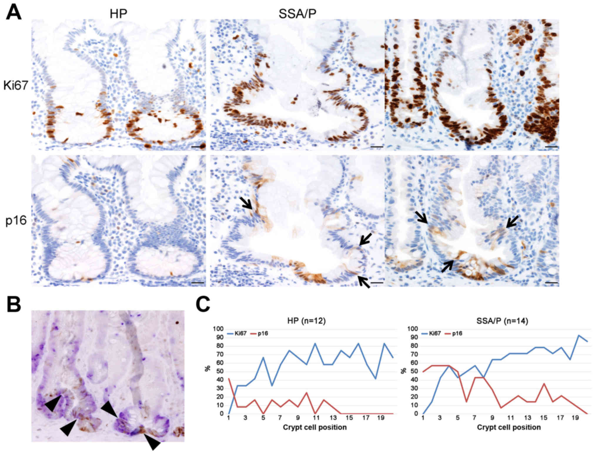
Chan TL, Zhao W, Leung SY and Yuen ST:
Cancer Genome Project: BRAF and KRAS mutations in colorectal
hyperplastic polyps and serrated adenomas. Cancer Res.
63:4878–4881. 2003.PubMed/NCBI
|
|
4
|
Iino H, Jass JR, Simms LA, Young J,
Leggett B, Ajioka Y and Watanabe H: DNA microsatellite instability
in hyperplastic polyps, serrated adenomas, and mixed polyps: A mild
mutator pathway for colorectal cancer? J Clin Pathol. 52:5–9. 1999.
View Article : Google Scholar : PubMed/NCBI
|
|
5
|
Park SJ, Rashid A, Lee JH, Kim SG,
Hamilton SR and Wu TT: Frequent CpG island methylation in serrated
adenomas of the colorectum. Am J Pathol. 162:815–822. 2003.
View Article : Google Scholar : PubMed/NCBI
|
|
6
|
Spring KJ, Zhao ZZ, Karamatic R, Walsh MD,
Whitehall VL, Pike T, Simms LA, Young J, James M, Montgomery GW, et
al: High prevalence of sessile serrated adenomas with BRAF
mutations: A prospective study of patients undergoing colonoscopy.
Gastroenterology. 131:1400–1407. 2006. View Article : Google Scholar : PubMed/NCBI
|
|
7
|
Yang S, Farraye FA, Mack C, Posnik O and
O'Brien MJ: BRAF and KRAS mutations in hyperplastic polyps and
serrated adenomas of the colorectum: Relationship to histology and
CpG island methylation status. Am J Surg Pathol. 28:1452–1459.
2004. View Article : Google Scholar : PubMed/NCBI
|
|
8
|
Carr NJ, Mahajan H, Tan KL, Hawkins NJ and
Ward RL: Serrated and non-serrated polyps of the colorectum: Their
prevalence in an unselected case series and correlation of BRAF
mutation analysis with the diagnosis of sessile serrated adenoma. J
Clin Pathol. 62:516–518. 2009. View Article : Google Scholar : PubMed/NCBI
|
|
9
|
Fujimori Y, Fujimori T, Imura J, Sugai T,
Yao T, Wada R, Ajioka Y and Ohkura Y: An assessment of the
diagnostic criteria for sessile serrated adenoma/polyps: SSA/Ps
using image processing software analysis for Ki67
immunohistochemistry. Diagn Pathol. 7:592012. View Article : Google Scholar : PubMed/NCBI
|
|
10
|
Higuchi T, Sugihara K and Jass JR:
Demographic and pathological characteristics of serrated polyps of
colorectum. Histopathology. 47:32–40. 2005. View Article : Google Scholar : PubMed/NCBI
|
|
11
|
O'Brien MJ, Yang S, Mack C, Xu H, Huang
CS, Mulcahy E, Amorosino M and Farraye FA: Comparison of
microsatellite instability, CpG island methylation phenotype, BRAF
and KRAS status in serrated polyps and traditional adenomas
indicates separate pathways to distinct colorectal carcinoma end
points. Am J Surg Pathol. 30:1491–1501. 2006. View Article : Google Scholar : PubMed/NCBI
|
|
12
|
Chetty R, Hafezi-Bakhtiari S, Serra S,
Colling R and Wang LM: Traditional serrated adenomas (TSAs) admixed
with other serrated (so-called precursor) polyps and conventional
adenomas: A frequent occurrence. J Clin Pathol. 68:270–273. 2015.
View Article : Google Scholar : PubMed/NCBI
|
|
13
|
Schmitt CA: Cellular senescence and cancer
treatment. Biochim Biophys Acta. 1775:5–20. 2007.PubMed/NCBI
|
|
14
|
Collado M, Blasco MA and Serrano M:
Cellular senescence in cancer and aging. Cell. 130:223–233. 2007.
View Article : Google Scholar : PubMed/NCBI
|
|
15
|
Campisi J and di d'Adda Fagagna F:
Cellular senescence: When bad things happen to good cells. Nat Rev
Mol Cell Biol. 8:729–740. 2007. View
Article : Google Scholar : PubMed/NCBI
|
|
16
|
Gutierrez-Reyes G, del Carmen Garcia, de
Leon M, Varela-Fascinetto G, Valencia P, Tamayo Pérez R, Rosado CG,
Labonne BF, Rochilin NM, Garcia RM, Valadez JA, et al: Cellular
senescence in livers from children with end stage liver disease.
PLoS One. 5:e102312010. View Article : Google Scholar : PubMed/NCBI
|
|
17
|
Dankort D, Filenova E, Collado M, Serrano
M, Jones K and McMahon M: A new mouse model to explore the
initiation, progression, and therapy of BRAFV600E-induced lung
tumors. Genes Dev. 21:379–384. 2007. View Article : Google Scholar : PubMed/NCBI
|
|
18
|
Kriegl L, Neumann J, Vieth M, Greten FR,
Reu S, Jung A and Kirchner T: Up and downregulation of p16(Ink4a)
expression in BRAF-mutated polyps/adenomas indicates a senescence
barrier in the serrated route to colon cancer. Mod Pathol.
24:1015–1022. 2011. View Article : Google Scholar : PubMed/NCBI
|
|
19
|
Shida Y, Ichikawa K, Fujimori T, Fujimori
Y, Tomita S, Fujii T, Sano Y, Oda Y, Goto H, Ohta A, et al:
Differentiation between sessile serrated adenoma/polyp and
non-sessile serrated adenoma/polyp in large hyper plastic polyp: A
Japanese collaborative study. Mol Clin Oncol. 1:53–58.
2013.PubMed/NCBI
|
|
20
|
Miyoshi H, Ajima R, Luo CT, Yamaguchi TP
and Stappenbeck TS: Wnt5a potentiates TGF-β signaling to promote
colonic crypt regeneration after tissue injury. Science.
338:108–113. 2012. View Article : Google Scholar : PubMed/NCBI
|
|
21
|
Powell DW, Adegboyega PA, Di Mari JF and
Mifflin RC: Epithelial cells and their neighbors I. Role of
intestinal myofibroblasts in development, repair, and cancer. Am J
Physiol Gastrointest Liver Physiol. 289:G2–G7. 2005. View Article : Google Scholar : PubMed/NCBI
|
|
22
|
Gregorieff A, Pinto D, Begthel H, Destrée
O, Kielman M and Clevers H: Expression pattern of Wnt signaling
components in the adult intestine. Gastroenterology. 129:626–638.
2005. View Article : Google Scholar : PubMed/NCBI
|
|
23
|
Rau TT, Atreya R, Aust D, Baretton G, Eck
M, Erlenbach-Wünsch K, Hartmann A, Lugli A, Stöhr R, Vieth M, et
al: Inflammatory response in serrated precursor lesions of the
colon classified according to WHO entities, clinical parameters and
phenotype-genotype correlation. J Pathol Clin Res. 2:113–124. 2016.
View Article : Google Scholar : PubMed/NCBI
|
|
24
|
Tanaka K, Tomita H, Hisamatsu K, Nakashima
T, Hatano Y, Sasaki Y, Osada S, Tanaka T, Miyazaki T, Yoshida K, et
al: ALDH1A1-overexpressing cells are differentiated cells but not
cancer stem or progenitor cells in human hepatocellular carcinoma.
Oncotarget. 6:24722–24732. 2015. View Article : Google Scholar : PubMed/NCBI
|
|
25
|
Jass JR: Serrated adenoma of the
colorectum and the DNA-methylator phenotype. Nat Clin Pract Oncol.
2:398–405. 2005. View Article : Google Scholar : PubMed/NCBI
|
|
26
|
Warner AS, Glick ME and Fogt F: Multiple
large hyperplastic polyps of the colon coincident with
adenocarcinoma. Am J Gastroenterol. 89:123–125. 1994.PubMed/NCBI
|
|
27
|
Tinmouth J, Henry P, Hsieh E, Baxter NN,
Hilsden RJ, McGregor Elizabeth S, Paszat LF, Ruco A, Saskin R,
Schell AJ, et al: Sessile serrated polyps at screening colonoscopy:
Have they been under diagnosed? Am J Gastroenterol. 109:1698–1704.
2014. View Article : Google Scholar : PubMed/NCBI
|
|
28
|
Dayi N, Baba HA, Schmid KW and Schmitz KJ:
Increased expression of α-methylacyl-coenzyme A racemase (AMACR;
p504s) and p16 in distal hyperplastic polyps. Diagn Pathol.
8:1782013. View Article : Google Scholar : PubMed/NCBI
|
|
29
|
Chetty R: Traditional serrated adenoma
(TSA): Morphological questions, queries and quandaries. J Clin
Pathol. 69:6–11. 2016. View Article : Google Scholar : PubMed/NCBI
|
|
30
|
Yuan Z, Li Q, Luo S, Liu Z, Luo D, Zhang
B, Zhang D, Rao P and Xiao J: PPARγ and Wnt signaling in adipogenic
and osteogenic differentiation of mesenchymal stem cells. Curr Stem
Cell Res Ther. 11:216–225. 2016. View Article : Google Scholar : PubMed/NCBI
|
|
31
|
Choi J, Kim S, Jung J, Lim Y, Kang K, Park
S and Kang S: Wnt5a-mediating neurogenesis of human adipose
tissue-derived stem cells in a 3D microfluidic cell culture system.
Biomaterials. 32:7013–7022. 2011. View Article : Google Scholar : PubMed/NCBI
|
|
32
|
Cardozo AJ, Gómez DE and Argibay PF:
Neurogenic differentiation of human adipose-derived stem cells:
Relevance of different signaling molecules, transcription factors,
and key marker genes. Gene. 511:427–436. 2012. View Article : Google Scholar : PubMed/NCBI
|
|
33
|
Hechtman JF and Harpaz N: Neurogenic
polyps of the gastrointestinal tract: A clinicopathologic review
with emphasis on differential diagnosis and syndromic associations.
Arch Pathol Lab Med. 139:133–139. 2015. View Article : Google Scholar : PubMed/NCBI
|