Introduction
Esophageal cancer is one of the most aggressive
gastrointestinal cancers ranking as the sixth leading cause of
cancer-related deaths worldwide (1). Approximately 70% of global esophageal
cancer cases occur in China, and squamous cell carcinoma accounts
for 90% of these histopathologically diagnosed cases (2). Early therapeutic intervention is often
hampered due to the insidious advancement of the disease, and poor
elucidation of its cellular and molecular mechanism underlying its
initiation and progression.
Carcinogenesis is a complex multistep process, in
which numerous studies propose that tumor-initiating cells (TICs)
or cancer stem cells (CSCs) are significant for its development
(3). CSCs were first identified in
human hematopoietic malignancies and then in several other solid
tumors such as cancers of the brain (4), breast (5), prostate (6), colon (7), pancreas (8) and lung (9). However, their presence and identity in
esophageal squamous cell carcinoma (ESCC) is yet to be defined.
Lack of appropriate biomarkers unusually expressed by this subset
of cancer cells is the main challenge.
Despite the fact that there are various studies
affirming the role of aberrant expression of cancer stem-related
markers CD90 (10), ALDH1 (11) and p75NTR (12) in the progression of ESCC and the
clinicopathological status of patients, the RNA-binding protein,
musashi1 (Msi1) which was initially identified in Drosophila
melanogaster by its ability to regulate asymmetric cell division of
neural and epithelial progenitor cells, has yet to be studied in
relation to this disease (13). In
mammals, Msi1 mainly expressed in stem and progenitor cells can
regulate memory (14). In recent
years, the role of Msi1 in tumors has attracted increasing
interest. Recently, it was recognized as candidate cancer stem cell
marker in pulmonary (15),
colorectal (16), intestinal
(17,18), endometrial (19), breast (20), gallbladder (21) and cervical squamous cell carcinomas
(22). In addition, the latest
studies show that Msi1, as the upstream protein of oncogenic and
epigenetic signals, promoted poor prognosis and chemoresistance
through the activation of the Akt pathway and IL-6 secretion
(23,24). Moreover, a recent study speculated
that Msi1 may be correlated with Notch1 expression in esophageal
cancer (25), but no experimental
studies have verified its impact on the development of esophageal
cancer. In the present study, we set out to investigate the
expression and clinicopathological significance of the putative
cancer stem cell marker Msi1 in ESCC clinical samples and determine
whether Msi1 plays a significant role in the proliferation,
apoptosis, sphere formation and migration of esophageal cancer cell
lines.
Materials and methods
Ethical standard and informed
consent
All procedures performed in the present study
involving human participants were in accordance with the ethical
standards of the Institutional and/or National Research Committee
and with the 1964 Declaration of Helsinki and its later amendments
or comparable ethical standards. Informed consent was obtained from
all individual participants included in the present study.
Cell lines
The TE-7 and KYSE70 cell lines (donated by Professor
Mingzhou Guo, General Hospital of the Chinese People's Liberation
Army) as well as TE-1, EC109, EC9706 and EC1 cell lines (donated by
Professor Qingxia Fan, Department of Oncology, The First Affiliated
Hospital of Zhengzhou University) in esophageal cancer research
were preserved in our laboratory and maintained in RPMI-1640 medium
supplemented with 10% fetal bovine serum (both from HyClone, Logan,
UT, USA), 100 U/ml of penicillin, and 100 µg/ml of streptomycin at
37°C and an atmosphere of 5% CO2.
Clinical samples for qPCR and
immunohistochemistry
Sixty-nine paired ESCC and adjacent non-cancerous
tissues were previously collected and stored (2012–2014) for qPCR.
Tissues were provided by the Department of Thoracic Surgery, The
First Affiliated Hospital of Zhengzhou University, with confirmed
histopathological results. Informed consent was obtained from each
patient, and the collection of the samples was approved by the
local Ethics Committee. Information pertaining to
clinicopathological parameters was also available. Thick (5-µm)
formalin-fixed paraffinized tissue sections were prepared from
carcinomas derived from 93 tumors and 20 matched adjacent normal
tissues. Informed consent was obtained from the patients or their
guardians. None of the patients received any radiotherapy or
chemotherapy before surgery.
RNA extraction, cDNA synthesis and
quantitative real-time PCR
Total RNA was extracted from the cell lines and
clinical samples using TRIzol reagent (Invitrogen Life
Technologies, Carlsbad, CA, USA). cDNA was synthesized from RNA
using PrimeScript RT reagent kit with gDNA Eraser (Takara, Shiga,
Japan). RT-PCR and qPCR were performed to detect the expression of
Msi1 in ESCC cell lines and clinical samples. GAPDH was used as an
internal control. With the ΔΔCq method, we compared the expression
level of clinical samples and cell lines (26).
Western blotting
Protein was extracted from the cell lines using an
Ultrasonic Cell Cracking Apparatus. After denaturation, the samples
were separated using 10% SDS-PAGE and transferred to NC membranes
(G&E) for 2 h. After incubation, the bands were exposed using
Bio-Rad with enhanced chemiluminescence (ECL). The exposure time
was ~1 min.
Immunohistochemistry
For subsequent immunohistochemical analysis,
aliquots of tissue sections were brought to 65°C for 30 min, and
then dewaxed in two consecutive xylene washes for 15 min each and
rehydrated in a series of decreasing alcohol concentrations.
Sections were washed with phosphate-buffered saline (PBS) (pH 7.4)
twice for 2 min. Antigen retrieval was achieved by steaming slides
for 20 min in citrate buffer. H2O2 (3%) was
applied for 30 min to suppress endogenous peroxidase activity.
Sections were stained with primary antibodies diluted with PBS (pH
7.4) according to the manufacturers instructions and incubated at
4°C overnight. The samples were brought to room temperature for 1 h
and signal amplification and visualization were performed using an
avidin-biotin complex (ABC) and the substrate 3,3-diaminobenzidine
(DAB), respectively. Sections were counterstained with hematoxylin
(DingGuo Biotechnology Co., Ltd., Beijing, China), washed in
running water for 10 min and treated with increasing serial
concentrations of alcohol and two consecutive xylene washes.
Finally, the samples were mounted for microscopic examination,
immunoreactivity was scored using Leica Application Suite Version 3
software, and microphotographs were captured using Leica inverted
Microscopy Digital Camera (magnification, ×200).
Sphere formation assay
TE-7 and KYSE-70 cells were resuspended in Dulbeccos
modified Eagles medium (DMEM)/F12 (Invitrogen Life Technologies)
supplemented with 4 µg/ml heparin (Sigma-Aldrich, St. Louis, MO,
USA), B27 (1:50; Gibco, Life Technologies, Carlsbad, CA, USA), 20
ng/ml epidermal growth factor (EGF) and 20 ng/ml basic fibroblast
growth factor (FGF) (both from PeproTech, Rocky Hill, NJ, USA),
penicillin 100 IU/ml and streptomycin 100 µg/ml, and then seeded
into 6-well ultra-low cluster plates (Corning Costar, Corning, NY,
USA). After being cultured for 6 days, we calculated the rate of
sphere formation and the spheres were collected and digested for
adherent culture and further analysis.
Immunocytochemistry
To validate the expression of Msi1 in adherent and
spheroid cells, immunocytochemistry was performed in the TE-7 and
KYSE70 cell lines. Collected spheroid and adherent cells were
transferred into the unused wells of the ultra-low attachment
24-well plate, and cultured for 8 h in stem culture medium. The
medium was carefully aspirated and the cells were gently rinsed
with PBS twice at room temperature and fixed using 4%
paraformaldehyde. Antibody staining and color visualization
processes were similar with those of immunohistochemistry. Finally,
the cells were maintained in PBS to avoid drying until
microphotographs were captured using Leica inverted Microscopy
Digital Camera (magnification, ×200).
Scoring and standardized IHC
staining
A semi-quantitative scoring approach which had
previously been used (27–29) was applied in the present study. Each
tumor was graded according to the intensity and the percentage of
stained cells. The intensity scores were as follows: 0, no
staining; +1, weak; +2, moderate; and +3, strong staining and the
percentage of stained cells was as follows: 0–5%, 0; 6–25%, +1;
26–50%, +2; 51–75%, +3; and 76–100%, +4. Finally, the scores were
combined by multiplying the intensity and the percentage of
positive cells. Using this method, the possible score range was
0–12. Accordingly, scores in the range of 0–4 (inclusive) were
considered as low (negative) and values >4 (6, 8, 9 and 12) as
high (positive).
Msi1 knockdown
Using Lipofectamine, siRNA was transfected in TE-7
and KYSE70 cells. Then, we determined the effectiveness of siRNA by
qPCR.
Flow cytometric (FACS) analysis
To assess the apoptosis of TE-7 and KYSE70 cells
after transfection with Msi1 siRNA or negative control we used flow
cytometry. The cells were collected and incubated with Annexin V
binding buffer and Annexin V antibody for half an hour at 4°C.
After being washed with buffer, the cells were resuspended in 300
µl of buffer, stained with propidium iodide (PI) and analyzed using
a flow cytometer and CellQuest software (BD Biosciences, San Diego,
CA, USA).
CCK-8 analysis
To investigate the effect of Msi1 on the
proliferation of TE-7 and KYSE70 cells, we collected and seeded
cells into a 96-well plate. After adherence, we added Cell Counting
Kit-8 (CCK-8) solution at 0, 24 and 48 h and detection followed
using a microplate reader 1 h later.
Migration assay
To detect the role of Msi1 in the process of ESCC
cell migration, we carried out a Transwell assay. In this
experiment, we resuspended KYSE70 or TE-7 cells in culture medium
without serum and placed them in the upper chamber. In addition,
complete medium was added in the training system. After 24 h, with
fixation and staining, we observed the number of cells that had
migrated to the lower chamber.
Statistical analysis
All statistical analyses were performed using SPSS
17.0 software. With paired or unpaired t-tests, we analyzed the
expression of Msi1 in cell lines and clinical samples, as well as
the difference of proliferation and apoptosis between si-Msi1 cells
and the negative control group. Overall survival rates were
determined from the time of curative surgery to the last follow-up
contact dates or reported dates of death and estimated using
Kaplan-Meier analysis. P-values <0.05 were considered to
indicate a statistically significant result.
Results
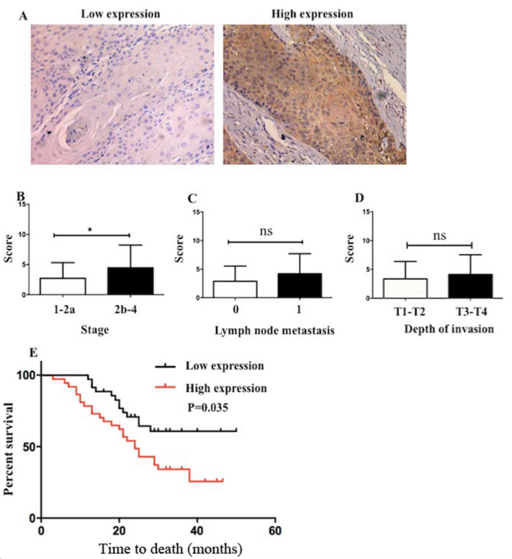
Msi1 expression is correlated with
clinical stage and lymphatic metastasis in patients with ESCC
Msi1 is considered as a cancer stem cell marker in
pulmonary, colorectal and breast cancer, and it regulates the
tumorigenesis in tumor models. We compared the expression of Msi1
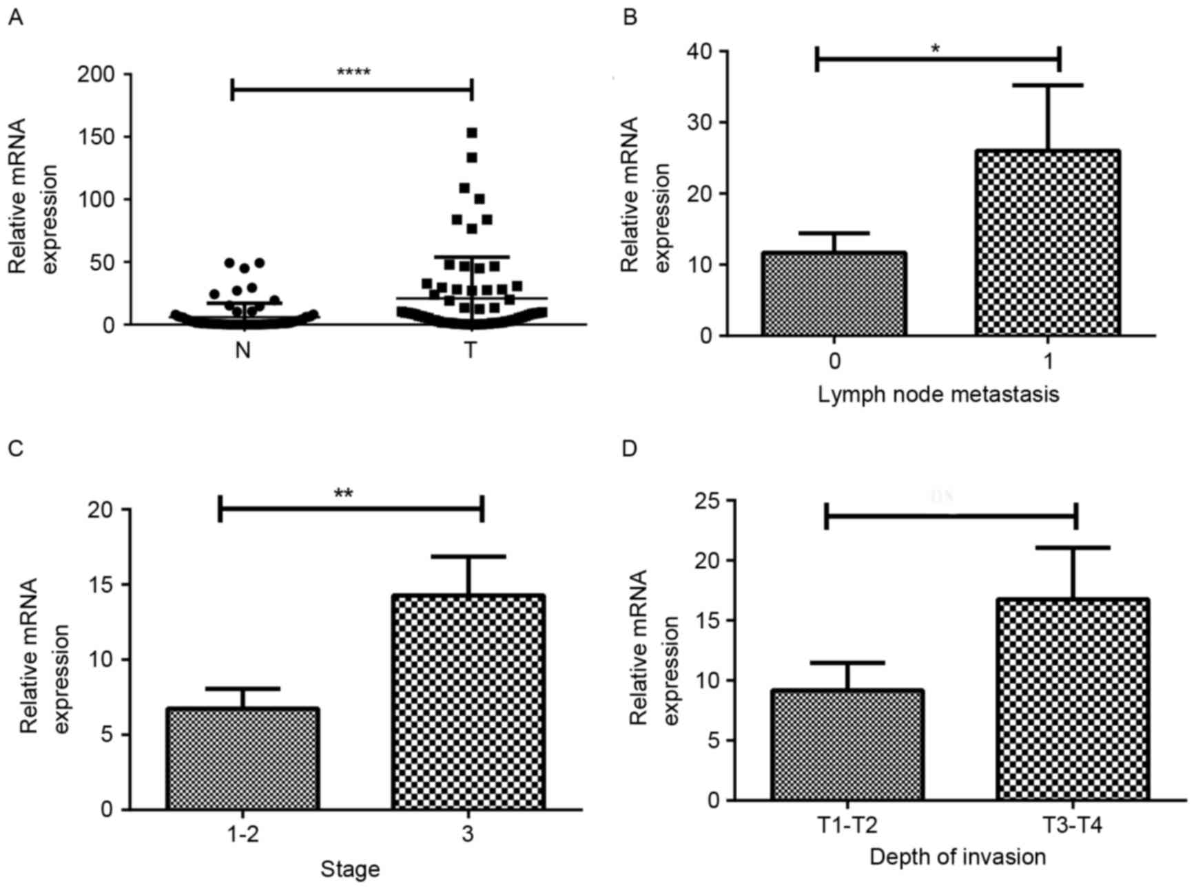
in tumor and adjacent non-cancerous tissues. As shown in Fig. 1A, Msi1 was found to be significantly
upregulated in cancer tissues when compared with the corresponding
non-tumor tissues (P<0.0001, n=67). We hypothesized that Msi1
plays a critical role in the progression of ESCC. In addition, we
found that there was a higher expression of Msi1 in the samples
from the patients suffering from lymph node metastasis or
advanced-stage ESCC (Fig. 1B,
P=0.04; Fig. 1C, P=0.005). However,
there was no statistical significance in the depth of invasion
(Fig. 1D). On the whole, the
aforementioned results revealed that Msi1 may be an oncogene and
act as a prognostic marker in ESCC patients.
Msi1 expression is enriched in
esophageal cancer stem cells
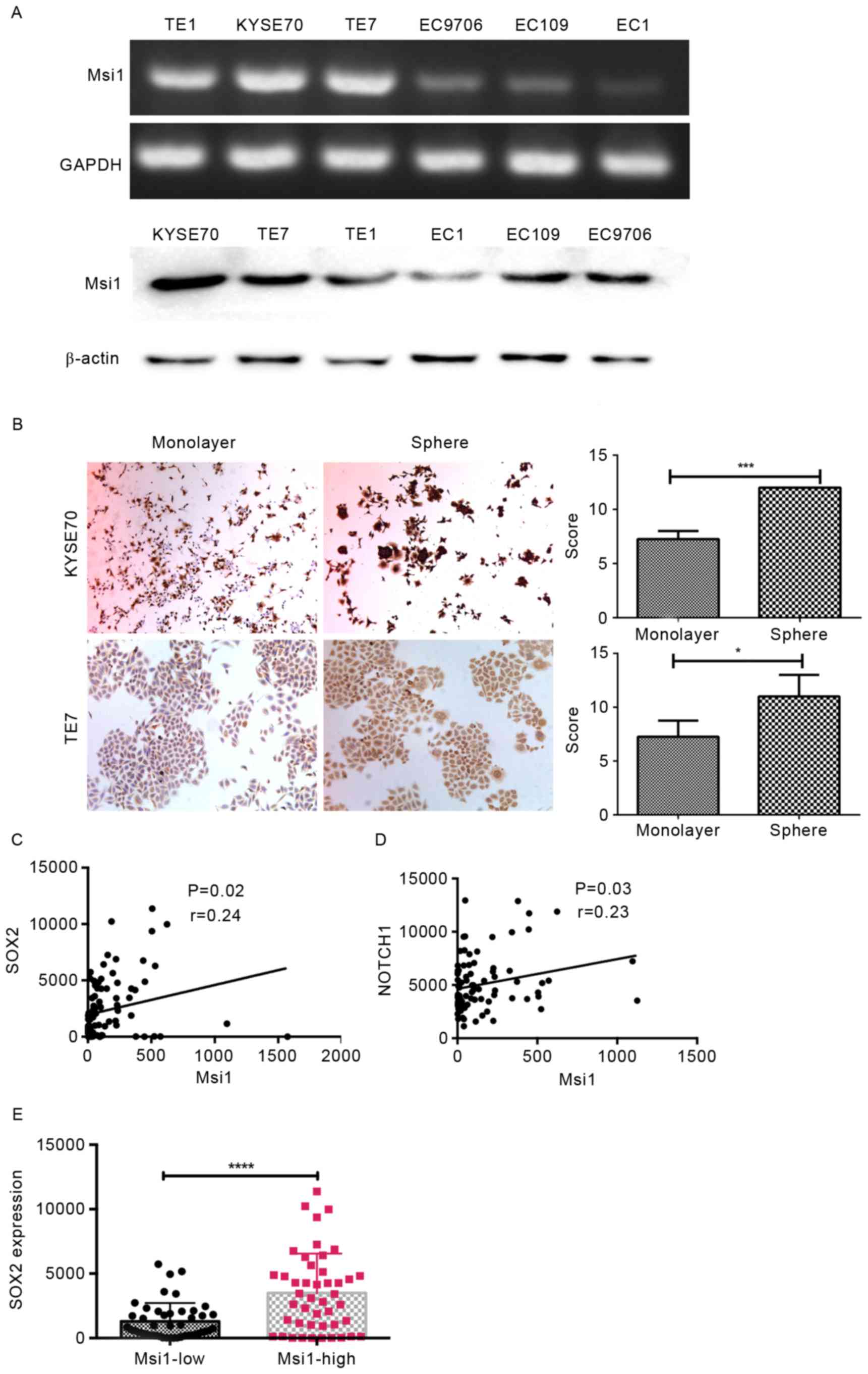
Msi1 had a higher expression in the poorly
differentiated ESCC cell lines TE-7, TE-1 and KYSE70 than in the
terminally differentiated cell lines EC109, EC9706 and EC-1
(Fig. 2A). Immunocytochemical
staining revealed that Msi1 was strongly positive in spheroid
cells, in TE-7 and KYSE70 cells when compared to the monolayer
(Fig. 2B, P=0.03 and P=0.02,
respectively). In addition, from TCGA analysis, we determined that
Msi1 expression had a significant positive correlation with SOX2
and Notch1 (Fig. 2C, P=0.02;
Fig. 2D, P=0.03) and that SOX2 had
a significantly different expression in the Msi1 high-expression
group compared with the low-expression group (Fig. 2E, P<0.0001). The latter two genes
were the key factors regulating cancer cell stemness. Moreover,
there was no similar phenomenon between the expression of Msi1 and
Notch1 (data not shown). According to previous studies and the
aforementioned results, we hypothesized that Msi1 plays a critical
role in ESCC stemness and its characteristics could have an effect
on the proliferation and apoptosis of cancer cells.
Decrease of Msi1 expression inhibits
esophageal cancer cell proliferation and promotes apoptosis
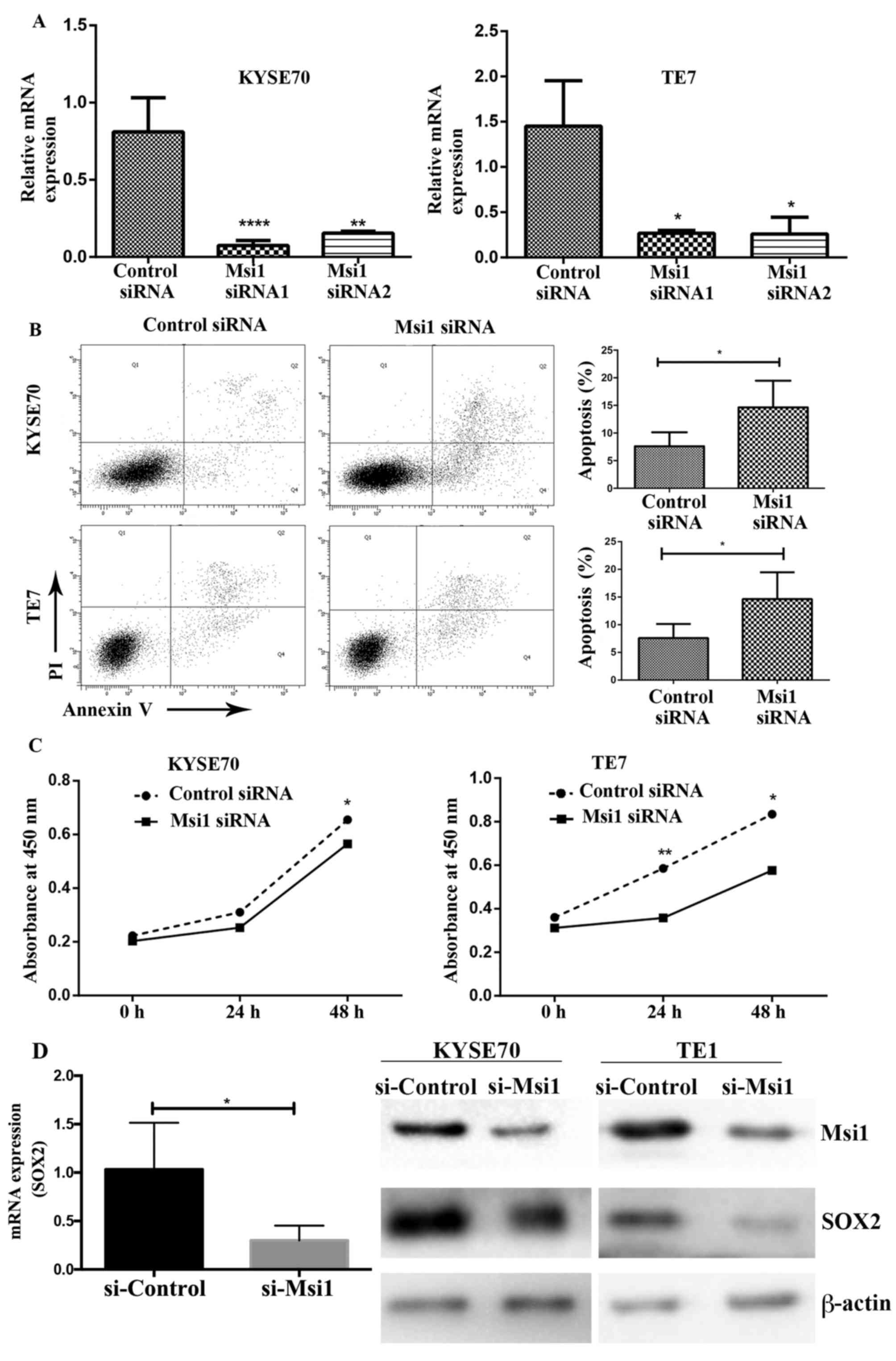
The role of Msi1 in the proliferation and apoptosis
of TE-7 and KYSE70 cells was examined following the transfection of
an Msi1 siRNA. RNA interference resulted in the decrease of Msi1
mRNA in TE-7 and KYSE70 cells compared to the control siRNA
(Fig. 3A). After transfection for
48 h, we collected the cells and detected apoptosis using flow
cytometry. Notably, we observed that knockdown of Msi1 increased
the apoptosis rates in TE-7 and KYSE70 cells (Fig. 3B). In addition, the proliferation of
TE-7 and KYSE70 cells transfected with Msi1-siRNA was observably
lower than that of the control group (Fig. 3C). Using PCR and western blotting,
we further determined that SOX2 expression was also decreased with
Msi1 interference (Fig. 3D).
Generally, Msi1 expression could promote the proliferation and
decrease the apoptosis of esophageal cancer cell lines.
Capacity of sphere formation and
migration is inhibited in Msi1 downregulated ESCC cells
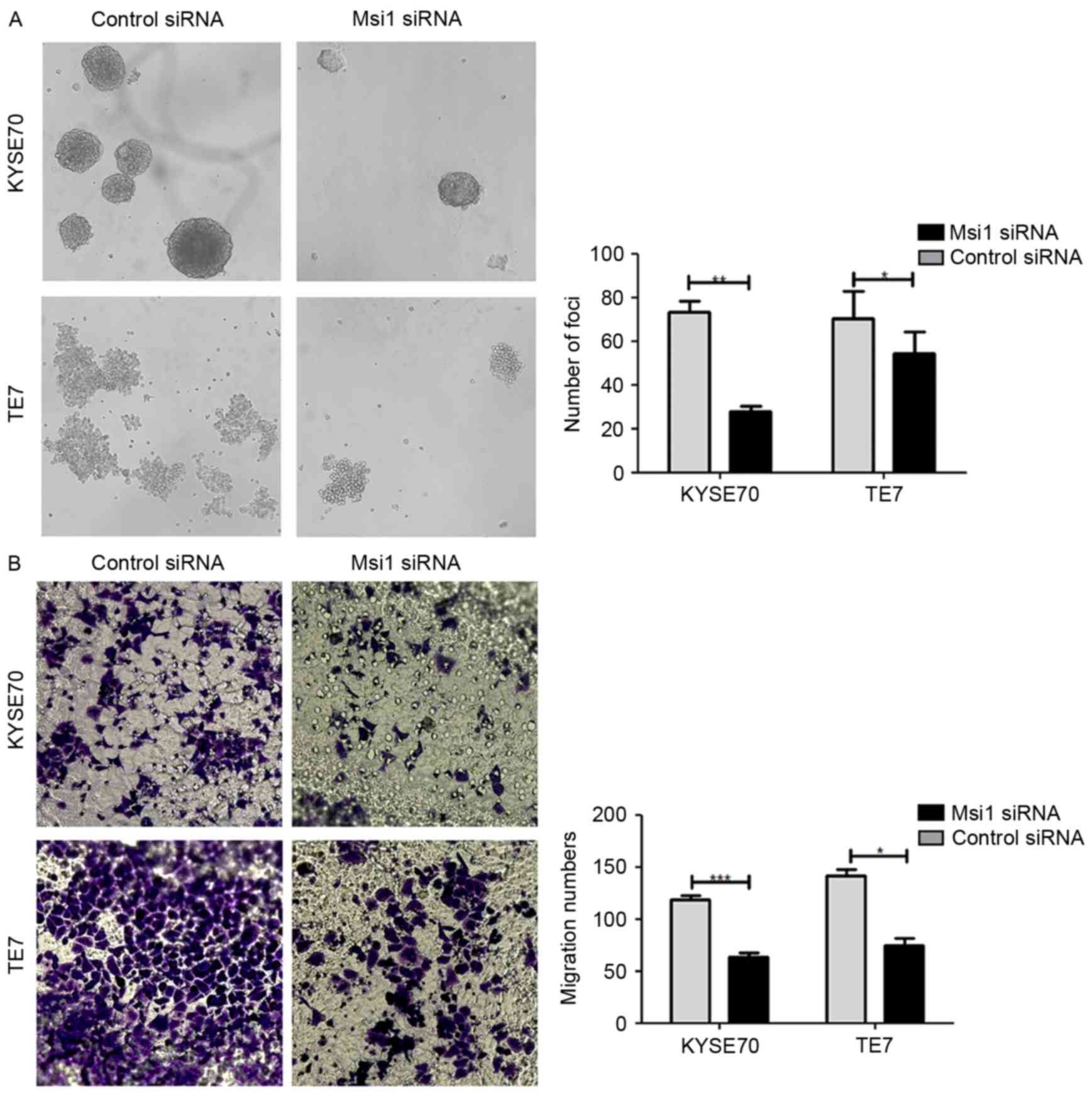
Using the same approach, we detected the influence
of knocked down Msi1 expression on the capacity of sphere formation
and migration in ESCC cells. We determined that in the si-Msi1
group the number of spheres formation was markedly decreased
(Fig. 4A). Similarly, with
Transwell assay, KYSE70 and TE-7 cells transfected with si-Msil
lacked of the ability to migrate to the lower chamber (Fig. 4B). The data demonstrated that Msi1
had an effect on tumor cell stemness and migration capacity.
Msi1 expression in ESCC tissues is
negatively correlated with the overall survival
To further evaluate the relationship of Msi1 with
the prognosis in ESCC, we investigated the protein expression of
Msi1 in ESCC samples by immunohistochemistry. As shown in Fig. 5A, Msi1 had a varying expression,
with patient tissues exhibiting high expression and low expression.
At the protein level, we also found that Msi1 was correlated with
late-stage ESCC. However, there was no statistical significance
with different lymph node metastasis and invasion of depth
(Fig. 5C-D). According to the
expression of Msi1 in tumor tissues, we divided the 93 patients
into two groups. There were 53 patients that had a high expression
of the stemness-associated gene Msi1, and the rest (40 patients)
had a low expression of Msi1 in tumor tissues. Moreover, compared
with the low expression group, the high expression group had a
significantly shorter overall survival (Fig. 5E; P=0.035).
Discussion
Despite the significant progress in ESCC diagnosis
and treatment, the dismal 5-year survival rate of ESCC has not
changed satisfyingly. Hence, the identification of important
molecules to assist in an early diagnosis and treatment targets of
ESCC are essential. Furthermore, with CSCs or TICs persisting in
tumor development, cancer patients cannot acquire anticipatory
prognosis after treatment with surgery, chemotherapy, radiotherapy
or combination therapy. Thus, it is necessary to identify the
pivotal point in ESCC development in order to target CSCs or
TICs.
Msi1, a regulatory factor in the process of
progenitor division, has also been identified as a regulator of
memory. Both reported physiological functions are relevant in
keeping the stemness of initial cells (14). Recently, an increasing number of
results in research revealed that Msi1 plays a significant role in
cancer development. In clinical samples, overexpression of Msi1 was
correlated with tumor-node-metastasis (TNM) stage and lymph node
metastasis. With Msi1 overexpression in tumor tissues, patients had
a decreased progression-free survival and overall survival.
Analogous results have been reported in lung, gastric and
gallbladder cancer, and colorectal carcinoma. Moreover, in
metastatic colorectal cancer cells it was demonstrated that NOTCH3
signaling regulated Msi1 expression, and played a key role in
decreasing the activity of NUMB thus activating DLL4 or NOTCH1 and
regulating of cancer cells (30).
As an important upstream molecule of NOTCH, it has also been
implicated in lung cancer. It has also been described as having a
similar role to ALDH1, the widely accepted cancer
stemness-associated gene in cancer development. In addition, a
previous study demonstrated that Msi1 promotes tumor growth by
targeting cell cycle checkpoint proteins p21, p27 and p53 (31).
Relevant studies revealed that CD271, CD133 and CD90
affected CSC development in ESCC, however, we are the first to
propose that Msi1, a well known molecule regulating cell division,
plays a significant role in ESCC development by promoting
proliferation and decreasing apoptosis of cancer cells. Firstly, as
revealed in other studies, Msi1 had a higher expression in ESCC
tumor tissues compared with adjacent non-cancerous tissues and its
distribution was also more diffuse (32,33).
As suggested in gastric cancer (34), due to the difference in
proliferation patterns between cancer cells and normal cells, Msi1
had a different expression in cancer and normal matched tissues.
Furthermore, we determined a negative correlation between Msi1
expression in tumor tissues and lymph node metastasis and TNM
stage. At the mRNA level, the patients with advanced-stage ESCC had
a higher expression of Msi1 than early-stage patients, and the
clinical samples with lymph node metastasis also exhibited a higher
expression of Msi1 compared with the non-metastatic samples. We
further investigated the expression patterns of Msi1 using
immunohistochemistry. As determined, tumor tissues exhibited a
higher expression of Msi1 compared to adjacent non-cancerous
tissues. Moreover advanced-stage ESCC tissues had an increased
expression of Msi1 when compared to ealy-stage patient tissues.
Patients with lymph node metastasis also had a higher expression of
Msi1, but the difference in expression with the non-metastatic
group was not statistically significant.
To further clarify the importance of Msi1 in tumor
development, we conducted a series of experiments in vitro
using various ESCC cell lines. Compared with TE-1, EC109, EC9706
and EC1, the poorly differentiated TE-7 and KYSE70 cell lines had a
higher expression of Msi1. We suspected that Msi1 was related to
cancer histological grade. Upon investigation this relationship was
not substantiated in the clinical specimens we observed due to the
diffuse expression patterns of Msi1, and therefore we may further
probe the expression of Msi1 in more ESCC patients. Notably, the
spheroid cells when compared with the monolayer ones, exhibited a
marked higher expression of Msi1. To the best of our knowledge,
sphere formation is an effective approach to acquire CSC-like
cells. Therefore, we believe that Msi1 can maintain the stemness of
ESCC cancer cells. Consistent with other studies, using TCGA
database, we found that Msi1 had a positive correlation with SOX2
in esophageal cancer (35). With
the interference of RNA, we knocked down the expression of Msi1 in
KYSE70 and TE-7 cells effectively. Then, significant changes were
observed in the cancer cells. There was a marked increase in
apoptosis in the si-Msi1 group and a decrease in proliferation. In
addition, si-Msi1 had an effect on sphere formation and migration
capacity. These results were similar to other types of cancer
investigated in previous studies (24,30,31).
Thus in clinical application, Msi1 can be considered as a target in
the prevention of the malignant biological behavior of tumor cells
and the improvement of the prognosis of cancer patients.
Furthermore, in future we need to construct an
animal model to demonstrate the impact of Msi1 on the development
of ESCC. In relation to the underlying mechanisms, in addition to
NOTCH signaling as previously aforementioned, we may investigate
the relationship with other cancer stemness-associated genes. We
may also probe the stemness-associated genes regulating the tumor
microenvironment as KLF4 which was studied in breast cancer
(36).
In conclusion, the present study illustrated that
Msi1 regulated the proliferation, apoptosis, sphere formation and
migration capacity of cancer cells, and had a higher expression in
spheroid cells. All the experimental results support that Msi1 may
act as a potential prognostic marker in patients with ESCC.
Furthermore, it could be used as a diagnostic and prognostic marker
in clinical conversions.
Acknowledgements
The present study was supported by grants from the
China-US (NFSC-NIH) Program for Biomedical Collaborative Research
(grant no. 812111102), and the National Natural Science Foundation
of China (grant no. 81171986).
References
|
1
|
Kamangar F, Dores GM and Anderson WF:
Patterns of cancer incidence, mortality, and prevalence across five
continents: Defining priorities to reduce cancer disparities in
different geographic regions of the world. J Clin Oncol.
24:2137–2150. 2006. View Article : Google Scholar : PubMed/NCBI
|
|
2
|
Song Y, Li L, Ou Y, Gao Z, Li E, Li X,
Zhang W, Wang J, Xu L, Zhou Y, et al: Identification of genomic
alterations in oesophageal squamous cell cancer. Nature. 509:91–95.
2014. View Article : Google Scholar : PubMed/NCBI
|
|
3
|
Visvader JE and Lindeman GJ: Cancer stem
cells in solid tumours: Accumulating evidence and unresolved
questions. Nat Rev Cancer. 8:755–768. 2008. View Article : Google Scholar : PubMed/NCBI
|
|
4
|
Singh SK, Clarke ID, Terasaki M, Bonn VE,
Hawkins C, Squire J and Dirks PB: Identification of a cancer stem
cell in human brain tumors. Cancer Res. 63:5821–5828.
2003.PubMed/NCBI
|
|
5
|
Ponti D, Costa A, Zaffaroni N, Pratesi G,
Petrangolini G, Coradini D, Pilotti S, Pierotti MA and Daidone MG:
Isolation and in vitro propagation of tumorigenic breast cancer
cells with stem/progenitor cell properties. Cancer Res.
65:5506–5511. 2005. View Article : Google Scholar : PubMed/NCBI
|
|
6
|
Patrawala L, Calhoun T,
Schneider-Broussard R, Li H, Bhatia B, Tang S, Reilly JG, Chandra
D, Zhou J, Claypool K, et al: Highly purified CD44+
prostate cancer cells from xenograft human tumors are enriched in
tumorigenic and metastatic progenitor cells. Oncogene.
25:1696–1708. 2006. View Article : Google Scholar : PubMed/NCBI
|
|
7
|
Ricci-Vitiani L, Lombardi DG, Pilozzi E,
Biffoni M, Todaro M, Peschle C and De Maria R: Identification and
expansion of human colon-cancer-initiating cells. Nature.
445:111–115. 2007. View Article : Google Scholar : PubMed/NCBI
|
|
8
|
Li C, Heidt DG, Dalerba P, Burant CF,
Zhang L, Adsay V, Wicha M, Clarke MF and Simeone DM: Identification
of pancreatic cancer stem cells. Cancer Res. 67:1030–1037. 2007.
View Article : Google Scholar : PubMed/NCBI
|
|
9
|
Eramo A, Lotti F, Sette G, Pilozzi E,
Biffoni M, Di Virgilio A, Conticello C, Ruco L, Peschle C and De
Maria R: Identification and expansion of the tumorigenic lung
cancer stem cell population. Cell Death Differ. 15:504–514. 2008.
View Article : Google Scholar : PubMed/NCBI
|
|
10
|
Tang KH, Dai YD, Tong M, Chan YP, Kwan PS,
Fu L, Qin YR, Tsao SW, Lung HL, Lung ML, et al: A CD90+
tumor-initiating cell population with an aggressive signature and
metastatic capacity in esophageal cancer. Cancer Res. 73:2322–2332.
2013. View Article : Google Scholar : PubMed/NCBI
|
|
11
|
Li S, Yue D, Chen X, Wang L, Li J, Ping Y,
Gao Q, Wang D, Zhang T, Li F, et al: Epigenetic regulation of
CD271, a potential cancer stem cell marker associated with
chemoresistance and metastatic capacity. Oncol Rep. 33:425–432.
2015.PubMed/NCBI
|
|
12
|
Huang SD, Yuan Y, Liu XH, Gong DJ, Bai CG,
Wang F, Luo JH and Xu ZY: Self-renewal and chemotherapy resistance
of p75NTR positive cells in esophageal squamous cell
carcinomas. BMC Cancer. 9:92009. View Article : Google Scholar : PubMed/NCBI
|
|
13
|
Okano H, Kawahara H, Toriya M, Nakao K,
Shibata S and Imai T: Function of RNA-binding protein Musashi-1 in
stem cells. Exp Cell Res. 306:349–356. 2005. View Article : Google Scholar : PubMed/NCBI
|
|
14
|
Hadziselimovic N, Vukojevic V, Peter F,
Milnik A, Fastenrath M, Fenyves BG, Hieber P, Demougin P, Vogler C,
de Quervain DJ, et al: Forgetting is regulated via Musashi-mediated
translational control of the Arp2/3 complex. Cell. 156:1153–1166.
2014. View Article : Google Scholar : PubMed/NCBI
|
|
15
|
Wang XY, Yu H, Linnoila RI, Li L, Li D, Mo
B, Okano H, Penalva LO and Glazer RI: Musashi1 as a potential
therapeutic target and diagnostic marker for lung cancer.
Oncotarget. 4:739–750. 2013. View Article : Google Scholar : PubMed/NCBI
|
|
16
|
Simon E, Petke D, Böger C, Behrens HM,
Warneke V, Ebert M and Röcken C: The spatial distribution of
LGR5+ cells correlates with gastric cancer progression.
PLoS One. 7:e354862012. View Article : Google Scholar : PubMed/NCBI
|
|
17
|
Rezza A, Skah S, Roche C, Nadjar J,
Samarut J and Plateroti M: The overexpression of the putative gut
stem cell marker Musashi-1 induces tumorigenesis through Wnt and
Notch activation. J Cell Sci. 123:3256–3265. 2010. View Article : Google Scholar : PubMed/NCBI
|
|
18
|
Li N, Yousefi M, Nakauka-Ddamba A, Li F,
Vandivier L, Parada K, Woo DH, Wang S, Naqvi AS, Rao S, et al: The
Msi family of RNA-binding proteins function redundantly as
intestinal oncoproteins. Cell Reports. 13:2440–2455. 2015.
View Article : Google Scholar : PubMed/NCBI
|
|
19
|
Götte M, Greve B, Kelsch R, Müller-Uthoff
H, Weiss K, Masouleh B Kharabi, Sibrowski W, Kiesel L and Buchweitz
O: The adult stem cell marker Musashi-1 modulates endometrial
carcinoma cell cycle progression and apoptosis via Notch-1 and
p21WAF1/CIP1. Int J Cancer. 129:2042–2049. 2011.
View Article : Google Scholar : PubMed/NCBI
|
|
20
|
Wang XY, Penalva LO, Yuan H, Linnoila RI,
Lu J, Okano H and Glazer RI: Musashi1 regulates breast tumor cell
proliferation and is a prognostic indicator of poor survival. Mol
Cancer. 9:2212010. View Article : Google Scholar : PubMed/NCBI
|
|
21
|
Liu DC, Yang ZL and Jiang S:
Identification of musashi-1 and ALDH1 as carcinogenesis,
progression, and poor-prognosis related biomarkers for gallbladder
adenocarcinoma. Cancer Biomark. 8:113–121. 2010.2011. View Article : Google Scholar
|
|
22
|
Hou T, Zhang W, Tong C, Kazobinka G, Huang
X, Huang Y and Zhang Y: Putative stem cell markers in cervical
squamous cell carcinoma are correlated with poor clinical outcome.
BMC Cancer. 15:7852015. View Article : Google Scholar : PubMed/NCBI
|
|
23
|
Fox RG, Lytle NK, Jaquish DV, Park FD, Ito
T, Bajaj J, Koechlein CS, Zimdahl B, Yano M, Kopp JL, et al:
Image-based detection and targeting of therapy resistance in
pancreatic adenocarcinoma. Nature. 534:407–411. 2016. View Article : Google Scholar : PubMed/NCBI
|
|
24
|
Chen HY, Lin LT, Wang ML, Lee SH, Tsai ML,
Tsai CC, Liu WH, Chen TC, Yang YP, Lee YY, et al: Musashi-1
regulates AKT-derived IL-6 autocrinal/paracrinal malignancy and
chemoresistance in glioblastoma. Oncotarget. 7:42485–42501. 2016.
View Article : Google Scholar : PubMed/NCBI
|
|
25
|
Moghbeli M, Rad A, Farshchian M,
Taghehchian N, Gholamin M and Abbaszadegan MR: Correlation between
Meis1 and Msi1 in esophageal squamous cell carcinoma. J
Gastrointest Cancer. 47:273–277. 2016. View Article : Google Scholar : PubMed/NCBI
|
|
26
|
Livak KJ and Schmittgen TD: Analysis of
relative gene expression data using real-time quantitative PCR and
the 2−ΔΔCT method. Methods. 25:402–408. 2001.
View Article : Google Scholar : PubMed/NCBI
|
|
27
|
McDonald JW and Pilgram TK: Nuclear
expression of p53, p21 and cyclin D1 is increased in
bronchioloalveolar carcinoma. Histopathology. 34:439–446. 1999.
View Article : Google Scholar : PubMed/NCBI
|
|
28
|
Xue LY, Hu N, Song YM, Zou SM, Shou JZ,
Qian LX, Ren LQ, Lin DM, Tong T, He ZG, et al: Tissue microarray
analysis reveals a tight correlation between protein expression
pattern and progression of esophageal squamous cell carcinoma. BMC
Cancer. 6:2962006. View Article : Google Scholar : PubMed/NCBI
|
|
29
|
Langan RC, Mullinax JE, Ray S, Raiji MT,
Schaub N, Xin HW, Koizumi T, Steinberg SM, Anderson A, Wiegand G,
et al: A pilot study assessing the potential role of non-CD133
colorectal cancer stem cells as biomarkers. J Cancer. 3:231–240.
2012. View
Article : Google Scholar : PubMed/NCBI
|
|
30
|
Pastò A, Serafin V, Pilotto G, Lago C,
Bellio C, Trusolino L, Bertotti A, Hoey T, Plateroti M, Esposito G,
et al: NOTCH3 signaling regulates MUSASHI-1 expression in
metastatic colorectal cancer cells. Cancer Res. 74:2106–2118. 2014.
View Article : Google Scholar : PubMed/NCBI
|
|
31
|
Liu X, Yang WT and Zheng PS: Msi1 promotes
tumor growth and cell proliferation by targeting cell cycle
checkpoint proteins p21, p27 and p53 in cervical carcinomas.
Oncotarget. 5:10870–10885. 2014. View Article : Google Scholar : PubMed/NCBI
|
|
32
|
Moghbeli M, Forghanifard MM, Sadrizadeh A,
Mozaffari HM, Golmakani E and Abbaszadegan MR: Role of Msi1 and
MAML1 in regulation of Notch signaling pathway in patients with
esophageal squamous cell carcinoma. J Gastrointest Cancer.
46:365–369. 2015. View Article : Google Scholar : PubMed/NCBI
|
|
33
|
Moghbeli M, Sadrizadeh A, Forghanifard MM,
Mozaffari HM, Golmakani E and Abbaszadegan MR: Role of Msi1 and
PYGO2 in esophageal squamous cell carcinoma depth of invasion. J
Cell Commun Signal. 10:49–53. 2016. View Article : Google Scholar : PubMed/NCBI
|
|
34
|
Kuang RG, Kuang Y, Luo QF, Zhou CJ, Ji R
and Wang JW: Expression and significance of Musashi-1 in gastric
cancer and precancerous lesions. World J Gastroenterol.
19:6637–6644. 2013. View Article : Google Scholar : PubMed/NCBI
|
|
35
|
Ying M, Wang S, Sang Y, Sun P, Lal B,
Goodwin CR, Guerrero-Cazares H, Quinones-Hinojosa A, Laterra J and
Xia S: Regulation of glioblastoma stem cells by retinoic acid: Role
for Notch pathway inhibition. Oncogene. 30:3454–3467. 2011.
View Article : Google Scholar : PubMed/NCBI
|
|
36
|
Yu F, Shi Y, Wang J, Li J, Fan D and Ai W:
Deficiency of Kruppel-like factor KLF4 in mammary tumor cells
inhibits tumor growth and pulmonary metastasis and is accompanied
by compromised recruitment of myeloid-derived suppressor cells. Int
J Cancer. 133:2872–2883. 2013.PubMed/NCBI
|