Introduction
Bladder cancer is one of the most common
genitourinary malignancies arising from the epithelial lining of
the urinary bladder. In China, the incidence and mortality rate
have been rapidly increasing in the last few decades (1,2).
Tobacco use, Schistosoma infection, chemical exposure, diet
and lifestyle trends and genetic susceptibility have been reported
to be risk factors for the tumorigenesis and progression of bladder
cancer (3–5). Standard treatment for bladder cancer
patients includes surgical resection or adjuvant chemotherapy and
radiation. However, despite advances in diagnosis and therapy,
there has been no improvement in the overall survival rate for
bladder cancer patients in recent decades. Therefore,
identification of novel molecular markers is critical to refining
our understanding of the pathogenesis of bladder cancer and for
developing more efficient treatment and surveillance
strategies.
The transient receptor potential (TRP) channel
family includes biological transmembrane proteins that play an
important role in various physiologic and pathologic processes by
modulating cytoplasmic signaling and cellular responses (6). The melastatin transient receptor
potential channel 7 (TRPM7), a member of the TRP channel family,
functions as a non-selective cation channel and a protein kinase
(7,8). Recent studies have reported that TRPM7
is highly expressed in various types of cancers such as breast,
ovarian and prostate cancer, glioblastoma and pancreatic cancer
(9–13). Accumulating evidence has also shown
that TRPM7 plays an important role in malignant progression
including the regulation of cell proliferation, adhesion,
apoptosis, gene expression, cell migration and metastasis (14–19).
It has been found that TRPM7 regulates cancer cell proliferation
and migration mainly by the channel activity mediating influxes of
both Ca2+ and Mg2+ (20,21).
Moreover, regulation of cell adhesion by TRPM7 includes the
combined effect of Ca2+ and kinase-dependent pathways on
actomyosin contractility (22).
However, the involvement of TRPM7 in the pathogenesis and
progression of bladder cancer remains to be detected.
The aim of the present study was to characterize the
expression and biological role of TRPM7 in bladder cancer. We found
that TRPM7 was overexpressed in bladder cancer. In addition, TRPM7
was strongly correlated with clinicopathological characteristics
and poor survival rates in bladder cancer. Furthermore, the effect
of TRPM7 on the biological behaviors of bladder cancer was
investigated by anti-TRPM7 small interfering RNA (siRNA) assays.
The data revealed that TRPM7 promoted bladder cancer proliferation,
migration and invasion.
Materials and methods
Tissue specimens
Bladder cancer and paired adjacent normal bladder
tissues used for immunohistochemistry were collected from 74
bladder cancer patients who underwent surgical resection in The
First Affiliated Hospital of China Medical University from 2008 to
2015. None of patients underwent chemotherapy, radiotherapy or
adjuvant treatment before surgery. The present study was approved
by the Ethics Committee of the China Medical University. Informed
consent was obtained from all patients. Twenty pairs of fresh
bladder cancer and adjacent non-tumor bladder tissues used for
quantitative PCR were collected from The First Affiliated Hospital
of China Medical University and snap frozen in liquid nitrogen
until use. The patients had not received any therapy before
admission.
Immunohistochemical staining
Immunohistochemistry for TRPM7 expression in bladder
cancer tissues was performed using standard methods. Sections were
deparaffinized in xylene and hydrated in a graded ethanol series.
The sections were then processed in 10 mmol/l citrate buffer (pH
6.0) and heated at 120°C for 5 min to retrieve the antigen. After
that, sections were soaking in 3% hydrogen peroxide for 20 min,
which served as a blocking agent for endogenous peroxidase
activity. After being rinsed in phosphate-buffered saline (PBS; pH
7.2), 10% goat serum was applied for 1 h at room temperature to
block non-specific reactions. Sections were incubated with
anti-TRPM7 goat polyclonal antibody (diluted 1:50; ab729; Abcam,
Cambridge, MA, USA) overnight at 4°C. Horseradish
peroxidase-conjugated anti-goat IgG was used as a secondary
antibody. After washing, the peroxidase reaction was developed with
3,3′-diaminobenzidine tetrahydrochloride (DAB) chromogen solution.
Finally, the sections were counterstained with hematoxylin,
dehydrated and coverslipped. All the slides were evaluated
independently by two pathologists using a conventional
semi-quantitative scoring system according to a previously defined
scoring system (16). Briefly, the
intensity of the staining in each section was assessed as very
strongly positive (3), moderately
positive (2), weakly positive
(1), or negative (0). The
percentage of positive tumor cells were scored as <5% (0), 5–20%
(1), 21–50% (2) and >50% (3) of cells. The final score was calculated
by multiplying the percentage and the intensity score. A score of
≥4 was defined as high TRPM7 expression and scores of <4 were
defined as low TRPM7 expression.
Cell culture and siRNA
transfection
Human bladder cancer cell lines (BIU87, 5637 and
T24) were cultured in RPMI-1640 medium with 10% fetal bovine serum
(FBS) (both from HyClone, Logan, UT, USA) at 37°C in 5%
CO2 incubator. We obtained TRPM7 siRNA and the negative
control siRNA (NC_siR) from GenePharma Co. Ltd. (Shanghai, China).
Before transfection, cells were plated overnight and grew to 30–50%
confluency. According to the manufacturer's instructions, the
siRNAs were transfected into 5637 and T24 cells using
Lipofectamine™ 3000 (Invitrogen, Carlsbad, CA, USA). The sequences
of siRNAs are shown in Table I. The
efficiency of siRNA was determined by the protein and mRNA levels
of TRPM7 in the 48-h post transfected cells.
 | Table I.Sequences of the primer pairs for
real-time RT-PCR and siRNAs targeting TRPM7. |
Table I.
Sequences of the primer pairs for
real-time RT-PCR and siRNAs targeting TRPM7.
| RNA names | Sequences (5–3′) |
|---|
| TRPM7 | F
5′-TAGCCTTTAGCCACTGGAC-3′ |
|
| R
5′-GCATCTTCTCCTAGATTTGC-3′ |
| GAPDH | F
5′-GAAGGTGAAGGTCGGAGTC-3′ |
|
| R
5′-GAAGATGGTGATGGGATTTC-3′ |
| TRPM7_siR 1 | S
GCGCUUUCCUUAUCCACUUTT |
|
| A
AAUGGAUAAGGAAAGCGCTT |
| TRPM7_siR 2 | S
CCAUAUCCCACAAUCUCAATT |
|
| A
UUGAGAUUGUGGGAUAUGGTT |
| TRPM7_siR 3 | S
GGUGUUCCCAGAAAGGCAATT |
|
| A
UUGCCUUUCUGGGAACACCTT |
| NC_siR | S
UUCUCCGAACGUGUCACGUTT |
|
| A
ACGUGACACGUUCGGAGAATT |
Immunofluorescence staining
BIU87, 5637 and T24 cells (5×10⁴
cells/ml) were seeded on 12-mm coverslips for 24 h, fixed with 4%
paraformaldehyde for 30 min, and then permeabilized for 20 min with
0.1% Triton X-100 solution and blocked in normal bovine serum for
30 min at room temperature. Cells were incubated overnight at 4°C
with anti-TRPM7 (1:50; Abcam) antibody in 2% bovine serum albumin
(BSA; BioShop, Burlington, ON, Canada), 2% FBS and 0.2% fish
gelatin (Sigma-Aldrich, St. Louis, MO, USA). The cells were then
incubated with FITC-labeled secondary antibody for 2 h at room
temperature. Nuclei were labeled with 4,6-diamidino-2-phenylindole
(DAPI) (2 µg/ml). Fluorescence was visualized with a confocal
microscope (Carl Zeiss Inc., Gottingen, Germany).
Quantitative real-time PCR
Total RNA was isolated from bladder cancer and
adjacent non-tumor bladder tissues with TRIzol reagent
(Invitrogen). cDNA was synthesized using a Transcriptor First
Strand cDNA Synthesis kit (Roche, Mannheim, Germany). Then,
SYBR-Green Real-Time PCR Master Mix (Applied Biosystems, Foster
City, CA, USA) was used. Real-time PCR was performed using Thermal
Cycler Dice™ Real-Time System TP800 (Takara, Tokyo, Japan).
Amplification conditions consisted of 2 min at 50°C for reverse
transcription, 5 min at 95°C for Taq activation followed by
45 cycles of 94°C for 40 sec, one cycle of 58°C for 20 sec and
elongation at 72°C for 30 sec. The sequence of primers designed for
TRPM7 and GAPDH are listed in Table
I. GAPDH served as the internal control for mRNA determination
of TRPM7. Results were normalized to GAPDH. The relative gene
expression was calculated using 2−ΔΔCt.
Western blot analysis
Cells were lysed by in ice-cold cell RIPA buffer.
Protein concentration was measured using the BCA protein assay kit
(Pierce, Rockford, IL, USA). Total cellular protein (30 µg) was
separated using sodium dodecyl sulfate-polyacrylamide gel
electrophoresis (SDS-PAGE) and transferred onto polyvinylidene
difluoride (PVDF) filter membranes (Millipore, Bedford, MA, USA).
The membrane was blocked with 5% non-fat milk in Tris-buffered
saline with Tween-20 (TBST) buffer for 2 h at room temperature and
incubated overnight at 4°C with a specific primary antibody against
TRPM7 (ab85016; 1:500 dilution; Abcam) and mouse-anti-human actin
(1:2,000 dilution; Santa Cruz Biotechnology, Inc., Santa Cruz, CA,
USA) as an internal control. Next, the membranes were incubated at
room temperature for 1 h with horseradish peroxidase-conjugated
goat anti-mouse IgG (1:2,500 dilution; Santa Cruz Biotechnology,
Inc.), and signals were developed using Western Blotting Luminol
Reagent (Gene Company Ltd., Hong Kong).
Cell proliferation assays
Cell proliferation was determined using the Cell
Counting Kit-8 (CCK-8) assay (Dojindo, Tokyo, Japan). The cells
(3×10³ cells/well) were seeded into 96-well plates. At
time points of 0, 6, 12, 24, 36 and 48 h, 10 µl of CCK-8 was
added/well and was incubated at 37°C for 1 h. The absorbance was
measured at 450 nm using a plate reader (Model 680; Bio-Rad
Laboratories, Hercules, CA, USA) to determine the number of viable
cells. All of the experiments were performed three times.
Wound healing assay
Cells (5×10⁵ cells/well) were seeded into
6-well plates. After the cells were grown to 80% confluency, wounds
were created by scraping the cells with a 100-µl pipette tip. The
6-well plates were incubated at 37°C and a microscope was used to
observe the migrated distance every 12 h.
Transwell assay
The invasion and migration of cells were measured
using 8-µm pore-size Transwell chambers with or without Matrigel
(BD Biosciences, San Diego, CA, USA). Cells (1×10⁵
cells/well) were seeded into the top chamber with the serum-free
medium while the bottom chambers were filled with 500 µl complete
RPMI-1640 medium. After 24 h of incubation, the cells remaining on
the upper side were carefully removed with cotton swabs, and those
cells that had migrated to the lower side were fixed and stained
with 1.0% crystal violet. The cells were quantified by counting all
of the cells that had migrated through the membrane in five random
fields under a light microscope (magnification, ×200). The mean
value was calculated from data obtained from three separate
chambers.
Flow cytometric analysis for
alteration in apoptosis
For apoptosis analysis, after transfection for 48 h,
cells were collected, washed with PBS, and stained with
FITC/Annexin V Apoptosis Detection Kit I (BD Biosciences), and
analyzed by flow cytometric analysis.
Cell cycle analysis
Cells were trypsinized and washed in ice-cold PBS,
and then fixed in ice-cold 75% ethanol in PBS overnight. PI/RNase
staining buffer (BD Biosciences) was added, and the cells were
incubated at 4°C for 30 min. Cell cycle profiles were analyzed
using a FACSCalibur flow cytometer (BD Biosciences).
Calculation and statistical
analysis
All data are presented as means ± standard deviation
(SD) and analyzed using SPSS 21.0 statistical software (SPSS, Inc.,
Chicago, IL, USA). t-test was used to determine the significance of
differences in multiple comparisons. Survival curves were estimated
by Kaplan-Meier analysis and compared by the log-rank test. All
tests performed were two-sided. P<0.05 was considered
statistically significant.
Results
TRPM7 is overexpressed in bladder
cancer tissues
To characterize the expression of TRPM7 in human
bladder cancer tissues, we firstly examined the mRNA and protein
levels of TRPM7 in 20 pairs of human bladder cancer, and the
adjacent non-tumor bladder tissues by qRT-PCR and western blotting.
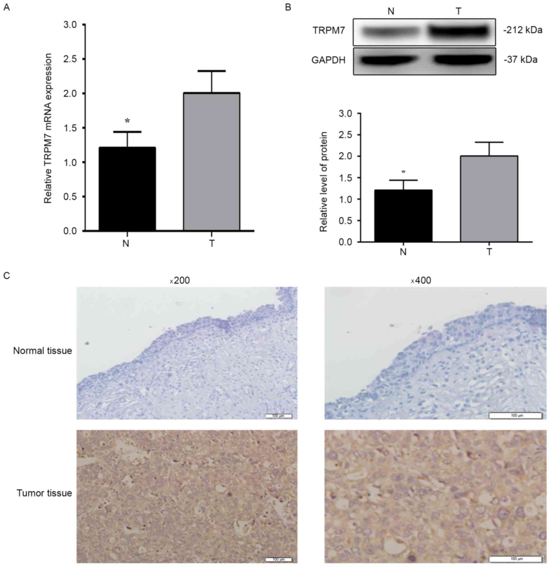
As shown in Fig. 1A and B, the mRNA
and protein levels of TRPM7 were significantly increased in the
bladder cancer tissues compared with levels in the paired adjacent
non-tumor bladder tissues (P<0.05). Next, we performed
immunohistochemical analysis to assess the protein expression and
subcellular localization of TRPM7 in 74 paraffin-embedded bladder
cancer tissues (Fig. 1C). High
TRPM7 expression was detected in 47 (63.5%) of the 74 bladder
cancer tissues and in 16 (25.80%) of the 62 adjacent non-tumor
tissues (P<0.05). These data suggest that TRPM7 is overexpressed
in bladder cancer tissues and may be a potential biomarker for
bladder cancer.
Relationship between TRPM7 expression
and clinicopathological variables in bladder cancer
To investigate the potential role of TRPM7 in
bladder cancer, the relationship between TRPM7 expression level and
clinicopathological factors was analyzed and is summarized in
Table II. As shown, TRPM7
overexpression was associated with recurrence (P<0.01), and
metastasis (P=0.021) of patients with bladder cancer. However,
TRPM7 exhibited no significant association with other
clinicopathological characteristics, such as age, sex, histologic
grade, tumor stage and multiplicity (all P>0.05).
| Table II.Relationship between the expression
of TRPM7 and clinicopathological factors in 74 bladder cancer
patients. |
Table II.
Relationship between the expression
of TRPM7 and clinicopathological factors in 74 bladder cancer
patients.
|
|
| TRPM7 expression,
n |
|
|---|
|
|
|
|
|
|---|
| Parameters | No. case | Low | High | P-value |
|---|
| Sex |
|
|
| 1.0 |
|
Male | 46 | 17 | 29 |
|
|
Female | 28 | 10 | 18 |
|
| Age, years |
|
|
| 0.432 |
|
<60 | 21 | 6 | 15 |
|
|
≥60 | 53 | 21 | 32 |
|
| Histological
grade |
|
|
| 0.226 |
|
Low | 45 | 19 | 26 |
|
|
High | 29 | 8 | 21 |
|
| Tumor stage |
|
|
| 0.333 |
|
Ta-T1 | 40 | 17 | 23 |
|
|
T2-T4 | 34 | 10 | 24 |
|
| Multiplicity |
|
|
| 0.054a |
|
Unifocal | 35 | 17 | 18 |
|
|
Multifocal | 39 | 10 | 29 |
|
| Recurrence |
|
|
|
<0.01a |
|
Yes | 47 | 3 | 44 |
|
| No | 27 | 24 | 3 |
|
| Metastasis |
|
|
| 0.021a |
|
Yes | 17 | 2 | 15 |
|
| No | 57 | 25 | 32 |
|
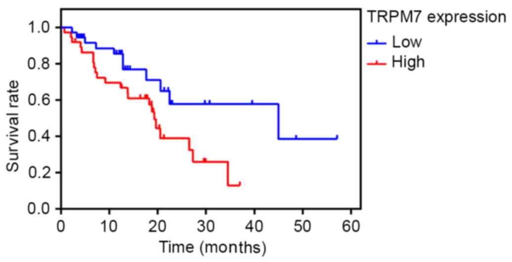
High TRPM7 expression predicts poor
prognosis of bladder cancer patients
The prognostic value of TRPM7 was evaluated by
Kaplan-Meier analysis. The survival curves indicated that high
TRPM7 expression was significantly associated with poor survival of
bladder cancer patients. Patients with TRPM7 high expression had
worse prognoses than those with low expression of TRPM7 (Fig. 2) (P<0.05).
Furthermore, univariate and multivariate Cox
regression analyses were applied to the clinicopathological
characteristics in regards to TRPM7 expression levels. As shown in
Table III, multiplicity
(P=0.017), recurrence (P<0.01), metastasis (P=0.049) and TRPM7
expression (P<0.01) were all significantly related to bladder
cancer patient poor survival. Multivariate analysis showed that
high TRPM7 expression was independently associated with the poor
prognosis of bladder cancer patients (P=0.035) (Table IV).
| Table III.Survival status and
clinicopathological parameters in 74 human bladder cancer
tissues. |
Table III.
Survival status and
clinicopathological parameters in 74 human bladder cancer
tissues.
|
|
| Survival status,
n |
|
|---|
|
|
|
|
|
|---|
| Parameters | Total | Alive | Dead | P-value |
|---|
| Sex |
|
|
| 1.000 |
|
Male | 46 | 18 | 28 |
|
|
Female | 28 | 11 | 17 |
|
| Age, years |
|
|
| 0.603 |
|
<60 | 21 | 7 | 14 |
|
|
≥60 | 53 | 22 | 31 |
|
| Histological
grade |
|
|
| 1.000 |
|
Low | 45 | 18 | 27 |
|
|
High | 29 | 11 | 18 |
|
| Tumor stage |
|
|
| 0.634 |
|
Ta-T1 | 40 | 17 | 23 |
|
|
T2-T4 | 34 | 12 | 22 |
|
| Multiplicity |
|
|
| 0.017a |
|
Unifocal | 35 | 19 | 16 |
|
|
Multifocal | 39 | 10 | 29 |
|
| Recurrence |
|
|
|
<0.01a |
|
Yes | 47 | 4 | 43 |
|
| No | 27 | 25 | 2 |
|
| Metastasis |
|
|
| 0.049a |
|
Yes | 17 | 3 | 14 |
|
| No | 57 | 26 | 31 |
|
| TRPM7 |
|
|
|
<0.01a |
| Low
expression | 27 | 26 | 1 |
|
| High
expression | 47 | 3 | 44 |
|
| Table IV.Contribution of various potential
prognostic factors to survival by Cox regression analysis on 74
human bladder cancer tissues. |
Table IV.
Contribution of various potential
prognostic factors to survival by Cox regression analysis on 74
human bladder cancer tissues.
|
| Hazard ratio | P-value | 95% CI |
|---|
| Multiplicity |
|
|
|
|
Unifocal vs. multifocal | 0.930 | 0.838 | 0.466–1.857 |
| Recurrence |
|
|
|
| Yes vs.
no | 5.542 | 0.043a | 1.052–29.194 |
| Metastasis |
|
|
|
| Yes vs.
no | 1.384 | 0.379 | 0.671–2.853 |
| TRPM7
expression |
|
|
|
| Low vs.
high | 0.089 | 0.035a | 0.009–0.846 |
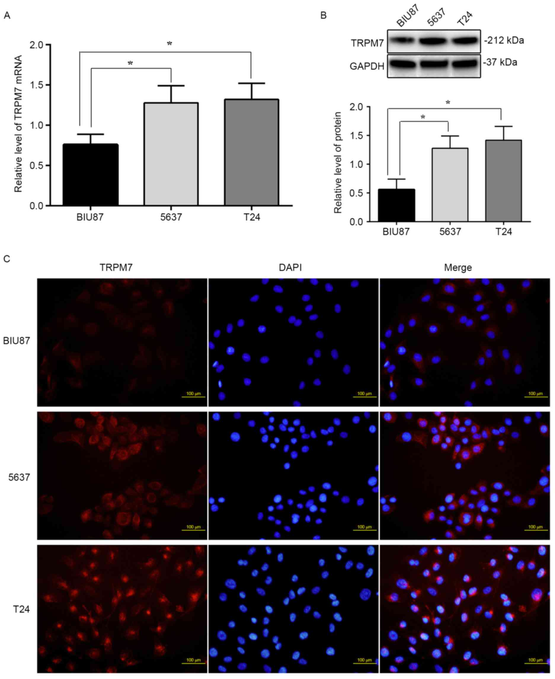
TRPM7 mRNA and protein levels are
higher in 5637 and T24 cells than BIU87 cells
To examine the mRNA and protein levels of TRPM7 in
BIU87, 5637 and T24 bladder cancer cell lines, RT-PCR and western
blotting were carried out, respectively. Fig. 3A shows that the levels of TRPM7 mRNA
in 5637 and T24 cells (normalized to GAPDH, 1.38±0.7, 1.45±0.6;
P<0.05) were significantly higher than that noted in the BIU87
cells (0.75±0.4; P<0.05). Western blotting demonstrated that
TRPM7 protein levels (normalized to GAPDH) were also higher in the
5637 and T24 cells (1.25±0.41, 1.36±0.49; P<0.05) compared to
that noted in the BIU87 cells (0.58±0.32) (Fig. 3B). TRPM7 protein levels were further
determined in bladder cancer cell lines using immunofluorescent
staining. As shown in Fig. 3C,
higher levels of TRPM7 protein were observed in the 5637 and T24
cells compared to that noted in the BIU87 cells in cell culture,
and the TRPM7 protein trend was to localize around the nuclear
membrane in the 5637 and T24 cells. The fluorescence intensity in
the 5637 and T24 cells was 25 or 30% higher than that in the BIU87
cells (P<0.05). Our results provide evidence that TRPM7 channels
are highly expressed in bladder cancer cells with a high degree of
malignancy, and enrichment of TRPM7 is around the nuclear
membrane.
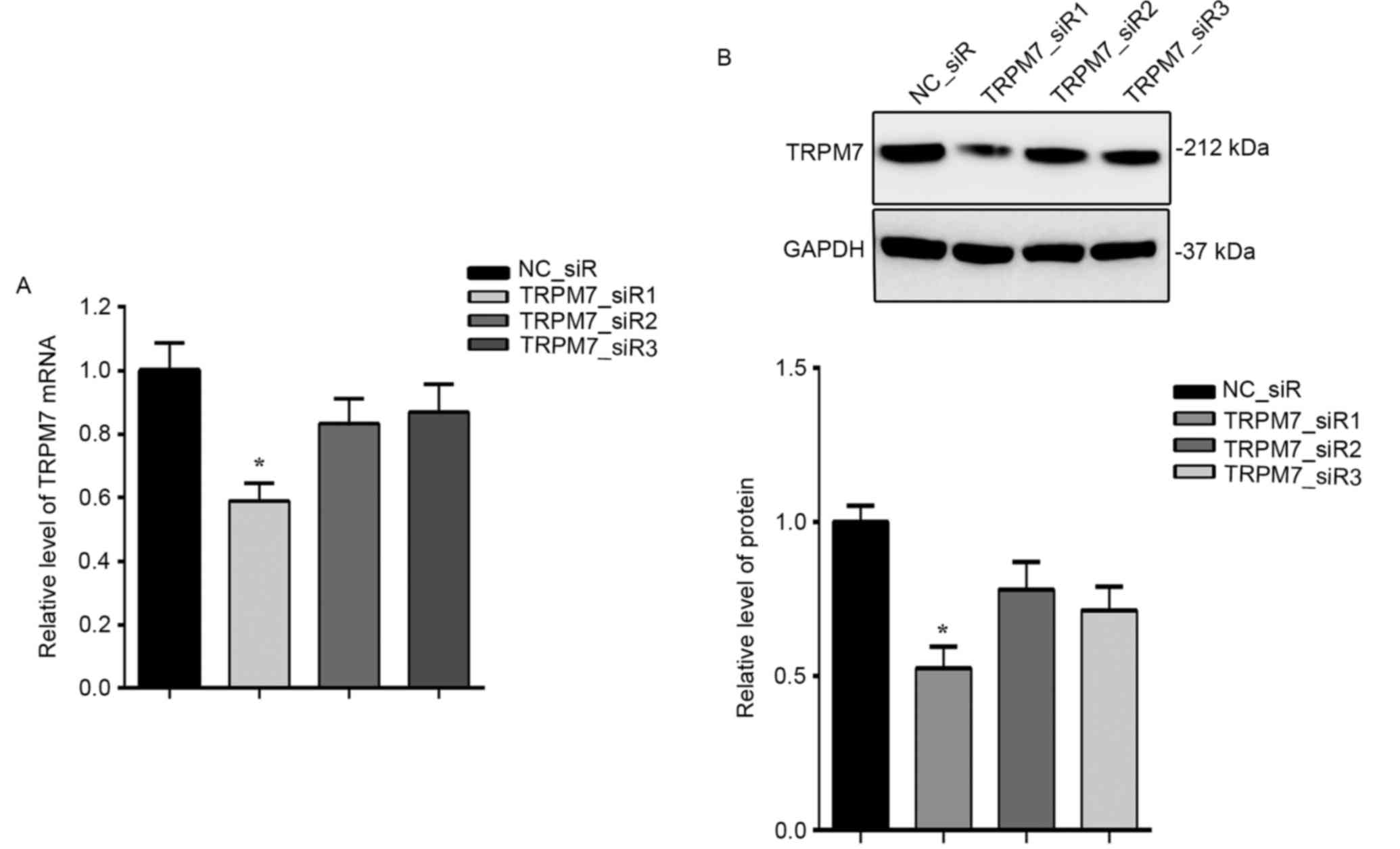
TRPM7 siRNA effectively suppresses
TRPM7 expression
To investigate the role of TRPM7 in bladder cancer,
TRPM7 was knocked down using RNAi method. T24 cells were
transfected with three siRNAs targeting TRPM7. As illustrated in
Fig. 4, the mRNA (Fig. 4A) and protein (Fig. 4B) expression levels of TRPM7 were
both significantly inhibited by TRPM7_siR1.
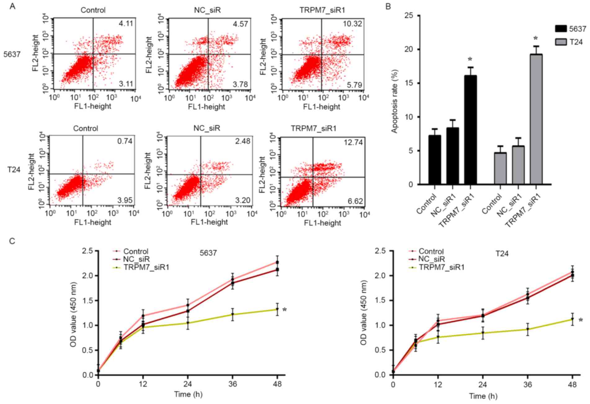
TRPM7 knockdown inhibits cellular
proliferation and induces cell apoptosis
We transfected the 5637 and T24 cells with
TRPM7_siR1 and NC_siR. Compared with NC_siR and control groups,
flow cytometric analysis revealed that downregulation of TRPM7
significantly induced apoptosis (Fig.
5A). Statistical analysis of apoptosis is shown in Fig. 5B. CCK-8 assay was carried out to
assess the effects of TRPM7 knockdown on T24 and 5637 cell
proliferation. The CCK-8 assay showed that after downregulation of
TRPM7 with TRPM7_siR1, T24 and 5637 cells exhibited a significant
decrease in cell proliferation compared with that noted in the
control siRNA group (Fig. 5C). Flow
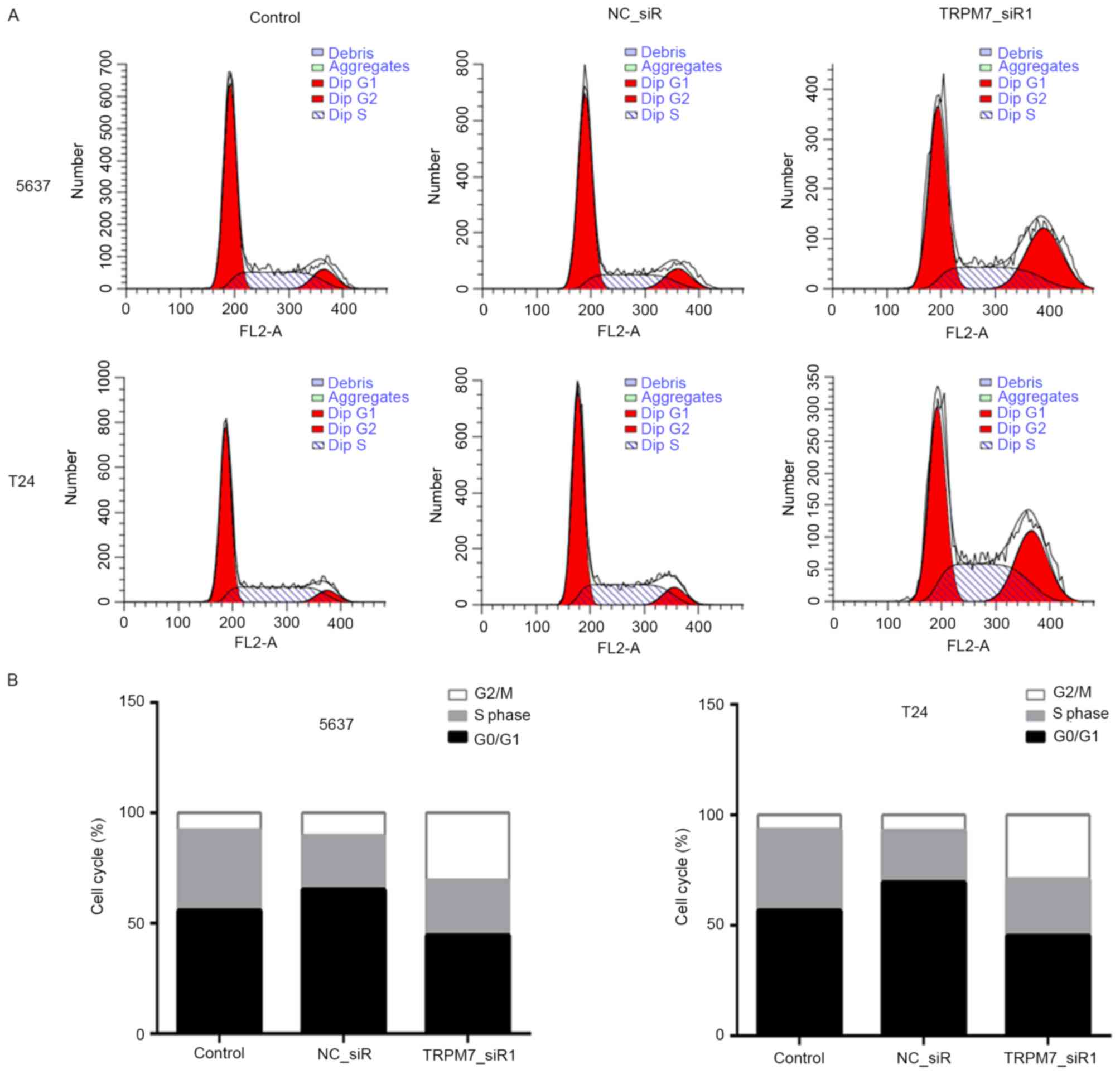
cytometric cell cycle analysis revealed that 5637 and T24 cells
were blocked in the G2/M phase after transfection with TRPM7_siR1
(Fig. 6A and B).
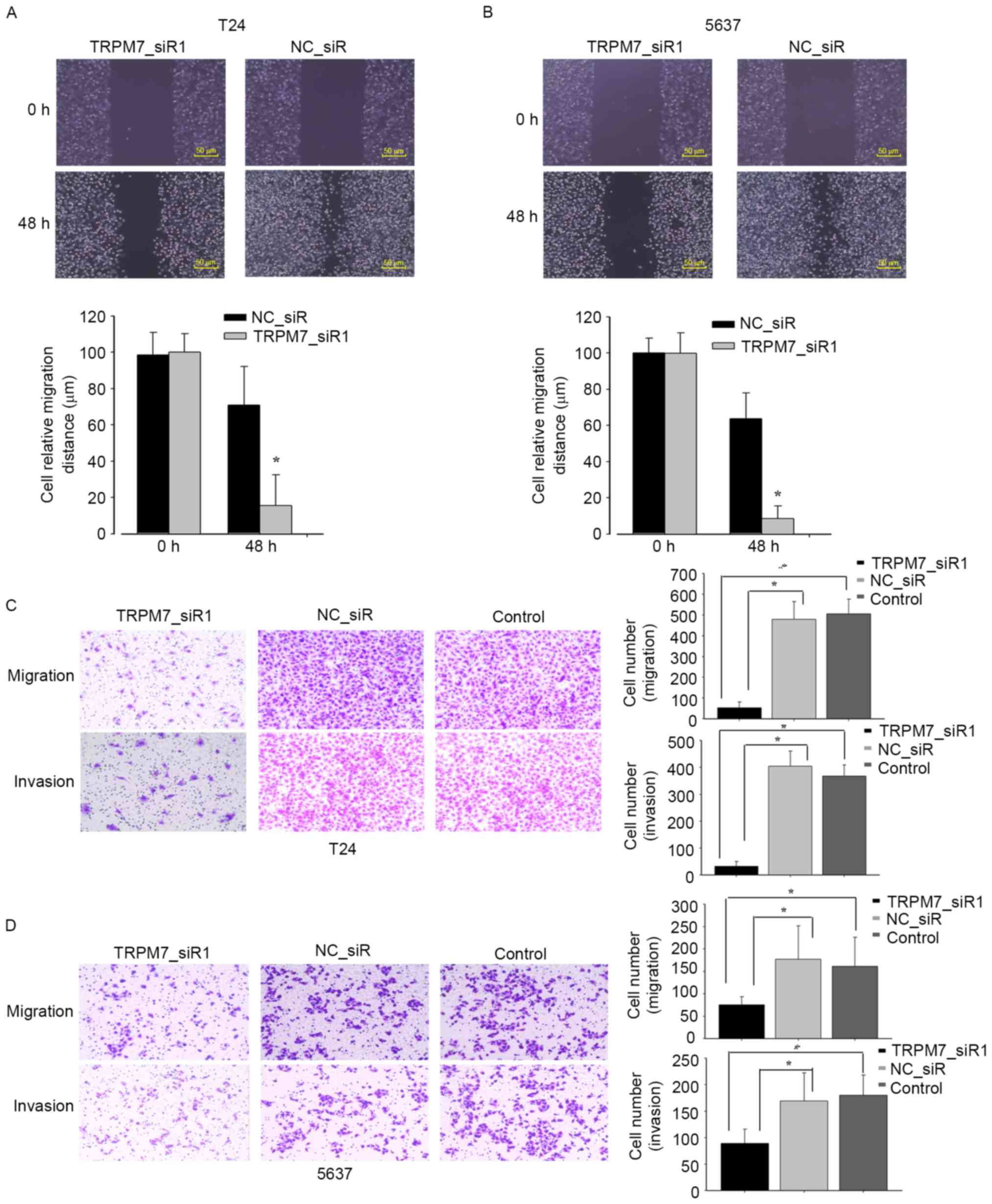
TRPM7 knockdown inhibits cell
motility
We next investigated the effect of TRPM7 on cell
motility with wound-healing and Transwell assays. After being
incubated with physical-wound and cultured in serum-free medium to
exclude the interference of proliferation, the percentage of wound
closure at 48 h was significantly lower in the TRPM7_siR1-treated
cells than that noted in the control cells (Fig. 7A and B). Transwell assays showed
that the silencing of TRPM7 measurably inhibited cell migration and
invasion in the Transwell assays (Fig.
7C and D). All the data support that TRPM7 stimulates bladder
cancer cell migration and invasion.
Discussion
It is believed that bladder tumorigenesis and
development are multistep pathologic processes involving numerous
genetic alterations, of which the inactivation of tumor repressors
and activation of oncogenes are critical events in the initiation
of bladder tumors. As one of the most fatal carcinomas worldwide,
the molecular mechanisms of bladder cancer remain unknown. It is
urgent to identify novel molecules which may serve as prognostic
factors and therapeutic targets for bladder cancer. In the present
study, we found that TRPM7 is highly expressed in bladder cancer
and is closely correlated with clinical stages and poor prognosis
of bladder cancer patients. However, TRPM7 accelerated bladder
cancer cell growth and migration. Taken together, these results
suggest that TRPM7 may be a potential regulator in bladder cancer
progression.
Calcium (Ca2+) and magnesium
(Mg2+) are two important metal elements that contribute
to a variety of tumor cell processes such as proliferation,
migration and apoptosis. Studies have reported that TRP channels
are involved in a variety of basic cellular processes and are
crucial for carcinogenesis and cancer development (22,23).
TRPM7, belonging to the TRP channel family, is a non-selective
cation channel mediating both Ca2+ and Mg2+
flow (7,8). TRPM7 is ubiquitously expressed and
essential for diverse physiological and pathophysiological
processes such as excitability, gene expression, muscle
contraction, cell volume regulation and hormone secretion (20,24).
Accumulating studies have confirmed that TRPM7 is aberrantly
overexpressed and plays a vital role in various types of cancers
(25–28). For example, TRPM7 is high expressed
in pancreatic cancer and correlates with tumor size and stage.
Moreover, TRPM7 is required for pancreatic cancer invasion
(13). However, the contribution of
TRPM7 to bladder cancer carcinogenesis remains largely unidentified
and needs to be determined.
In the present study, we firstly detected TRPM7
expression in human bladder cancer tissues by RT-PCR, western blot
and immunohistochemical analyses. The expression levels of TRPM7
mRNA and protein were significantly higher in bladder cancer than
levels in the non-tumor tissues. In addition, a similar consequence
was detected in bladder cancer cell lines (BIU87, 5637 and T24).
Next, the correlation analysis demonstrated that higher TRPM7
expression level was closely associated with recurrence (P<0.01)
and metastasis (P=0.021). All these data suggest that TRPM7
functions as a potential oncogene and plays an important role in
the progression of bladder cancer.
Previous research has demonstrated that TRPM7 is
correlated with the poor prognosis of various malignancies such as
neuroblastoma (19,29). Thus, we performed Kaplan-Meier
analysis to investigate the prognostic role of TRPM7 in bladder
cancer patients. We found that overexpression of TRPM7 was
correlated with the patient overall survival time. In addition,
univariate and multivariate analyses revealed that TRPM7 is a
significant independent prognostic predictor for bladder cancer
patients.
Thus, to further explore the functions of TRPM7 in
bladder cancer, we determined the effect of TRPM7 on the behaviors
of bladder cancer cells (T24 and 5637). TRPM7 was downregulated via
RNAi strategy, the siRNAs targeting TRPM7 were designed and
screened in bladder cancer cells, and the most efficient silencing
siRNA (TRPM7_siR1) was used for following observations. After TRPM7
was silenced, the CCK-8 assay showed that the proliferation of T24
and 5637 cells was significant inhibited and flow cytometric cell
cycle analysis exhibited that cells were blocked in the G2/M phase,
and flow cytometric analysis revealed that downregulation of TRPM7
induced apoptosis.
Bladder cancer has a tendency to metastasize, and
metastasis is an important characteristic which influences bladder
cancer patient prognosis. We used wound-healing and Transwell
assays to study the role of TRPM7 in bladder cancer migration and
invasion. The loss-of-function experiments showed that the
silencing of TRPM7 expression inhibited cell migratory and invasive
abilities. All the results suggest that TRPM7 may be a potential
tumor promoter in bladder cancer.
In summary, our data confirmed that TRPM7 is
overexpressed in bladder cancer tissue. Furthermore, high TRPM7
expression may be involved in the clinical development and poor
prognosis of bladder cancer patients. Moreover, we showed that
TRPM7 may be involved in cell proliferation, apoptosis, migration
and invasion abilities of bladder cancer cells. The present study
indicated that TRPM7 has potent oncogenic activity in bladder
cancer and TRPM7 channel functions may uncover new strategies in
the future to prevent the progression of bladder cancer
diseases.
Acknowledgements
The present study was supported by the The National
Natural Science Foundation of China (81372723), and The Key Urology
Laboratory Foundation of Shenyang City of China (F13-293-1-00).
References
|
1
|
Pang C, Guan Y, Li H, Chen W and Zhu G:
Urologic cancer in China. Jpn J Clin Oncol. 46:497–501. 2016.
View Article : Google Scholar : PubMed/NCBI
|
|
2
|
Ye F, Wang L, Castillo-Martin M, McBride
R, Galsky MD, Zhu J, Boffetta P, Zhang DY and Cordon-Cardo C:
Biomarkers for bladder cancer management: Present and future. Am J
Clin Exp Urol. 2:1–14. 2014.PubMed/NCBI
|
|
3
|
Al-Zalabani AH, Stewart KF, Wesselius A,
Schols AM and Zeegers MP: Modifiable risk factors for the
prevention of bladder cancer: A systematic review of meta-analyses.
Eur J Epidemiol. 31:811–851. 2016. View Article : Google Scholar : PubMed/NCBI
|
|
4
|
Burger M, Catto JW, Dalbagni G, Grossman
HB, Herr H, Karakiewicz P, Kassouf W, Kiemeney LA, La Vecchia C,
Shariat S, et al: Epidemiology and risk factors of urothelial
bladder cancer. Eur Urol. 63:234–241. 2013. View Article : Google Scholar : PubMed/NCBI
|
|
5
|
Guillaume L and Guy L: Epidemiology of and
risk factors for bladder cancer and for urothelial tumors. Rev
Prat. 64(1372–1374): 1378–1380. 2014.(In French).
|
|
6
|
Clapham DE: TRP channels as cellular
sensors. Nature. 426:517–524. 2003. View Article : Google Scholar : PubMed/NCBI
|
|
7
|
Clapham DE, Runnels LW and Strübing C: The
TRP ion channel family. Nat Rev Neurosci. 2:387–396. 2001.
View Article : Google Scholar : PubMed/NCBI
|
|
8
|
Runnels LW, Yue L and Clapham DE:
TRP-PLIK, a bifunctional protein with kinase and ion channel
activities. Science. 291:1043–1047. 2001. View Article : Google Scholar : PubMed/NCBI
|
|
9
|
Guilbert A, Gautier M, Dhennin-Duthille I,
Rybarczyk P, Sahni J, Sevestre H, Scharenberg AM and
Ouadid-Ahidouch H: Transient receptor potential melastatin 7 is
involved in oestrogen receptor-negative metastatic breast cancer
cells migration through its kinase domain. Eur J Cancer.
49:3694–3707. 2013. View Article : Google Scholar : PubMed/NCBI
|
|
10
|
Wang J, Liao QJ, Zhang Y, Zhou H, Luo CH,
Tang J, Wang Y, Tang Y, Zhao M, Zhao XH, et al: TRPM7 is required
for ovarian cancer cell growth, migration and invasion. Biochem
Biophys Res Commun. 454:547–553. 2014. View Article : Google Scholar : PubMed/NCBI
|
|
11
|
Lin CM, Ma JM, Zhang L, Hao ZY, Zhou J,
Zhou ZY, Shi HQ, Zhang YF, Shao EM and Liang CZ: Inhibition of
transient receptor potential melastain 7 enhances apoptosis induced
by TRAIL in PC-3 cells. Asian Pac J Cancer Prev. 16:4469–4475.
2015. View Article : Google Scholar : PubMed/NCBI
|
|
12
|
Chen WL, Barszczyk A, Turlova E, Deurloo
M, Liu B, Yang BB, Rutka JT, Feng ZP and Sun HS: Inhibition of
TRPM7 by carvacrol suppresses glioblastoma cell proliferation,
migration and invasion. Oncotarget. 6:16321–16340. 2015. View Article : Google Scholar : PubMed/NCBI
|
|
13
|
Yee NS, Kazi AA, Li Q, Yang Z, Berg A and
Yee RK: Aberrant over-expression of TRPM7 ion channels in
pancreatic cancer: Required for cancer cell invasion and implicated
in tumor growth and metastasis. Biol Open. 4:507–514. 2015.
View Article : Google Scholar : PubMed/NCBI
|
|
14
|
Mizuno H, Suzuki Y, Watanabe M, Sokabe T,
Yamamoto T, Hattori R, Gotoh M and Tominaga M: Potential role of
transient receptor potential (TRP) channels in bladder cancer
cells. J Physiol Sci. 64:305–314. 2014. View Article : Google Scholar : PubMed/NCBI
|
|
15
|
Meng X, Cai C, Wu J, Cai S, Ye C, Chen H,
Yang Z, Zeng H, Shen Q and Zou F: TRPM7 mediates breast cancer cell
migration and invasion through the MAPK pathway. Cancer Lett.
333:96–102. 2013. View Article : Google Scholar : PubMed/NCBI
|
|
16
|
Middelbeek J, Kuipers AJ, Henneman L,
Visser D, Eidhof I, van Horssen R, Wieringa B, Canisius SV, Zwart
W, Wessels LF, et al: TRPM7 is required for breast tumor cell
metastasis. Cancer Res. 72:4250–4261. 2012. View Article : Google Scholar : PubMed/NCBI
|
|
17
|
Aarts M, Iihara K, Wei WL, Xiong ZG,
Arundine M, Cerwinski W, MacDonald JF and Tymianski M: A key role
for TRPM7 channels in anoxic neuronal death. Cell. 115:863–877.
2003. View Article : Google Scholar : PubMed/NCBI
|
|
18
|
Jin J, Desai BN, Navarro B, Donovan A,
Andrews NC and Clapham DE: Deletion of Trpm7 disrupts
embryonic development and thymopoiesis without altering
Mg2+ homeostasis. Science. 322:756–760. 2008. View Article : Google Scholar : PubMed/NCBI
|
|
19
|
Lehen'kyi V, Shapovalov G, Skryma R and
Prevarskaya N: Ion channnels and transporters in cancer. 5. Ion
channels in control of cancer and cell apoptosis. Am J Physiol Cell
Physiol. 301:C1281–C1289. 2011. View Article : Google Scholar : PubMed/NCBI
|
|
20
|
Romani A: Regulation of magnesium
homeostasis and transport in mammalian cells. Arch Biochem Biophys.
458:90–102. 2007. View Article : Google Scholar : PubMed/NCBI
|
|
21
|
de Baaij JH, Hoenderop JG and Bindels RJ:
Magnesium in man: Implications for health and disease. Physiol Rev.
95:1–46. 2015. View Article : Google Scholar : PubMed/NCBI
|
|
22
|
Guilbert A, Gautier M, Dhennin-Duthille I,
Haren N, Sevestre H and Ouadid-Ahidouch H: Evidence that TRPM7 is
required for breast cancer cell proliferation. Am J Physiol Cell
Physiol. 297:C493–C502. 2009. View Article : Google Scholar : PubMed/NCBI
|
|
23
|
Clark K, Langeslag M, van Leeuwen B, Ran
L, Ryazanov AG, Figdor CG, Moolenaar WH, Jalink K and van Leeuwen
FN: TRPM7, a novel regulator of actomyosin contractility and cell
adhesion. EMBO J. 25:290–301. 2006. View Article : Google Scholar : PubMed/NCBI
|
|
24
|
Wang J, Xiao L, Luo CH, Zhou H, Hu J, Tang
YX, Fang KN and Zhang Y: Overexpression of TRPM7 is associated with
poor prognosis in human ovarian carcinoma. Asian Pac J Cancer Prev.
15:3955–3958. 2014. View Article : Google Scholar : PubMed/NCBI
|
|
25
|
Kim BJ, Nah SY, Jeon JH, So I and Kim SJ:
Transient receptor potential melastatin 7 channels are involved in
ginsenoside Rg3-induced apoptosis in gastric cancer cells. Basic
Clin Pharmacol Toxicol. 109:233–239. 2011. View Article : Google Scholar : PubMed/NCBI
|
|
26
|
Chen JP, Luan Y, You CX, Chen XH, Luo RC
and Li R: TRPM7 regulates the migration of human nasopharyngeal
carcinoma cell by mediating Ca2+ influx. Cell Calcium.
47:425–432. 2010. View Article : Google Scholar : PubMed/NCBI
|
|
27
|
Hanano T, Hara Y, Shi J, Morita H,
Umebayashi C, Mori E, Sumimoto H, Ito Y, Mori Y and Inoue R:
Involvement of TRPM7 in cell growth as a spontaneously activated
Ca2+ entry pathway in human retinoblastoma cells. J
Pharmacol Sci. 95:403–419. 2004. View Article : Google Scholar : PubMed/NCBI
|
|
28
|
Yee NS, Kazi AA and Yee RK: Cellular and
developmental biology of TRPM7 channel-kinase: Implicated roles in
cancer. Cells. 3:751–777. 2014. View Article : Google Scholar : PubMed/NCBI
|
|
29
|
Lange I and Koomoa DL: MycN promotes TRPM7
expression and cell migration in neuroblastoma through a process
that involves polyamines. FEBS Open Bio. 4:966–975. 2014.
View Article : Google Scholar : PubMed/NCBI
|