Introduction
Osteosarcoma, the most common primary malignancy of
bones, is histologically characterized by the presence of malignant
mesenchymal cells with a high rate of pulmonary metastasis
(1,2). In the past, the 5-year survival rate
for osteosarcoma was ~10–20%, indicating a very poor prognosis
(3). Multidisciplinary treatment,
including limb salvage surgery and multidrug neoadjuvant
chemotherapy, has improved the cumulative 5-year survival to 60–80%
(4). Thus, osteosarcoma is no
longer incurable. However, the overall survival has remained nearly
unchanged in the last three decades owing to genetic, epigenetic
and biological complexities (5).
Therefore, elucidating the molecular mechanisms underlying
osteosarcoma is critical for developing new therapeutic strategies
to improve the prognosis of patients with osteosarcoma.
Basic leucine zipper and W2 domains 2 (BZW2) is a
member of the bZIP superfamily of transcription factors. It is
involved in cell-cell adhesion via cadherin binding. Guo et
al reported that BZW2 is a novel E-cadherin-interacting protein
based on a quantitative proteomic analysis (6). BZW1, another member of the bZIP
superfamily, is a novel proliferation regulator in salivary
mucoepidermoid carcinoma. The knockdown of BZW1 significantly
inhibited the proliferation and metastasis of Mc3 cells and a high
expression of BZW1 has been detected in high-grade mucoepidermoid
carcinoma (7). However, for BZW2,
the potential roles and underlying mechanisms in tumorigenesis
remain elusive.
Accumulating studies have shown the importance of
the PI3K/AKT/mTOR signaling pathway in tumorigenesis (8,9). Sun
et al reported that the activation of the PI3K/AKT/mTOR
signaling pathway is correlated with tumor progression and
decreases patient survival for those with urothelial carcinoma of
the urinary bladder (10).
Similarly, overexpression of the PI3K/Akt/mTOR pathway proteins in
sialyl-Tn antigen-positive muscle invasive bladder cancer is
independently associated with a high risk of cancer-related deaths
(11). However, it is not clear
whether the Akt/mTOR signaling pathways are involved in the
BZW2-induced effects on osteosarcoma.
In the present study, we investigated the expression
of BZW2 in human osteosarcoma cell lines and an osteoblast cell
line. BZW2 was overexpressed in osteosarcoma cell lines and
osteosarcoma tissues. The functions of BZW2 were further examined
in osteosarcoma cells. Our results demonstrated that the knockdown
of BZW2 hinders osteosarcoma cell proliferation and
colony-formation ability by inhibiting the G2/M cell cycle
transition. The mechanism underlying the function of BZW2 was also
investigated. Decreased BZW2 downregulated the Akt/mTOR signaling
pathway in osteosarcoma cells. Notably, we found that BZW2
expression was positively correlated with the Enneking stage and
the recurrence of osteosarcoma. To the best of our knowledge, this
is the first evidence that the overexpression of BZW2 is a
prerequisite for the development and progression in human
osteosarcoma.
Materials and methods
Cell lines and cell culture
Two osteosarcoma cell lines (MNNG/HOS and U2OS) and
one human osteoblast cell line (hFOB 1.19) were used in the present
study. All three cell lines were obtained from the Cell Bank of the
Chinese Academy of Sciences (Shanghai, China). They were cultured
in either Dulbecco's modified Eagles medium (MNNG/HOS) or RPMI-1640
medium (U2OS) containing 10% fetal bovine serum (Bio-west, Logan,
UT, USA), 100 U/ml penicillin, and 100 mg/ml streptomycin (both
from Sigma-Aldrich, St. Louis, MO, USA). The cells were maintained
at 37°C in a humidified atmosphere containing 5% CO2.
The hFOB 1.19 cell line was cultured according to the established
American Type Culture Collection (ATCC; Mannasas, VA, USA)
protocols.
Human osteosarcoma samples
From 2015 to 2016, 50 patients with osteosarcoma
were treated at Shanghai Jiaotong University Affiliated Sixth
People's Hospital. They received primary surgical treatment and
preoperative and postoperative neoadjuvant therapy. Each
osteosarcoma sample along with a corresponding adjacent non-tumor
tissue sample was obtained during surgery. The samples were
immediately frozen in liquid nitrogen after resection and stored at
−80°C in a refrigerator. Ethics approval was obtained from the
local hospital Ethics Committees, and written informed consent was
obtained from each patient prior to sample collection (YS-2016-064,
24 February 2016).
RNA extraction and qRT-PCR
analysis
Total RNAs of human tissue samples and cultured
cells were purified using TRIzol reagent (Invitrogen, Carlsbad, CA,
USA). cDNA was synthesized using a PrimeScript RT Reagent kit
(Takara, Shiga, Japan). qRT-PCR was performed using SYBR-Green
Premix Ex Taq (Takara) on an ABI 7500 PCR system (Applied
Biosystems, Foster City, CA). All reactions were performed in 10-µl
reaction volumes in triplicate. The primer sequences were as
follows: BZW2 forward, 5′-GGAGCTTTCCGACTTCCTCC-3′ and
reverse, 5′-AAGCACCACCTCCTTGATCG-3′; β-actin forward,
5′-TTGTTACAGGAAGTCCCTTGCC-3′ and reverse,
5′-ATGCTATCACCTCCCCTGTGTG-3′.
Protein extraction and western blot
analysis
Lysates were extracted from cultured cells using
T-PER Protein Extraction Reagent (Thermo Fisher Scientific, Inc.,
Waltham, MA, USA) containing PhosSTOP (Roche, Basel, Switzerland)
and Complete Mini. Equal amounts of proteins were electrophoresed
and transferred to nitrocellulose membranes or polyvinylidene
fluoride membranes (Millipore, Billerica, MA, USA). After they were
blocked in 5% non-fat milk, the membranes were incubated with the
following primary antibodies: BZW2 (polyclonal; 1:500; #21001-1-AP;
Proteintech, Wuhan, China), mTOR (total) (monoclonal; 1:1,000;
#2983), mTOR (Ser2448) (monoclonal; 1:1,000; #5536), Akt (total)
(monoclonal; 1:1,000; #4691), Akt (Thr308) (monoclonal; 1:1,000;
#13038) (all from Cell Signaling Technology), or β-actin
(polyclonal; 1:5,000; #20536-1-AP; Proteintech). The secondary
antibody was goat anti-rabbit IgG (1:5,000; #A0545; Sigma-Aldrich).
Visualization was performed using SuperSignal West Femto Maximum
Sensitivity Substrate (Thermo Fisher Scientific, Inc.).
siRNA transfection
Human BZW2 siRNA and non-specific control
(NC) siRNA were synthesized by RiboBio (Guangzhou, China) and were
transfected into cells using Lipofectamine 2000 reagent
(Invitrogen) following the manufacturer's protocol. The sequence of
the siRNA targeting BZW2 was GGTCTTCTGTGGACATGTA. For
proliferation and cell cycle analyses as well as RNA extraction and
western blotting, cells were used 48 h after transfection.
Cell proliferation and colony
formation assays
For the cell proliferation assay, 48 h after
transfection, the cells were seeded into a 96-well plate (3,000
cells/well). A 10-µl aliquot of Cell Counting Kit-8 (CCK-8)
solution (Dojindo, Kumamoto, Japan) was added to triplicate wells
and incubated for 2 h. The absorbance at 450 nm was assessed. Each
measurement was performed in triplicate, and experiments were
repeated twice. For the colony formation assay, 48 h after siRNA
transfection, MNNG/HOS or U2OS cells (1×10³ cells/well)
were cultured in 6-well plates for 10 days, and then subjected to
100% methanol fixation for 30 min and 0.1% crystal violet staining
for 30 min. The cell colonies were counted and all assays were
independently performed in triplicate.
Cell cycle analysis
Forty-eight hours after transfection, cells were
collected and fixed with 70% ethanol. The cells were then stained
with 50 µg/ml of propidium iodide containing RNase I (both from
Kaiji Biological Inc., Nanjing, China), followed by analysis using
a FACSCalibur flow cytometer (BD Biosciences, San Jose, CA, USA).
The results were analyzed using ModFit software (BD Biosciences).
Assays were independently performed three times.
Intracellular signaling array
Forty-eight hours after transfection, MNNG/HOS and
U2OS cells were harvested and lysed on ice with 0.1 ml of cell
lysis buffer containing a cocktail of protease inhibitors for 5
min. The lysates were centrifuged at 10,000 × g at 4°C for 10 min.
Intracellular signaling molecules were detected using a
PathScan® Intracellular Signaling Array kit (#7744; Cell
Signaling Technology) according to the manufacturer's procedure.
Fluorescent images of the slides were captured using the
Odyssey® Infrared Imaging System (LI-COR, Lincoln, NE,
USA) and the intensities of the spots were quantified using Image
Studio analysis software.
Immunohistochemistry
A standard immunohistochemistry (IHC) staining
procedure was followed. Briefly, paraffin-embedded sections were
cut at 4 µm, dewaxed in xylene, and heated in a microwave at 60°C
for 20 min in EDTA buffer (pH 9.0) for antigen retrieval. For each
slide, endogenous peroxidase activity was blocked by 10 min of
incubation in 0.3% H2O2, followed by
incubation at 37°C with a 1:100 dilution of the primary antibody
against BZW2 (Proteintech) or p-AKT (Thr308) (1:200; Cell Signaling
Technology). Slides were rinsed three times in phosphate-buffered
saline (PBS) and incubated for 30 min using the EnVision Staining
kit (DAKO, Glostrup, Denmark), followed by three additional washes
in PBS and color development over 3–10 min in a moist chamber at
20°C using 3,3′-diaminobenzidine (DAB). The slides were
counterstained with hematoxylin and dehydrated in a graded ethyl
alcohol series (70, 90 and 100%). For the sections used as negative
controls, PBS was used as a substitute for the primary antibody.
IHC signal intensities were scored as follows: 0 (no staining), 1
(staining in <1% of cells), 2 (staining in 1–10% of cells), or 3
(staining in >10% of cells). The samples classified as 0 and 1
were considered negative, while the samples classified as 2 and 3
were considered positive. Assessment of IHC staining was
independently performed by two expert pathologists. Any discordance
was resolved by discussion and consensus.
Statistical evaluation
Data were compiled and analyzed using SPSS version
21.0 (SPSS, Inc., Chicago, IL, USA). The differences between groups
were evaluated using a two-tailed Student's t-test. The
correlations between the IHC results for BZW2 and the
clinicopathological parameters were determined using Chi-square
tests. The correlation analysis between BZW2 and p-Akt was
performed using the Spearman correlation test. P<0.05 was
considered significant.
Results
BZW2 is overexpressed in osteosarcoma
cell lines and human osteosarcoma tissue samples
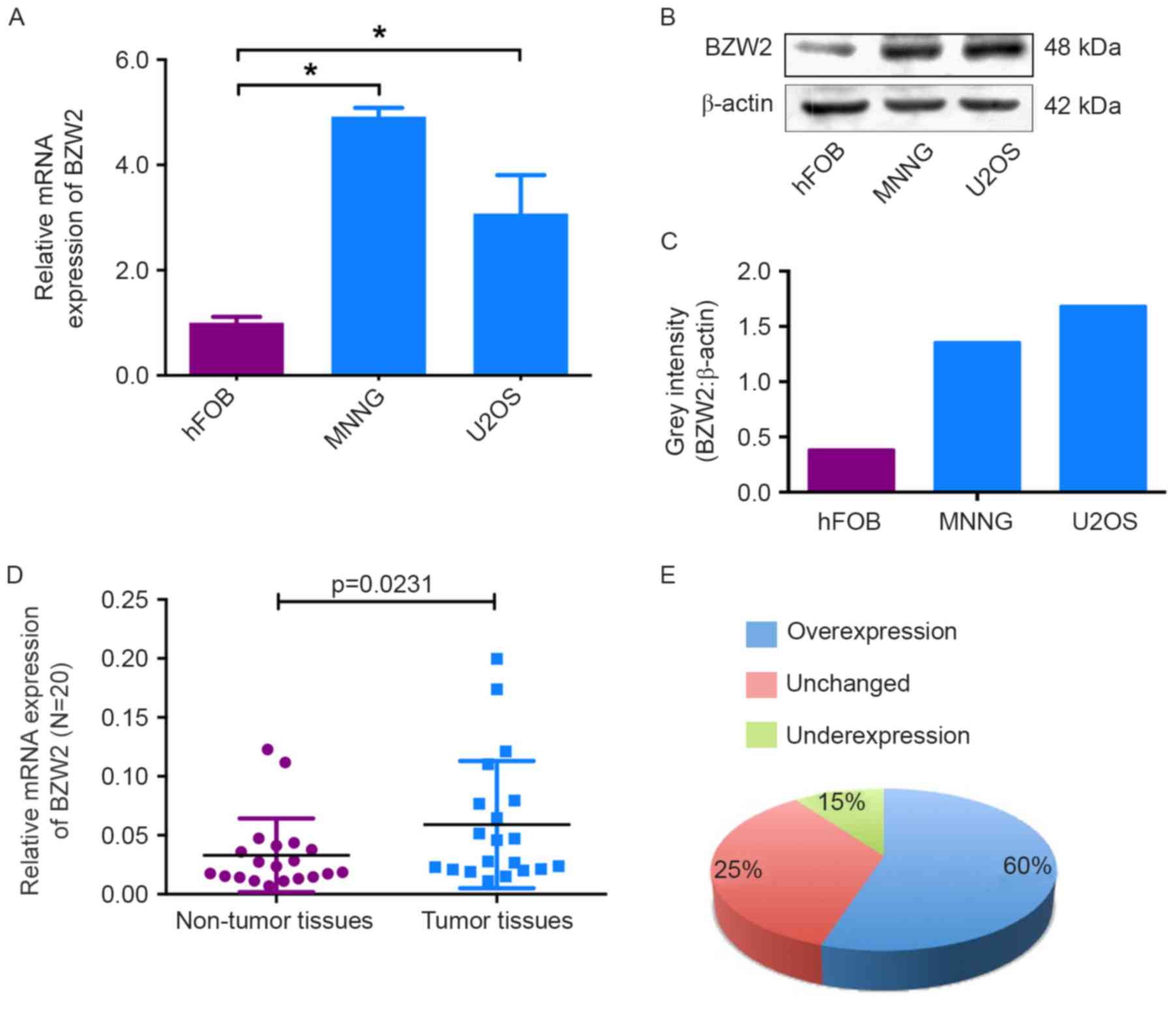
To determine the role of BZW2 in osteosarcoma, we
first detected the mRNA and protein expression levels of BZW2 in
osteosarcoma cell lines and an osteoblast cell line by qRT-PCR and
western blot analyses. Compared with the osteoblast cell line, BZW2
overexpression was detected at both the mRNA and protein levels in
osteosarcoma cell lines (Fig.
1A-C). To further confirm its role, BZW2 expression levels were
investigated in 20 paired osteosarcoma and adjacent non-tumor
tissues. As shown in Fig. 1D and E,
BZW2 expression was significantly upregulated in 60% (12/20) of
osteosarcoma tissues compared with adjacent non-tumor tissues.
These results show that BZW2 may play an important role in
osteosarcoma tumorigenesis and thus warrants further
investigation.
Knockdown of BZW2 inhibits
osteosarcoma cell proliferation in vitro
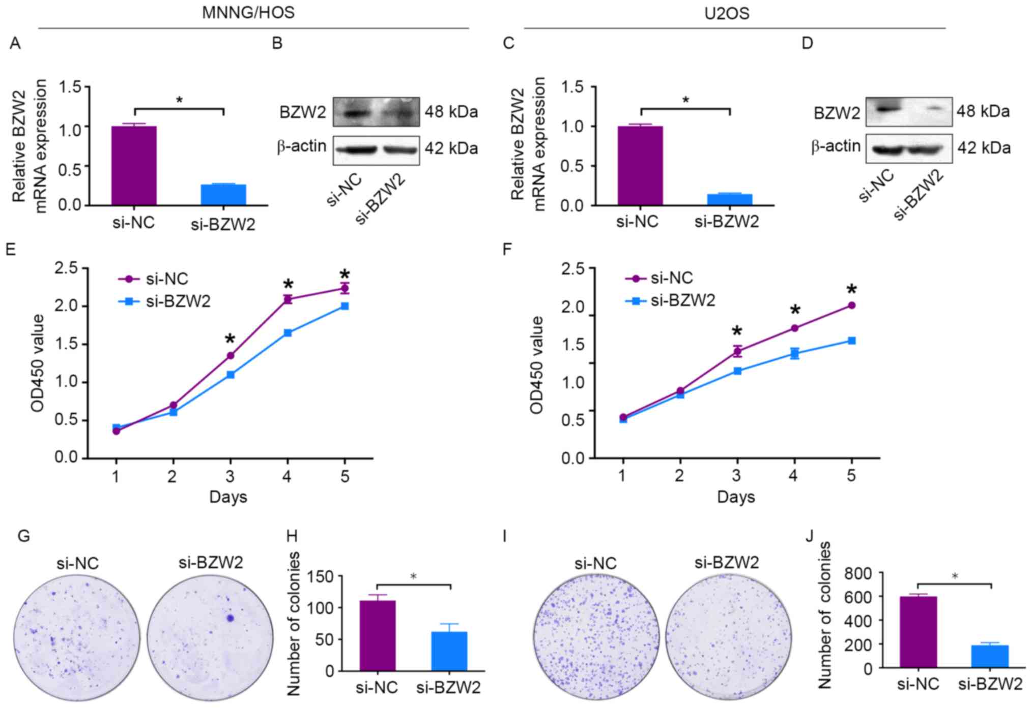
To explore the functional significance of BZW2 in
osteosarcoma, BZW2-specific siRNA was used to knockdown BZW2
in osteosarcoma cells. BZW2 knockdown was validated by qRT-PCR and
western blotting. BZW2 mRNA and protein expression levels
were significantly decreased after transfection with
BZW2-specific siRNA in MNNG/HOS and U2OS cells (Fig. 2A-D). Next, a CCK-8 assay was used to
detect cell proliferation. A 5-day growth curve analysis showed
that the knockdown of BZW2 significantly inhibited the growth of
osteosarcoma cells (Fig. 2E and F).
A colony formation assay was carried out to determine the
colony-forming capacity of osteosarcoma cells after the knockdown
of BZW2. The number and size of colonies were both obviously
decreased in the BZW2-knockdown group in comparison with the
control group (Fig. 2G-J). These
results demonstrate that BZW2 plays an oncogenic role in
osteosarcoma.
Knockdown of BZW2 inhibits the G2/M
cell cycle transition in osteosarcoma cells
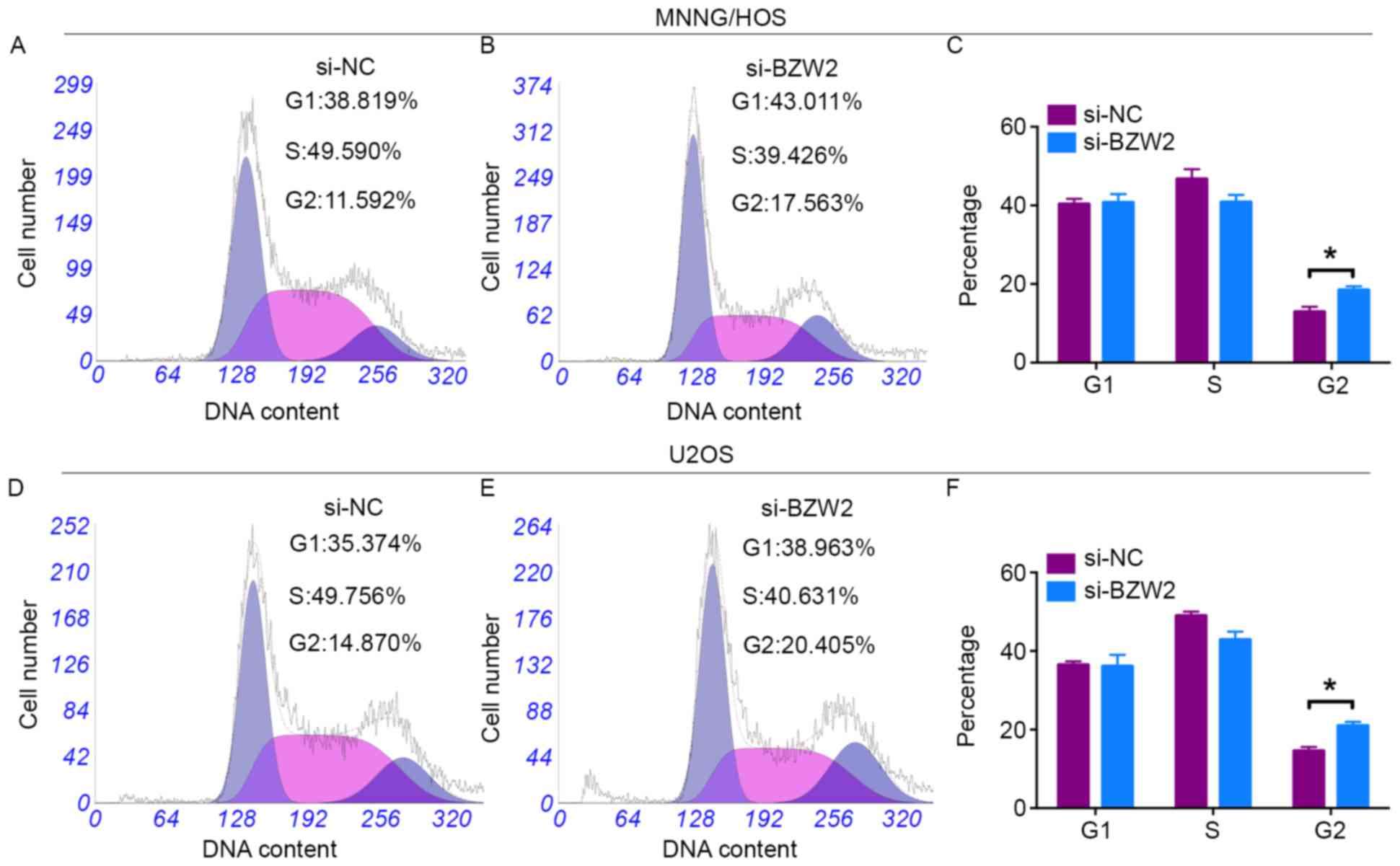
Cell cycle changes in response to BZW2 knockdown
were analyzed using flow cytometry. A cell cycle assay revealed
that si-BZW2-transfected cells had a significant higher percentage
of cells in the G2/M phase than that of si-NC-transfected cells
(Fig. 3A-F). However, no
significant difference was found in the G1 or S cell populations
between BZW2-knockdown and control-transfected cells. In
conclusion, these data revealed that BZW2 knockdown attenuated
osteosarcoma cell proliferation by inhibiting the G2/M cell cycle
transition.
Knockdown of BZW2 inhibits
osteosarcoma cell proliferation via the AKT/mTOR signaling
pathway
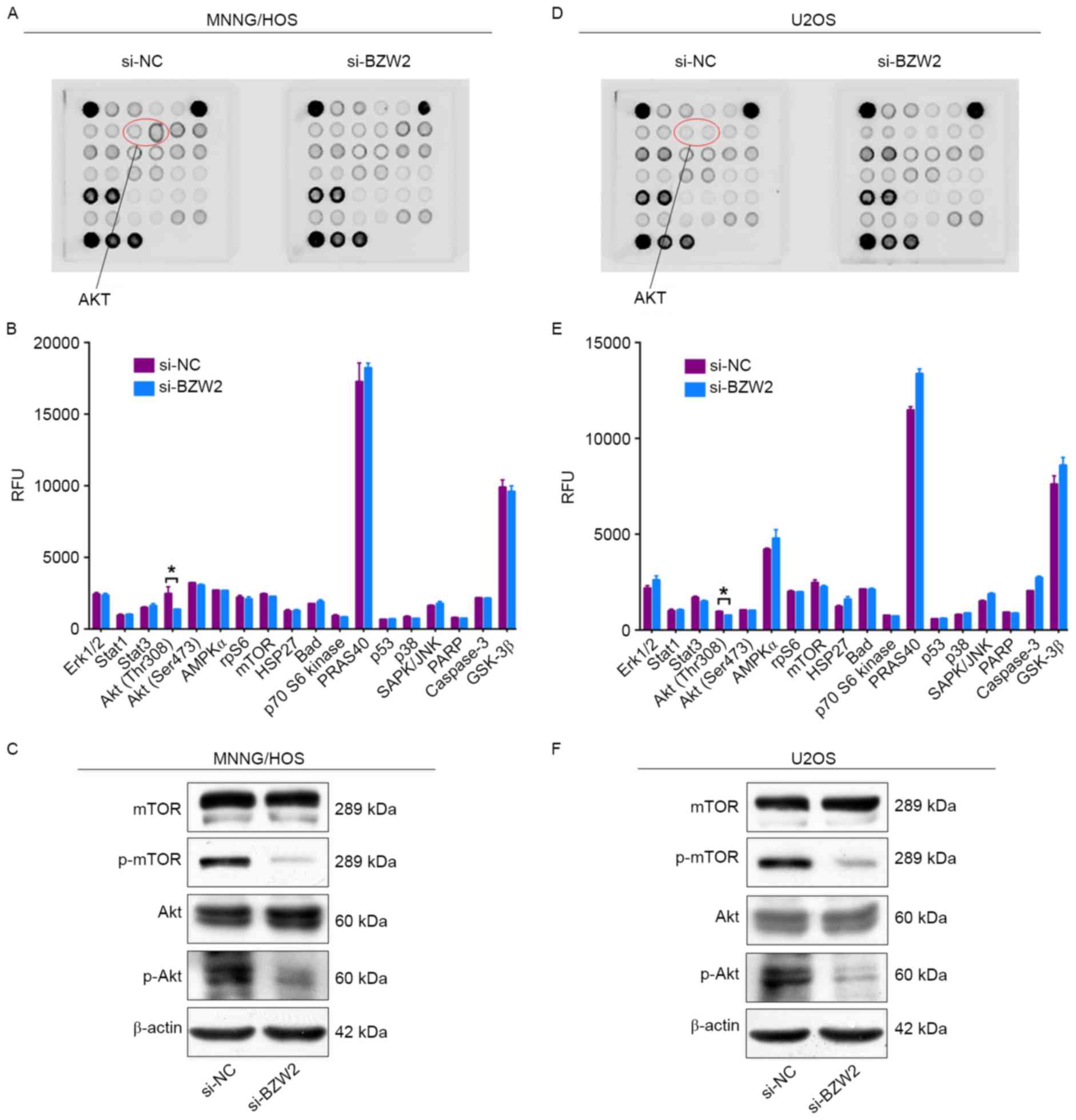
To determine the mechanism by which BZW2 affects the
proliferation of osteosarcoma cells, intracellular signaling arrays
were used to detect protein expression after BZW2 knockdown in
MNNG/HOS and U2OS cells. A small decrease in the phosphorylation of
Akt-Thr308 was detected in osteosarcoma cells (Fig. 4A-D). We then examined whether BZW2
influences the AKT/mTOR signaling pathway in osteosarcoma cells
using western blot analysis. Based on this analysis, BZW2 knockdown
inhibited the phosphorylation of Akt and mTOR. These results
support the conclusion that BZW2 plays a crucial role in
osteosarcoma cell growth by influencing the Akt/mTOR signaling
pathway.
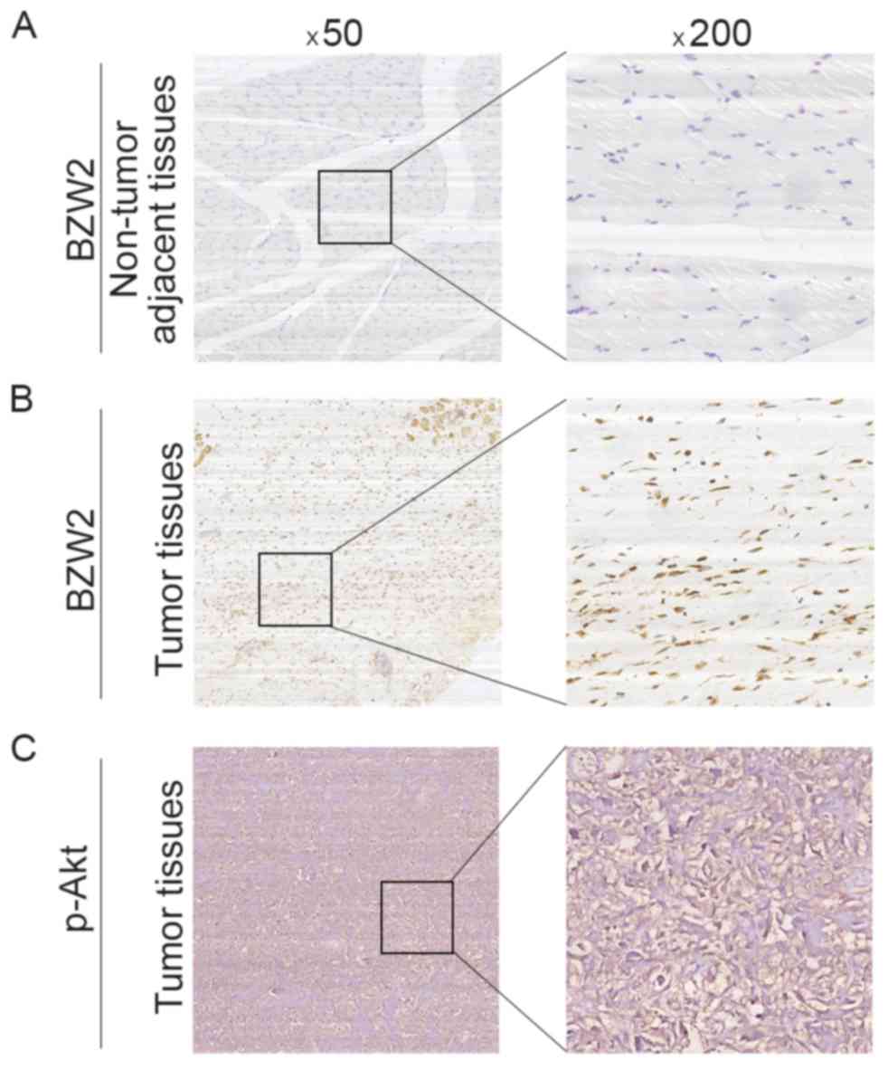
BZW2 expression is correlated to
Enneking stage and tumor recurrence in osteosarcoma
To further determine the clinicopathological
significance of BZW2 in osteosarcoma, we performed an IHC analysis
of BZW2 using 50 human osteosarcoma tissue samples and
corresponding non-tumor tissues. Representative IHC images of BZW2
expression in osteosarcoma tissues and adjacent non-tumor tissues
are shown in Fig. 5. Correlations
between the BZW2 expression level and the clinicopathological
characteristics of patients with osteosarcoma are summarized in
Table I. BZW2 expression levels
were higher in osteosarcoma tissue samples than in corresponding
non-tumor tissues (P=0.021). The expression level of BZW2 was
higher in patients at a clinically advanced Enneking stage than in
those at an early stage (P=0.009). The BZW2 expression level was
also positively correlated with recurrence (P=0.013). No
correlation was found between BZW2 and other demographic and
clinical factors, including sex, age, tumor location, tumor
necrosis rate, cortical destruction and metastasis. Finally, we
examined p-Akt expression in osteosarcoma tissues. Representative
images of p-Akt expression in osteosarcoma tissues are shown in
Fig. 5C. We found that there was a
significant correlation between the expression of both BZW2 and
p-Akt in osteosarcoma tissues (R=0.380, P=0.006). Collectively,
these results indicate that BZW2 is upregulated in osteosarcoma and
plays a potentially important role in osteosarcoma progression.
 | Table I.Correlation analyses of BZW2 protein
expression in relation to clinicopathological variables of 50
patients with osteosarcoma. |
Table I.
Correlation analyses of BZW2 protein
expression in relation to clinicopathological variables of 50
patients with osteosarcoma.
|
|
| BZW2 expression
level |
|
|
|---|
|
|
|
|
|
|
|---|
| Clinicopatho-logical
parameters | No. of cases | Negative | Positive | χ2 | P-value |
|---|
| Sex |
|
|
| 1.602 | 0.206 |
| Male | 29 | 19 | 10 |
|
|
|
Female | 21 | 10 | 11 |
|
|
| Age (years) |
|
|
| 0.512 | 0.474 |
|
<18 | 28 | 15 | 13 |
|
|
| ≥18 | 22 | 14 | 8 |
|
|
| Location |
|
|
| 1.667 | 0.434 |
|
Femur | 24 | 12 | 12 |
|
|
|
Tibia | 14 | 10 | 4 |
|
|
|
Elsewhere | 12 | 7 | 5 |
|
|
| Tumor necrosis rate
(%) |
|
|
| 0.006 | 0.939 |
|
<90 | 36 | 21 | 15 |
|
|
| ≥90 | 14 | 8 | 6 |
|
|
| Cortical
destruction |
|
|
| 0.487 | 0.485 |
|
Yes | 38 | 21 | 17 |
|
|
| No | 12 | 8 | 4 |
|
|
| Recurrence |
|
|
| 6.226 | 0.013a |
|
Yes | 23 | 9 | 14 |
|
|
| No | 27 | 20 | 7 |
|
|
| Metastasis |
|
|
| 0.112 | 0.738 |
|
Yes | 32 | 18 | 14 |
|
|
| No | 18 | 11 | 7 |
|
|
| Ennecking
stage |
|
|
| 6.912 | 0.009a |
| II | 34 | 24 | 10 |
|
|
|
III | 16 | 5 | 11 |
|
|
Discussion
Osteosarcoma exhibits a propensity to affect
children and adolescents. The treatment for osteosarcoma has
advanced from amputation to complex limb salvage surgery combined
with systemic multiagent chemotherapy (12). However, robust evidence indicates
that in the 30 years since the dramatic increase in survival in the
late 1980s, the 5-year survival rate for osteosarcoma has not
improved substantially (13). Thus,
it is important to identify additional potential therapeutic
targets. ROCK1 has been reported as a potential therapeutic target
in osteosarcoma. The knockdown of ROCK1 inhibits proliferation and
induces apoptosis in osteosarcoma cell lines (14). GLI2 regulates metastasis as well as
progression in osteosarcoma. The depletion of GLI2 may be an
effective therapeutic method for preventing osteosarcoma metastasis
(15). In addition, ROR2 and Trps1
are potential therapeutic targets (16,17).
In the present study, we demonstrated that BZW2 was
upregulated in osteosarcoma cell lines and osteosarcoma tissue
samples in comparison with corresponding adjacent non-tumor
tissues. The knockdown of BZW2 impaired osteosarcoma cell
proliferation as well as colony-formation ability, and led to cell
cycle arrest in the G2/M phase. These results indicated that
BZW2 plays a critical role in osteosarcoma cell growth and
functions as an oncogene in osteosarcoma. Notably, in the present
study, we found that the expression of BZW2 is positively
correlated with osteosarcoma recurrence and Enneking stage
according to IHC staining results. BZW2 plays a role in the
regulation of translation initiation. Singh et al reported
that BZW2, also termed 5MP1, interacts with human translation
factor eIF2 and eIF3 in vitro and promotes ternary complex
formation (18). Subsequent
research revealed that the expression of BZW2 could induce ATF4
translation, which plays an important role in eukaryotic gene
regulation (19). ATF4 is a
pro-oncogenic transcription factor that drives the transcription of
various genes involved in amino acid synthesis, nutrient uptake,
autophagy and the inhibition of apoptosis (20).
According to a combined analysis of murine and human
gene expression using microarray and chip analyses, BZW2 is
associated with the ability of MYC to maintain tumorigenesis
(21). However, the potential
mechanism by which BZW2 functions in osteosarcoma remains far from
clear. In the present study, to further investigate the underlying
mechanisms of BZW2, we used the PathScan Intracellular Signaling
Antibody Array kit to identify the host signaling pathway affected
by BZW2 depletion. These data revealed a small decrease in the
phosphorylation of some components of the Akt/mTOR pathway in BZW2
knockdown cells. The Akt/mTOR signaling pathway is tightly
associated with progression in various types of cancer, such as
breast (8), lung (22) and prostate cancer (23). Recently, the Akt/mTOR signaling
pathway was also found to participate in the tumorigenesis of
osteosarcoma. Song et al reported that p53 suppresses
osteosarcoma cell growth, metastasis and angiogenesis via the
inhibition of the PI3K/AKT/mTOR signaling pathway (24). Upon phosphorylation, activated mTOR
contributes to osteosarcoma cellular transformation and a poor
prognosis (25). We detected
changes in the Akt/mTOR pathway after BZW2 knockdown by western
blotting. Specifically, the knockdown of BZW2 inhibited the
expression of phosphorylated Akt and mTOR, indicating that BZW2
influences osteosarcoma progression partly via the Akt/mTOR
signaling pathway. The possible mechanism of BZW2 interaction with
Akt/mTOR signaling pathway is as follows. It was reported that BZW2
induced ATF4 translation (19).
ATF4 and phosphorylated Akt are important endoplasmic reticulum
(ER) stress proteins (26).
Therefore, BZW2 may interact with ATF4 to regulate the Akt/mTOR
signaling pathway.
In conclusion, we demonstrated for the first time
that BZW2 is upregulated in human osteosarcoma cell lines and
clinical tissues. High expression of BZW2 was positively correlated
to the Enneking stage and the recurrence of osteosarcoma, and these
correlations may be explained by its effects on cell proliferation.
More importantly, we demonstrated that BZW2 has an oncogenic
function by affecting the Akt/mTOR signaling pathway in
osteosarcoma cells. These data provide compelling evidence that
BZW2 is a potential therapeutic target for osteosarcoma.
Acknowledgements
The manuscript was edited by Elsevier English
language editing service.
References
|
1
|
Morrow JJ and Khanna C: Osteosarcoma
genetics and epigenetics: Emerging biology and candidate therapies.
Crit Rev Oncog. 20:173–197. 2015. View Article : Google Scholar : PubMed/NCBI
|
|
2
|
Klein MJ and Siegal GP: Osteosarcoma:
Anatomic and histologic variants. Am J Clin Pathol. 125:555–581.
2006. View Article : Google Scholar : PubMed/NCBI
|
|
3
|
Yamamoto N and Tsuchiya H: Chemotherapy
for osteosarcoma - where does it come from? What is it? Where is it
going? Expert Opin Pharmacother. 14:2183–2193. 2013. View Article : Google Scholar : PubMed/NCBI
|
|
4
|
Ferrari S and Serra M: An update on
chemotherapy for osteosarcoma. Expert Opin Pharmacother.
16:2727–2736. 2015. View Article : Google Scholar : PubMed/NCBI
|
|
5
|
Geller DS and Gorlick R: Osteosarcoma: A
review of diagnosis, management, and treatment strategies. Clin Adv
Hematol Oncol. 8:705–718. 2010.PubMed/NCBI
|
|
6
|
Guo Z, Neilson LJ, Zhong H, Murray PS,
Zanivan S and Zaidel-Bar R: E-cadherin interactome complexity and
robustness resolved by quantitative proteomics. Sci Signal.
7:rs72014. View Article : Google Scholar : PubMed/NCBI
|
|
7
|
Li S, Chai Z, Li Y, Liu D, Bai Z, Li Y, Li
Y and Situ Z: BZW1, a novel proliferation regulator that
promotes growth of salivary muocepodermoid carcinoma. Cancer Lett.
284:86–94. 2009. View Article : Google Scholar : PubMed/NCBI
|
|
8
|
Polo Ml, Riggio M, May M, Rodríguez MJ,
Perrone MC, Stallings-Mann M, Kaen D, Frost M, Goetz M, Boughey J,
et al: Activation of PI3K/Akt/mTOR signaling in the tumor stroma
drives endocrine therapy-dependent breast tumor regression.
Oncotarget. 6:22081–22097. 2015. View Article : Google Scholar : PubMed/NCBI
|
|
9
|
Sharma N, Nanta R, Sharma J, Gunewardena
S, Singh KP, Shankar S and Srivastava RK: PI3K/AKT/mTOR and sonic
hedgehog pathways cooperate together to inhibit human pancreatic
cancer stem cell characteristics and tumor growth. Oncotarget.
6:32039–32060. 2015. View Article : Google Scholar : PubMed/NCBI
|
|
10
|
Sun CH, Chang YH and Pan CC: Activation of
the PI3K/Akt/mTOR pathway correlates with tumour progression and
reduced survival in patients with urothelial carcinoma of the
urinary bladder. Histopathology. 58:1054–1063. 2011. View Article : Google Scholar : PubMed/NCBI
|
|
11
|
Costa C, Pereira S, Lima L, Peixoto A,
Fernandes E, Neves D, Neves M, Gaiteiro C, Tavares A, da Costa Gil
RM, et al: Abnormal protein glycosylation and activated
PI3K/Akt/mTOR pathway: Role in bladder cancer prognosis and
targeted therapeutics. PLoS One. 10:e01412532015. View Article : Google Scholar : PubMed/NCBI
|
|
12
|
Isakoff MS, Bielack SS, Meltzer P and
Gorlick R: Osteosarcoma: Current treatment and a collaborative
pathway to success. J Clin Oncol. 33:3029–3035. 2015. View Article : Google Scholar : PubMed/NCBI
|
|
13
|
Whelan J, McTiernan A, Cooper N, Wong YK,
Francis M, Vernon S and Strauss SJ: Incidence and survival of
malignant bone sarcomas in England 1979–2007. Int J Cancer.
131:E508–E517. 2012. View Article : Google Scholar : PubMed/NCBI
|
|
14
|
Liu X, Choy E, Hornicek FJ, Yang S, Yang
C, Harmon D, Mankin H and Duan Z: ROCK1 as a potential therapeutic
target in osteosarcoma. J Orthop Res. 29:1259–1266. 2011.
View Article : Google Scholar : PubMed/NCBI
|
|
15
|
Nagao-Kitamoto H, Nagata M, Nagano S,
Kitamoto S, Ishidou Y, Yamamoto T, Nakamura S, Tsuru A, Abematsu M,
Fujimoto Y, et al: GLI2 is a novel therapeutic target for
metastasis of osteosarcoma. Int J Cancer. 136:1276–1284. 2015.
View Article : Google Scholar : PubMed/NCBI
|
|
16
|
Morioka K, Tanikawa C, Ochi K, Daigo Y,
Katagiri T, Kawano H, Kawaguchi H, Myoui A, Yoshikawa H, Naka N, et
al: Orphan receptor tyrosine kinase ROR2 as a potential therapeutic
target for osteosarcoma. Cancer Sci. 100:1227–1233. 2009.
View Article : Google Scholar : PubMed/NCBI
|
|
17
|
Li Z, Jia M, Wu X, Cui J, Pan A and Li L:
Overexpression of Trps1 contributes to tumor angiogenesis and poor
prognosis of human osteosarcoma. Diagn Pathol. 10:1672015.
View Article : Google Scholar : PubMed/NCBI
|
|
18
|
Singh CR, Watanabe R, Zhou D, Jennings MD,
Fukao A, Lee B, Ikeda Y, Chiorini JA, Campbell SG, Ashe MP, et al:
Mechanisms of translational regulation by a human eIF5-mimic
protein. Nucleic Acids Res. 39:8314–8328. 2011. View Article : Google Scholar : PubMed/NCBI
|
|
19
|
Hiraishi H, Oatman J, Haller SL, Blunk L,
McGivern B, Morris J, Papadopoulos E, Gutierrez W, Gordon M,
Bokhari W, et al: Essential role of eIF5-mimic protein in animal
development is linked to control of ATF4 expression. Nucleic Acids
Res. 42:10321–10330. 2014. View Article : Google Scholar : PubMed/NCBI
|
|
20
|
Harding HP, Zhang Y, Zeng H, Novoa I, Lu
PD, Calfon M, Sadri N, Yun C, Popko B, Paules R, et al: An
integrated stress response regulates amino acid metabolism and
resistance to oxidative stress. Mol Cell. 11:619–633. 2003.
View Article : Google Scholar : PubMed/NCBI
|
|
21
|
Wu CH, Sahoo D, Arvanitis C, Bradon N,
Dill DL and Felsher DW: Combined analysis of murine and human
microarrays and ChIP analysis reveals genes associated with the
ability of MYC to maintain tumorigenesis. PLoS Genet.
4:e10000902008. View Article : Google Scholar : PubMed/NCBI
|
|
22
|
Yu G, Huang B, Chen G and Mi Y:
Phosphatidylethanolamine-binding protein 4 promotes lung cancer
cells proliferation and invasion via PI3K/Akt/mTOR axis. J Thorac
Dis. 7:1806–1816. 2015.PubMed/NCBI
|
|
23
|
Xia Q, Zheng Y, Jiang W, Huang Z, Wang M,
Rodriguez R and Jin X: Valproic acid induces autophagy by
suppressing the Akt/mTOR pathway in human prostate cancer cells.
Oncol Lett. 12:1826–1832. 2016.PubMed/NCBI
|
|
24
|
Song R, Tian K, Wang W and Wang L: P53
suppresses cell proliferation, metastasis, and angiogenesis of
osteosarcoma through inhibition of the PI3K/AKT/mTOR pathway. Int J
Surg. 20:80–87. 2015. View Article : Google Scholar : PubMed/NCBI
|
|
25
|
Hu K, Dai HB and Qiu ZL: mTOR signaling in
osteosarcoma: Oncogenesis and therapeutic aspects (Review). Oncol
Rep. 36:1219–1225. 2016. View Article : Google Scholar : PubMed/NCBI
|
|
26
|
Hasanain M, Bhattacharjee A, Pandey P,
Ashraf R, Singh N, Sharma S, Vishwakarma AL, Datta D, Mitra K and
Sarkar J: α-Solanine induces ROS-mediated autophagy through
activation of endoplasmic reticulum stress and inhibition of
Akt/mTOR pathway. Cell Death Dis. 6:e18602015. View Article : Google Scholar : PubMed/NCBI
|