Introduction
Mantle cell lymphoma is an aggressive mature B-cells
derived non-Hodgkin lymphoma (NHL) with genetic hallmark of
translocation t(11;14)(q13;q32), resulting in overexpression of
cyclin D1 (1). Most of MCL patients
have an aggressive clinical behavior and poor survival rates.
However, the potent and effective therapeutic agents are still
lacked. The Wnt/β-catenin pathway is responsible for growth and
development in normal cells, constitutively activation of
Wnt/β-catenin pathway was demonstrated in MCL and appeared to
promote tumorigenesis (2). With the
dysregulation of Wnt inhibitory factors and inactivation of GSK3β,
β-catenin accumulated in the cytoplasm and translocated to the
nucleus, in which it promotes transcription of downstream target
genes including cyclin D1 that could contribute to oncogenesis
(3). Moreover, there is a growing
body of evidence that abnormal epigenetic regulations occur in the
hematological diseases (4),
methyltransferase-1 (DNMT-1) has become a highlight of the
methylation study. Recent studies suggest that magnitude of DNA
methylation variations could be used as an independent prognostic
factor for MCL prognosis (5). It is
interesting to note that promoter methylation was reported to be
responsible for the silence and dysregulation of Wnt inhibitory
factors and a certain number of tumor suppressor genes (6).
Arsenic derivatives were first used as therapeutic
compounds or poison in ancient Greek and China (7). In the 1990s, clinical trails in acute
promyelocytic leukemia (APL) with pure arsenic trioxide (ATO)
demonstrated complete remission was achieved in 66% patients with a
survival rate of 60% at 7 years (8). It is noteworthy that ATO and oral
trans- retinoic acid (ATRA) combination has been the
standard of care for patients with non-high-risk APL that were
newly diagnosed (9). The
therapeutic potential of ATO has also been developed in many other
hematologic malignancies. Although disappointing results were
identified in elderly patients with acute nonpromyelocytic leukemia
(ANPL) who were tested with ATO alone or in combination with other
agents (10), recent reports
suggested that ATO and ATRA combination may be a potential
treatment for AML patients with NPM1 (11). Two multicenter trails also evaluated
ATO usage in patients with MDS and provided evidence for ATO
activity in MDS treatment depending on the risk group (12). A randomized phase 2 trail manifested
that the combination of ATO and bortezomib was safe and
well-tolerated in multiple myeloma patients (13). However, there are rare reports on
the effects and mechanism of ATO used in mantle cell lymphoma. In
this study, we investigated the mechanism of ATO especially the
effect on Wnt/β-catenin pathway and DNMT-1 in MCL and aimed to
provide better treatment options for MCL patients.
Materials and methods
Ethics statement
The study was approved by the Medical Ethical
Committee of the Provincial Hospital Affiliated to Shandong
University. All human samples were obtained after informed consents
had been given, according to the Declaration of Helsinki.
Antibodies and regents
Primary rabbit antibody against β-catenin (D10A8),
c-Myc (D3N8F) and DNMT-1 (D63A6) were obtained from Cell Signaling
Technology (Danvers, MA, USA). GAPDH was purchased from Zhongshan
Goldenbridge (ZSGB-BIO, Beijing, China). ATO was purchased from
Yida Medicine Co. (Haerbin, China), LiCl was from Sigma-Aldrich
(Sigma, St. Louis, MO, USA), 5-azacytidine (5-azaC) was also from
Sigma-Aldrich.
Cell culture and treatments
Human MCL cell lines Jeko-1, Mino, SP53 and Grant519
were used in the present study. Unless otherwise specified, Jeko-1,
Mino, SP53 and Grant519 were cultured in RPMI-1640 (Gibco, Grand
Island, NY, USA) with 10% fetal calf serum (FBS, Hyclone, Logan,
UT, USA) maintained at 37°C in 5% carbon dioxide. Jeko-1 was
purchased from Cell Bank of Chinese Academy of Sciences (Shanghai,
China). Mino, SP53 and Grant519 were kind gifts from Dr Michael
Wang (Department of Lymphoma and Myeloma, the University of Texas
MD Anderson Cancer Center, Houston, TX, USA). All cells were
cultured with the needed concentrations of ATO, LiCl, 5-azaC for
necessary time, and the medium was changed every 48 h. Peripheral
blood from healthy volunteers was collected by heparin
anticoagulation and then peripheral blood mononuclear cells (PBMCs)
were separated by Ficoll-Hypaque density gradient centrifugation
method (TBD Science, Tianjin, China) and used as normal
control.
Protein extraction and western blot
analyses
Total protein was extracted from cell lines, PBMCs
by using RIPA and 1% PMSF (Shenergy Biocolor, China). BCA assay
(Shenergy Biocolor) was used to measure the concentrations of total
protein according to the manufacturer's instructions.
Electrophoresis was performed on 5 or 10% sodium dodecyl
sulfate-polyacrylamide gel electrophoresis (SDS-PAGE) with 30 µg
total protein, and then the protein was transferred on
polyvinylidene difluoride (PVDF) membranes. After incubated with
10% milk for 1 h in room temperature, the membranes were covered
with primary antibodies at 4°C overnight. All of the primary
antibodies were used at 1:1,000 dilution. After one hour incubation
with anti-rabbit/mouse IgG horseradish peroxidase (HRP) conjugated
antibodies, immunodetection was done with enhanced
chemiluminescence detection kit (Millipore, Billerica, MA, USA).
GAPDH was used as the loading control. Protein bands were detected
by laser densitometry and analyzed by Multi Gauge Ver.4.0 software
(Fujifilm Life Science, Japan).
Immunohistochemistry analysis
Twenty cases of human MCL lymph nodes tissue and 15
cases of reactive hyperplasia of lymph node tissues embedded by
paraffin were enrolled in the present study. All of the lymph node
tissue samples detected were obtained from patients who attended
Shandong Provincial Hospital Affiliated to Shandong University
(Shandong, China) from 2010 to 2016. MCL diagnosis was based on
established clinical criteria (14). Paraffin-embedded tissue sections
were processed and incubated with primary antibodies at 1:100
dilution overnight, PBS were used in negative controls sections
instead. After incubation with the second antibody which was
purchased from SP reagent kit (Zhongshan Goldenbridge Biotechnology
Company, Beijing, China). Five high-power fields were randomly
captured at ×400 magnification. According to the proportion of
positively stained tumor cells, tumors displaying staining in 30%
or more of the cells were categorized as positive, on the contrary,
were categorized as negative.
Cell viability and apoptosis
assay
Cell Counting Kit-8 (CCK8; EnoGene, China) was
utilized to detect the viability of cells. Cells
(5×10⁴)/100 µl were seeded into each well of the 96-well
plates and treated with necessary concentrations of ATO for 24 h,
and then the cells were incubated with 10 µl CCK8 at 37°C for 4 h.
The absorbance at 450 nm was measured with a SpectraMax M2
Multi-Mode Microplate Reader (Molecular Devices, Sunnyvale, CA,
USA).
An amount of 1×10⁶ cells treated with
different concentrations of ATO were stained with Annexin V-FITC
and PI for 15 min at room temperature in the dark, and then
immediately analyzed with FACScan flow cytometer (Beckman Coulter,
Chicago, IL, USA). The data were analyzed with FlowJo Version 7.6
software (Tree Star Inc., Ashland, OR, USA).
RNA extraction and quantitative
real-time PCR (qRT–PCR)
Total RNA was extracted from cell lines with TRIzol
reagent (Takara, Dalian, China), PrimeScript RT reagent kit
(Takara) was used to conduct reverse transcription of complementary
DNA (cDNA) according to the manufacturer's instructions.
Amplification reaction was performed on LightCycler 480 real-time
PCR system (Roche Dignostics, Mannheim, Germany). Data were
analyzed by using the 2−ΔΔCt method with LightCycler 480
Gene Scanning Version 1.5 Software (Roche Diagnostics). Actin was
used as an internal control, the primer sequences are listed in
Table I.
 | Table I.Primer sequences. |
Table I.
Primer sequences.
| Gene | Primer sequences |
|---|
| Cyclin D1-F |
5′-CAAATGGAGCTGCTCCTGGTG-3′ |
| Cyclin D1-R |
5′-CTTCGATCTGCTCCTGGCAGG-3′ |
| MMP7-F |
5′-AGATGTGGAGTGCCAGATGT-3′ |
| MMP7-R |
5′-TAGACTGCTACCATCCGTCC-3′ |
| β-actin-F |
5′-TGACGTGGACATCCGCAAAG-3′ |
| β-actin-R |
5′-CTGGAAGGTGGACAGCGAGG-3′ |
Statistical analysis
Statistics was analyzed by SPSS (version 17.0, SPSS,
Chicago, IL, USA) for windows. Data represent the mean from at
least three independent experiments for all results. Kaplan-Meier
method was used for survival analysis. Data which complied with
normal distribution were expressed as mean ± standard deviation
(SD). The numerical data were statistically analyzed by Student's
t-test. P<0.05 was considered statistically significant.
Results
ATO promotes cell apoptosis and
inhibits cell viability
To elucidate the effects of ATO in MCL, the
apoptosis rate of MCL cell lines (Jeko-1 and Mino) treated with ATO
(0, 0.5, 2.0 and 5.0 µM) for 24 h was measured by flow cytometry.
Significant increase of apoptosis rates were detected in Jeko-1 and
Mino cells with higher concentrations of ATO (Fig. 1A-C, P<0.05) compared with 0.5 µM
ATO-treated group. Early apoptosis rates in the two cell lines
increased slightly with higher concentrations of ATO, however, the
increasement of late apoptotic cells was more remarkable. Moreover,
the CCK8 assay was performed on MCL cell lines Jeko-1, Mino and
SP53. As shown in the figure, the viability of cells was notably
inhibited by ATO with increased concentrations (0, 0.5, 2.0, 5.0,
7.5 and 10.0 µM) (Fig. 1D). In
conclusion, these data manifested that ATO treatment induced MCL
apoptosis and inhibited cell viability in a dose-dependent
manner.
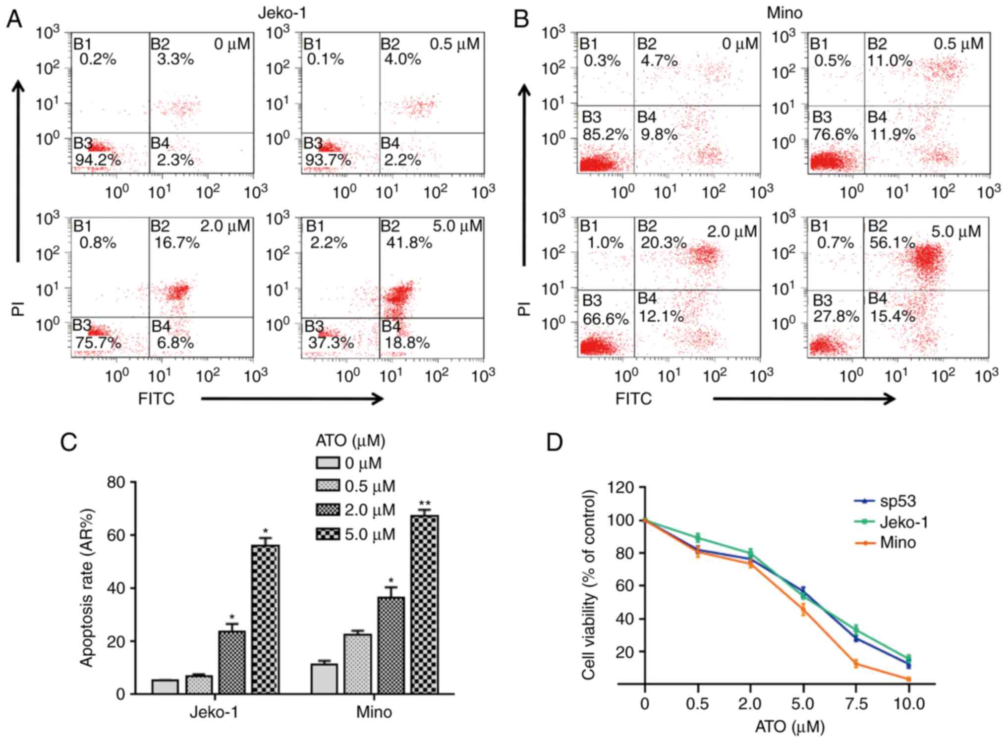 | Figure 1.ATO promotes cell apoptosis and
inhibits cell viability. (A and B) Apoptosis rates of Jeko-1 (A)
and Mino (B) cells treated with ATO in gradient concentrations (0,
0.5, 2.0 and 5 µM) for 24 h were quantified by flow cytometry. (C)
Statistical analysis of (A) and (B). Solvent group served as
control, Student's t-test, **P<0.01, *P<0.05. (D) After MCL
cell lines Jeko-1, Mino and SP53 were cultivated with ATO for 24 h
in gradient concentration (0, 0.5, 2.0, 5.0, 7.5 and 10.0 µM), the
viability of the cells was evaluated by CCK-8 assay. Data represent
the mean ± SD from three independent experiments. |
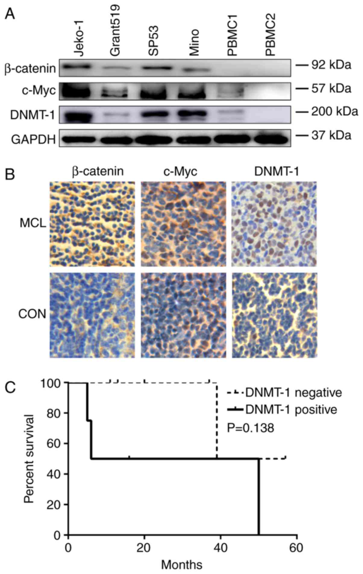
Wnt/β-catenin pathway is active and
DNMT-1 is upregulated in MCL
As the dysregulation of Wnt/β-catenin pathway could
promote lymphomagenesis, we examined the protein expression of
β-catenin, which is the core molecule of the Wnt/β-catenin pathway,
and the downstream molecule c-Myc in MCL. In MCL cell lines Jeko-1,
Grant519, SP53 and Mino, the expression of β-catenin and c-Myc in
all cell lines were notably higher compared with PBMCs from healthy
volunteers (Fig. 2A). Furthermore,
the conclusion was also confirmed in MCL tissue samples by
immunohistochemical staining (Fig.
2B).
Recent reports also indicated that methylation
related silence of Wnt inhibitory factors contributed to the
activation the Wnt/β-catenin pathway (6), we also showed that protein expression
of DNMT-1 in MCL cell lines and MCL patients' lymphoma tissues were
extremely higher than PBMCs from healthy volunteers and reactive
hyperplasia of lymph node tissues manifested by western blot
analysis and immunohistochemical staining, respectively (Fig. 2A and B). The upregulation of DNMT-1
further reduced expression of tumor suppressor genes which in turn
contributed to tumorigenesis. In our follow-up and survival
analysis, we found that the survival rate of MCL patients with
negative DNMT-1 expression was higher than the postive expression
group before 39 months according to the survival curve, although
the expression of DNMT-1 was not closely associated with MCL
patients' prognosis (Fig. 2C,
P>0.05).
ATO suppresses Wnt/β-catenin pathway
in MCL
To explore the inhibition mechanism of ATO in MCL,
we investigated the effect of ATO on Wnt/β-catenin pathway and
DNMT-1 which were demonstrated upregulated in MCL. The protein
expression of β-catenin and c-myc were found decreased with the
increased concentrations of ATO (0, 0.5, 2.0 and 5.0 µM) for 24 h
in Jeko-1, Mino and SP53 cells (Fig.
3A-F, P<0.05). We further investigated cyclin D1 and MMP7,
the downstream molecules of the Wnt/β-catenin pathway, were all
decreased in a dose-dependent manner with ATO in the transcript
level (Fig. 3G and H, P<0.05).
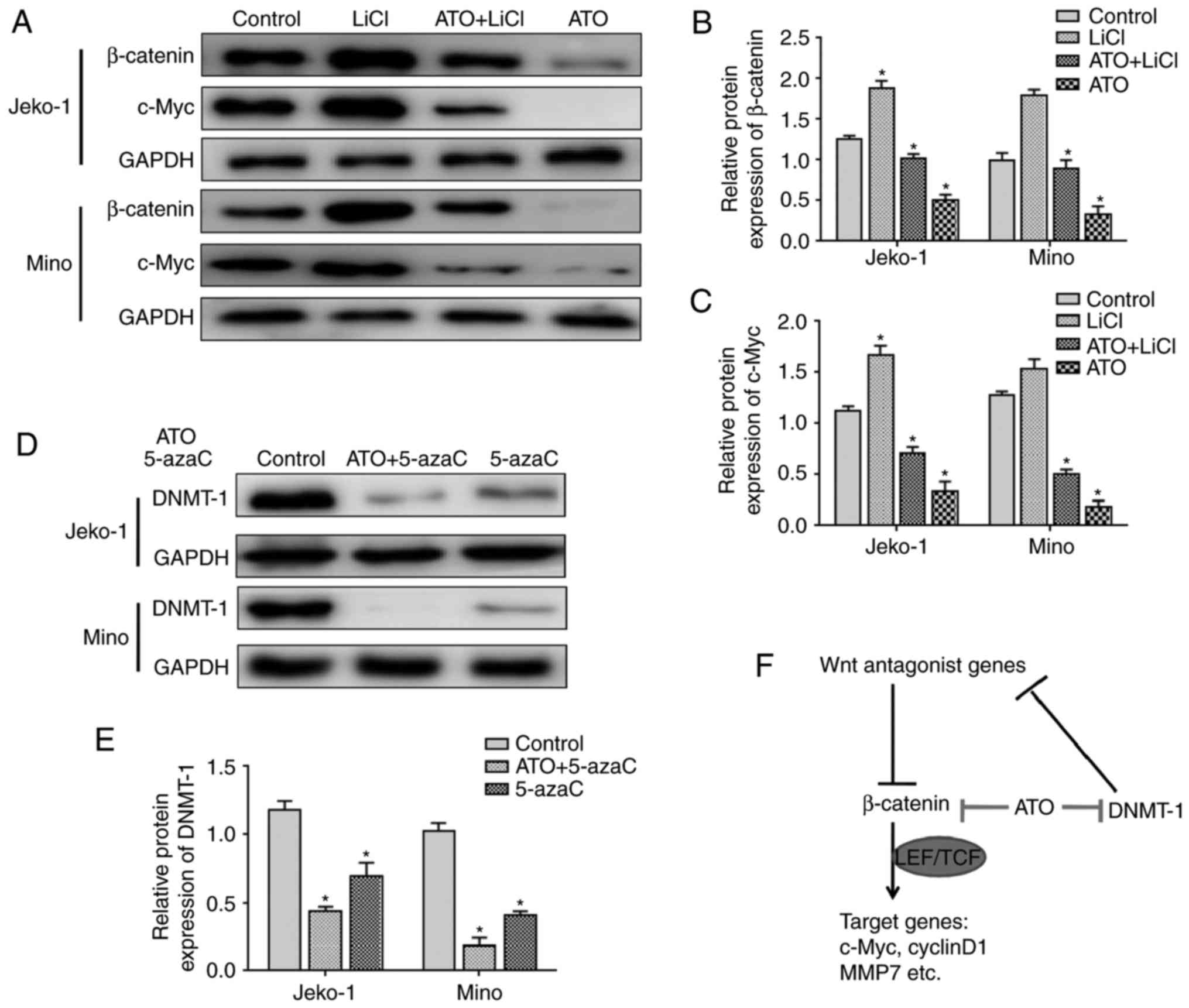
In addition, to verify the suppression effect on Wnt/β-catenin
pathway of ATO, LiCl which is a classical activator of
Wnt/β-catenin pathway was added. However, ATO attenuated
upregulation of β-catenin and c-myc expression after LiCl
stimulation in MCL cell lines (Fig.
4A-C, P<0.05).
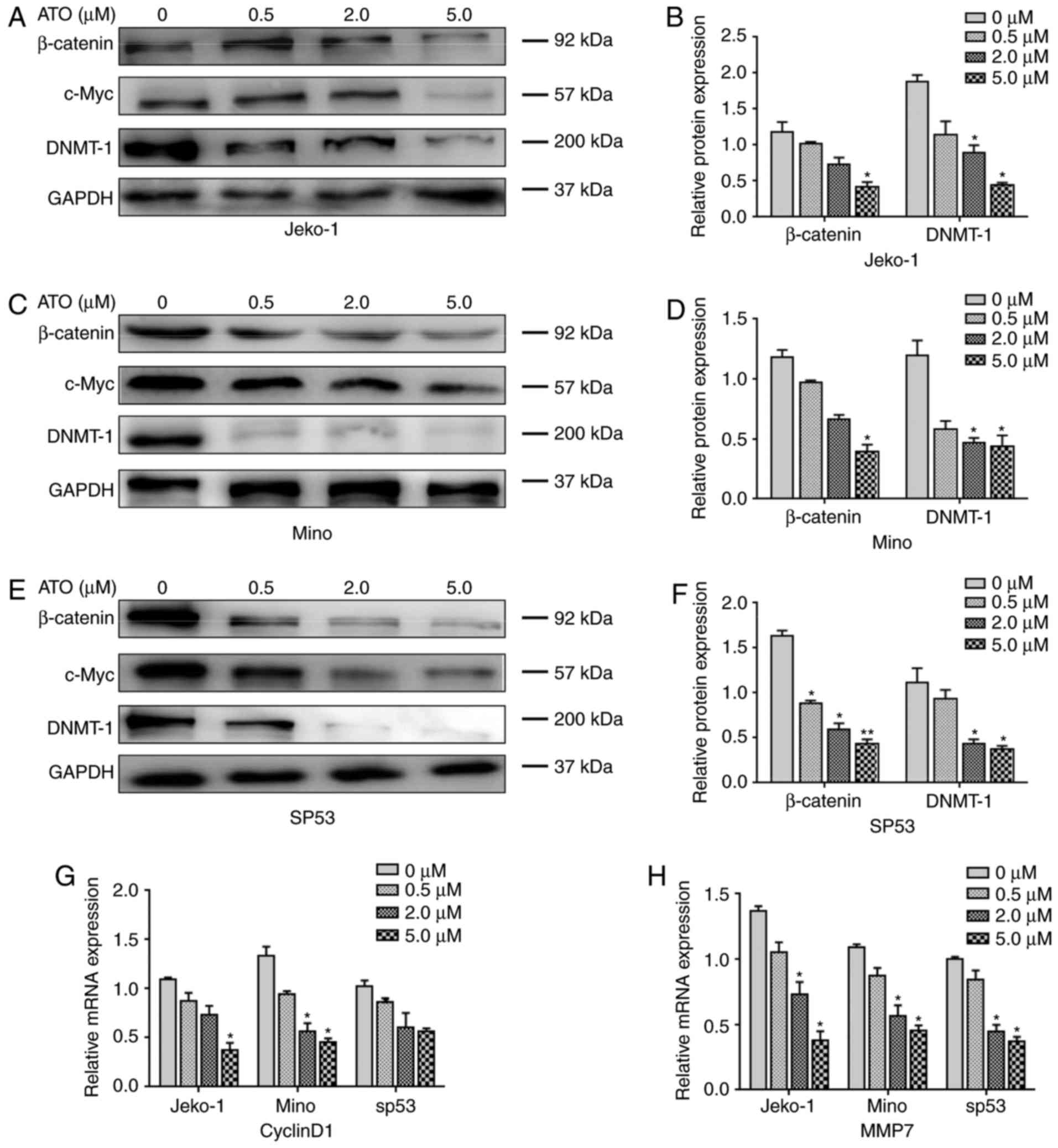 | Figure 3.ATO supresses Wnt/β-catenin pathway
and DNMT-1 in MCL. (A-F) After cultivated with ATO for 48 h in
gradient concentration (0, 1.0, 2.0 and 5.0 µM), protein
expressions of β-catenin, c-Myc and DNMT-1 in MCL cell lines Jeko-1
(A), Mino (C) and SP53 (E) were assayed by western blotting. (B, D
and F) Statistical analysis of (A), (C) and (E). Solvent group
served as control, Student's t-test, **P<0.01, *P<0.05. (G
and H) After cultivated with ATO for 48 h in gradient concentration
(0, 1.0, 2.0 and 5.0 µM), mRNA expression of cyclin D1 and MMP7 in
MCL cell lines Jeko-1, Mino and SP53 were quantified by qRT–PCR.
Solvent group served as control, Student's t-test, *P<0.05. Data
represent the mean ± SD from three independent experiments. |
ATO inhibits DNMT-1 expression in
MCL
The western blot analysis showed that the protein
expression of DNMT-1 was also downregulated by ATO in a
dose-dependent manner (0, 0.5, 2.0 and 5.0 µM) in MCL cell lines
Jeko-1, Mino and SP53 (Fig. 3A-F,
P<0.05). The combination of ATO and demethylation agent 5-azaC
was more potent than 5-azaC used alone in the inhibition of DMNT-1
expression (Fig. 4D and E,
P<0.05).
Discussion
In this study, we demonstrated that ATO could
promote apoptosis and inhibited cell viability in MCL cell lines.
Wnt/β-catenin pathway and DNMT-1 were constitutively activated in
MCL, however, the expression of DNMT-1, β-catenin and the
downstream molecules of Wnt/β-catenin pathway such as c-myc, cyclin
D1 and MMP7 were all downregulated in a dose-dependent manner with
the treatment of ATO. ATO also attenuated upregulation of β-catenin
after LiCl stimulation and provided synergistic effect with 5-azaC
on the DNMT-1 inhibition. Our results indicated that ATO suppressed
MCL by targeting Wnt/β-catenin pathway and DNMT-1. These findings
may provide better treatment options for MCL patients (Fig. 4F).
ATO has been found effective in solid tumors and
hematologic malignancies, studies revealed ATO interacts with
multiple molecules and signaling pathways (15,16).
In APL, at lower concentrations (0.1–0.5 µM), ATO promotes
differentiation; however, it induces apoptosis at higher
concentrations (0.5–2.0 µM) (8).
The apoptosis effect of ATO is associated with targeting and
inducing degradation of the PML-RARα protein and downregulation of
human telomerase gene (17). It is
underlined that significant increase of apoptosis rates and
inhibition of cell viability were also detected in Jeko-1 and Mino
cells with high concentrations of ATO compared with 0.5 µM
ATO-treated group in our result. ATO also has a promising role in
relapsed multiple myeloma (13).
Previous studies showed that ATO could inhibit capillary tubule
growth and vessel branching by reducing VEGF production (18). A report from Zhou et al
(19) indicated that ATO can
decrease the expression of β-catenin and induce synergic activity
with Bortezomib in multiple myeloma.
Activated Wnt/β-catenin pathway is a crucial pathway
for leukemic transformation, proliferation and survival (20). Previous reports mentioned
constitutively activation of the pathway and upregulation of
signaling molecules such as Wnt3 and Wnt10 in MCL (2,21).
Consistent with the previous investigation, our results manifested
enhancement of β-catenin and c-myc in the MCL cell lines and MCL
patients' lymph tissues. The expression of β-catenin and the
downstream molecules such as c-myc, cyclin D1 and MMP7 which are
associated with cell proliferation, cell cycle and invasion were
all downregulated in a dose-dependent manner with ATO. The
upregulation of β-catenin after LiCl stimulation was also
attenuated by ATO in MCL cell lines. Zheng et al also
considered ATO probably inhibited the Wnt/β-catenin pathway through
demethylating and reactivating the Wnt inhibitor, SFPR1 (22). Our result showed that DNMT-1 was
inhibited by ATO in a dose-dependent manner in MCL lines. DNMT-1,
DNMT-3a and DNMT3b are important methyltransferases for the
establishment and maintenance of DNA methylation status (23). DNMT-1 is responsible for the
maintenance of established DNA methylation patterns (24). Recent findings indicated that ATO
was catalyzed and converted into methylated products and then
produced S-adenosylhomocysteine which could bring about DNA
hypomethylation in liver and a small part in kidney and lung,
however, the process utilized S-adenosylmethionine which is an
essential co-factor of DNMTs, so that ATO participated in the
regulation of the methylated tumor suppressor genes by direct
inhibiting DNMTs and a depletion of the methyl donor (25). The tumor suppressor genes
demethylated by ATO may perform function by targeting signaling
pathways such as Wnt/β-catenin pathway. Furthermore, our follow-up
and survival analysis indicated that the survival rate of MCL
patients with negative DNMT-1 expression was higher than the
positive expression group before 39 months. The combination of ATO
and demethylation agent 5-azaC was more effective than 5-azaC used
alone in the inhibition of DMNT-1 expression. It is known that
5-azaC exerts a hypomethylating role in low concentration but a
cytotoxic activity with high concentration. The synergistic effect
of ATO with 5-azaC on the DNMT-1 inhibition suggested it may be an
efficient therapeutic strategy in MCL.
In conclusion, this study indicated that ATO has an
anticancer effect on MCL by targeting Wnt/β-catenin pathway and
DNMT-1. The synergistic effect of ATO and other agents such as
5-azaC may guide drug usage of ATO in clinical therapy for MCL.
However, the mechanism and unique metabolism of ATO still need
further research to explore its potential.
Acknowledgements
This study was supported in part by: National
Natural Science Foundation (nos. 81473486 and 81270598), National
Public Health Grand Research Foundation (no. 201202017), Natural
Science Foundations of Shandong Province (nos. ZR2012HZ003 and
2009ZRB14176), Technology Development Projects of Shandong Province
(nos. 2014GSF118021 and 2010GSF10250, and no. 2008GG2NS02018),
Program of Shandong Medical Leading Talent, and Taishan Scholar
Foundation of Shandong Province.
Glossary
Abbreviations
Abbreviations:
|
MCL
|
mantle cell lymphoma
|
|
ATO
|
arsenic trioxide
|
|
APL
|
acute promyelocytic leukemia
|
|
DNMT-1
|
DNA methyltransferase-1
|
|
5-azaC
|
5-azacytidine
|
References
|
1
|
Lu K, Chen N, Zhou XX, Ge XL, Feng LL, Li
PP, Li XY, Geng LY and Wang X: The STAT3 inhibitor WP1066
synergizes with vorinostat to induce apoptosis of mantle cell
lymphoma cells. Biochem Biophys Res Commun. 464:292–298. 2015.
View Article : Google Scholar : PubMed/NCBI
|
|
2
|
Gelebart P, Anand M, Armanious H, Peters
AC, Bard J Dien, Amin HM and Lai R: Constitutive activation of the
Wnt canonical pathway in mantle cell lymphoma. Blood.
112:5171–5179. 2008. View Article : Google Scholar : PubMed/NCBI
|
|
3
|
Hoffmeyer K, Raggioli A, Rudloff S, Anton
R, Hierholzer A, Del Valle I, Hein K, Vogt R and Kemler R:
Wnt/β-catenin signaling regulates telomerase in stem cells and
cancer cells. Science. 336:1549–1554. 2012. View Article : Google Scholar : PubMed/NCBI
|
|
4
|
Wang LQ, Wong KY, Rosèn A and Chim CS:
Epigenetic silencing of tumor suppressor miR-3151 contributes to
Chinese chronic lymphocytic leukemia by constitutive activation of
MADD/ERK and PIK3R2/AKT signaling pathways. Oncotarget.
6:44422–44436. 2015. View Article : Google Scholar : PubMed/NCBI
|
|
5
|
Queirós AC, Beekman R, Vilarrasa-Blasi R,
Duran-Ferrer M, Clot G, Merkel A, Raineri E, Russiñol N, Castellano
G, Beà S, et al: Decoding the DNA methylome of mantle cell lymphoma
in the light of the entire B cell lineage. Cancer Cell. 30:806–821.
2016. View Article : Google Scholar : PubMed/NCBI
|
|
6
|
Wang L, Shalek AK, Lawrence M, Ding R,
Gaublomme JT, Pochet N, Stojanov P, Sougnez C, Shukla SA, Stevenson
KE, et al: Somatic mutation as a mechanism of Wnt/β-catenin pathway
activation in CLL. Blood. 124:1089–1098. 2014. View Article : Google Scholar : PubMed/NCBI
|
|
7
|
Huang A, Yue D, Liao D, Cheng L, Ma J, Wei
Y, Tong A and Cheng P: Survivin T34A increases the therapeutic
efficacy of arsenic trioxide in mouse hepatocellular carcinoma
models. Oncol Rep. 36:3283–3290. 2016. View Article : Google Scholar : PubMed/NCBI
|
|
8
|
Falchi L, Verstovsek S, Ravandi-Kashani F
and Kantarjian HM: The evolution of arsenic in the treatment of
acute promyelocytic leukemia and other myeloid neoplasms: Moving
toward an effective oral, outpatient therapy. Cancer.
122:1160–1168. 2016. View Article : Google Scholar : PubMed/NCBI
|
|
9
|
Lo-Coco F, Avvisati G, Vignetti M, Thiede
C, Orlando SM, Iacobelli S, Ferrara F, Fazi P, Cicconi L, Di Bona
E, et al: Gruppo Italiano Malattie Ematologiche dell'Adulto;
German-Austrian Acute Myeloid Leukemia Study Group; Study Alliance
Leukemia: Retinoic acid and arsenic trioxide for acute
promyelocytic leukemia. N Engl J Med. 369:111–121. 2013. View Article : Google Scholar : PubMed/NCBI
|
|
10
|
Aldoss I, Mark L, Vrona J, Ramezani L,
Weitz I, Mohrbacher AM and Douer D: Adding ascorbic acid to arsenic
trioxide produces limited benefit in patients with acute myeloid
leukemia excluding acute promyelocytic leukemia. Ann Hematol.
93:1839–1843. 2014. View Article : Google Scholar : PubMed/NCBI
|
|
11
|
Martelli MP, Gionfriddo I, Mezzasoma F,
Milano F, Pierangeli S, Mulas F, Pacini R, Tabarrini A, Pettirossi
V, Rossi R, et al: Arsenic trioxide and all-trans retinoic acid
target NPM1 mutant oncoprotein levels and induce apoptosis in
NPM1-mutated AML cells. Blood. 125:3455–3465. 2015. View Article : Google Scholar : PubMed/NCBI
|
|
12
|
Bejanyan N, Tiu RV, Raza A, Jankowska A,
Kalaycio M, Advani A, Chan J, Saunthararajah Y, Mooney L,
Maciejewski JP, et al: A phase 2 trial of combination therapy with
thalidomide, arsenic trioxide, dexamethasone, and ascorbic acid
(TADA) in patients with overlap myelodysplastic/myeloproliferative
neoplasms (MDS/MPN) or primary myelofibrosis (PMF). Cancer.
118:3968–3976. 2012. View Article : Google Scholar : PubMed/NCBI
|
|
13
|
Sharma M, Khan H, Thall PF, Orlowski RZ,
Bassett RL Jr, Shah N, Bashir Q, Parmar S, Wang M, Shah JJ, et al:
A randomized phase 2 trial of a preparative regimen of bortezomib,
high-dose melphalan, arsenic trioxide, and ascorbic acid. Cancer.
118:2507–2515. 2012. View Article : Google Scholar : PubMed/NCBI
|
|
14
|
Dreyling M, Thieblemont C, Gallamini A,
Arcaini L, Campo E, Hermine O, Kluin-Nelemans JC, Ladetto M, Le
Gouill S, Iannitto E, et al: ESMO Consensus conferences: guidelines
on malignant lymphoma. part. 2:marginal zone lymphoma, mantle cell
lymphoma, peripheral T–cell lymphoma. Ann Oncol 24: 857–877.
2013.
|
|
15
|
Zhang S, Ma C, Pang H, Zeng F, Cheng L,
Fang B, Ma J, Shi Y, Hong H, Chen J, et al: Arsenic trioxide
suppresses cell growth and migration via inhibition of miR-27a in
breast cancer cells. Biochem Biophys Res Commun. 469:55–61. 2016.
View Article : Google Scholar : PubMed/NCBI
|
|
16
|
Zhang Z, Liu H, Zhou H, Zhu X, Zhao Z, Chi
X, Shan H and Gao J: A facile route to core-shell nanoparticulate
formation of arsenic trioxide for effective solid tumor treatment.
Nanoscale. 8:4373–4380. 2016. View Article : Google Scholar : PubMed/NCBI
|
|
17
|
Chou WC, Hawkins AL, Barrett JF, Griffin
CA and Dang CV: Arsenic inhibition of telomerase transcription
leads to genetic instability. J Clin Invest. 108:1541–1547. 2001.
View Article : Google Scholar : PubMed/NCBI
|
|
18
|
Roboz GJ, Dias S, Lam G, Lane WJ, Soignet
SL, Warrell RP Jr and Rafii S: Arsenic trioxide induces dose- and
time-dependent apoptosis of endothelium and may exert an
antileukemic effect via inhibition of angiogenesis. Blood.
96:1525–1530. 2000.PubMed/NCBI
|
|
19
|
Zhou L, Hou J, Fu W, Wang D, Yuan Z and
Jiang H: Arsenic trioxide and 2-methoxyestradiol reduce
beta-catenin accumulation after proteasome inhibition and enhance
the sensitivity of myeloma cells to Bortezomib. Leuk Res.
32:1674–1683. 2008. View Article : Google Scholar : PubMed/NCBI
|
|
20
|
Kühnl A, Valk PJ, Sanders MA, Ivey A,
Hills RK, Mills KI, Gale RE, Kaiser MF, Dillon R, Joannides M, et
al: Downregulation of the Wnt inhibitor CXXC5 predicts a better
prognosis in acute myeloid leukemia. Blood. 125:2985–2994. 2015.
View Article : Google Scholar : PubMed/NCBI
|
|
21
|
Mathur R, Sehgal L, Braun FK, Berkova Z,
Romaguerra J, Wang M, Rodriguez MA, Fayad L, Neelapu SS and
Samaniego F: Targeting Wnt pathway in mantle cell
lymphoma-initiating cells. J Hematol Oncol. 8:632015. View Article : Google Scholar : PubMed/NCBI
|
|
22
|
Zheng L, Jiang H, Zhang ZW, Wang KN, Wang
QF, Li QL and Jiang T: Arsenic trioxide inhibits viability and
induces apoptosis through reactivating the Wnt inhibitor secreted
frizzled related protein-1 in prostate cancer cells. Onco Targets
Ther. 9:885–894. 2016.PubMed/NCBI
|
|
23
|
Mizuno S, Chijiwa T, Okamura T, Akashi K,
Fukumaki Y, Niho Y and Sasaki H: Expression of DNA
methyltransferases DNMT1, 3A, and 3B in normal hematopoiesis and in
acute and chronic myelogenous leukemia. Blood. 97:1172–1179. 2001.
View Article : Google Scholar : PubMed/NCBI
|
|
24
|
Haas BW, Filkowski MM, Cochran RN, Denison
L, Ishak A, Nishitani S and Smith AK: Epigenetic modification of
OXT and human sociability. Proc Natl Acad Sci USA. 113:pp.
E3816–E3823. 2016; View Article : Google Scholar : PubMed/NCBI
|
|
25
|
Khaleghian A, Ghaffari SH, Ahmadian S,
Alimoghaddam K and Ghavamzadeh A: Metabolism of arsenic trioxide in
acute promyelocytic leukemia cells. J Cell Biochem. 115:1729–1739.
2014. View Article : Google Scholar : PubMed/NCBI
|