Introduction
Hepatocellular carcinoma (HCC) is the fifth most
diagnosed cancer and the second most frequent cause of
cancer-related deaths in males worldwide. In women, it is the
seventh most commonly diagnosed cancer and the sixth leading cause
of cancer-related deaths (1). The
prognosis remains poor in HCC patients since most systemic
therapies for this disease are ineffective. Therefore, new
treatment options, including molecular-targeted therapies, are
urgently needed.
LOX (lysyl oxidase) proteins belong to the LOX
family, which comprises five different enzymes (i.e., lysyl
oxidase, lysyl oxidase-like 1, lysyl oxidase-like 2, lysyl
oxidase-like 3 and lysyl oxidase-like 4), some of which have been
identified as oncogenes and regulators of cell growth (2–4). LOX
and LOXL1-4 are extracellular matrix-modifying enzymes that
catalyse crosslinking in collagen and elastin (5). Among these proteins, LOXL2 has been
reported to play a crucial role in metastasis in a variety of
malignancies (6–9). Increased LOXL2 levels lead to tumor
progression and metastasis, probably by promoting tumor cell
invasion and the remodelling of the tumor microenvironment
(2,10–14).
Previous studies revealed that LOXL2 was associated
with epithelial-mesenchymal transition (EMT) in tumor specimens
obtained from patients (9,15). Peinado et al (15) reported that LOXL2 mediated the
induction of EMT by repressing E-cadherin, indicating that LOXL2
contributes to tumor progression. EMT was originally proposed as a
process of organogenesis that was characterized by the combined
loss of expression of epithelial cell junction proteins, such as
E-cadherin, and the gain of expression of mesenchymal markers, such
as vimentin (16–18).
In the present study, we demonstrated that LOXL2 is
involved in the invasion of HCC and is expressed at higher levels
in HCC than in normal tissues. The clinical significance and
biological involvement of LOXL2 in the progression of HCC has not
yet been determined. Therefore, we investigated HCC specimens and
the effect of LOXL2 overexpression. Since LOXL2 plays important
roles in tumor cell motility and invasiveness, we analysed the
association between LOXL2 expression and prognosis in addition to
several clinicopathological factors.
Since Zhu et al have already reported that
the upregulation of LOX was associated with poor prognosis on HCC
patients (19), we further
investigated cell proliferation, invasive potential and wound
healing in LOXL2-inhibited HCC cell lines by BAPN, which is a
potent and irreversible inhibitor of lysyl oxidase (20,21).
Furthermore, we revealed that LOXL2 promoted HCC invasion and is
correlated with EMT and is therefore, an attractive therapeutic
target.
Materials and methods
Cell lines and culture conditions
Human HCC cell lines (SNU-182, SNU-387, SNU-398,
SNU-423 and SNU-449) were obtained from the American Type Culture
Collection (ATCC; Rockville, MD, USA). The HCC cell lines SK-HEP-1,
PLC/PRF/5 and Hep3B were kindly provided by Barrie Bode, Professor
and Chair of the Department of Biological Sciences at Northern
Illinois University (DeKalb, IL, USA). HuH-1, HuH-2 and HuH-7 cell
lines were provided by Jake Liang (NIDDK, National Institutes of
Health, Bethesda, MD, USA) and the HLE and HLF cell lines were
provided by Sato J (National Institutes of Biomedical Innovation,
Health and Nutrition, Japan). FOCUS, an another hepatoma cell line,
was provided by Jack Wands (Brown University, Providence, RI, USA).
All cell lines were cultured in Dulbecco's modified Eagle's medium
(Sigma-Aldrich, St. Louis, MO, USA), supplemented with 10% foetal
bovine serum (FBS) at 37°C in an atmosphere containing 5%
CO2.
Patients and specimens
Cancerous tissues and surrounding non-cancerous
hepatic parenchyma tissues were obtained from 150 patients with
primary HCC who underwent resection surgery at Nagoya University
Hospital, from May 1994 to December 2003. The present study was
approved by the Ethics Committee of Nagoya University Hospital, and
written informed consent was obtained from all patients. The mean
follow-up period during the prognosis study was 51.9 months.
Quantitative reverse transcription
polymerase chain reaction (qRT-PCR)
Total RNA was isolated from primary HCC tissues and
the corresponding non-cancerous tissues and used to generate
complementary DNA. The cDNAs were amplified using PCR primers that
were specific for LOX, LOXL1, LOXL2, LOXL3 LOXL4, E-cadherin and
vimentin. Glyceraldehyde-3-phosphate dehydrogenase (GAPDH) was used
as an internal control. RNA expression levels were determined by
qRT-PCR. Primers are listed in Table
I (22). The level of mRNA was
assessed by standard curves. Real-time detection of the emission
intensity of SYBR®-Green was performed using StepOne
Plus (Applied Biosystems, Foster City, CA, USA). Each qRT-PCR was
performed at least three times and a no-template control was used
as a negative control.
 | Table I.Primer sets designed for the qRT-PCR
assay. |
Table I.
Primer sets designed for the qRT-PCR
assay.
| Primer | Sequence | Genome
position | Length (bp) |
|---|
| LOX: F |
GCGGCGGAGGAAAACTGT | 964–981 | 19 |
| LOX: R |
AGCAGCACCCTGTGATCATAATC | 1015–1037 | 24 |
| LOXL1: F |
GACTGCCAGTGGATCGACATAA | 1875–1896 | 22 |
| LOXL1: R |
CTCCAAAACAATATACTTTGGGTTCA | 1936–1961 | 26 |
| LOXL2: F |
CTCCCAGATCCACAACAATGG | 2139–2159 | 21 |
| LOXL2: R |
AGCAGGTCATAGTGGGTGAACA | 2240–2261 | 22 |
| LOXL3: F |
TGCAAATATGATGGACATAGAATCTG | 1809–1834 | 26 |
| LOXL3: R |
CAAACCTCCTGTTGGCCTCTT | 1870–1890 | 21 |
| LOXL4: F |
GGGCCCCGGGAATTATATCT | 2215–2234 | 20 |
| LOXL4: R |
GCATATTGTTGGAGAAATCTGACTCT | 2269–2294 | 26 |
| GAPDH: F |
GCATATTGTTGGAGAAATCTGACTCT | 19–40 | 26 |
| GAPDH: R |
GCATATTGTTGGAGAAATCTGACTCT | 69–94 | 26 |
The EMT status of each patient tumor was calculated
using the mRNA expression levels of E-cadherin and vimentin as
follows: vimentin/E-cadherin ratio <2, epithelial type (E);
vimentin/E-cadherin ≥2, mesenchymal type (M).
Cell migration and invasion
assays
Control and transfected cells (HLF and SK-HEP-1)
were transplanted into a 35-mm culture-insert µ-Dish for migration
assays (Ibidi GmbH, Martinsried, Germany). The cells were plated in
triplicate at a density of 5,000 cells/well in a micro-dish. After
24 h, medium containing 350 µM BAPN (Sigma-Aldrich), which has been
reported as an inhibitor of LOXL2 or control was added at the
optimized concentrations (23,24).
After 24 h, frames were removed, and wounds were made in the
plates. Cell migration towards the wound was monitored at 24 and 48
h.
For the invasion assays, the cells were trypsinized
in 0.25% trypsin containing EDTA. Subsequently, the cells were
suspended and then implanted in a Transwell insert with 8-µm pores
that was previously coated with 50 µl Matrigel (Corning Life
Sciences, Tewksbury, MA, USA) and 200 µl of medium containing 5%
FBS was added to the bottom chamber. Migration was allowed for 24 h
and a cotton swab was then used to remove the non-migrated cells in
the upper chamber. The filters were then individually fixed with 4%
polysorbate and dyed with Diff-Quik stain (Dade Behring Holdings,
Inc., Deerfield, IL, USA). Migrated cells were counted in five
random fields in each chamber under a light microscope (Olympus
DP-70; Olympus Corp., Tokyo, Japan).
Cell proliferation assay
Cell proliferation of the HLF and SK-HEP-1 cell
lines was evaluated in vitro using
3-(4,5-dimethylthiazol-2-yl)-2,5 diphenyl tetrazolium bromide (MTT)
assays. Control and transfected cells were seeded in 96-well plates
at an initial cell density of 5,000 cells/well. The cells were
cultured in DMEM containing 5% FBS for 24, 48, 72 and 96 h. At the
indicated time-point, the culture medium was replaced with 800
µg/ml MTT solution and the cells were then incubated for another 3
h. After the MTT solution was removed, 100 µl of dimethyl sulfoxide
(DMSO) was added to each well to dissolve the blue formazan. The
plates were shaken for 5 min and absorbance was assessed at a
wavelength of 570 nm.
Cell culture supernatants
The culture supernatants of HCC cell lines,
including SK-HEP-1, PLC/PRF/5, Hep-3B, HLE, HLF, HuH-2 and HuH-7
cells, were investigated using a CircuLex Human LOXL2 ELISA kit
(Medical & Biological Laboratories, Co., Ltd., Nagoya, Japan).
A total of 1×10⁸ cells were transplanted and cultured in
DMEM containing 10% FBS for 24 or 48 h. After 48 h, the culture
supernatant was separated using a centrifuge and the amount of
LOXL2 protein in the culture supernatant was assessed using an
ELISA kit.
Statistical analysis
Differences in numerical data between two groups
were evaluated using Fisher's exact test or χ2 test.
Survival rates were calculated using the Kaplan-Meier method and
the difference in survival curves was analysed using the log-rank
test. Independent prognostic factors were analysed using the Cox
proportional hazards regression model. Data are expressed as the
mean ± SD. P<0.05 is considered to indicate a statistically
significant difference. The data were analysed using JMP version 11
software (JMP; SAS Institute, Cary, NC, USA).
Results
The expression of LOX and LOXL family
members in human HCC cell lines
Real-time PCR analyses were used to determine the
mRNA expression levels of LOX and LOXL family members in human HCC
cell lines (Fig. 1A). Furthermore,
the mRNA expression levels of E-cadherin and vimentin were assessed
to determine the extent of EMT and to evaluate the correlation
between the EMT status and the expression levels of LOX and LOXL
family members. A positive correlation between EMT status and LOXL2
expression was observed in human HCC cell lines (Fig. 1B).
LOXL2 expression in resected HCC
specimens and its clinicopathological features
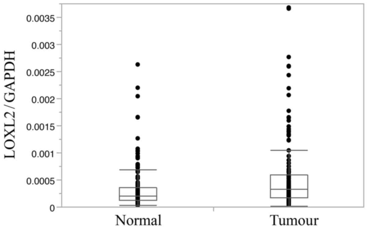
Subsequently, the expression of LOXL2 was assessed
in cancerous and normal liver tissues obtained from 150 resected
HCC specimens. LOXL2 expression levels were significantly higher in
cancerous tissues than in normal tissues (Fig. 2).
The demographics of the 150 patients that were
subjected to HCC resection are listed in Table II. The enrolled patients were
assigned to two groups (high and low expression) according to the
average mRNA level of LOXL2. The high and low expression groups
included 40 and 110 patients, respectively. No significant
correlations were found between LOXL2 expression levels and
clinicopathological parameters (Table
III).
| Table II.Patient demographics. |
Table II.
Patient demographics.
| Age, years (mean ±
SD) | 62.3 (±10.1) |
| Sex
(male/female) | 131/19 |
| Etiology (HBV vs.
HCV vs. HBV + HCV vs. others) | 28/97/2/23 |
| Histological type
of tumor (mod/well/poor) | 111/30/9 |
| Tumor size
(cm) | 4.8 (±3.1) |
| Tumor multiplicity
(solitary vs. multiple) | 113/37 |
| Pattern of tumor
growth (expansive vs. infiltrative) | 124/26 |
| Formation of
fibrous capsule (present vs. absent) | 113/37 |
| Septal formation
(present vs. absent) | 99/51 |
| Child-Pugh
classification (A/B) | 135/21 |
| Liver damage
(A/B) | 107/43 |
| Pathological T
category (T1/T2/T3/T4) | 15/81/38/16 |
| Portal vein
invasion [(+) vs. (−)] | 26/124 |
| Venous invasion
[(+) vs. (−)] | 9/141 |
| Serum AFP level
(±SD) (ng/ml) | 62.3 (±10.1) |
| Table III.Correlation between LOXL2 expression
and clinicopathological characteristics of HCC patients. |
Table III.
Correlation between LOXL2 expression
and clinicopathological characteristics of HCC patients.
|
Characteristics | LOXL2 high
(N=40) | LOXL2 low
(N=110) | P-value |
|---|
| Age, years (mean ±
SD) | 18/22 | 54/56 | 0.716 |
| Sex
(male/female) | 33/7 | 98/12 | 0.283 |
| Etiology (HBV vs.
HCV vs. HBV + HCV vs. others) | 8/26/0/6 | 20/71/2/17 | 0.853 |
| Histological type
of tumor (mod/well/poor) | 28/9/3 | 83/21/6 | 0.782 |
| Tumor size (mean ±
SD), (cm) | 30/10 | 64/46 | 0.117 |
| Tumor mutiplicity
(solitary vs. multiple) | 29/11 | 84/26 | 0.627 |
| Pattern of tumor
growth (expansive vs. infiltrative) | 36/4 | 88/28 | 0.153 |
| Formation of
fibrous capsule (present vs. absent) | 32/8 | 81/29 | 0.424 |
| Septal formation
(present vs. absent) | 23/17 | 76/34 | 0.185 |
| Child-pugh
classification (A/B) | 29/4 | 94/6 | 0.322 |
| Liver damage
(A/B) | 26/14 | 81/29 | 0.301 |
| Pathological T
category (T1/T2/T3/T4) | 1/22/13/4 | 14/59/25/13 | 0.315 |
| Portal vein
invasion [(+) vs. (−)] | 35/5 | 89/21 | 0.346 |
| Venous invasion
[(+) vs. (−)] | 37/3 | 100/10 | 0.759 |
| Serum AFP level
(mean ± SD), (ng/ml) | 2245±1125 | 1703±740.5 | 0.578 |
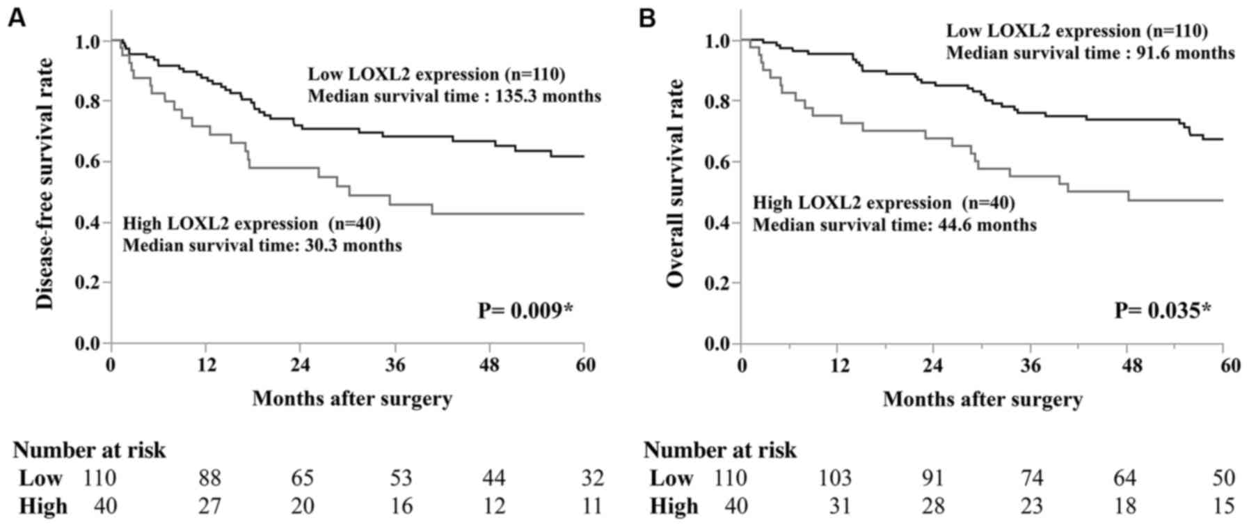
Survival analysis of the expression of
LOXL2 in HCC-resected patients
To determine whether LOXL2 affects survival in
patients with HCC, we analysed disease-free survival (DFS) and
overall survival (OS). For DFS, the median survival time (MST) of
the patients with low and high LOXL2 expression was 135.3 and 30.3
months, respectively (P=0.009; Fig.
3A). For OS, the MST of patients with low and high LOXL2
expression was 91.6 and 44.6 months, respectively (P=0.035;
Fig. 3B). These results revealed
that there was a significant difference in survival time in
resected HCC patients.
The clinical variables associated with HCC prognoses
were evaluated using COX regression models. A univariate analysis
revealed that the following prognostic factors were significant for
overall survival: tumor multiplicity, pathological stage, portal
vein invasion, venous invasion, serum AFP level and LOXL2
expression. A multivariate analysis demonstrated that portal vein
invasion (HR, 2.363, 95% CI, 1.19–4.566, P=0.015), venous invasion
(HR, 1.673, 95% CI, 1.051–2.710, P=0.026), serum AFP level (HR,
2.363, 95% CI, 1.098–2.871, P=0.019), and LOXL2 expression (HR,
1.782, 95%CI, 1.003–3.117, P=0.009), were independent prognostic
factors (Table IV).
| Table IV.Univariate and multivariate analyses
of prognostic factors on overall survival. |
Table IV.
Univariate and multivariate analyses
of prognostic factors on overall survival.
|
| Univariate
analysis | Multivariate
analysis |
|---|
|
|
|
|
|---|
|
| HR | 95% CI | P-value | HR | 95% CI | P-value |
|---|
| Age, years (≥65 vs.
<65) | 1.219 | 0.772–1.933 | 0.393 |
|
|
|
| Sex (male vs.
female) | 1.294 | 0.659–2.927 | 0.478 |
|
|
|
| Tumor size (high
vs. low) | 1.596 | 0.987–2.662 | 0.057 |
|
|
|
| Tumor multiplicity
(multiple vs. solitary) | 1.735 | 1.066–2.767 | 0.027a | 1.621 | 1.066–2.767 | 0.083 |
| Pattern of tumor
growth (expansive vs. infiltrative) | 1.036 | 0.579–2.019 | 0.911 |
|
|
|
| Formation of
fibrous capsule (absent vs. present) | 1.583 | 0.941–2.575 | 0.082 |
|
|
|
| Septal formation
(absent vs. present) | 1.344 | 0.826–2.144 | 0.229 |
|
|
|
| Child-Pugh
classification (B vs. A) | 1.544 | 0.828–2.682 | 0.163 |
|
|
|
| Liver damage (B vs.
A) | 1.388 | 0.856–2.208 | 0.179 |
|
|
|
| Pathological stage
(III, IV vs. I, II) | 1.816 | 1.140–2.871 | 0.012a | 0.094 | 0,487-1.664 | 0.747 |
| Portal vein
invasion [(+) vs. (−)] | 2.216 | 1.197–3.579 | 0.011a | 2.363 | 1.09–4.566 | 0.015a |
| Venous invasion
[(+) vs. (−)] | 2.656 | 1.219–5.135 | 0.016a | 1.673 | 1.051–2.71 | 0.026a |
| Serum AFP level
(high vs. low) | 1.673 | 1.051–2.710 | 0.029a | 1.760 | 1.098–2.871 | 0.019a |
| LOXL2 expression
(high vs. low) | 1.672 | 1.017–2.681 | 0.043a | 1.782 | 1.003–3.117 | 0.009a |
Migration and invasion assay of LOXL2
suppressed cell lines
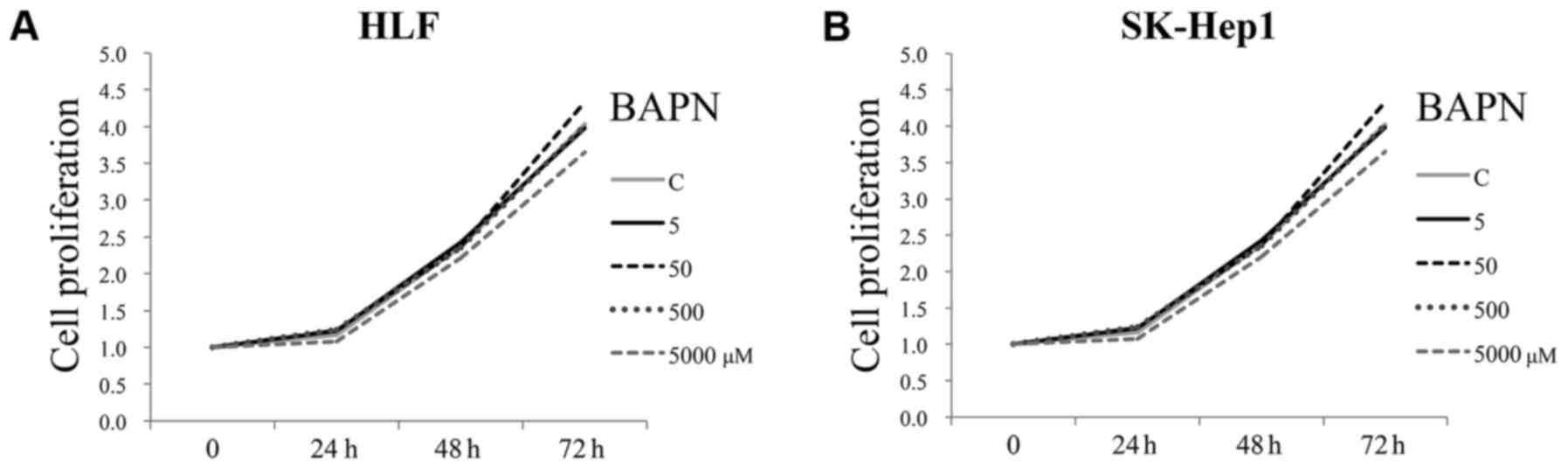
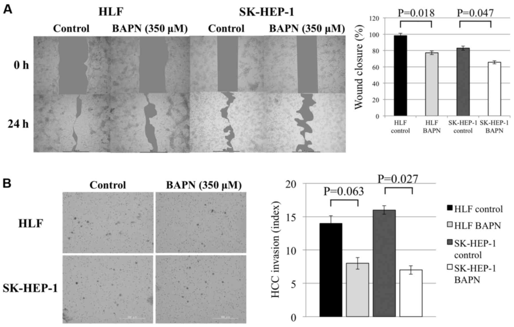
A functional analysis of LOXL2 expression was
performed in HCC cell lines using BAPN. LOXL2 was overexpressed in
HLF and SK-HEP-1 cells and the results of the MTT assays revealed
that BAPN did not suppress proliferation in HCC cells. In both cell
lines, even when the BAPN concentration was increased, the
proliferation rate was not affected (Fig. 4). In wound healing assays, wound
closure was suppressed by BAPN in both cell lines (Fig. 5A) and in invasion assays, the
invasion ability was significantly suppressed by BAPN (Fig. 5B).
Characterization of the expression of
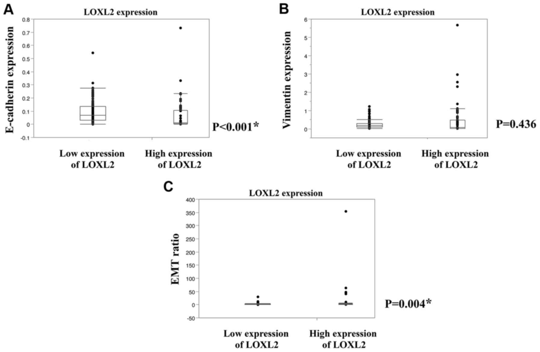
LOXL2 in HCC patients and correlation with EMT status
The mRNA expression levels of E-cadherin and
vimentin were evaluated using real-time PCR in cancerous tissues
obtained from 150 resected-HCC patients and then, the EMT status
was determined based on the vimentin to E-cadherin ratio, as
described in Materials and methods section. Although there was no
significant correlation between LOXL2 expression and vimentin,
there was a significant correlation between LOXL2 expression and
both E-cadherin levels and EMT (Fig.
6).
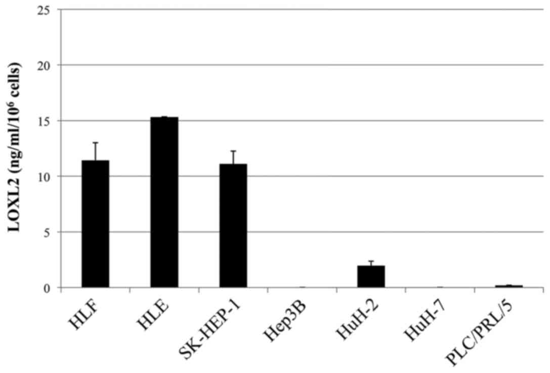
LOXL2 levels in cultured
supernatants
Finally, we assessed the expression levels of LOXL2
in the culture supernatants of HCC cell lines. The results of these
assays are displayed in Fig. 7. In
HLE, HLF and SK-HEP-1 cells, the expression levels of LOXL2 were
high, similar to those of LOXL2 in HCC cell lines.
Discussion
LOX is a key enzyme that controls extracellular
matrix, collagen and elastin maturation, however its role is not
limited to these functions. It also plays a critical role in cancer
development and invasion. Previous studies have reported that LOX
is downregulated in various malignancies, including basal and
squamous cell, colon, oesophageal, gastric, head and neck squamous
cell, pancreatic, and prostatic cancer (21,25–27).
In particular, LOXL2 has been reported to be an important molecule
during metastasis in a variety of malignancies (6–9).
Several studies have reported that LOXL2 is associated with tumor
development and invasion in HCC (19,28,29).
In the present study, we demonstrated that LOXL2 levels are
certainly correlated with EMT status in our cell line libraries as
previously reported, and that LOXL2 inhibition by BAPN reduced the
migration and invasion ability of HCC cells.
Whether the LOX family is clinically implicated in
cancer development and progression has remained controversial
(30). Increased expression of
LOXL2 leads to tumor progression and metastasis, probably because
LOXL2 promotes tumor cell invasion and the remodelling of the tumor
microenvironment. Previous studies have revealed that LOXL2 was
associated with EMT in tumor specimens obtained from patients
(9,15). Peinado et al (15) reported that LOXL2 mediated the
induction of EMT by suppressing E-cadherin, indicating that LOXL2
contributes to tumor progression. EMT was originally proposed as a
process involved in organogenesis that was characterized by the
combined loss of expression of epithelial cell junction proteins,
such as E-cadherin, and gain of expression of mesenchymal markers,
such as vimentin (16–18). Therefore, targeting the LOX family
to control EMT has been suggested as a new therapeutic strategy for
treating cancer (14).
We investigated the proliferative activities and
invasive and migratory potential in HCC cells using BAPN, a potent
irreversible lysyl oxidase inhibitor. Several previous studies have
used BAPN to inhibit LOXL2 both in vitro and in vivo
(10,14,31).
In the present study, we found that HCC cell proliferation was not
inhibited by BAPN, whereas migration and invasion were
significantly inhibited. We think this may have occurred because
LOXL2 has been reported to regulate intracellular and extracellular
proteins (5,32), and induce fibrosis, tumor invasion
and metastasis (6–9,13,15,28).
To better understand the clinical association
between LOXL2 and EMT in HCC prognosis, we assessed E-cadherin and
vimentin expression levels as markers of epithelial and mesenchymal
cells, respectively. Previous studies have revealed that patients
with high expression levels of E-cadherin had better overall
survival than those with low E-cadherin levels. Therefore,
E-cadherin may be a prognostic marker in HCC patients (33,34).
In a previous study, we reported that EMT status was a critical
prognostic factor in pancreatic (17), gastric (35) and colorectal cancer (36). When EMT status was determined using
the vimentin/E-cadherin ratio (17), patients could be divided into
epithelial and mesenchymal groups. The patients in the mesenchymal
group had significantly poorer survival than those in the
epithelial group. Furthermore, there was significant correlation
between the expression of LOXL2 and vimentin. However, there was a
significant correlation between LOXL2 expression and E-cadherin
levels or EMT in the 150 surgically resected specimens examined in
this study. In a previous study, tumor metastasis was induced as a
result of the intracellular LOXL2-induced oxidation of the
transcription factor Snail, which subsequently induced EMT
(11,15,37).
These findings are consistent with our results, indicating that our
method of predicting HCC prognosis could be useful. In order to
eliminate the possibility of BAPN side-effects, we think that these
knocked-down experiments should be confirmed by siRNA
experiments.
Several limitations of the present study should be
acknowledged. Firstly, the study was retrospectively designed,
although the results were in accordance with previous studies
(28,38). Secondly, we only assessed the mRNA
expression. An analysis of a larger cohort based on the protein
expression is required to confirm the present exploratory study. In
addition, further studies are required to investigate the
background mechanisms involved in the association between LOXL2 and
EMT in HCC.
In conclusion, we have demonstrated that LOXL2
levels are a critical prognostic factor that is correlated with EMT
in HCC patients. Further investigations are necessary to determine
the relationship between EMT and LOXL2 and its value as a novel
therapeutic target.
Acknowledgements
The present study was supported by the Department of
Gastroenterological Surgery, Nagoya University Graduate School of
Medicine, the Charitable Trust Soyu Medical Foundation. We thank
Yoko Nishikawa for collecting clinical samples and for helping to
perform the experiments.
Funding
The study was funded by the The Charitable Trust
Soyu Medical Foundation.
Availability of data and materials
The datasets used during the present study are
available from the corresponding author upon the reasonable
request.
Authors' contributions
GN and SY conceived and designed the study. SY and
YK provided administrative support. GN, SY, MH, MS, HT, HS and TF
provided the study materials and patients. GN, SY, ST, YN, MK, NI,
CT and DK collected and assembled the data. GN, SY, GN, MK and MF
analysed and interpreted the data. GN, SY, MH, MS and YK wrote the
manuscript. All authors read and approved the manuscript and agree
to be accountable for all aspects of the research in ensuring that
the accuracy or integrity of any part of the work are appropriately
investigated and resolved.
Ethics approval and consent to
participate
All experimental protocols were approved by the
Ethics Committee of Nagoya University Hospital (Nagoya, Japan).
Consent for publication
Not applicable.
Competing interests
The authors declare that they have no competing
interests.
Glossary
Abbreviations
Abbreviations:
|
LOX
|
lysyl oxidase
|
|
LOXL2
|
lysyl oxidase-like 2
|
|
EMT
|
epithelial-to-mesenchymal
transition
|
|
HCC
|
hepatocellular carcinoma
|
|
mRNA
|
messenger RNA
|
|
qRT-PCR
|
quantitative reverse transcription
polymerase chain reaction
|
|
BAPN
|
β-aminopropionitrile
|
|
MTT
|
3-(4,5-dimethylthiazol-2-yl)-2,5
diphenyl tetrazolium bromide
|
|
AFP
|
α-fetoprotein
|
References
|
1
|
Jemal A, Bray F, Center MM, Ferlay J, Ward
E and Forman D: Global cancer statistics. CA Cancer J Clin.
61:69–90. 2011. View Article : Google Scholar : PubMed/NCBI
|
|
2
|
Akiri G, Sabo E, Dafni H, Vadasz Z,
Kartvelishvily Y, Gan N, Kessler O, Cohen T, Resnick M, Neeman M
and Neufeld G: Lysyl oxidase-related protein-1 promotes tumor
fibrosis and tumor progression in vivo. Cancer Res. 63:1657–1666.
2003.PubMed/NCBI
|
|
3
|
Geach TJ and Dale L: Members of the lysyl
oxidase family are expressed during the development of the frog
Xenopus laevis. Differentiation. 73:414–424. 2005.
View Article : Google Scholar : PubMed/NCBI
|
|
4
|
Xu X, Wang B and Xu Y: Expression of lysyl
oxidase in human osteosarcoma and its clinical significance: A
tumor suppressive role of LOX in human osteosarcoma cells. Int J
Oncol. 43:1578–1586. 2013. View Article : Google Scholar : PubMed/NCBI
|
|
5
|
Kim YM, Kim EC and Kim Y: The human lysyl
oxidase-like 2 protein functions as an amine oxidase toward
collagen and elastin. Mol Biol Rep. 38:145–149. 2011. View Article : Google Scholar : PubMed/NCBI
|
|
6
|
Fong SF, Dietzsch E, Fong KS, Hollosi P,
Asuncion L, He Q, Parker MI and Csiszar K: Lysyl oxidase-like 2
expression is increased in colon and esophageal tumors and
associated with less differentiated colon tumors. Genes Chromosomes
Cancer. 46:644–655. 2007. View Article : Google Scholar : PubMed/NCBI
|
|
7
|
Offenberg H, Brünner N, Mansilla F, Torben
Orntoft F and Birkenkamp-Demtroder K: TIMP-1 expression in human
colorectal cancer is associated with TGF-B1, LOXL2, INHBA1,
TNF-AIP6 and TIMP-2 transcript profiles. Mol Oncol. 2:233–240.
2008. View Article : Google Scholar : PubMed/NCBI
|
|
8
|
Peng L, Ran YL, Hu H, Yu L, Liu Q, Zhou Z,
Sun YM, Sun LC, Pan J, Sun LX, et al: Secreted LOXL2 is a novel
therapeutic target that promotes gastric cancer metastasis via the
Src/FAK pathway. Carcinogenesis. 30:1660–1669. 2009. View Article : Google Scholar : PubMed/NCBI
|
|
9
|
Rückert F, Joensson P, Saeger HD,
Grützmann R and Pilarsky C: Functional analysis of LOXL2 in
pancreatic carcinoma. Int J Colorectal Dis. 25:303–311. 2010.
View Article : Google Scholar : PubMed/NCBI
|
|
10
|
Erler JT and Giaccia AJ: Lysyl oxidase
mediates hypoxic control of metastasis. Cancer Res. 66:10238–10241.
2006. View Article : Google Scholar : PubMed/NCBI
|
|
11
|
Peinado H, Moreno-Bueno G, Hardisson D,
Pérez-Gómez E, Santos V, Mendiola M, de Diego JI, Nistal M,
Quintanilla M, Portillo F and Cano A: Lysyl oxidase-like 2 as a new
poor prognosis marker of squamous cell carcinomas. Cancer Res.
68:4541–4550. 2008. View Article : Google Scholar : PubMed/NCBI
|
|
12
|
Barry-Hamilton V, Spangler R, Marshall D,
McCauley S, Rodriguez HM, Oyasu M, Mikels A, Vaysberg M, Ghermazien
H, Wai C, et al: Allosteric inhibition of lysyl oxidase-like-2
impedes the development of a pathologic microenvironment. Nat Med.
16:1009–1017. 2010. View
Article : Google Scholar : PubMed/NCBI
|
|
13
|
Barker HE, Chang J, Cox TR, Lang G, Bird
D, Nicolau M, Evans HR, Gartland A and Erler JT: LOXL2-mediated
matrix remodeling in metastasis and mammary gland involution.
Cancer Res. 71:1561–1572. 2011. View Article : Google Scholar : PubMed/NCBI
|
|
14
|
Barker HE, Cox TR and Erler JT: The
rationale for targeting the LOX family in cancer. Nat Rev Cancer.
12:540–552. 2012. View
Article : Google Scholar : PubMed/NCBI
|
|
15
|
Peinado H, Del Carmen Iglesias-de la Cruz
M, Olmeda D, Csiszar K, Fong KS, Vega S, Nieto MA, Cano A and
Portillo F: A molecular role for lysyl oxidase-like 2 enzyme in
snail regulation and tumor progression. EMBO J. 24:3446–3458. 2005.
View Article : Google Scholar : PubMed/NCBI
|
|
16
|
Thiery JP: Epithelial-mesenchymal
transitions in tumour progression. Nat Rev Cancer. 2:442–454. 2002.
View Article : Google Scholar : PubMed/NCBI
|
|
17
|
Yamada S, Fuchs BC, Fujii T, Shimoyama Y,
Sugimoto H, Nomoto S, Takeda S, Tanabe KK, Kodera Y and Nakao A:
Epithelial-to-mesenchymal transition predicts prognosis of
pancreatic cancer. Surgery. 154:946–954. 2013. View Article : Google Scholar : PubMed/NCBI
|
|
18
|
Yamada S, Okumura N, Wei L, Fuchs BC,
Fujii T, Sugimoto H, Nomoto S, Takeda S, Tanabe KK and Kodera Y:
Epithelial to mesenchymal transition is associated with shorter
disease-free survival in hepatocellular carcinoma. Ann Surg Oncol.
21:3882–3890. 2014. View Article : Google Scholar : PubMed/NCBI
|
|
19
|
Zhu J, Huang S, Wu G, Huang C, Li X, Chen
Z, Zhao L and Zhao Y: Lysyl oxidase is predictive of unfavorable
outcomes and essential for regulation of vascular endothelial
growth factor in hepatocellular carcinoma. Dig Dis Sci.
60:3019–3031. 2015. View Article : Google Scholar : PubMed/NCBI
|
|
20
|
Tang SS, Trackman PC and Kagan HM:
Reaction of aortic lysyl oxidase with beta-aminopropionitrile. J
Biol Chem. 258:4331–4338. 1983.PubMed/NCBI
|
|
21
|
Kirschmann DA, Seftor EA, Fong SF, Nieva
DR, Sullivan CM, Edwards EM, Sommer P, Csiszar K and Hendrix MJ: A
molecular role for lysyl oxidase in breast cancer invasion. Cancer
Res. 62:4478–4483. 2002.PubMed/NCBI
|
|
22
|
Rahn DD, Good MM, Roshanravan SM, Shi H,
Schaffer JI, Singh RJ and Word RA: Effects of preoperative local
estrogen in postmenopausal women with prolapse: A randomized trial.
J Clin Endocrinol Metab. 99:3728–3736. 2014. View Article : Google Scholar : PubMed/NCBI
|
|
23
|
Gunasekaran S, Weinstein P and Chvapil MJ:
Effect of topical beta APN application on evoked potential
conduction in rat sciatic nerve and spinal cord. Surg Neurol.
28:201–207. 1987. View Article : Google Scholar : PubMed/NCBI
|
|
24
|
da Silva R, Uno M, Marie SK and Oba-Shinjo
SM: The lysyl oxidase inhibitor, beta-aminopropionitrile,
diminishes the metastatic colonization potential of circulating
breast cancer cells. PLoS One. 10:e01197812015. View Article : Google Scholar : PubMed/NCBI
|
|
25
|
Payne SL, Hendrix MJ and Kirschmann DA:
Paradoxical roles for lysyl oxidases in cancer-a prospect. J Cell
Biochem. 101:1338–1354. 2007. View Article : Google Scholar : PubMed/NCBI
|
|
26
|
Bouez C, Reynaud C, Noblesse E, Thépot A,
Gleyzal C, Kanitakis J, Perrier E, Damour O and Sommer P: The lysyl
oxidase LOX is absent in basal and squamous cell carcinomas and its
knockdown induces an invading phenotype in a skin equivalent model.
Clin Cancer Res. 12:1463–1469. 2006. View Article : Google Scholar : PubMed/NCBI
|
|
27
|
Kaneda A, Wakazono K, Tsukamoto T,
Watanabe N, Yagi Y, Tatematsu M, Kaminishi M, Sugimura T and
Ushijima T: Lysyl oxidase is a tumor suppressor gene inactivated by
methylation and loss of heterozygosity in human gastric cancers.
Cancer Res. 64:6410–6415. 2004. View Article : Google Scholar : PubMed/NCBI
|
|
28
|
Wong CC, Tse AP, Huang YP, Zhu YT, Chiu
DK, Lai RK, Au SL, Kai AK, Lee JM, Wei LL, et al: Lysyl
oxidase-like 2 is critical to tumor microenvironment and metastatic
niche formation in hepatocellular carcinoma. Hepatology.
60:1645–1658. 2014. View Article : Google Scholar : PubMed/NCBI
|
|
29
|
Zheng Y, Wang X, Wang H, Yan W, Zhang Q
and Chang X: Expression of the lysyl oxidase propeptide in
hepatocellular carcinoma and its clinical relevance. Oncol Rep.
31:1669–1676. 2014. View Article : Google Scholar : PubMed/NCBI
|
|
30
|
Pez F, Dayan F, Durivault J, Kaniewski B,
Aimond G, Le Provost GS, Deux B, Clézardin P, Sommer P, Pouysségur
J and Reynaud C: The HIF-1-inducible lysyl oxidase activates HIF-1
via the Akt pathway in a positive regulation loop and synergizes
with HIF-1 in promoting tumor cell growth. Cancer Res.
71:1647–1657. 2011. View Article : Google Scholar : PubMed/NCBI
|
|
31
|
Bondareva A, Downey CM, Ayres F, Liu W,
Boyd SK, Hallgrimsson B and Jirik FR: The lysyl oxidase inhibitor,
beta-aminopropionitrile, diminishes the metastatic colonization
potential of circulating breast cancer cells. PLoS One.
4:e56202009. View Article : Google Scholar : PubMed/NCBI
|
|
32
|
Cano A, Santamaría PG and Moreno-Bueno G:
LOXL2 in epithelial cell plasticity and tumor progression. Future
Oncol. 8:1095–1108. 2012. View Article : Google Scholar : PubMed/NCBI
|
|
33
|
Singh AB, Sharma A, Smith JJ, Krishnan M,
Chen X, Eschrich S, Washington MK, Yeatman TJ, Beauchamp RD and
Dhawan P: Claudin-1 up-regulates the repressor ZEB-1 to inhibit
E-cadherin expression in colon cancer cells. Gastroenterology.
141:2140–2153. 2011. View Article : Google Scholar : PubMed/NCBI
|
|
34
|
Chen X, Wang Y, Xia H, Wang Q, Jiang X,
Lin Z, Ma Y, Yang Y and Hu M: Loss of E-cadherin promotes the
growth, invasion and drug resistance of colorectal cancer cells and
is associated with liver metastasis. Mol Biol Rep. 39:6707–6714.
2012. View Article : Google Scholar : PubMed/NCBI
|
|
35
|
Murai T, Yamada S, Fuchs BC, Fujii T,
Nakayama G, Sugimoto H, Koike M, Fujiwara M, Tanabe KK and Kodera
Y: Epithelial-to-mesenchymal transition predicts prognosis in
clinical gastric cancer. J Surg Oncol. 109:684–689. 2014.
View Article : Google Scholar : PubMed/NCBI
|
|
36
|
Mashita N, Yamada S, Nakayama G, Tanaka C,
Iwata N, Kanda M, Kobayashi D, Fujii T, Sugimoto H, Koike M, et al:
Epithelial to mesenchymal transition might be induced via CD44
isoform switching in colorectal cancer. J Surg Oncol. 110:745–751.
2014. View Article : Google Scholar : PubMed/NCBI
|
|
37
|
Moreno-Bueno G, Salvador F, Martín A,
Floristán A, Cuevas EP, Santos V, Montes A, Morales S, Castilla MA,
Rojo-Sebastián A, et al: Lysyl oxidase-like 2 (LOXL2), a new
regulator of cell polarity required for metastatic dissemination of
basal-like breast carcinomas. EMBO Mol Med. 3:528–544. 2011.
View Article : Google Scholar : PubMed/NCBI
|
|
38
|
Wu L, Zhang Y, Zhu Y, Chong Q, Xiang Y and
Fu L: The effect of LOXL2 in hepatocellular carcinoma. Mol Med
Repl. 14:1923–1932. 2016. View Article : Google Scholar
|