Introduction
Regucalcin was originally found as a novel
calcium-binding protein in 1978 (1–4). This
protein plays a manifold role as a suppressor of various signaling
processes in the regulation of cellular function in various types
of cells and tissues (5–7). The regucalcin gene (rgn) is
localized on the X chromosome and is identified in over 15 species
consisting of regucalcin family in vertebrate and invertebrate
species (7–10). The expression of rgn was
regulated by the activity of various physiological factors
including peptide and steroid hormones as well as cytokines
(11,12) and the process of this gene
expression was related to various transcription factors including
AP-1, NF1-A1, RGPR-p117, β-catenin and other factors (12). This process was implicated in the
phosphorylation and dephosphorylation of various intracellular
signaling factors in the cytoplasm and nucleus in vitro
(12). Regucalcin was largely
present in the cytoplasm, and was translocated into the nucleus
through mechanisms which depend on the activation of calcium
signaling related to protein kinase C in cells. In addition,
nuclear regucalcin regulated transcription activity (13). Regucalcin exerted multifunctional
effects in maintaining cellular calcium homeostasis, inhibition of
manifold protein kinases, protein phosphatases and protein
synthesis in the cytoplasm and nucleus, and nuclear function in
various types of cells (5–7,13).
Notably, regucalcin has been demonstrated to inhibit cell
proliferation and apoptotic cell death, which were mediated through
the stimulation of signaling factors (14,15).
Accumulating evidence indicated that regucalcin played a pivotal
role in maintaining cell homeostasis as a modulator protein in the
cell signaling process implicated in transcription activity
(14,15).
Furthermore, regucalcin has been demonstrated to
play a pathophysiological role in metabolic disorders (16–19).
Notably, regucalcin played a crucial role as a suppressor in cell
proliferation and carcinogenesis (14,19).
Endogenous regucalcin was demonstrated to suppress cell
proliferation of cloned-rat normal kidney NRK52E cells (20) and rat hepatoma H4-II-E cells in
vitro (21) due to inducing G1
and G2/M phase cell cycle arrest (20,21).
Mechanically, the suppressive effects of overexpressed endogenous
regucalcin on cell proliferation were shown to be mediated through
the suppression of the activities of various protein kinases,
protein phosphatases and PI3 kinase implicated in various signaling
pathways (14,19). Furthermore, endogenous regucalcin
has been observed to enhance the expression of p53 and
Rb mRNAs (22),
tumor-suppressor genes, and suppress the expression of
c-Myc, Ha-ras, c-jun and Chk2 mRNAs (23), enhancer genes of tumorigenesis in
hepatoma cells (13,19,24).
In addition, regucalcin was revealed to inhibit cytoplasmic protein
synthesis in the cytoplasm and DNA and RNA synthesis in the nucleus
of liver and hepatoma cells (13,19,24).
Thus, endogenous regucalcin was demonstrated to inhibit cell
proliferation implicated in multifunctional pathways in cancer
cells (17,19).
Notably, the expression of rgn and its
protein levels were downregulated in tumor tissues of human
subjects and cancer cells (19,25).
Survival rates were demonstrated to be prolonged in patients with
pancreatic, breast, liver and lung cancers with increased
rgn expression (26–29). Overexpressed endogenous regucalcin
was revealed to suppress the proliferation of human pancreatic
cancer MiaPaCa-2 (26), MDA-MB-231
human breast cancer (27), liver
cancer HepG2 (28) and human lung
adenocarcinoma A549 cells (28)
in vitro. Regucalcin has been proposed to reveal a potential
activity as a suppressor of human carcinogenesis.
Regucalcin, which is produced from the tissues
including liver, is present in the serum of human subjects and
animals (18,30). Extracellular regucalcin may play a
part in the regulation of cell function. However, this has been
poorly understood. The aim of the present study was to investigate
whether exogenous regucalcin revealed a suppressive effect on the
growth of human liver cancer cells. We observed that exogenous
regucalcin suppressed the growth of human liver cancer HepG2 cells
in vitro.
Materials and methods
Materials
The α-minimum essential medium (α-MEM; with
glutamine) with antibiotics (penicillin and streptomycin) were
purchased from Gibco Life Technologies Corporation (Grand Island,
NY, USA). Fetal bovine serum (FBS) was obtained from Omega
Scientific Inc. (Tarzana, CA, USA). Tumor necrosis factor-α (TNF-α)
was obtained from R&D Systems (Minneapolis, MN, USA). PD98059,
staurosporine, Bay K 8644, worthomannin or 5,
6-dichloro-1-β-D-ribofuranosylbenzimidazole (DRB), crystal violet
and all other reagents were purchased from Sigma-Aldrich (St.
Louis, MO, USA) unless otherwise specified. Gemcitabine was
obtained from Hospira, Inc. (Lake Forest, IL, USA) and it was
diluted in phosphate-buffered saline (PBS; Sigma-Aldrich).
Regucalcin
Regucalcin was isolated from rat liver cytosol as
previously described (1). Rat
livers were perfused with Tris-HCl buffer (pH 7.4), containing 100
mM Tris, 120 mM NaCl, 4 mM KCl, cooled at 4°C to remove blood.
Subsequently, the livers were immediately removed, cut into small
pieces, suspended 1:4 (weight/volume) in Tris-HCl buffer (pH 7.4)
and homogenized in a Potter-Elvehjem homogenizer (Takashima
Corporation, Tokyo, Japan) with a Teflon pestle (Thomas Scientific,
Swedesboro, NJ, USA) with cooling at 4°C (1). The homogenate was spun at 5,500 × g in
a refrigerated centrifuge for 10 min, and the supernatant was spun
at 105,00 × g for 60 min at 4°C. The resulting supernatant was
isolated to electorophoretic homogeneity by gel filtration on
Sephadex G-75 and G-50 (Santa Cruz Biotechnology, Dallas, TX, USA),
followed by ion-exchange chromatography on diethylaminoethyl
(DEAE)-cellulose as previously described (1). The purity of the isolated regucalcin
was confirmed using SDS-gel electrophoresis and western blot
analysis (1). Isolated regucalcin
was used in the following experiments.
Human liver cancer cells
We used human hepatoblastoma liver cancer HepG2
cells which were obtained from the American Type Culture Collection
(Rockville, MD, USA). The HepG2 cell line was derived from a
15-year-old child with primary hepatoblastoma (31), and its derivative C3A was not from
hepatocellular carcinoma (31,32).
Cell proliferation
HepG2 cells (1×10⁵/ml per well) were
cultured using a 24-well plate in α-MEM (containing 10% FBS, 1%
penicillin plus streptomycin, and 1% fungizone) in the presence or
absence of regucalcin (0.01, 0.1, 0.5, 1 or 10 nM) for 1, 2, 3 and
6 days (20,21). In separate experiments, cells
(1×10⁵/ml per well) were cultured for 3 days in DMEM
containing 10% FBS and 1% P/S in the presence of TNF-α (1 ng/ml),
Bay K 8644 (1 µM), PD98059 (1 µM), staurosporin (0.1 µM),
worthomannin (1 µM), DRB (1 µM) or gemcitabine (10 nM), which were
at an effective concentration. After the culture, the cells on
dishes were detached to determine the cell number.
Cell death
HepG2 cells (1×10⁵/ml per well) were
cultured using a 24-well plate in α-MEM (containing 10% FBS, 1%
penicillin plus streptomycin, and 1% fungizone) in the absence of
regucalcin for 3 days to reach subconfluence, and then the cells
were cultured in the presence or absence of regucalcin (0.01, 0.1,
0.5, 1 or 10 nM) with or without gemcitabine (10 nM) for 24 or 48 h
(15). After the culture, the cells
on dishes were detached to determine the cell number.
Cell counting
Following trypsinization of each of culture dishes
using 0.05% trypsin plus EDTA in
Ca2+/Mg2+-free PBS for 2 min at 37°C, cells
attached on dishes were collected by pipetting (20,21).
The cells were suspended on PBS solution and stained with eosin.
Cell numbers were counted under a microscope (Nikon TMS; Nikon,
Tokyo, Japan) using a hemocytometer plate (Sigma-Aldrich). We took
the average of two countings for each dish. Cell number is shown as
the number per well of each plate.
Colony formation assay
HepG2 cells were seeded into 6-well dishes at a
density of 1×10³/well and cultured in medium containing
10% FBS under 5% CO2 at 37°C for 14 days, when visible
clones were formed on the plates (33). Obtained colonies were washed with
PBS (2 ml, 3 times) and fixed with methanol (0.5 ml/well) for 20
min at room temperature, and then washed 3 times with PBS.
Subsequently, the colonies were stained with 0.1% crystal violet (1
ml) for 30 min at room temperature. Stained cells were washed 4
times with PBS (2 ml). The plate was air-dried for 2 h at room
temperature. The colony containing more than 50 cells was counted
under a microscope (Nikon TMS; Nikon).
Statistical analysis
Statistical significance was evaluated using
GraphPad InStat version 3 for Windows XP (GraphPad Software Inc.,
La Jolla, CA, USA). Multiple comparisons were performed using
one-way analysis of variance (ANOVA) with Tukey-Kramer multiple
comparisons post test for indicated parametric data. P<0.05 was
considered to indicate a statistically significant difference.
Results
In the present study we examined whether suppressive
effects of exogenous regucalcin on cell proliferation were revealed
in human hepatoblastoma liver cancer HepG2 cells in vitro.
HepG2 cells were cultured with the addition of either vehicle (PBS)
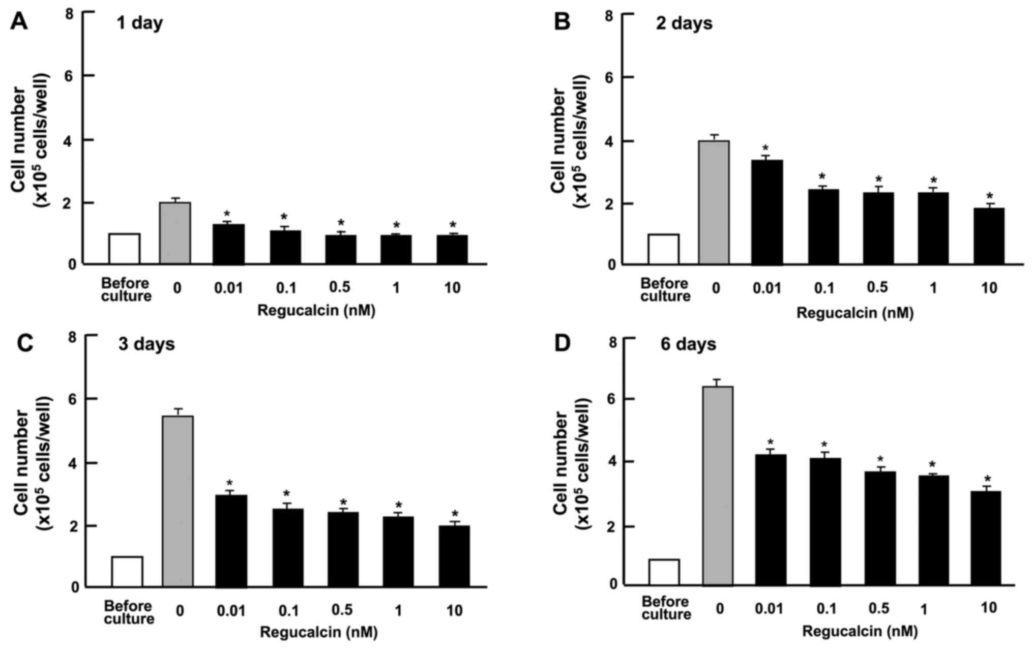
or exogenous regucalcin (0.01–10 nM) for 1–6 days (Fig. 1). An increasing of culture periods
raised cell number in the control group. Culture with the addition
of exogenous regucalcin suppressed the elevation of cell numbers
(Fig. 1), revealing that the
proliferation of HepG2 cells was suppressed with the physiological
levels of regucalcin which is present in the serum (18,30).
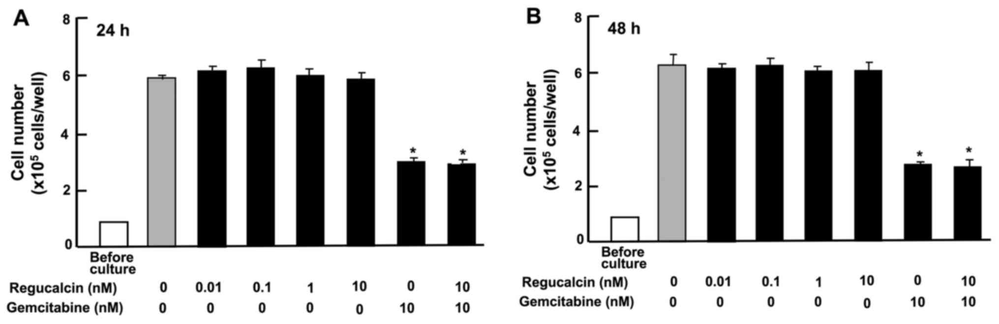
The effect of exogenous regucalcin on the death of
HepG2 cells in vitro is displayed in Fig. 2. Cells were cultured for 3 days upon
reaching subconfluency, and then the cells were cultured for 24 or
48 h, after the addition of either vehicle (PBS) or regucalcin
(0.01–10 nM) with or without gemcitabine (10 nM). The number of
HepG2 cells was not changed in the presence of exogenous
regucalcin, although the addition of gemcitabine caused the death
of cells (Fig. 2). Thus, exogenous
regucalcin exhibited a suppressive effect on cell proliferation
independently of the death of HepG2 cells.
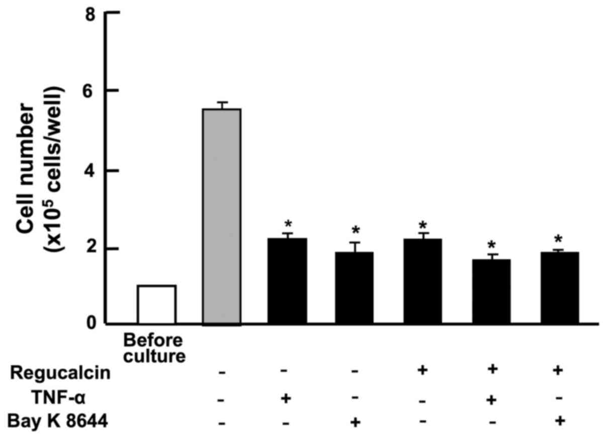
The suppressive effects of exogenous regucalcin on
the proliferation of HepG2 cells were compared with the effects of
other factors that inhibit cell growth (Fig. 3). The effects of exogenous
regucalcin (10 nM) suppressing the proliferation of HepG2 cells
were not potentiated by the addition of TNF-α (1 ng/ml), an inducer
of nuclear factor-κB (NF-κB) signaling (34), or Bay K 8644 (1 µM), an agonist of
Ca2+ entry into cells (35), that caused a decrease in the number
of cells (Fig. 3).
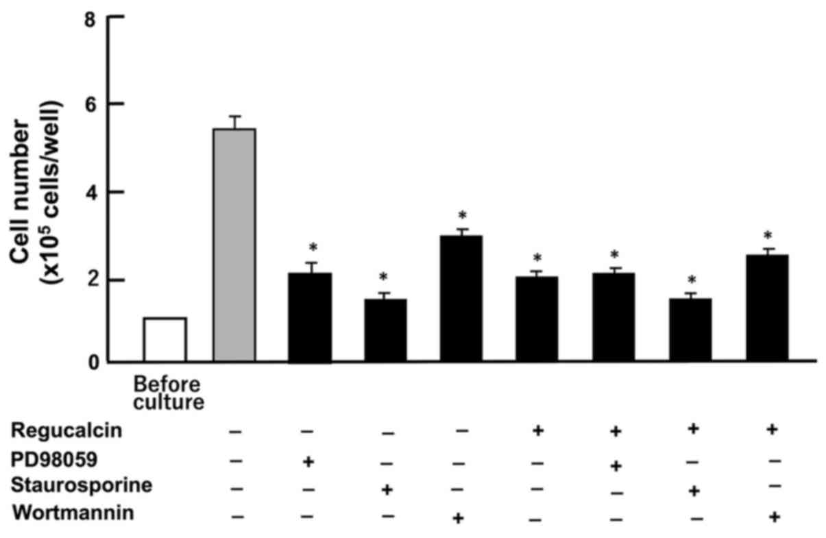
Subsequently, we determined whether the suppressive
effects of exogenous regucalcin on the proliferation of HepG2 cells
were implicated in intracellular signaling pathways. The effects of
exogenous regucalcin in suppressing cell proliferation were not
enhanced by the addition of PD98059 (1 µM), an extracellular
signal-regulated kinase (ERK) inhibitor (36), staurosporin (0.1 µM), an inhibitor
of protein kinase C (37) and
worthomannin (1 µM), an inhibitor of phosphatidylinositol 3-kinase
(PI3K) (38) (Fig. 4).
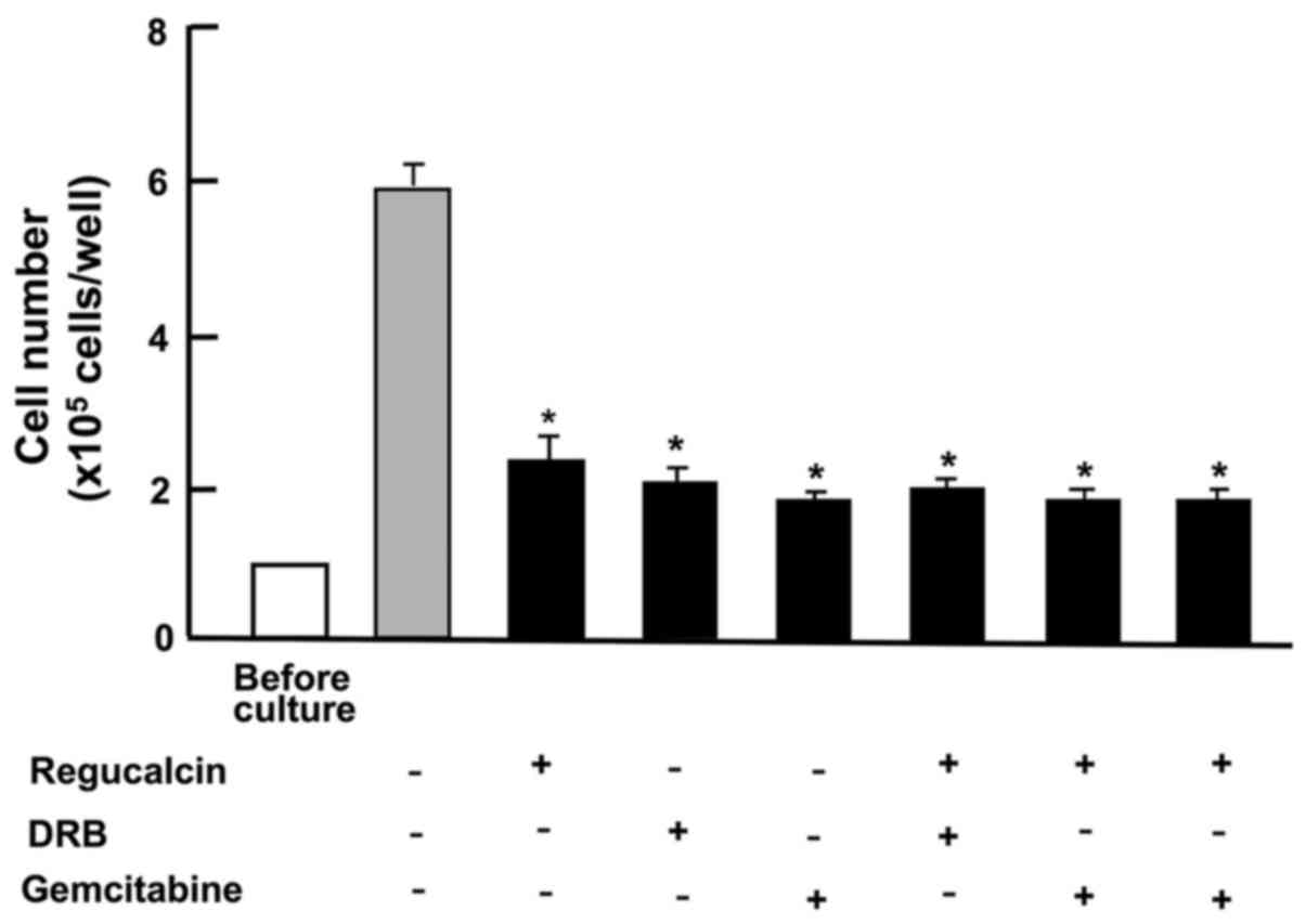
Subsequently, to determine whether the suppressive
effects of exogenous regucalcin on the proliferation of HepG2 cells
were implicated in nuclear function, we used DRB (1 µM), an
inhibitor of transcription activity with RNA polymerase II
inhibition (39), or gemcitabine
(10 nM), an antitumor drug that induces the damage of nuclear DNA
(40). The suppressive effects of
exogenous regucalcin on the proliferation of HepG2 cells were not
altered by the addition of DRB or gemcitabine, which induced
suppression of the proliferation of HepG2 cells (Fig. 5).
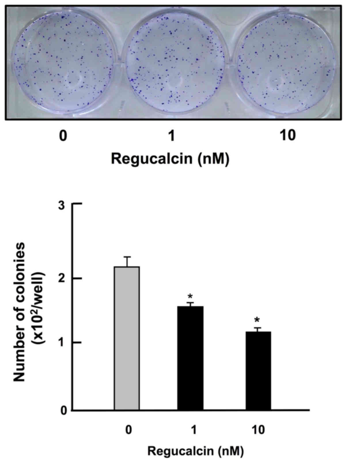
Furthermore, exogenous regucalcin was shown to
decrease colony formation of HepG2 cells in vitro (Fig. 6). Culture with exogenous regucalcin
(1 and 10 nM) led to a reduction of the number of colonies of HepG2
cells (Fig. 6). Thus, exogenous
regucalcin was demonstrated to exhibit suppressive effects on
colony formation due to inhibiting the proliferation of HepG2 cells
in vitro.
Discussion
Regucalcin is produced from tissues including liver,
and it has been shown to be present in the serum of human subjects
and animals (18,30). Whether or not extracellular
regucalcin is important in the regulation of cell function has been
poorly understood. Exogenous regucalcin was demonstrated to bind
the plasma membranes and activate plasma membrane
(Ca2+-Mg2+)-adenosine triphosphatase in rat
liver cells in vitro (41).
Exogenous regucalcin may regulate the function of hepatoma cells.
In the present study, we found that culture with exogenous
regucalcin suppressed the growth in human hepatoblastoma liver
cancer HepG2 cells, suggesting its role in the regulation of cell
proliferation in vitro.
The effects of exogenous regucalcin in suppressing
the proliferation of HepG2 cells were not enhanced by the addition
of TNF-α, an enhancer of NF-κB signaling (34), Bay K 8644, an agonist of
Ca2+ entry in the cells (35), staurosporin, an inhibitor of
calcium-dependent protein kinase C (36), PD98059, an ERK inhibitor (37) and wortmannin, an inhibitor of PI3
kinase (38). The suppressive
effects of exogenous regucalcin on the proliferation of HepG2 cells
were not potentiated by the treatment of various inhibitors that
regulate intracellular signaling pathways related to proliferation
in vitro. Furthermore, the effects of exogenous regucalcin
in suppressing cell proliferation were not potentiated in the
presence of DRB, an inhibitor of transcription activity with RNA
polymerase II inhibition (39). The
effects of exogenous regucalcin in inhibiting the proliferation of
HepG2 cells were implicated with various intracellular signaling
processes. Exogenous regucalcin binds to the plasma membranes of
HepG2 cells, and bound regucalcin may induce the generation of
signaling factors that lead to the suppression of transcription
activity-related signaling processes in the nucleus of HepG2 cells.
In addition, it is hypothesized that exogenous regucalcin bound to
liver plasma membranes may be internalized into hepatoma cells, and
that internalized regucalcin demonstrates suppressive effects on
the signaling pathways implicated to cell proliferation (7,13,14).
The exact mechanisms of action remain to be elucidated in further
studies.
The effects of exogenous regucalcin in suppressing
the proliferation of HepG2 cells were compared with that of
gemcitabine, which is an antitumor drug, which causes nuclear DNA
damage and apoptosis (40).
Suppressive effects of exogenous regucalcin on the proliferation of
HepG2 cells revealed similar effects with gemcitabine. Exogenous
regucalcin did not cause the death of HepG2 cells in vitro,
supporting the hypothesis that regucalcin does not possess an
effect in inducing apoptotic cell death. Revealing the effects of
exogenous regucalcin in inducing the proliferation of HepG2 cells
was not based on apoptotic cell death. The mechanism by which
exogenous regucalcin suppressed the proliferation of HepG2 cells
may be based on different mode of action compared to that of
gemcitabine. Exogenous regucalcin may be a useful tool to
potentiate antitumor effects on human liver cancer cells in
combination with gemcitabine.
Notably, culture with exogenous regucalcin was
demonstrated to suppress colony formation of HepG2 cells in
vitro. This effect may be based on exogenous regucalcin-induced
suppression of the proliferation of HepG2 cells. Thus, exogenous
regucalcin plays a suppressive role on the growth of human liver
cancer cells. We used human hepatoblastoma liver cancer HepG2 cells
in the present study. However, this is unlikely to affect our
conclusions that exogenous regucalcin demonstrated suppressive
effects on the colony formation and proliferation of liver cancer
cells. In addition, it is possible that exogenous regucalcin
demonstrated a suppressive effect on hepatocellular carcinoma and
hepatoblastoma. However, this remains to be elucidated by using
human hepatocellular carcinoma tumor cells.
Our previous studies demonstrated that exogenous
regucalcin demonstrated suppressive effects on the proliferation of
human pancreatic cancer MiaPaCa-2 cells (42) and MDA-MB-231 human breast cancer
cells (43) in vitro.
Furthermore, the present study revealed that exogenous regucalcin
inhibited the growth of human liver cancer cells in vitro.
Thus, exogenous regucalcin, which is produced in the tissues, may
suppress the growth in various types of human cancer cells.
Exogenous regucalcin has been suggested to contribute as a
suppressor in the development of carcinogenesis, thus proposing a
therapeutic strategy with regucalcin gene therapy.
Acknowledgements
The authors thank Dr Oliver Hankinson for his
encouragement, David Geffen School of Medicine, University of
California, California (UCLA).
Funding
The present study was supported in part from the
Foundation for Biomedical Research on Regucalcin, Japan.
Availability of data and materials
The datasets used during the present study are
available from the corresponding author upon reasonable
request.
Authors' contributions
MY conceived designed the study. MY and MT performed
the experiment and discussed with TM. MY wrote the manuscript and
MT reviewed and edited the manuscript. All authors read and
approved the manuscript and agree to be accountable for all aspects
of the research in ensuring that the accuracy or integrity of any
part of the work are appropriately investigated and resolved.
Ethics approval and consent to
participate
All experimental protocols consisted of cells
cultured in vitro.
Consent for publication
Not applicable.
Competing interests
The authors state that they have no competing
interests.
References
|
1
|
Yamaguchi M and Yamamoto T: Purification
of calcium binding substance from soluble fraction of normal rat
liver. Chem Pharm Bull. 26:1915–1918. 1978. View Article : Google Scholar : PubMed/NCBI
|
|
2
|
Yamaguchi M and Sakurai T: Inhibitory
effect of calcium-binding protein regucalcin on
Ca2+-activated DNA fragmentation in rat liver nuclei.
FEBS Lett. 279:281–284. 1991. View Article : Google Scholar : PubMed/NCBI
|
|
3
|
Shimokawa N and Yamaguchi M: Molecular
cloning and sequencing of the cDNA coding for a calcium-binding
protein regucalcin from rat liver. FEBS Lett. 327:251–255. 1993.
View Article : Google Scholar : PubMed/NCBI
|
|
4
|
Misawa H and Yamaguchi M: The gene of
Ca2+-binding protein regucalcin is highly conserved in
vertebrate species. Int J Mol Med. 6:191–196. 2000.PubMed/NCBI
|
|
5
|
Yamaguchi M: Role of regucalcin in calcium
signaling. Life Sci. 66:1769–1780. 2000. View Article : Google Scholar : PubMed/NCBI
|
|
6
|
Yamaguchi M: Role of regucalcin in
maintaining cell homeostasis and function (Review). Int J Mol Med.
15:371–389. 2005.PubMed/NCBI
|
|
7
|
Yamaguchi M: Regucalcin and cell
regulation: Role as a suppressor in cell signaling. Mol Cell
Biochem. 353:101–137. 2011. View Article : Google Scholar : PubMed/NCBI
|
|
8
|
Shimokawa N, Matsuda Y and Yamaguchi M:
Genomic cloning and chromosomal assignment of rat regucalcin gene.
Mol Cell Biochem. 151:157–163. 1995. View Article : Google Scholar : PubMed/NCBI
|
|
9
|
Thiselton DL, McDowall J, Brandau O,
Ramser J, d'Esposito F, Bhattacharya SS, Ross MT, Hardcastle AJ and
Meindl A: An integrated, functionally annotated gene map of the
DXS8026-ELK1 interval on human Xp11.3-Xp11.23: Potential hotspot
for neurogenetic disorders. Genomics. 79:560–572. 2002. View Article : Google Scholar : PubMed/NCBI
|
|
10
|
Yamaguchi M, Makino R and Shimokawa N: The
5 end sequences and exon organization in rat regucalcin gene. Mol
Cell Biochem. 165:145–150. 1996. View Article : Google Scholar : PubMed/NCBI
|
|
11
|
Yamaguchi M: Hormonal regulation of
regucalcin gene expression: Involvement in cell metabolism. Horm
Stud. 1:12013. View Article : Google Scholar
|
|
12
|
Yamaguchi M: The transcriptional
regulation of regucalcin gene expression. Mol Cell Biochem.
346:147–171. 2011. View Article : Google Scholar : PubMed/NCBI
|
|
13
|
Yamaguchi M: Role of regucalcin in cell
nuclear regulation: Involvement as a transcription factor. Cell
Tissue Res. 354:331–341. 2013. View Article : Google Scholar : PubMed/NCBI
|
|
14
|
Yamaguchi M: Suppressive role of
regucalcin in liver cell proliferation: Involvement in
carcinogenesis. Cell Prolif. 46:243–253. 2013. View Article : Google Scholar : PubMed/NCBI
|
|
15
|
Yamaguchi M: The anti-apoptotic effect of
regucalcin is mediated through multisignaling pathways. Apoptosis.
18:1145–1153. 2013. View Article : Google Scholar : PubMed/NCBI
|
|
16
|
Yamaguchi M: Regucalcin and metabolic
disorder: Osteoporosis and hyperlipidemia are induced in regucalcin
transgenic rats. Mol Cell Biochem. 327:53–63. 2010. View Article : Google Scholar
|
|
17
|
Yamaguchi M and Murata T: Involvement of
regucalcin in lipid metabolism and diabetes. Metabolism.
62:1045–1051. 2013. View Article : Google Scholar : PubMed/NCBI
|
|
18
|
Yamaguchi M: Regucalcin as a potential
biomarker for metabolic and neuronal diseases. Mol Cell Biochem.
391:157–166. 2014. View Article : Google Scholar : PubMed/NCBI
|
|
19
|
Yamaguchi M: Involvement of regucalcin as
a suppressor protein in human carcinogenesis: Insight into the gene
therapy. J Cancer Res Clin Oncol. 141:1333–1341. 2015. View Article : Google Scholar : PubMed/NCBI
|
|
20
|
Nakagawa T, Sawada N and Yamaguchi M:
Overexpression of regucalcin suppresses cell proliferation of
cloned normal rat kidney proximal tubular epithelial NRK52E cells.
Int J Mol Med. 16:637–643. 2005.PubMed/NCBI
|
|
21
|
Yamaguchi M and Daimon Y: Overexpression
of regucalcin suppresses cell proliferation in cloned rat hepatoma
H4-II-E cells: Involvement of intracellular signaling factors and
cell cycle-related genes. J Cell Biochem. 95:1169–1177. 2005.
View Article : Google Scholar : PubMed/NCBI
|
|
22
|
Tsurusaki Y and Yamaguchi M: Role of
regucalcin in liver nuclear function: Binding of regucalcin to
nuclear protein or DNA and modulation of tumor-related gene
expression. Int J Mol Med. 14:277–281. 2004.PubMed/NCBI
|
|
23
|
Tsurusaki Y and Yamaguchi M:
Overexpression of regucalcin modulates tumor-related gene
expression in cloned rat hepatoma H4-II-E cells. J Cell Biochem.
90:619–626. 2003. View Article : Google Scholar : PubMed/NCBI
|
|
24
|
Misawa H, Inagaki S and Yamaguchi M:
Suppression of cell proliferation and deoxyribonucleic acid
synthesis in the cloned rat hepatoma H4-II-E cells overexpressing
regucalcin. J Cell Biochem. 84:143–149. 2001. View Article : Google Scholar : PubMed/NCBI
|
|
25
|
Murata T and Yamaguchi M: Alternatively
spliced variants of the regucalcin gene in various human normal and
tumor tissues. Int J Mol Med. 34:1141–1146. 2014. View Article : Google Scholar : PubMed/NCBI
|
|
26
|
Yamaguchi M, Osuka S, Weitzmann MN,
El-Rayes BF, Shoji M and Murata T: Prolonged survival in pancreatic
cancer patients with increased regucalcin gene expression:
Overexpression of regucalcin suppresses the proliferation in human
pancreatic cancer MIA PaCa-2 cells in vitro. Int J Oncol.
48:1955–1964. 2016. View Article : Google Scholar : PubMed/NCBI
|
|
27
|
Yamaguchi M, Osuka S, Weitzmann MN, Shoji
M and Murata T: Increased regucalcin gene expression extends
survival in breast cancer patients: Overexpression of regucalcin
suppresses the proliferation and metastatic bone activity in
MDA-MB-231 human breast cancer cells in vitro. Int J Oncol.
49:812–822. 2016. View Article : Google Scholar : PubMed/NCBI
|
|
28
|
Yamaguchi M, Osuka S, Weitzmann MN,
El-Rayes BF, Shoji M and Murata T: Prolonged survival in
hepatocarcinoma patients with increased regucalcin gene expression:
HepG2 cell proliferation is suppressed by overexpression of
regucalcin in vitro. Int J Oncol. 49:1686–1694. 2016.
View Article : Google Scholar : PubMed/NCBI
|
|
29
|
Yamaguchi M, Osuka S, Shoji M, Weitzmann
MN and Murata T: Survival of lung cancer patients is prolonged with
higher regucalcin gene expression: Suppressed proliferation of lung
adenocarcinoma A549 cells in vitro. Mol Cell Biochem. 430:37–46.
2017. View Article : Google Scholar : PubMed/NCBI
|
|
30
|
Yamaguchi M and Isogai M: Tissue
concentration of calcium-binding protein regucalcin in rats by
enzyme-linked immunoadsorbent assay. Mol Cell Biochem. 122:65–68.
1993. View Article : Google Scholar : PubMed/NCBI
|
|
31
|
Knowles BB, Howe CC and Aden DP: Human
hepatocellular carcinoma cell lines secrete the major plasma
proteins and hepatitis B surface antigen. Science. 209:497–499.
1980. View Article : Google Scholar : PubMed/NCBI
|
|
32
|
Ao L, Guo Y, Song X, Guan Q, Zheng W,
Zhang J, Huang H, Zou Y, Guo Z and Wang X: Evaluating
hepatocellular carcinoma cell lines for tumour samples using
within-sample relative expression orderings of genes. Liver Int.
37:1688–1696. 2017. View Article : Google Scholar : PubMed/NCBI
|
|
33
|
Fang Z, Tang Y, Fang J, Zhou Z, Xing Z,
Guo Z, Guo X, Wang W, Jiao W, Xu Z, et al: See comment in PubMed
Commons belowSimvastatin inhibits renal cancer cell growth and
metastasis via AKT/mTOR, ERK and JAK2/STAT3 pathway. PLoS One.
8:e628232013. View Article : Google Scholar : PubMed/NCBI
|
|
34
|
Lee ZH, Kwack K, Kim KK, Lee SH and Kim
HH: Activation of c-Jun N-terminal kinase and activator protein 1
by receptor activator of nuclear factor kappaB. Mol Pharmacol.
58:1536–1545. 2000. View Article : Google Scholar : PubMed/NCBI
|
|
35
|
Cano-Abad MF, Villarroya M, García AG,
Gabilan NH and López MG: Calcium entry through L-type calcium
channels causes mitochondrial disruption and chromaffin cell death.
J Biol Chem. 276:39695–39704. 2001. View Article : Google Scholar : PubMed/NCBI
|
|
36
|
Chen S, Wang Y, Ruan W, Wang X and Pan C:
Reversing multidrug resistance in hepatocellular carcinoma cells by
inhibiting extracellular signal-regulated kinase/mitogen-activated
protein kinase signaling pathway activity. Oncol Lett. 8:2333–2339.
2014. View Article : Google Scholar : PubMed/NCBI
|
|
37
|
Chen QW, Edvinsson L and Xu CB: Role of
ERK/MAPK in endothelin receptor signaling in human aortic smooth
muscle cells. BMC Cell Biol. 10:522009. View Article : Google Scholar : PubMed/NCBI
|
|
38
|
Serrano-Nascimento C, da Silva Teixeira S,
Nicola JP, Nachbar RT, Masini-Repiso AM and Nunes MT: The acute
inhibitory effect of iodide excess on sodium/iodide symporter
expression and activity involves the PI3K/Akt signaling pathway.
Endocrinology. 155:1145–1156. 2014. View Article : Google Scholar : PubMed/NCBI
|
|
39
|
Palangat M, Grass JA, Langelier MF,
Coulombe B and Landick R: The RPB2 flap loop of human RNA
polymerase II is dispensable for transcription initiation and
elongation. Mol Cell Biol. 31:3312–3325. 2011. View Article : Google Scholar : PubMed/NCBI
|
|
40
|
Tang SC and Chen YC: Novel therapeutic
targets for pancreatic cancer. World J Gastroenterol.
20:10825–10844. 2014. View Article : Google Scholar : PubMed/NCBI
|
|
41
|
Yamaguchi M, Mori S and Kato S:
Calcium-binding protein regucalcin is an activator of
(Ca2+-Mg2+)-adenosine triphosphatase in the
plasma membranes of rat liver. Chem Pharm Bull (Tokyo).
36:3532–3539. 1988. View Article : Google Scholar : PubMed/NCBI
|
|
42
|
Yamaguchi M and Murata T: Suppressive
effects of exogenous regucalcin on the proliferation of human
pancreatic cancer MIA PaCa-2 cells in vitro. Int J Mol Med.
35:1773–1778. 2015. View Article : Google Scholar : PubMed/NCBI
|
|
43
|
Yamaguchi M and Murata T: Exogenous
regucalcin suppresses the proliferation of human breast cancer
MDA-MB-231 bone metastatic cells in vitro. Mol Med Rep.
12:7801–7805. 2015. View Article : Google Scholar : PubMed/NCBI
|