Introduction
Gastric cancer is a common malignant disease that
seriously threatens human life and health (1). In addition to surgery, chemotherapy,
and radiotherapy, PDT is another promising curative and palliative
treatment approach to eliminate tumor tissue (2). Given its high efficiency, safety,
synergy compatibility, repeatability, and relatively low cost, PDT
has become attractive to researcher (3). In PDT, the photosensitizer, a
light-sensitive drug is administered via systemic injection and is
subsequently illuminated by suitable light at the target tissue,
leading to generation of reactive oxygen species (ROS), notably
singlet oxygen (SOG), thereby causing oxidative stress to damage
cellular organelles and membranes and ultimately lead to apoptosis
or necrosis of neoplastic tissue (4,5). PDT
can be used as monotherapy and in combination with surgery,
chemotherapy, or standard radiation therapy (6–9).
Monotherapy usually suffers from incomplete tumor killing to
ultimately induce poor prognoses and unsuccessful therapy. The
combination treatment strategies revealed a more effective
antitumor effect preclinically and clinically (10). In the combination of PDT with
chemotherapy, chemotherapy can enhance the antitumor effect of PDT
by targeting surviving cancer cells and inhibiting regrowth of
damaged tumor blood vessels. Furthermore, PDT-mediated vascular
permeabilization can enhance the accumulation of
chemotherapeuticdrugs in tumors, thereby improving chemotherapy
efficacy. Therefore, the method of combination of PDT with
chemotherapy may be an effective treatment strategy for gastric
cancermanagement.
AlPcS4, derived from photofrin, is a
second-generation photosensitizer. It exhibits near-infrared
absorption (position of absorption maxima at 675 nm), thereby
providing relatively low tissue absorption and deeper tissue
penetration. Furthermore, higher quantum yields, good
photostability, and little photobleaching result in the wide use of
the photosensitizer in the treatment of coelom cancer in PDT
(11,12). It has been reported that
AlPcS4 has a superior PDT effect in various cancer cell
lines, including breast, pancreatic, ovarian, and colon cancer
(13–19). However, to date, whether it can
inhibit the growth of gastric cancer cells has not been
demonstrated. The antitumor effect of AlPcS4/PDT in
combination with chemotherapeuticdrugs on gastric cancer cells is
also less known.
Although many researchers have reported that
PDT-combined chemotherapy is an effective and feasible therapy for
cancer, the combined antitumor effect depends on the selected drug
and the dose of the drug. Choosing different chemical drugs can
induce different antitumor effects. Thus, researching different
chemical drugs for combination with AlPcS4/PDT for an
optimal antitumor effect on gastric cancer is important. 5-FU, DOX,
CDDP, MMC and VCR are common gold-standard chemotherapeutic agents
that are clinically recommended for gastric cancer. In particular,
5-FU, as a thymidylate synthase inhibitor, can rapidly divide cells
to cease replication and die by preventing the production of dTMP
(20). DOX, an anthracycline
chemotherapeutic agent, intercalates into DNA and halts the process
of DNA replication by inhibiting the progression of topoisomeraseII
(21). CDDP is a metallic
coordination compound and can interfere with the DNA repair
mechanism, thereby causing DNA damage and inducing apoptosis, which
causescell death (22). MMC is an
antibiotic that was isolated from Gram-negative bacteria
Streptomyces caespitosus and can cross-link double-stranded
DNA at adenosine and guanine during the G1 or S phase. This
antibiotic prevents DNA stranding from separating during the DNA
replication process and then halting mitosis. The antibiotic can
also bind to the promoter sites of inducible genes, thereby
suppressing the synthesis of cellular RNA and protein to control
diseases (23). VCR as a vinca
alkaloid can interact with β-tubulin in a region adjacent to the
GTP-binding site to prevent the formation of spindle microtubules,
thereby disabling the function of the cell for aligning and moving
the chromosomes to further induce high frequency of micronuclei,
chromosome aberration, sister chromatid exchange, DNA damage, and
interference with DNA, RNA, and protein synthesis. All of these
processes cause cancer cell death (24). Overall, all of these
chemotherapeutic agents have an anti-growth effect on cancer cells
via DNA or RNA dysfunction. Using them in combination with
AlPcS4/PDT for synergistic therapy is expected to
achieve a significant antitumor effect on gastric cancer.
Chemotherapy is effective in antitumor treatment.
However, chemotherapy requires multiple drug doses that can easily
result in severe toxic side effects and multi-drug resistance
(25). The chemotherapy agents
aforementioned are no exception. Hence, using low-dose
chemotherapeutic drugs in combination with AlPcS4/PDT
therapy may effectively reduce toxic side effects and multi-drug
resistance problems. The low-dose chemical therapy also leads to
significant inhibition of the growth activities of gastric cancer
cells with the aid of PDT-mediated vascular permeabilization
(26–28).
Therefore, in this present study, we attempted to
investigate the inhibition of the growth effect by combination
treatment between low-dose chemotherapeutic agents (5-FU, DOX,
CDDP, MMC and VCR) and AlPcS4/PDT on SGC-7901 gastric
cancer cells and compare the antitumor effect between them in order
to find promising combination treatment schemes with high
anticancer efficiency and low toxic side effects. Given that
AlPcS4 was dominant in our design scheme, we evaluated
the influence of AlPcS4 intracellular uptake ability and
ROS and SOG generation abilities in the presence of low-dose
chemotherapeutic agents. The apoptosis-inducing and
necrosis-inducing ability was further demonstrated. Low-dose 5-FU,
DOX and MMC combination treatment had significant antitumor effects
with low dark-cytotoxicity. This combination increased
AlPcS4 intracellular uptake ability and ROS and SOG
generation abilities, thereby inducing significant apoptosis and
necrosis. Low-dose CDDP and VCR combination treatment had a
relatively inferior increasing inhibition effect in terms of
increasing apoptosis activities. However, low-dose CDDP and VCR
indicated a slight adverse effect on AlPcS4
intracellular uptake ability and SOG generation ability.
Materials and methods
Reagents
5-FU, DOX, CDDP, MMC and VCR were purchased from
Sigma-Aldrich; Merck (St. Louis, MO, USA) and dissolved in dimethyl
sulfoxide (DMSO; Sigma-Aldrich; Merck) or sterile PBS (HyClone; GE
Healthcare Life Sciences, Logan, UT, USA). The materials were
stored at 4°C and then diluted as needed in RPMI-1640 medium
(HyClone; GE Healthcare Life Sciences) on the day of use.
AlPcS4 was purchased from Frontier Scientific (Logan,
UT, USA) and dissolved in sterile PBS with a concentration of 2
mg/ml and stored at 4°C in the dark. Then, AlPcS4 was
diluted to a range of 1–32 µg/ml following a gradient dilution
method in RPMI-1640 medium on the day of use.
Cells
SGC-7901 cells, which are part of a human
moderately-differentiated gastric carcinoma cell line, were donated
by the State Key Laboratory of Cancer Biology, Digestion
Department, Xijing Hospital, affiliated with the Fourth Military
Medical University, Xi'an, China. The cells were cultured in
RPMI-1640 medium that was supplemented with 10% fetal bovine serum
(Sijiqing Co., Ltd., Hangzhou, China) and 1%
penicillin/streptomycin in a humidified incubator (Heracell™ 150i
CO2 with copper chambers; Thermo Fisher Scientific,
Inc., Waltham, MA, USA) at 37°C with 5% CO2.
Measurement of the absorption spectra
and fluorescence spectra of a mixture of AlPcS4 and
chemotherapeutic agents
The absorption spectra of free-AlPcS4 (8
µg/ml), AlPcS4 (8 µg/ml) in the presence of 5-FU (20
µM), DOX (0.4 µg/ml), CDDP (5 µM), MMC (0.5 µg/ml) and VCR (0.1
µg/ml) were assessed at 1-h intervals for 6 h by an
ultraviolet-visible spectrophotometer (V-550 UV/VIS; Jasco
International, Co., Ltd., Tokyo, Japan). Fluorescence spectra of
free-AlPcS4, AlPcS4 in the presence of 5-FU,
DOX, CDDP, MMC, and VCR were recorded using a fluorescence
spectrophotometer (F-4500; Hitachi, Ltd., Tokyo, Japan).
CCK-8 assay
The dark cytotoxicity and anti-growth effect of
free-AlPcS4, AlPcS4 in the presence of 5-FU,
DOX, CDDP, MMC and VCR at different doses on SGC-7901 cells was
assessed by CCK-8 assay. Briefly, 1×10⁴ SGC-7901
cells/well were seeded in sterile 96-well flat-bottomed plates and
incubated overnight at 37°C. Then, diluted free-AlPcS4,
AlPcS4 in the presence of 5-FU (20 µM), DOX (0.4 µg/ml),
CDDP (5 µM), MMC (0.5 µg/ml), and VCR (0.1 µg/ml) were added to
each well with final concentrations in the range of 1–32 µg/ml.
Wells with cells were divided into different groups. Wells without
drug and irradiation treatment comprised the control group, those
with drug and no irradiation comprised the dark cytotoxicity group,
those with drug and irradiation comprised the synergistic
therapeutic group, and wells that contained only complete culture
media comprised the blank control group. In the synergistic
therapeutic group, the cells were incubated with
free-AlPcS4, AlPcS4 in the presence of 5-FU,
DOX, CDDP, MMC and VCR and then illuminated by a 635-nm laser light
with a power density of 100 mW/cm2. All the cells
treated with drugs were incubated for 6 h and then washed with PBS
twice. Subsequently, they were incubated with fresh RPMI-1640
complete medium for 24 h. Following incubation, the solution
containing 100 µl RPMI-1640 medium and 10 µl CCK-8 reagent (Dojindo
Molecular Technologies, Inc., Kumamoto, Japan) was added to each
well. The cells were then incubated for 1 h at 37°C again. Finally,
the absorbance levels of the cells were assessed at 450 nm using a
microplate reader (Infinite® M200 PRO; Tecan Group,
Ltd., Mannedorf, Switzerland). The dark cytotoxicity and
anti-growth effect of free-AlPcS4, AlPcS4 in
the presence of 5-FU, DOX, CDDP, MMC and VCR at the doses used was
calculated as: [(OD of the drug treated-OD of the blank
control)/(OD of the control-OD of the blank control)] ×100%.
Detection of intracellular uptake
ability of AlPcS4
Intracellular uptake ability of AlPcS4
was evaluated by a fluorescence spectrophotometer (F-4500; Hitachi,
Ltd.). In the incubator, 2.5×10⁵ SGC-7901 cells were
seeded in six-well plates and allowed to attach overnight. Then,
the cells were treated with RPMI-1640 complete media that
containedfree-AlPcS4, AlPcS4 in the presence
of 5-FU, DOX, CDDP, MMC and VCR at the same concentrations as
aforementioned. Then the cells were incubated for 6 h at 37°C with
5% CO2. Following incubation, the cells were washed with
PBS twice, digested with trypsin enzyme (HyClone; GE Healthcare
Life Sciences, Logan, UT, USA) and collected by centrifugation.
Then, the emission spectra at 635 nm excitation wavelength were
measured.
Detection of cell apoptosis and
necrosis by Hoechst 33342/PI assay
Induction of apoptosis and necrosis was detected
using Hoechst 33324/PI nuclear staining kit following the
manufacturer's instructions. In the incubator, 2.5×10⁵
cells/ml SGC-7901 cells were seeded on sterile coverslips in 6-well
plates and allowed to attach overnight. Then, the cells were
treated with free-AlPcS4, AlPcS4 in the
presence of 5-FU (20 µM), DOX (0.4 µg/ml), CDDP (5 µM), MMC (0.5
µg/ml), and VCR (0.1 µg/ml) at 37°C with 5% CO2 for 6 h.
Following incubation, the cells were washed with PBS two times, and
the medium was replaced with fresh RPMI-1640 culture medium. The
cells were irradiated with 635-nm laser light for 5 min and
incubated at 37°C with 5% CO2 for 24 h. Following
treatment, the cells were stained with 5 µl Hoechst 33324 and 5 µl
PI reagents for 10 min. After removing the coverslips, the cells
were washed with PBS again, mounted on slides with glycerol, and
imaged with a fluorescence microscope (Nikon Corp., Tokyo, Japan).
To quantify the percentage of apoptosis and necrosis, we counted
the number of cells with apoptotic and necrotic characteristics
among 200 cells at a high-power field in accordance with the
results of stained cell nucleus. All statistical analyses were
performed using SPSS 18.0 (SPSS, Inc., Chicago, IL, USA). The
statistical difference between the means was analyzed by Student's
t-distribution test. Significance was set at the 5% level. Data are
presented as the mean ± SD of three replicate experiments.
Generation of ROS and SOG assay
Generation of ROS and SOG were assessed by a DCFH-DA
fluorescence probe (Beyotime Institute of Biotechnology, Shanghai,
China), and singlet oxygen sensor green reagent (SOSGR; Thermo
Fisher Scientific, Inc.) via a fluorescence spectrophotometer
(F-4500; Hitachi, Ltd.). SGC-7901 cells were seeded in six-well
plates at a density of 2.5×10⁵ cells/well and incubated
to adhere securely. Then, the cells were treated with
free-AlPcS4, AlPcS4 in the presence of 5-FU
(20 µM), DOX (0.4 µg/ml), CDDP (5 µM), MMC (0.5 µg/ml), and VCR
(0.1 µg/ml) at 1–32 µg/ml for 6 h. The cells were washed twice with
PBS and irradiated with laser systems for 5 min. For the detection
of ROS, the cells were harvested, incubated with 10 µmol/l DCFH-DA
for 20 min at 37°C in complete darkness again, washed with PBS
twice, and assessment was conducted using a fluorescence
spectrophotometer under an excitation of 488-nm of light. For the
detection of SOSGR, the cells were harvested, permeabilized with
0.5% Triton X-100 in PBS for 10 min, centrifuged at 70 × g for 5
min, washed with PBS, mixed with SOSGR, irradiated with a 635-nm
laser system for 5 min, and then detected using a fluorescence
spectrophotometer under an excitation of 504-nm of light.
Results
Characterization of AlPcS4
and the chemotherapeutic agent-AlPcS4 mixture
To ensure the influence of AlPcS4 itself
by chemotherapeutic agent, the UV-vis absorption spectra and
fluorescence intensity of AlPcS4 and the
chemotherapeutic agent-AlPcS4 mixture and
free-AlPcS4 were assessed. Fig. 1A revealed that the absorption levels
of AlPcS4 were derived from the AlPcS4
mixture. The Figure also revealed that the chemotherapeutic
agent-AlPcS4 mixture had weaker reduction compared to
free-AlPcS4 in deionized water as time changed. Compared
to deionized water, the main absorption levels of AlPcS4
were obtained from free-AlPcS4 and the chemotherapeutic
agent-AlPcS4 mixture was much more stable in a culture
medium that contained FBS (Fig.
1B). However, the main absorption levels of AlPcS4
in the chemotherapeutic agent-AlPcS4 mixture or
free-AlPcS4 in a culture medium that contained FBS were
lower than those in deionized water. A possible reason for this
finding is that AlPcS4 binds nonspecifically to serum
albumin based on electrostatic interaction. The inverted absorption
peaks (near 550 nm) appeared in the absorption spectra of the
chemotherapeutic agent-AlPcS4 mixture and
free-AlPcS4 in culture medium as time increased
(Fig. 1B). These peaks may be
caused by coagulation at different degrees. The fluorescence
intensity of AlPcS4 was derived from AlPcS4,
and the chemotherapeutic agent-AlPcS4 mixture also
indicated no significant change even after 6 h compared with
free-AlPcS4 (Fig. 1C and
D). Only the fluorescence intensity of AlPcS4+VCR
was slightly reduced after 5 h (Fig.
1D). Furthermore, whether in deionized water or culture medium
that contained FBS, the fluorescence intensities markedly increased
after dilution of the chemotherapeutic agent-AlPcS4
mixture for 1 h. These may be induced by AlPcS4 at high
concentrations, and it had a concentration-dependent fluorescent
quenching effect. Overall, the selected chemotherapeutic agents in
the present study did not influence AlPcS4 itself. In
culture medium, AlPcS4 and the chemotherapeutic
agent-AlPcS4 mixture scarcely influenced the absorption
levels and fluorescence intensity of AlPcS4. However,
the main absorption levels and fluorescence intensity of
AlPcS4 (near 675 nm) were not influenced by a
chemotherapeutic agent.
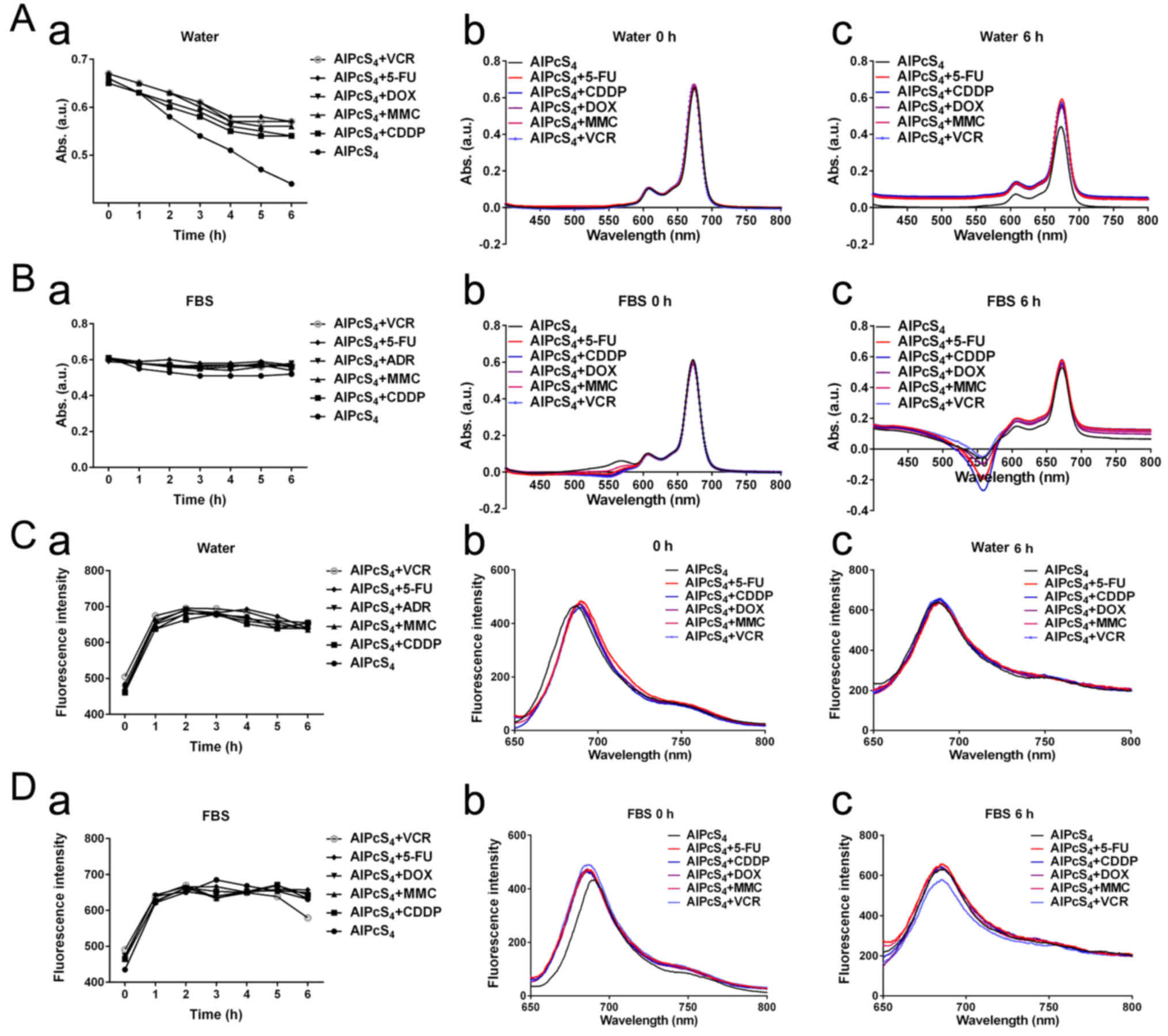 | Figure 1.UV-vis absorption spectra and
fluorescence intensity of AlPcS4 mixture with 5-FU (20
µm), CDDP (5 µm), DOX (0.4 µm/ml), MMC (0.5 µm/ml), and VCR (0.1
µm/ml) or free-AlPcS4 at 8 µm/ml. (A-a and B-a) Maximum
OD values near 675 nm absorption spectra of corresponding agents in
deionized water and RPMI-1640 culture medium that contained FBS at
1–6 h. (A-b and B-b) UV-vis absorption spectra of corresponding
agents in deionized water and RPMI-1640 culture medium that
contained FBS at 0 h. (A-c and B-c) UV-Vis absorption spectra of
corresponding agents in deionized water and RPMI-1640 culture
medium that contained FBS at 6 h. (C-a and D-a) Maximum
fluorescence intensity near 687 nm fluorescence spectra of
corresponding agents in deionized water and RPMI-1640 culture
medium that contained FBS at 1–6 h. (C-b and D-b) Fluorescence
spectra of corresponding agents in deionized water and RPMI-1640
culture medium that contained FBS at 0 h. (C-c and D-c)
Fluorescence spectra of corresponding agentsin deionized water and
RPMI-1640 culture medium that contained FBS at 6 h.
AlPcS4, Al(III) phthalocyanine chloride tetrasulfonic
acid; 5-FU, 5-fluorouracil; DOX, doxorubicin; CDDP, cisplatin; MMC,
mitomycin C; VCR, vincristine; ROS. |
Cytotoxicity and anti-growth effect of
single or combination treatment
To determine whether the selected chemotherapeutic
agents and AlPcS4 under laser light exposure had
synergistic antitumor effects on SGC-7901 cells, dark cytotoxicity,
and photo-cytotoxicity were determined by CCK-8 assay. As shown in
Fig. 2, without light irradiation,
the cell viability of SGC-7901 cells incubated with various
chemotherapeutic agents, different concentrations of
free-AlPcS4, and the mixture of chemotherapeutic agents
with different concentrations of AlPcS4 was mostly
higher than 80, 90 and 80% respectively. The dark cytotoxicity of
AlPcS4 with or without low-dose chemotherapeutic agents
was markedly low. The dark cytotoxicity induced by the combination
treatment of AlPcS4 with a low-dose chemotherapeutic
agent was lower than the summation of the dark cytotoxicity induced
by free-AlPcS4 as well as the low-dose chemotherapeutic
agent alone. The dark cytotoxicity was even lower than that from
the low-dose chemotherapeutic agent alone. Hence, without laser
irradiation, the antagonistic effects in the combination treatment
of AlPcS4/PDT with low-dose chemotherapeutic agents were
obtained. The antagonistic effects of AlPcS4+VCR and
AlPcS4+CDDP were higher than those of
AlPcS4+5-FU, AlPcS4+DOX, and
AlPcS4+MMC.
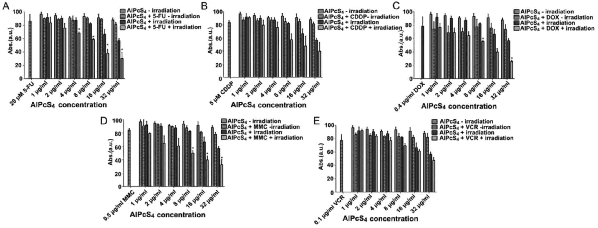 | Figure 2.Antitumor growth effect on SGC-7901
cells in single and combination treatment therapy as determined by
CCK-8 assay. Dark cytotoxicity and antitumor growth effect on
SGC-7901 cells by (A) AlPcS4 + 5-FU, (B)
AlPcS4 + CDDP, (C) AlPcS4 + DOX, (D)
AlPcS4 + MMC or (E) AlPcS4 + VCR. The cells
were treated with 1–32 µm/ml free-AlPcS4 or
AlPcS4 + 5-FU (20 µm), AlPcS4 + CDDP (5 µm),
AlPcS4 + DOX (0.4 µm/ml), AlPcS4 + MMC (0.5
µm/ml) or AlPcS4 + VCR (0.1 µm/ml) for 6 h. The cells
were then incubated again for 24 h with or without 635-nm laser
irradiation at 100 mW/cm2 illumination dosage for 5 min.
*P<0.05, represents a statistical difference in antitumor effect
between the combination of AlPcS4 with a chemical agent
and free-AlPcS4. AlPcS4, Al(III)
phthalocyanine chloride tetrasulfonic acid; 5-FU, 5-fluorouracil;
DOX, doxorubicin; CDDP, cisplatin; MMC, mitomycin C; VCR,
vincristine. |
After 635 nm laser irradiation of 100
mW/cm2 illumination dosage for 5 min, the cell viability
of free-AlPcS4 slightly decreased as the concentration
increased. Even at 32 µg/ml, the cell viabilities of cells remained
at ~57%. However, the viability of cells treated with
AlPcS4+5-FU, AlPcS4+DOX,
AlPcS4+CDDP, AlPcS4+MMC and
AlPcS4+VCR after irradiation by laser light had a
significant decrease, especially at high concentrations. At 32
µg/ml, the cell viabilities of cells were decreased to roughly 30,
25, 39, 32 and 47%, respectively. Notably, the anti-growth effect
by PDT combined with chemical therapy of AlPcS4+5-FU,
AlPcS4+DOX, and AlPcS4+MMC was higher than
the efficiency summation of free-AlPcS4 and 5-FU, DOX,
and MMC. When we discarded the antitumor growth effect induced by
free-AlPcS4 at the highest concentration and 5-FU, DOX,
or MMC, the inhibitory effects of the combination treatment
increased at average values of 12.49, 14.67 and 10.3%,
respectively. The results of the statistical analysis revealed that
there were significant differences between free-AlPcS4
and AlPcS4+5-FU, AlPcS4+DOX, and
AlPcS4+MMC concerning the antitumor effect, compared
with AlPcS4+CDDP andAlPcS4+VCR.
Apoptosis/necrosis-inducing abilities
of single or combination treatment
Apoptosis is the main cause of death in PDT. Hence,
to ascertain whether the selected chemotherapeutic agents and
AlPcS4 under laser light exposure induced apoptosis in
the cells, a Hoechst 33324 and PI staining assay was conducted. As
shown in Figs. 3–5, cell shrinkage and nuclear fragmentation
appeared in SGC-7901 cells induced by both free-AlPcS4
and the chemotherapeutic agent-AlPcS4 mixture after
irradiation at 6, 12 and 24 h, only the number of cell induced by
free-AlPcS4 was low. In other words, apoptosis is active
in AlPcS4/PDT. The apoptosis-inducing abilities were
increased in SGC-7901 cells in AlPcS4/PDT synergistic
treatment with low-dose chemical drug agents. Apoptosis induced by
single AlPcS4/PDT was mainly active at 6–12 h.
Furthermore, given the presence of chemotherapeutic agents,
especially 5-FU, DOX and MMC, the duration of apoptosis-inducing
time increased. Even at 24 h, higher apoptosis-inducing abilities
were obtained (Fig. 7). In
addition, the increased apoptosis-inducing abilities were obtained
in the combination therapy even without the synergistic effects.
However, apoptosis was induced quickly at 6 h (Fig. 3). The increasing trend may be due to
the low-dose chemotherapeutic agent. Hence, although the
apoptosis-inducing abilities decreased at 12 and 24 h (Figs. 4 and 5), the final inhibitory effects of cell
viabilities in combination treatment of AlPcS4/PDT with
low-dose CDDP and VCR increased.
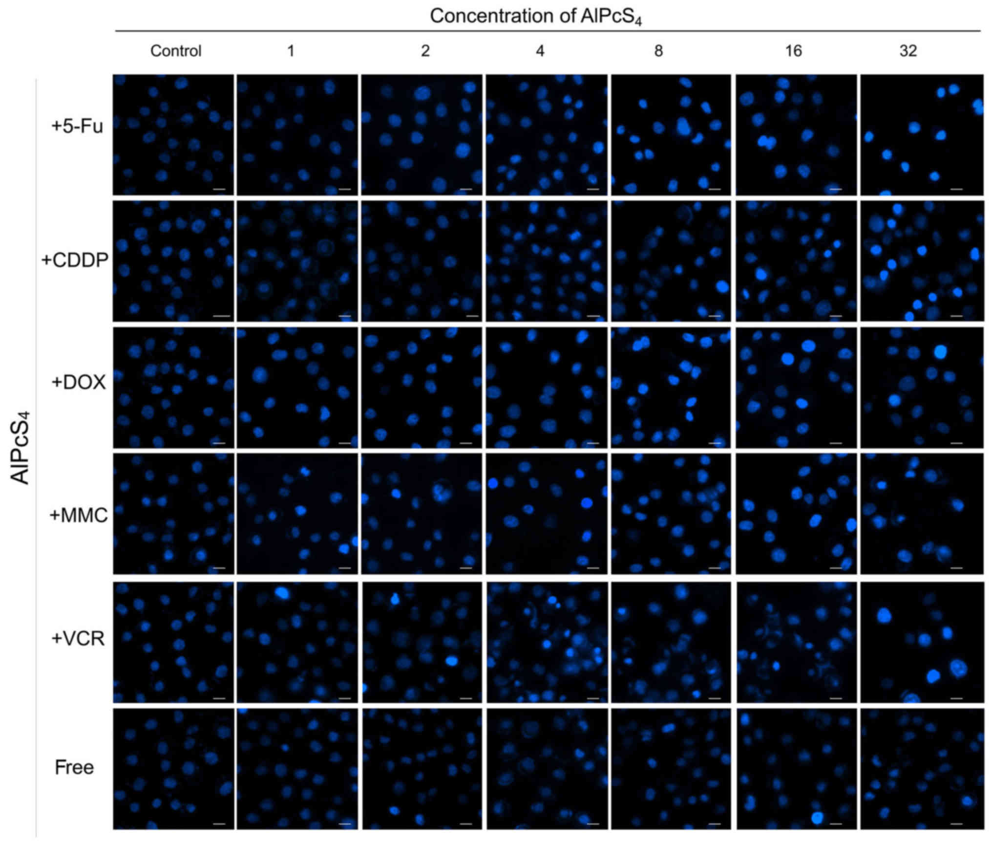 | Figure 3.Apoptosis induced by
AlPcS4 + 5-FU, AlPcS4 + CDDP,
AlPcS4 + DOX, AlPcS4 + MMC, AlPcS4
+ VCR and free-AlPcS4 in SGC-7901 cells after being
irradiated for 6 h. The cells were then treated with 1–32 µm/ml
free-AlPcS4 or AlPcS4 + 5-FU (20 µm),
AlPcS4 + CDDP (5 µm), AlPcS4 + DOX (0.4
µm/ml), AlPcS4 + MMC (0.5 µm/ml), or AlPcS4 +
VCR (0.1 µm/ml) for 6 h. These samples were then irradiated with
635-nm laser irradiation at 100 mW/cm2 illumination
dosage for 5 min, incubated for 6 h, stained with Hoechst 33342
probe, and then imaged using afluorescence microscope. All the
Hoechst staining images were acquired at an ×400 magnification. The
scale bar represented 20 µm. AlPcS4, Al(III)
phthalocyanine chloride tetrasulfonic acid; 5-FU, 5-fluorouracil;
DOX, doxorubicin; CDDP, cisplatin; MMC, mitomycin C; VCR,
vincristine. |
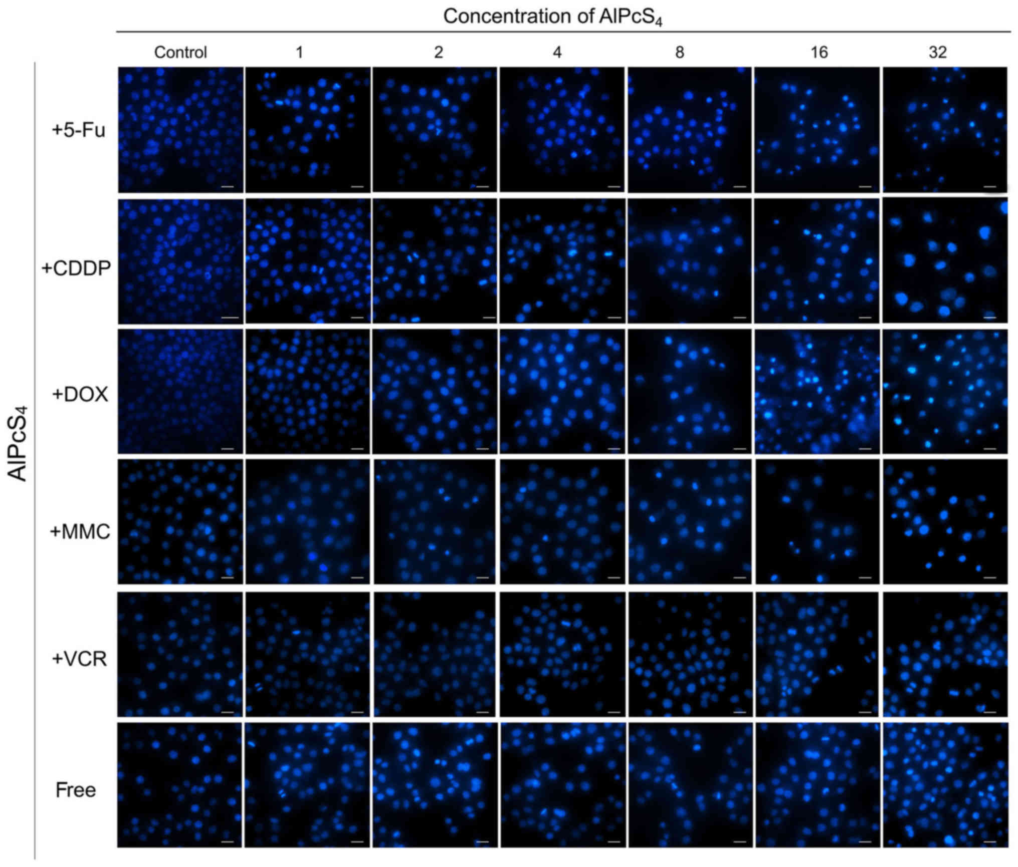 | Figure 5.Apoptosis induced by
AlPcS4 + 5-FU, AlPcS4 + CDDP,
AlPcS4 + DOX, AlPcS4 + MMC, AlPcS4
+ VCR and free-AlPcS4 in SGC-7901 cells after being
irradiated for 24 h. The cells were then treated with 1–32 µm/ml
free-AlPcS4 or AlPcS4 + 5-FU (20 µm),
AlPcS4 + CDDP (5 µm), AlPcS4 + DOX (0.4
µm/ml), AlPcS4 + MMC (0.5 µm/ml) or AlPcS4 +
VCR (0.1 µm/ml) for 6 h. The cells were then irradiated with 635-nm
laser irradiation at 100 mW/cm2 illumination dosage for
5 min, incubated for 24 h, stained with Hoechst 33342 probe, and
then imaged using afluorescence microscope. All the Hoechst
staining images were acquired at an ×400 magnification. The scale
bar represented 20 µm. AlPcS4, Al(III) phthalocyanine
chloride tetrasulfonic acid; 5-FU, 5-fluorouracil; DOX,
doxorubicin; CDDP, cisplatin; MMC, mitomycin C; VCR,
vincristine. |
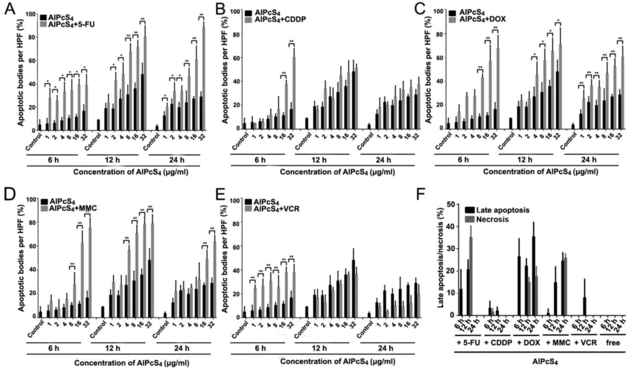 | Figure 7.Statistical analysis of apoptosis and
necrosis induced by AlPcS4 + 5-FU, AlPcS4 +
CDDP, AlPcS4 + DOX, AlPcS4 + MMC,
AlPcS4 + VCR and free-AlPcS4 in SGC-7901
cells after being irradiated for 6, 12 and 24 h. (A-E) The
histograms represent the percentage of cells with apoptotic and
necrotic characteristics among 200 cells at a high-power field. The
data represent the average of three experiments. The bar is the SD.
*P<0.05 and **P<0.01 represented a statistically significant
difference in the number of apoptotic bodies between the
combination of AlPcS4 with a chemical agent and
free-AlPcS4. AlPcS4, Al(III) phthalocyanine
chloride tetrasulfonic acid; 5-FU, 5-fluorouracil; DOX,
doxorubicin; CDDP, cisplatin; MMC, mitomycin C; VCR,
vincristine. |
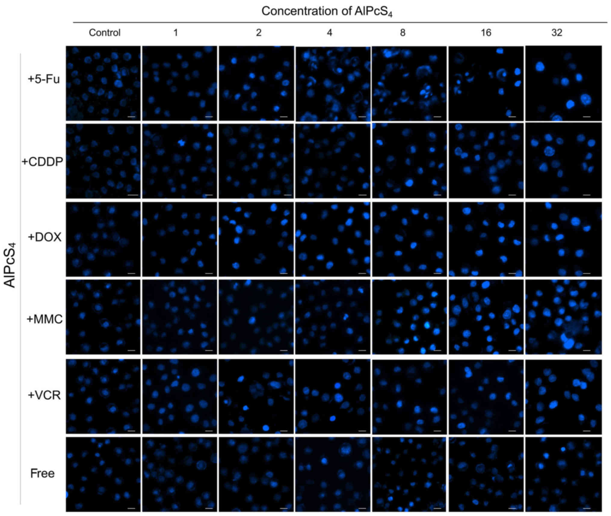 | Figure 4.Apoptosis induced by
AlPcS4 + 5-FU, AlPcS4 + CDDP,
AlPcS4 + DOX, AlPcS4 + MMC, AlPcS4
+ VCR, and free-AlPcS4 in SGC-7901 cells after being
irradiated for 12 h. The cells were treated with 1–32 µm/ml
free-AlPcS4 or AlPcS4 + 5-FU (20 µm),
AlPcS4 + CDDP (5 µm), AlPcS4 + DOX (0.4
µm/ml), AlPcS4 + MMC (0.5 µm/ml) or AlPcS4 +
VCR (0.1 µm/ml) for 6 h. The cells were then irradiated with 635-nm
laser irradiation at 100 mw/cm2 illumination dosage for
5 min, incubated for 12 h, stained with Hoechst 33342 probe, and
then imaged using afluorescence microscope. All the Hoechst
staining images were acquired at an ×400 magnification. The scale
bar represented 20 µm. AlPcS4, Al(III) phthalocyanine
chloride tetrasulfonic acid; 5-FU, 5-fluorouracil; DOX,
doxorubicin; CDDP, cisplatin; MMC, mitomycin C; VCR,
vincristine. |
In the synergistic therapy, higher
apoptosis-inducing abilities induced by AlPcS4/PDT+DOX
were obtained, especially at high concentrations of
AlPcS4 at 24 h, compared to AlPcS4/PDT+MMC
(Fig. 5). In the treatment of
AlPcS4+DOX or AlPcS4+MMC and irradiation with
laser light after 6 and 24 h, the percentage of apoptotic bodies
increased at average values of 3.8-, 1.9-, 2.8- and 1.7-fold, at 6
and 24 h, respectively (Fig. 7).
Additional late apoptotic and necrotic cells were obtained even at
6 and 12 h after treatment by AlPcS4/PDT+DOX (Fig. 6). In contrast to
AlPcS4/PDT+DOX and AlPcS4/PDT+MMC, apoptosis
was induced quickly by AlPcS4/PDT+5-FU at 6 h. This
increasing trend was similar to that of AlPcS4/PDT+VCR.
In addition, the apoptosis-inducing abilities increased (Figs. 3 and 7). At 32 µg/ml, the percentage of
apoptotic bodies of AlPcS4+5-FU at 6, 12 and 24 h
reached ~38, 80 and 88%, respectively (Fig. 7). The apoptosis-inducing abilities
at12 and 24 h were higher than those in AlPcS4/PDT+DOX
and AlPcS4/PDT+MMC. In addition, more necrotic cells
were observed at 12 h after treatment with
AlPcS4/PDT+5-FU (Fig.
6).
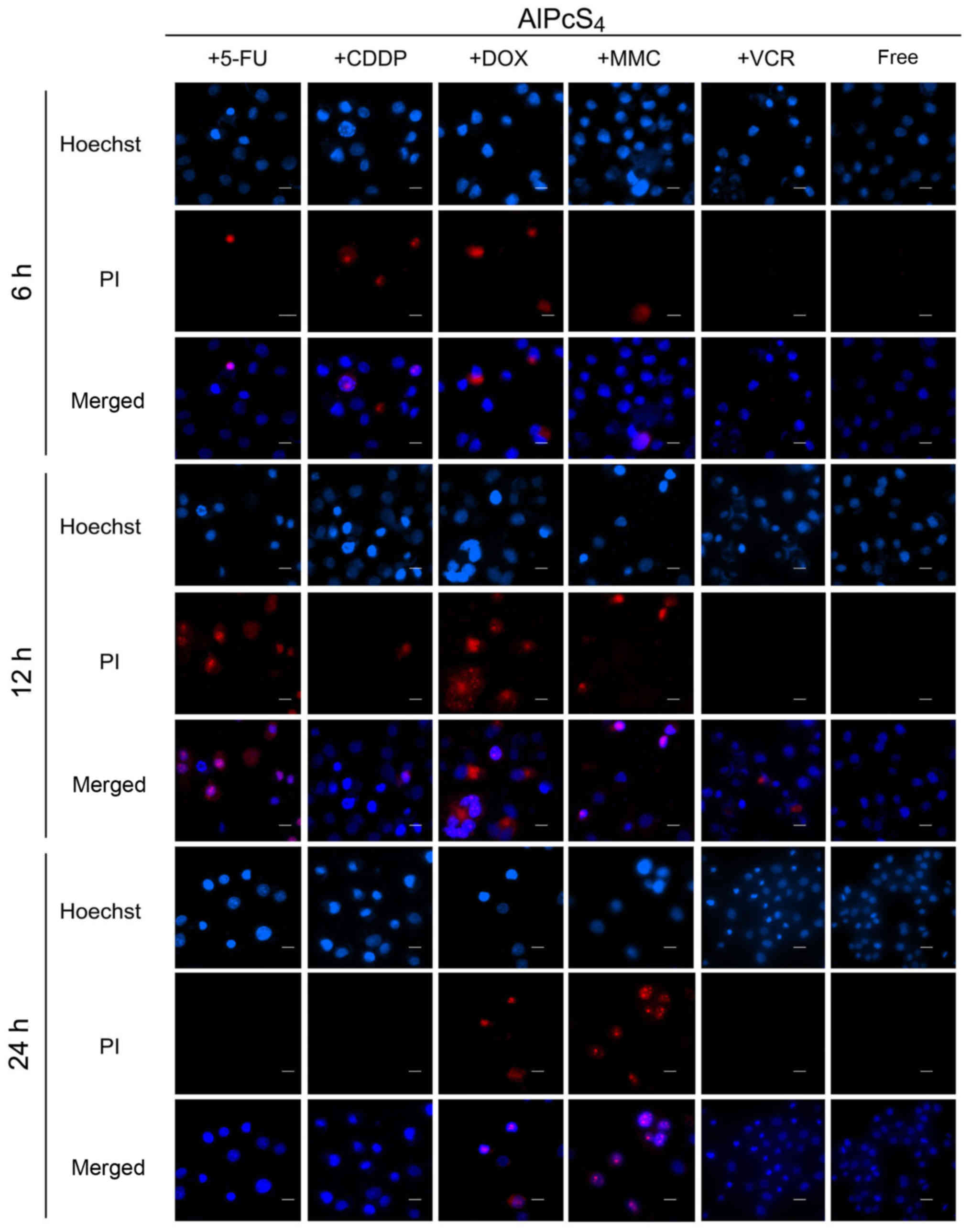 | Figure 6.Apoptosis and necrosis induced by
AlPcS4 + 5-FU, AlPcS4 + CDDP,
AlPcS4 + DOX, AlPcS4 + MMC, AlPcS4
+ VCR and free-AlPcS4 in SGC-7901 cells after being
irradiated for 6, 12 and 24 h. The cells were treated with 1–32
µm/ml free-AlPcS4 or AlPcS4 + 5-FU (20 µm),
AlPcS4 + CDDP (5 µm), AlPcS4 + DOX (0.4
µm/ml), AlPcS4 + MMC (0.5 µm/ml) or AlPcS4 +
VCR (0.1 µm/ml) for 6 h. The cells were then irradiated with 635-nm
laser irradiation at 100 mW/cm2 illumination dosage for
5 min. The cells were incubated for 6, 12 and 24 h, stained with
Hoechst 33342 and PI probes, and imaged usingfluorescence
microscopy. Below 32 µm/ml, no necrosis was obtained. Therefore,
the Hoechst staining and PI images are shown at 32 µm/ml. All the
Hoechst staining and PI images were acquired at an ×400
magnification. The scale bar represented 20 µm. AlPcS4,
Al(III) phthalocyanine chloride tetrasulfonic acid; 5-FU,
5-fluorouracil; DOX, doxorubicin; CDDP, cisplatin; MMC, mitomycin
C; VCR, vincristine. |
In addition, the apoptosis assay results revealed
that AlPcS4/PDT+5-FU evidently improved
apoptosis-inducing abilities even at low concentrations of
AlPcS4. AlPcS4/PDT+DOX slightly increased
apoptosis-inducing abilities at low concentrations of
AlPcS4 at 24 h. AlPcS4/PDT+MMC indicated
fewer increased apoptosis-inducing abilities at a low concentration
of AlPcS4. At 4 µg/ml AlPcS4, the percentage
of apoptotic bodies of AlPcS4/PDT+MMC reached~16 and
29%, at 6 and 24 h, respectively. Generally, 5-FU and DOX have
optimal apoptosis-inducing abilities of AlPcS4 even at
low concentrations. MMC markedly improved the apoptosis-inducing
abilities of AlPcS4 at a high concentration. CDDP and
VCR slightly improved apoptosis-inducing abilities even when
significantly inhibited.
Influence of AlPcS4
fluorescence intensity on combination treatment
To estimate the influence of AlPcS4
delivery efficiency on combination treatment, a fluorescence
intensity assay of AlPcS4 was evaluated after treatment
with different chemotherapeutic agents and AlPcS4. As
revealed in Fig. 8, the
fluorescence intensity of AlPcS4 significantly increased
with the help of low-dose 5-FU and DOX. Notably,
AlPcS4+5-FU exhibited a significant difference at all
the used concentrations compared to free-AlPcS4, but
AlPcS4+DOX exhibited a statistical difference only at a
high concentration. At a high concentration of AlPcS4,
the increasing fluorescence intensity trend in the presence of DOX
was greater than 5-FU. At the highest concentration of
AlPcS4, DOX and 5-FUfluorescence intensity was increased
by 300 and 270%, respectively. Compared with DOX and 5-FU, MMC
resulted in inferior increases by 150% on average. Furthermore,
only at 32 µg/ml AlPcS4, did the fluorescence intensity
exhibit a statistical difference between AlPcS4+MMC and
free-AlPcS4. Conversely, CDDP and VCR did not markedly
improve the fluorescent intensity of AlPcS4. VCR even
reduced the fluorescence intensity of AlPcS4. The
fluorescent intensity of AlPcS4 could be used to reveal
the efficiency of cellular internalization of AlPcS4.
Thus, DOX and 5-FU could prominently increase the efficiency of
cellular internalization of AlPcS4. MMC could slightly
increase the efficiency of cellular internalization of
AlPcS4. CDDP and VCR could not increase the efficiency
of cellular internalization of AlPcS4, even when
reduced.
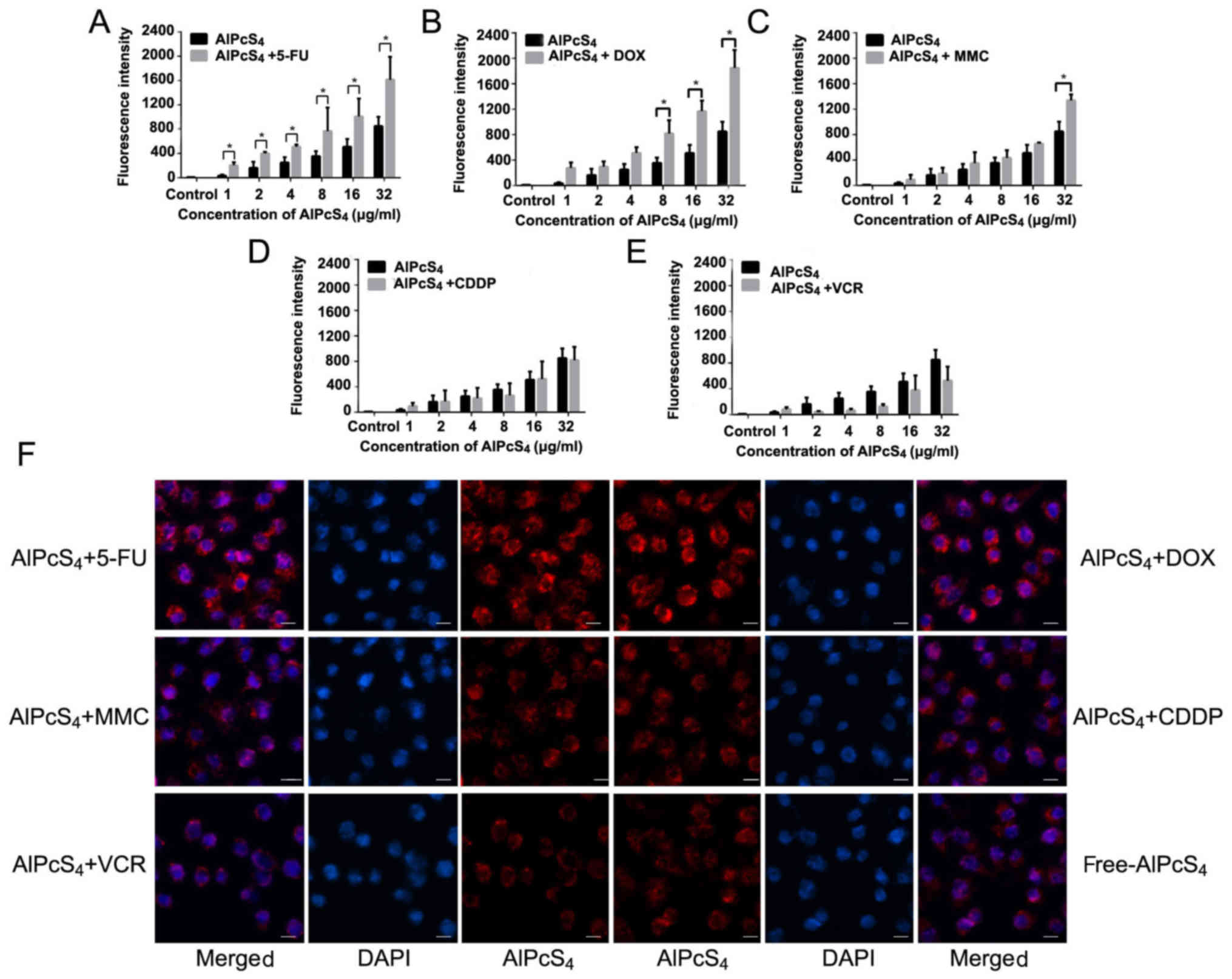 | Figure 8.Fluorescence intensity analysis and
fluorescence imaging of AlPcS4 in SGC-7901 cells after
treatment with AlPcS4 + 5-FU, AlPcS4 + DOX,
AlPcS4 + MMC, AlPcS4 + CDDP,
AlPcS4 + VCR and free-AlPcS4. (A-E)
Fluorescent intensity analysis of AlPcS4 in SGC-7901
cells after treatment with 1–32 µm/ml free-AlPcS4,
AlPcS4 + 5-FU (20 µm), AlPcS4 + DOX (0.4 µm),
AlPcS4 + MMC (0.5 µm/ml), AlPcS4 + CDDP (5
µm) or AlPcS4 + VCR (0.1 µm/ml) for 6 h and measured by
using a fluorescence spectrophotometer. The data represents the
average of three experiments and the bar is the SD. *P<0.05 and
**P<0.01 represented a statistically significant difference in
the fluorescence intensity of AlPcS4 in cells between
the combination therapy of AlPcS4 with a chemical agent
and single therapy of free-AlPcS4. (F) The fluorescent
images of AlPcS4 in SGC-7901 cells after treatment with
32 µm/ml free-AlPcS4, AlPcS4 + 5-FU (20 µm),
AlPcS4 + DOX (0.4 µm/ml), AlPcS4 + MMC (0.5
µm/ml), AlPcS4 + CDDP (5 µm) or AlPcS4 + VCR
(0.1 µm/ml) for 6 h and measured using a fluorescence microscope.
All the images were acquired at an ×400 magnification. The scale
bar represented 20 µm. AlPcS4, Al(III) phthalocyanine
chloride tetrasulfonic acid; 5-FU, 5-fluorouracil; DOX,
doxorubicin; CDDP, cisplatin; MMC, mitomycin C; VCR,
vincristine. |
Influence of AlPcS4
intracellular location on combination treatment
The intracellular location of the photosensitizer is
a significant factor that influences the PDT effect. To observe the
intracellular location of AlPcS4 and evaluate the
influence of AlPcS4 intracellular location in the
presence of low-dose used chemical agents, fluorescence imaging was
carried out. As shown in Fig. 8F,
intracellular staining remained mainly in the cytoplasm and did not
change compared with free-AlPcS4. However, compared with
free-AlPcS4, the fluorescence signal significantly
increased after treatment with AlPcS4+5-FU and
AlPcS4+DOX. These results were consistent with the
results of fluorescent intensity assay.
SOG generation of single or
combination treatment
Underlight activation, the photosensitizer generated
SOG to induce cancer cell death. Thus, the concentration of SOG in
SGC-7901 cells induced by single (free-AlPcS4) or
combination (AlPcS4+chemical agent) treatment was
assessed. 5-FU and DOX improved SOG generation abilities of
AlPcS4 (Fig. 9).
Compared withfree-AlPcS4, 5-FU and DOX resulted in
significant increases at average values of 1.7- and 1.9-fold and
exhibited statistical differences. MMC resulted in inferior
increases, it could improve AlPcS4 SOG generation
abilities by 120% on average but it did not exhibit statistical
differences (Fig. 9). Compared with
5-FU, DOX and MMC, CDDP and VCR were not able to increase the
concentration of SOG (Fig. 9). At
high concentrations, VCR reduced SOG generation abilities of
AlPcS4. These results were consistent with the results
obtained in the fluorescent intensity assay of
AlPcS4+VCR.
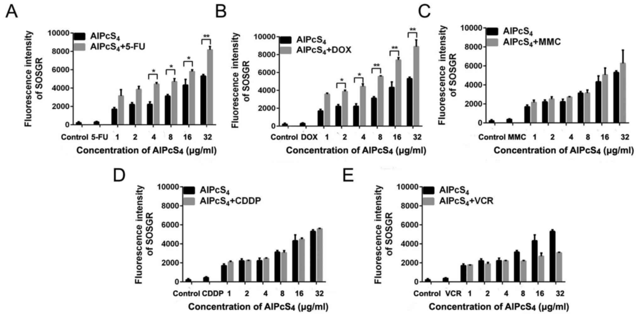 | Figure 9.SOG production in SGC-7901 cells
treated with AlPcS4 + 5-FU, AlPcS4 + CDDP,
AlPcS4 + DOX, AlPcS4 + MMC, AlPcS4
+ VCR and free-AlPcS4. (A-E) Fluorescence intensities of
SOSGR probes were measured to analyze SOG in SGC-7901 cells after
treatment with 1–32 µm free-AlPcS4. (A)
AlPcS4 + 5-FU (20 µm), (B) AlPcS4 + DOX (0.4
µm/ml), (C) AlPcS4 + MMC (0.5 µm/ml), (D)
AlPcS4 + CDDP (5 µm) or (E) AlPcS4 + VCR (0.1
µm/ml) and irradiation with 635-nm laser light for 5 min. Data
represent the average of three experiments and the bar is the SD.
*P<0.05 and **P<0.01 represent a statistically significant
difference in the fluorescence intensity of SOSGR in cells between
the combination therapy of AlPcS4 with a chemical agent
and single therapy of free-AlPcS4. AlPcS4,
Al(III) phthalocyanine chloride tetrasulfonic acid; 5-FU,
5-fluorouracil; DOX, doxorubicin; CDDP, cisplatin; MMC, mitomycin
C; VCR, vincristine; SOG, singlet oxygen. |
ROS generation of single or
combination treatment
Given the help of SOG generated by the
photosensitizer, the ROS concentration associated with reticulum
stress may be easily increased by chemical agents and the
photosensitizer, thereby triggering apoptosis pathway activation
and leading to cell death (29,30).
Hence, the evaluation of the ROS generation induced by
AlPcS4 combination treatment with chemical agentswas
necessary. As shown in Fig. 10,
after pre-treatment with AlPcS4 and chemical agents and
irradiation with laser light, DCFH-DA fluorescence intensity of
SGC-7901 cells increased at different levels. At the highest
concentration of AlPcS4, treatments with
AlPcS4/PDT +5-FU, AlPcS4/PDT +DOX,
AlPcS4/PDT +MMC, AlPcS4/PDT +CDDP, and
AlPcS4/PDT+VCR resulted in increases by 10.5-, 8.7-,
3.1-, 2.9- and 2-fold on average, respectively, compared with
free-AlPcS4/PDT. At all concentrations of
AlPcS4, treatments with AlPcS4/PDT +5-FU,
AlPcS4/PDT +DOX, AlPcS4/PDT +MMC,
AlPcS4/PDT +CDDP, and AlPcS4/PDT+VCR resulted
in 6.3-, 5.6-, 2.9-, 2- and 1.6-fold, increases, respectively,
compared with free-AlPcS4/PDT. In contrast to MMC, CDDP,
and VCR, 5-FU and DOX increased ROS concentration in a
dose-dependent manner, and the increasing fluorescence intensity
trend of 5-FU was higher than that of DOX. Statistical analysis
also revealed that only AlPcS4+5-FU and
AlPcS4+DOX exhibited significant differences compared
with free-AlPcS4.
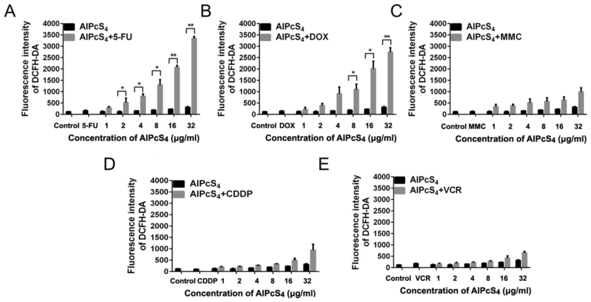 | Figure 10.ROS production in SGC-7901 cells
treated with AlPcS4 + 5-FU, AlPcS4 + CDDP,
AlPcS4 + DOX, AlPcS4 + MMC, AlPcS4
+ VCR and free-AlPcS4. (A-E) Fluorescence intensities of
DCFH-DA probes were measured to analyze ROS in SGC-7901 cells after
treatment with 1–32 µm/ml free-AlPcS4. (A)
AlPcS4 + 5-FU (20 µm/ml), (B) AlPcS4 + DOX
(0.4 µm/ml), (C) AlPcS4 + MMC (0.5 µm/ml), (D)
AlPcS4 + CDDP (5 µm) or (E) AlPcS4 + VCR (0.1
µm/ml) and irradiation with 635-nm laser light for 5 min. Data
represent the average of three experiments and the bar is the SD.
*P<0.05 and **P<0.01 represent a statistically significant
difference in the fluorescence intensity of DCFH-DA in cells
between the combination therapy of AlPcS4 with a
chemical agent and single therapy of free-AlPcS4.
AlPcS4, Al(III) phthalocyanine chloride tetrasulfonic
acid; 5-FU, 5-fluorouracil; DOX, doxorubicin; CDDP, cisplatin; MMC,
mitomycin C; VCR, vincristine; ROS, reactive oxygen species. |
Discussion
AlPcS4 is a second-generation
photosensitizer that may be a promising antitumor agent in PDT for
gastric cancer therapy due to its emission spectra in the NIR
region, deep penetration in tissue, high quantum yields, good
photostability, and little photobleaching. However, its low
delivery efficiency induces slight penetration ability in cancer
cells, thereby leading to a limited PDT effect on gastric cancer
cells. These issues warrant resolution. Gantchev et al
proposed a combination treatment strategy via AlPcS4/PDT
and etoposide, an antitumor drug, on K562 human leukemic cells to
improve the antitumor effect of AlPcS4/PDT (31). This proposal which points to the
combination treatment of AlPcS4/PDT with chemical
agents, may be a promising method for gastric cancer therapy.
Little research has focused on the combination of PDT with
conventional chemotherapeutics for gastric cancer therapy. Thus,
using different chemical drugs for combination with
AlPcS4/PDT to optimize treatment effects on gastric
cancer should be investigated.
Nonaka et al demonstrated that the
combination of Photofrin/PDT with CDDP enhanced cytotoxic and
apoptotic effects, thereby inhibiting the cell growth in lymphoma
cancer and esophageal carcinoma (32). Casas et al evaluated the
synergistic effect between 5-ALA/PDT and DOX on mammary
adenocarcinomas, and the anticancer effect was significantly
enhanced by the combined treatment. Datta et al observed
that combined 5-ALA/PDT and MMC treatment was a workable
therapeutic approach on superficial bladder cancer (33). Martin et al demonstrated that
the enhancement of antitumor growth effectiveness could be obtained
by combining 5-ALA/PDT and 5-FU in non-melanoma skin cancer
(34). Dima et al revealed
that a clearly positive inhibition effect was obtained by using the
combination of Photofrin/PDT with VCR in ovarian cancer (35). CDDP, DOX, MMC, 5-FU and VCR are also
common gold-standard chemotherapeutic agents that are clinically
recommended for gastric cancer. Hence, we can infer that CDDP, DOX,
MMC, 5-FU, and VCR may be used for AlPcS4/PDT
combination treatment to improve the effect of
AlPcS4/PDT. However, all the aforementioned
chemotherapeutic agents have high systemic toxicity, drug
resistance, high rates of tumor metastasis, and recurrence during
use. Therefore, a combined treatment must be used to enhance
therapeutic efficacy, reduce toxic side effects, and elude drug
resistance. Ilaria et al investigated the antitumor growth
effect of the combination of indocyanine green/PDT with low-dose
CDDP on breast cancer cells. Wei et al evaluated the
inhibitory effect of the combination of 5-ALA/PDT with low-dose
CDDP on HeLa cells (28,36). The results revealed that indocyanine
green or 5-ALA/PDT combination with low-dose CDDP have mutual
reinforcement of antitumor efficacy while having few toxic side
effects. Therefore, in the present study, we investigated the
combined effect of AlPcS4/PDT with low-dose CDDP, DOX, MMC, 5-FU
and VCR on SGC-7901 gastric cancer cells, respectively.
The inhibition of cell viability induced by the
combination treatment of AlPcS4/PDT with low-dose
chemotherapeutic agents without 635-nm laser irradiation was lower
than the summation of the inhibition of cell viability induced by
free-AlPcS4 or alone and low-dose chemotherapeutic
agents without 635-nm laser irradiation. This value was even lower
than the inhibition of cell viability induced by low-dose
chemotherapeutic agents used alone. The antagonistic effects in the
combination treatment of AlPcS4/PDT with low-dose
chemotherapeutic agents without laser irradiation were obtained.
The antagonistic effect of low-dose VCR in combination therapy
without 635-nm laser irradiation was more pronounced, whereas the
effect of low-dose CDDP was inferior. Compared with VCR and CDDP,
the inhibition of cell viability induced by the combination
treatment of AlPcS4/PDT with low-dose 5-FU, DOX, and MMC
without 635-nm laser irradiation was more evident. The antagonistic
effects of low-dose 5-FU, DOX and MMC in combination therapy
without 635-nm laser irradiation were weaker. The degree of the
antagonistic effect of low-dose DOX was higher than 5-FU and was
much higher than MMC. Low-dose chemotherapy usually induced
resistance emergence to limit the treatment effect of the antitumor
drugs (37). Hence, the
antagonistic effect in combination therapy without laser
irradiation may be caused by drug resistance.
Furthermore, the antitumor growth effects of the
combination treatment of AlPcS4/PDT with low-dose
chemotherapeutic agents used in our study with 635-nm laser
irradiation were assessed. The combination treatment of
AlPcS4/PDT with low-dose 5-FU, DOX and MMC had
significant inhibitory effects on gastric cancer cells. The
inhibitory effects of combination treatment were higher than the
efficiency summation of free-AlPcS4 and 5-FU, DOX, or
MMC, respectively. In other words, the synergistic anticancer
activity via the combination of AlPcS4/PDT with low-dose
DOX was optimal; at low-dosages, 5-FU or MMC was inferior. Compared
with 5-FU, DOX, or MMC, the inhibitory effect of the combination
treatment of AlPcS4/PDT with low-dose CDDP or VCR was
lower than the efficiency summation of free-AlPcS4 and
CDDP or VCR, respectively. However, the antitumor growth effect
continued to increase at average values of 16.5 and 8.7% compared
with free-AlPcS4. The increased antitumor effect in
combination therapy with laser irradiation was contrary to the
antagonistic effects in the combination therapy without laser
irradiation. Therefore, irradiation of laser light and PDT therapy
may improve the drug resistance of low-dose chemotherapeutic
agents.
To determine the reason for the enhancement of
therapeutic efficacy, the fluorescence intensity of
AlPcS4 with low-dose chemotherapeutic agents, including
CDDP, DOX, MMC, 5-FU and VCR, in SGC-7901 cells was determined. The
increasing trend of DOX was higher than that of 5-FU and much
higher than that of MMC, thereby revealing that DOX was superior in
effectively improving the efficiency of cellular internalization of
AlPcS4. The antitumor effect of the combination of
AlPcS4/PDT with low-dose DOX was optimal. Thus, the mass
of increased antitumor effect in AlPcS4/PDT+DOX may be
induced by the increased AlPcS4/PDT effect. The results
of apoptosis-inducing and necrosis-inducing abilities revealed that
AlPcS4 with low-dose chemotherapeutic agents increased
apoptosis and necrosis abilities compared with
free-AlPcS4. In addition, the results of the
apoptosis-inducing ability revealed that low-dose chemotherapeutic
agents quickly increased the apoptosis-inducing ability.
Furthermore, in the presence of low-dose chemotherapeutic agents,
the apoptosis activation time was prolonged. High
apoptosis-inducing ability was induced.
The results of SOG generation demonstrated that the
increased SOG effect of AlPcS4/PDT+DOX was higher than
that of AlPcS4/PDT+5-FU and much higher than that of
AlPcS4/PDT+MMC. The results of ROS generation related to
apoptosis revealed that the increased ability of ROS generation
induced by AlPcS4/PDT+5-FU was higher than that by
AlPcS4/PDT+DOX and much higher than that by
AlPcS4/PDT+MMC. These results revealed that the
combination treatment of AlPcS4/PDT with low-dose
chemotherapeutic agents exhibited a higher antitumor growth effect
not only by improving the apoptosis-inducing activities of the
chemotherapeutic agents but also by increasing the
apoptosis-inducing activities of AlPcS4/PDT. In
addition, the results of the synergistic effect revealed that the
combination antitumor growth effect of AlPcS4/PDT+DOX
was higher than that of AlPcS4/PDT+DOX and much higher
than that of AlPcS4/PDT+MMC. Hence, the increased degree
of the apoptosis-inducing activities of AlPcS4/PDT was
much more significant. However, the general trend of the ROS
generation and antitumor growth effect revealed that ROS-related
triggers that induce apoptosis are important factors that lead to
cell death in the combination treatment of AlPcS4/PDT
with low-dose chemotherapeutic agents.
Therefore, the possible molecular pathway involved
in the combination treatment-induced apoptosis may involve the
activation of the mitochondrial apoptosis pathway or via the
endoplasmic reticulum (ER) stress-induced apoptosis pathway
(38,39). AlPcS4 can be accumulated
in the mitochondria, and local damage induced by AlPcS4
may be propagated to the mitochondria by various means; chemical
therapy can also activate mitochondrial apoptosis pathways to lead
cell apoptosis (40). Mitochondrial
ROS can damage DNA and activate an aberrant apoptosis signaling
pathway (41). Furthermore, the
generation of ROS could trigger ER stress (42). This stress can further trigger
several specific signaling pathways, including ER-associated
protein degradation and the unfolded protein response (38). The unfolded protein response can
trigger apoptosis activities by inducing cytoprotective and
destructive functions when ER stress is prolonged or adaptive
responses fail. In a future study, the expression levels of a
series of proteins involved in the mitochondrial apoptosis pathway
and ER-stress apoptosis pathway will be detected by western blot
analysis in combination therapy, such as Bcl-2 family proteins,
caspase-related proteins, phosphorylated eIF2α, GADD153, ATF6,
GRP78 and GRP94. Our aim is to verify the enhanced synergistic
antitumor activity induced by activating the mitochondrial
apoptosis pathway and the ER stress-induced apoptosis pathway.
Finally, we conclude that the combination treatment
of AlPcS4/PDT with low-dose chemotherapeutic agents may
provide promising treatment strategies to increase the weak
delivery efficiency of AlPcS4in gastric cancer cells and
further effectively improve the antitumor effect on gastric cancer.
The treatment also decreases the toxic side effects. Hence, we
investigated the antitumor growth effect on the SGC-7901 gastric
cancer cell line by combination treatment of AlPcS4/PDT
with various low-dose chemotherapeutic agents, including 5-FU, DOX,
CDDP, MMC and VCR. The antitumor growth effect could be increased
through a combination treatment of AlPcS4/PDT with a
low-dose chemotherapeutic agent. An evident synergistic effect was
obtained in the combination treatment of AlPcS4/PDT with
low-dose 5-FU, DOX, and MMC. These combination treatments increased
AlPcS4 intracellular uptake ability and ROS and SOG
generation abilities, thereby inducing significant apoptosis and
necrosis. In addition, low-dose chemotherapeutic agents improved
apoptosis-inducing abilities quickly and prolong the
apoptosis-inducing period of AlPcS4/PDT. In general, the
combination treatments of AlPcS4/PDT with low-dose
chemotherapeutic agents had a significant antitumor growth effect
and a low dark-cytotoxicity effect on gastric cancer via the
increase of AlPcS4 intracellular uptake ability,
improving the apoptosis-inducing abilities induced by
chemotherapeutic agents in a short time and prolonging the
apoptosis-inducing period of AlPcS4/PDT.
Acknowledgements
We gratefully acknowledge Professor Kaichun Wu and
Yongzhan Nie of the Department of Gastroenterology and State Key
Laboratory of Cancer Biology, Xijing Hospital, Fourth Military
Medical University for kindly providing us with the SGC-7901 cell
line. The TEM study was undertaken at the International Center for
Dielectric Research, Xi'an Jiaotong University, Xi'an, China. We
also thank Mr. Chuansheng Ma for his help in using the TEM
facility.
Funding
The present study was supported by the National
Natural Science Foundation of China under grant nos. 61505159,
61575156, 61775178, 61335012, 61727823 and 61705177, and the China
Postdoctoral Science Foundation (grant nos. 2015M572570 and
2017M613107).
Availability of data and materials
The datasets used during the present study are
available from the corresponding author upon reasonable
request.
Authors' contributions
JX, ZZ and CY conceived and designed the study. JX,
SW, BX, YH, JW and SW performed the experiments. JX wrote the
paper. LZ and LS review and edited the manuscript. All authors read
and approved the final manuscript and agree to be accountable for
all aspects of the research in ensuring that the accuracy or
integrity of any part of the work are appropriately investigated
and resolved.
Ethics approval and consent to
participate
Not applicable.
Consent for publication
Not applicable.
Competing interests
The authors declare that they have no competing
interests.
Glossary
Abbreviations
Abbreviations:
|
AlPcS4
|
Al(III) phthalocyanine chloride
tetrasulfonic acid
|
|
PDT
|
photodynamic therapy
|
|
5-FU
|
5-fluorouracil
|
|
DOX
|
doxorubicin
|
|
CDDP
|
cisplatin
|
|
MMC
|
mitomycin C
|
|
VCR
|
vincristine
|
|
ROS
|
reactive oxygen species
|
|
SOG
|
singlet oxygen
|
|
ER
|
endoplasmic reticulum
|
References
|
1
|
Torre LA, Bray F, Siegel RL, Ferlay J,
Lortet-Tieulent J and Jemal A: Global cancer statistics, 2012. CA
Cancer J Clin. 65:87–108. 2015. View Article : Google Scholar : PubMed/NCBI
|
|
2
|
Dougherty TJ, Gomer CJ, Henderson BW, Jori
G, Kessel D, Korbelik M, Moan J and Peng Q: Photodynamic therapy. J
Natl Cancer Inst. 90:889–905. 1998. View Article : Google Scholar : PubMed/NCBI
|
|
3
|
Vrouenraets MB, Visser GW, Snow GB and van
Dongen GA: Basic principles, applications in oncology and improved
selectivity of photodynamic therapy. Anticancer Res. 23:505–522.
2003.PubMed/NCBI
|
|
4
|
Shafirstein G, Battoo A, Harris K, Baumann
H, Gollnick SO, Lindenmann J and Nwogu CE: Photodynamic therapy of
non-small cell lung cancer. Narrative review and future directions.
Ann Am Thorac Soc. 13:265–275. 2016.PubMed/NCBI
|
|
5
|
Moor AC: Signaling pathways in cell death
and survival after photodynamic therapy. J Photochem Photobiol B.
57:1–13. 2000. View Article : Google Scholar : PubMed/NCBI
|
|
6
|
Rigual NR, Shafirstein G, Frustino J,
Seshadri M, Cooper M, Wilding G, Sullivan MA and Henderson B:
Adjuvant intraoperative photodynamic therapy in head and neck
cancer. JAMA Otolaryngol Head Neck Surg. 139:706–711. 2013.
View Article : Google Scholar : PubMed/NCBI
|
|
7
|
Lucena SR, Salazar N, Gracia-Cazaña T,
Zamarrón A, González S, Juarranz Á and Gilaberte Y: Combined
treatments with photodynamic therapy for non-melanoma skin cancer.
Int J Mol Sci. 16:25912–25933. 2015. View Article : Google Scholar : PubMed/NCBI
|
|
8
|
Zimmermann A, Ritsch-Marte M and Kostron
H: mTHPC-mediated photodynamic diagnosis of malignant brain tumors.
Photochem Photobiol. 74:611–616. 2001. View Article : Google Scholar : PubMed/NCBI
|
|
9
|
Wang GD, Nguyen HT, Chen H, Cox PB, Wang
L, Nagata K, Hao Z, Wang A, Li Z and Xie J: X-Ray induced
photodynamic therapy: A combination of radiotherapy and
photodynamic therapy. Theranostics. 6:2295–2305. 2016. View Article : Google Scholar : PubMed/NCBI
|
|
10
|
Luo D, Carter KA, Miranda D and Lovell JF:
Chemophototherapy: An emerging treatment option for solid tumors.
Adv Sci. 4:16001062017. View Article : Google Scholar
|
|
11
|
Rosenthal I: Phthalocyanines as
photodynamic sensitizers. Photochem Photobiol. 53:859–870. 1991.
View Article : Google Scholar : PubMed/NCBI
|
|
12
|
Samuni A, Samuni A and Swartz HM:
Evaluation of dibromonitrosobenzene sulfonate as a spin trap in
biological systems. Free Radic Biol Med. 7:37–43. 1989. View Article : Google Scholar : PubMed/NCBI
|
|
13
|
da Silva NS, Ribeiro Cde M, Machado AH and
Pacheco-Soares C: Ultrastructural changes in Tritrichomonas foetus
after treatments with AlPcS4 and photodynamic therapy. Vet
Parasitol. 146:175–181. 2007. View Article : Google Scholar : PubMed/NCBI
|
|
14
|
Derycke AS, Kamuhabwa A, Gijsens A,
Roskams T, De Vos D, Kasran A, Huwyler J, Missiaen L and de Witte
PA: Transferrin-conjugated liposome targeting of photosensitizer
AlPcS4 to rat bladder carcinoma cells. J Natl Cancer Instit.
96:1620–1630. 2004. View Article : Google Scholar
|
|
15
|
Plaetzer K, Kiesslich T, Krammer B and
Hammerl P: Characterization of the cell death modes and the
associated changes in cellular energy supply in response to
AlPcS4-PDT. Photochem Photobiol Sci. 1:172–177. 2002. View Article : Google Scholar : PubMed/NCBI
|
|
16
|
Gijsens A, Derycke A, Missiaen L, De Vos
D, Huwyler J, Eberle A and de Witte P: Targeting of the
photocytotoxic compound AlPcS4 to Hela cells by transferrin
conjugated PEG-liposomes. Int J Cancer. 101:78–85. 2002. View Article : Google Scholar : PubMed/NCBI
|
|
17
|
Rück A, Heckelsmiller K, Kaufmann R,
Grossman N, Haseroth E and Akgün N: Light-induced apoptosis
involves a defined sequence of cytoplasmic and nuclear calcium
release in AlPcS4-photosensitized rat bladder RR 1022 epithelial
cells. Photochem Photobiol. 72:210–216. 2000. View Article : Google Scholar : PubMed/NCBI
|
|
18
|
Moor AC, Wagenaars-van Gompel AE, Hermanns
RC, van der Meulen J, Smit J, Wilschut J, Brand A, Dubbelman TM and
VanSteveninck J: Inhibition of various steps in the replication
cycle of vesicular stomatitis virus contributes to its
photoinactivation by AlPcS4 or Pc4 and red light. Photochem
Photobiol. 69:353–359. 1999. View Article : Google Scholar : PubMed/NCBI
|
|
19
|
Peng Q, Moan J, Farrants GW, Danielsen HE
and Rimington C: Location of P-II and AlPCS4 in human tumor LOX in
vitro and in vivo by means of computer-enhanced video fluorescence
microscopy. Cancer Lett. 58:37–47. 1991. View Article : Google Scholar : PubMed/NCBI
|
|
20
|
Longley DB, Harkin DP and Johnston PG:
5-fluorouracil: Mechanisms of action and clinical strategies. Nat
Rev Cancer. 3:330–338. 2003. View Article : Google Scholar : PubMed/NCBI
|
|
21
|
Ottewell PD, Woodward JK, Lefley DV, Evans
CA, Coleman RE and Holen I: Anticancer mechanisms of doxorubicin
and zoledronic acid in breast cancer tumor growth in bone. Mol
Cancer Ther. 8:2821–2832. 2009. View Article : Google Scholar : PubMed/NCBI
|
|
22
|
Dasari S and Tchounwou PB: Cisplatin in
cancer therapy: Molecular mechanisms of action. Eur J Pharmacol.
740:364–378. 2014. View Article : Google Scholar : PubMed/NCBI
|
|
23
|
Li NY, Chen F, Dikkers FG and Thibeault
SL: Dose-dependent effect of mitomycin C on human vocal fold
fibroblasts. Head Neck. 36:401–410. 2014. View Article : Google Scholar : PubMed/NCBI
|
|
24
|
Mohammadgholi A, Rabbani-Chadegani A and
Fallah S: Mechanism of the interaction of plant alkaloid
vincristine with DNA and chromatin: Spectroscopic study. DNA Cell
Biol. 32:228–235. 2013. View Article : Google Scholar : PubMed/NCBI
|
|
25
|
Zhang D and Fan D: Multidrug resistance in
gastric cancer: Recent research advances and ongoing therapeutic
challenges. Expert Rev Anticancer Ther. 7:1369–1378. 2007.
View Article : Google Scholar : PubMed/NCBI
|
|
26
|
Castano AP, Mroz P, Wu MX and Hamblin MR:
Photodynamic therapy plus low-dose cyclophosphamide generates
antitumor immunity in a mouse model. Proc Natl Acad Sci USA.
105:5495–5500. 2008. View Article : Google Scholar : PubMed/NCBI
|
|
27
|
Chibber S, Hassan I, Farhan M, Salman M
and Naseem I: White light augments chemotherapeutic potential of
cyclophosphamide: An in vitro study. Biometals. 26:23–31. 2013.
View Article : Google Scholar : PubMed/NCBI
|
|
28
|
Wei XQ, Ma HQ, Liu AH and Zhang YZ:
Synergistic anticancer activity of 5-aminolevulinic acid
photodynamic therapy in combination with low-dose cisplatin on Hela
cells. Asian Pac J Cancer Prev. 14:3023–3028. 2013. View Article : Google Scholar : PubMed/NCBI
|
|
29
|
Broekgaarden M, Weijer R, van Gulik TM,
Hamblin MR and Heger M: Tumor cell survival pathways activated by
photodynamic therapy: A molecular basis for pharmacological
inhibition strategies. Cancer Metastasis Rev. 34:643–690. 2015.
View Article : Google Scholar : PubMed/NCBI
|
|
30
|
Farooqi AA, Li KT, Fayyaz S, Chang YT,
Ismail M, Liaw CC, Yuan SS, Tang JY and Chang HW: Anticancer drugs
for the modulation of endoplasmic reticulum stress and oxidative
stress. Tumour Biol. 36:5743–5752. 2015. View Article : Google Scholar : PubMed/NCBI
|
|
31
|
Gantchev TG, Brasseur N and van Lier JE:
Combination toxicity of etoposide (VP-16) and photosensitisation
with a water-soluble aluminium phthalocyanine in K562 human
leukaemic cells. Br J Cancer. 74:1570–1577. 1996. View Article : Google Scholar : PubMed/NCBI
|
|
32
|
Nonaka M, Ikeda H and Inokuchi T: Effect
of combined photodynamic and chemotherapeutic treatment on lymphoma
cells in vitro. Cancer Lett. 184:171–178. 2002. View Article : Google Scholar : PubMed/NCBI
|
|
33
|
Datta SN, Allman R, Loh C, Mason M and
Matthews PN: Effect of photodynamic therapy in combination with
mitomycin C on a mitomycin-resistant bladder cancer cell line. Br J
Cancer. 76:312–317. 1997. View Article : Google Scholar : PubMed/NCBI
|
|
34
|
Martin G: Prospective, case-based
assessment of sequential therapy with topical Fluorouracil cream
0.5% and ALA-PDT for the treatment of actinic keratosis. J Drugs
Dermatol. 10:372–378. 2011.PubMed/NCBI
|
|
35
|
Dima VF, Mihăilescu IN, Dima SV, Chivu L,
Stirbeţ M, Udrea M and Popa A: Studies of the effects of associated
photodynamic therapy and drugs on macromolecular synthesis of
tumoral cells grown in vitro. Arch Roum Pathol Exp Microbiol.
49:155–175. 1990.PubMed/NCBI
|
|
36
|
Crescenzi E, Varriale L, Iovino M,
Chiaviello A, Veneziani BM and Palumbo G: Photodynamic therapy with
indocyanine green complements and enhances low-dose cisplatin
cytotoxicity in MCF-7 breast cancer cells. Mol Cancer Ther.
3:537–544. 2004.PubMed/NCBI
|
|
37
|
Day T and Read AF: Does high-dose
antimicrobial chemotherapy prevent the evolution of resistance?
PLoS Comput Biol. 12:e10046892016. View Article : Google Scholar : PubMed/NCBI
|
|
38
|
Ron D and Walter P: Signal integration in
the endoplasmic reticulum unfolded protein response. Nat Rev Mol
Cell Biol. 8:519–529. 2007. View Article : Google Scholar : PubMed/NCBI
|
|
39
|
Fogg VC, Lanning NJ and Mackeigan JP:
Mitochondria in cancer: At the crossroads of life and death. Chin J
Cancer. 30:526–539. 2011. View Article : Google Scholar : PubMed/NCBI
|
|
40
|
Buytaert E, Dewaele M and Agostinis P:
Molecular effectors of multiple cell death pathways initiated by
photodynamic therapy. Biochim Biophys Acta. 1776:86–107.
2007.PubMed/NCBI
|
|
41
|
Ott M, Gogvadze V, Orrenius S and
Zhivotovsky B: Mitochondria, oxidative stress and cell death.
Apoptosis. 12:913–922. 2007. View Article : Google Scholar : PubMed/NCBI
|
|
42
|
Xu C, Bailly-Maitre B and Reed JC:
Endoplasmic reticulum stress: Cell life and death decisions. J Clin
Invest. 115:2656–2664. 2005. View Article : Google Scholar : PubMed/NCBI
|


