Introduction
The digestive system is one of the systems that is
most likely to be disordered, including infection, inflammation,
atypical hyperplasia, metaplasia and carcinogenesis. According to
the World Cancer Report 2014, cancers of the digestive system were
the most commonly diagnosed cancers and the most common cause of
cancer-related deaths in 2012 (1).
Increased attention should be paid to the diagnosis, therapy and
prognosis of these cancer patients to prolong survival time and
improve their quality of life.
For cell survival, it is essential for cells to
avoid excessive change in cell volume, which jeopardizes structural
integrity and the stability of the intracellular milieu (2). Cell volume regulation plays pivotal
roles in all types of cellular functions, such as epithelial
transport, metabolism, excitation, hormone release, cell migration,
cell proliferation and cell death (2–4).
Channel proteins on the cell membrane govern the
movements of water and electrolytes for the regulation of cell
volume. Although it has been confirmed that channel proteins play
important roles in a wide variety of physiological and/or
pathophysiological processes, the molecular entity for the
volume-regulated anion channel (VRAC) is not entirely confirmed.
The search for the molecular identity of VRAC has been long and has
yielded multiple potential candidates, all of which eventually
turned out to have properties not fully compatible with those of
VRAC (5). Recently, 2 groups have
independently reported that the leucine-rich repeat-containing 8A
(LRRC8A) is a main molecular determinant of VRAC current (6,7), which
has been immediately confirmed in different cells or tissue by
other groups (8–11). However, in human retinal pigment
epithelium cells, bestrophin 1, but not LRRC8A is essential for
volume regulation (12). This idea
concerning a non-essential contribution of LRRC8A to volume
regulation has also been supported by others (13,14).
These findings indicate that the molecular determinants of VRAC may
be more complex and that VRAC could be formed by a cell
type-specific or tissue-specific subunit component.
Given its importance in cancer cell proliferation,
apoptosis, migration and/or invasion, and drug resistance, VARC has
been considered to be a promising target for cancer diagnosis,
prognosis and therapy (15–17). However, the biological functions of
the LRRC8 family proteins remain poorly understood. LRRC8A was
isolated and identified in a girl with congenital
agammaglobulinemia (18),
indicating that it may be involved in B- and T-cell development
(19–21). In vascular smooth muscle cells, it
was found that LRRC8A takes part in inflammation via supporting
tumor necrosis factor-α-induced superoxide production (22). In human ovarian (A2780) and alveolar
(A549) cancer cells, a reduced expression of LRRC8A may contribute
to the acquisition of cisplatin resistance (23,24).
The contribution of LRRC8A varies and is largely dependent on the
cell type; thus, the precise role of LRRC8A warrants further
investigation.
In the present study, we first disclosed that the
high expression of LRRC8A may be correlated with the short survival
time of colon cancer patients by enhancing the growth and
metastasis of cancer cells.
Materials and methods
Cell culture, transfection and
infection
Human colon cancer cells (HCT116) were routinely
grown in Roswell Park Memorial Institute (RPMI)-1640 culture medium
with 10% fetal bovine serum (FBS), 100 IU/ml penicillin and 100
µg/ml streptomycin in an atmosphere with 100% humidity, 5%
CO2, and 95% O2 at 37°C. The cells were
trypsinized and subcultured every 2 days.
The small interfering RNAs (siRNAs) were chemically
synthesized and labeled with or without FAM carboxyfluorescein
(Shanghai GenePharma Co., Ltd., Shanghai, China). The sense strand
of LRRC8A siRNA was 5′-ACCAAGCUCAUCGUCCUCAACtt-3′; and, the sense
strand of the negative control siRNA was
5′-UUCUCCGAACGUGUCACGUtt-3′. The short hairpin RNA (shRNA) was
designed according to the sequence of siRNA, inserted into the
pGLVH1 lentivirus shRNA vector containing the GFP gene, and then
packaged as infectious lentivirus by the Shanghai GenePharma Co.
The sequence of LRRC8A shRNA was,
5′-CCGGACCAAGCTCATCGTCCTCAACCTCGAGGTTGAGGACGATGAGCTTGGTTTTTTG-3′
(12).
For siRNA transfection, HCT116 cells were cultured
in RPMI-1640 medium without antibiotics in 12-well culture plates
for 24 h and reached 30–40% confluence. The cells were transfected
with 100 nM siRNA in the presence of Invitrogen™ Lipofectamine 2000
(2.5 µl in 1,000 µl medium; Thermo Fisher Scientific, Inc.,
Waltham, MA, USA) in serum and antibiotic-free culture medium for 6
h, and were incubated in normal RPMI-1640 medium for 72 h. For
lentivirus infection, when cells reached 70% confluence, the
lentivirus particles [multiplicity of infection (MOI=5)] were
diluted and added into the culture medium; after a 12-h incubation,
the cells were incubated in normal medium and subcultured for 1
week. The transfection efficiencies were detected using flow
cytometry.
Animal feeding and tumor cell
implantation
The animal experimental protocol was approved by the
Laboratory Animal Administration Committee of Xi'an Jiaotong
University and was carried out in accordance with the Guidelines
for Animal Experimentation of Xi'an Jiaotong University and the
Guide for the Care and Use of Laboratory Animals published by the
US National Institutes of Health (NIH Publication no. 85–23,
revised 2011).
Male BALB/c nude mice, 4 weeks of age, were provided
by the Laboratory Animal Center of Xi'an Jiaotong University and
maintained under a 12-h dark/light cycle with ad libitum
self-feeding in specific pathogen-free conditions (55% humidity and
22°C). Every nude mouse was subcutaneously injected with the
scramble and LRRC8A shRNA lentivirus-infected HCT116 cells
(1×10⁶) at the armpits of the right and left forelimb,
respectively. The initial body weight of 10 mice was 14.7±0.3 g and
then measured once a week; after 3 weeks, the mice were
anesthetized with 1.5% isoflurane inhalation, and then the
tumorigenesis (indicated by green fluorescence) was detected using
the in vivo non-invasive small animal molecular imaging
system (Xenogen; Caliper Life Sciences, Inc., Hopkinton, MA, USA);
according to the IACUC guidelines for mice, tumor cannot reach the
maximum allowable size (diameter, 1.5 cm; area, 1.8 cm2;
and volume 1.8 cm3). Next, the solid tissues of the
neoplasms were dissected and weighed; the tissues were fixed in 10%
formalin solution and embedded with paraffin.
Tissue microarray, immunostaining, and
hematoxylin and eosin staining
Tissue microarrays were commercial and purchased
from the Shanghai Outdo Biotech Co., Ltd. (Shanghai, China)
(HDgS-C120PT-01, HOrg-C120PG-02, HDgS-C140PT-01, HRec-Ade180Sur-01
and HCol-Ade180Sur-02); ethics approval for research using human
tissue was obtained from the Ethics Committee in Taizhou Hospital,
Zhejiang Province, and included a waiver for consent. Patients
involved in HRec-Ade180Sur-01 underwent surgery during the period
from July 2006 to August 2007, and then were followed up in August
2013. Patients involved in HCol-Ade180Sur-02 underwent surgery
during the period from May 2007 to April 2008, and were followed up
in September 2014. The tissues or tissue microarrays were
immunolabeled using the UltraSensitive™ SP system (KIT-9710; Fuzhou
Maixin Biotech Co., Ltd., Fuzhou, China) and stained with
hematoxylin and eosin (H&E; C0105; Beyotime Institute of
Biotechnology, Haimen, China) following the manufacturer's
instructions; the dilution of primary antibodies for LRRC8A (cat.
no. ab157489; Abcam, Cambridge, MA, USA) and CD31 (cat. no.
ab134168; Abcam) was 1:100. Images were captured under the digital
pathology whole slide scanners. Using Aperio ImageScope software
(Leica Microsystems, Inc., Buffalo Grove, IL, USA), parenchyma in
cancer and cancer adjacent tissues were depicted, and the intensity
and distribution of the immunostaining reaction (LRRC8A) were
analyzed.
Solutions and chloride current
recording
The isotonic bath solution contained (in mM) the
following: 70 NaCl, 0.5 MgCl2, 2 CaCl2, 10
N-2-hydroxyethylpiperazine-N-2-ethanesulfonic acid (HEPES), and 140
D-mannitol. The hypotonic bath solution (47% hypotonic, compared
with the isotonic solution) was obtained by omitting the D-mannitol
from the isotonic solution. The pipette solution contained (in mM)
70 N-methyl-d-glucamine chloride (NMDG-Cl), 1.2
MgCl2, 10 HEPES, 1 ethylene
glycol-bis(2-aminoethylether)-N,N,N′,N′-tetraacetic acid
(EGTA), 140 D-mannitol and 2 ATP. The osmolarity of solutions was
detected with an automatic cryoscopic osmometer (Osmomat 030;
Gonotec, Berlin, Germany). The pH of the bath and pipette solutions
was adjusted to 7.40 and 7.25, respectively.
Coverslips with cells were put in a bath chamber,
and whole-cell chloride currents were recorded with 4–6 MΩ pipettes
and an EPC-7 patch clamp amplifier (HEKA Electronik, Lambrecht,
Germany) at 20–24°C. The membrane potential was held at the
Cl− equilibrium potential (0 mV) and stepped to the 200
msec-pulses of ±80, ±40 and 0 mV in sequence and repeatedly, with a
4-sec interval between pulses. Currents were measured at 10 msec
after the onset of each voltage step. Electrophysiological signals
were recorded with a sampling rate of 3 kHz, filtered in 2 steps
(filter 1, 10 kHz; filter 2, 3 kHz), and transferred to a computer
via a 1401 interface (CED; Cambridge Electronic Design Ltd.,
Cambridge, UK). Data were collected and analyzed using the EPC
software package (CED; Cambridge Electronic Design Ltd.).
Western blotting
HCT116 cells or tumor tissues were lysed using
radio-immunoprecipitation assay (RIPA) lysis buffer. The collected
supernatant was quantified using bicinchoninic acid (BCA) protein
assay (cat. no. 23227; Thermo Fisher Scientific, Inc., Waltham, MA,
USA) and boiled with the loading buffer for 5 min to denature the
proteins. The proteins were loaded onto sodium
dodecylsulfate-polyacrylamide gel electrophoresis (12% SDS-PAGE)
gels, separated and transferred onto a polyvinylidene difluoride
(PVDF) membrane. The PVDF membrane was blocked with 3% non-fat dry
milk for 1 h at 37°C, soaked in TBS-Tween-20 (TBST) solutions
containing the primary antibodies (1:1,000 for LRRC8A; cat. no.
ab157489; Abcam) overnight at 4°C, rinsed with TBS to wash the
unbound primary antibodies away, and exposed to TBST solutions
containing peroxidase-labeled secondary antibodies (1:5,000; cat.
no. ab6721; Abcam) for 1 h at 37°C. After washing the unbound
second antibodies away, the chemiluminescent method was used to
detect the expression of LRRC8A (cat. no. 34079; Thermo Fisher
Scientific, Inc.). Images were obtained and analyzed by the
ChemiDoc™ XRS+ system with Image Lab™ software (Bio-Rad
Laboratories, Inc., Hercules, CA, USA).
Cell migration
The cell migratory potential was assessed by ex
vivo wound-scratch experiments. HCT116 cells were seeded in
24-well culture plates and allowed to reach 80–90% confluence at 1
week after lentivirus infection. The monolayer cells were scratched
with a 200-µl pipette tip. The cells were incubated in the medium
containing 10 ng/ml epidemic (EGF; E9644; Sigma-Aldrich; Merck
KGaA, Darmstadt, Germany) for 48 h. The images were captured under
an inverted fluorescence microscope (Nikon Eclipse Ti-U) and the
widths of wounds were measured by the Nikon NIS-Elements Basic
Research software (both from Nikon Corp., Tokyo, Japan).
Apoptosis detection
TdT-mediated dUTP nick end labeling (TUNEL) was used
to detect cell apoptosis in the neoplasm tissue. The Colorimetric
TUNEL Apoptosis Assay kit (C1091; Beyotime Institute of
Biotechnology) was used according to the manufacturer's protocol.
The images were captured under an upright Olympus BX51 microscope
with a DP71 digital camera (Olympus Corp., Tokyo, Japan).
Statistical analysis
Values are expressed as the mean ± standard error.
The differences among the multiple groups were assessed by one-way
analysis of variance; all pairwise multiple comparison procedures
were tested by Holm-Sidak method. The differences in 2 groups were
analyzed by the Mann-Whitney U test or the Student's t-test. The
survival curves were plotted according to the Kaplan-Meier method
and checked by the log-rank test. The comparisons of ratios in
different groups were analyzed by the Chi-square test. All tests
were computed using SPSS 13.0 software (SPSS, Inc., Chicago, IL,
USA). P<0.05 was considered to be indicative of statistical
significance.
Results
LRRC8A proteins are widely expressed
in the digestive system
VRAC is widely distributed in mammals. LRRC8A was
recently found to be a main molecular determinant of VRAC (6,7), which
has been confirmed to be expressed in the lymphocytes, vascular
smooth muscle cells, astrocytes and several cancer cell lines. Yet,
there is little knowledge concerning the expression of LRRC8A in
the human digestive system. Thus, the distribution of LRRC8A was
observed first.
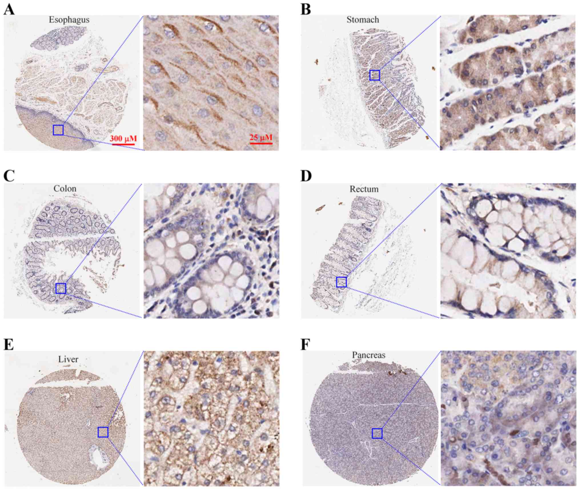
The expression of LRRC8A was immunolabeled in the
normal human tissues including the esophagus (n=3), stomach (n=6),
colon (n=5), rectum (n=2), liver (n=2) and pancreas (n=2). In the
stratified squamous epithelium of the esophagus (Fig. 1A) and the columnar epithelium and
glands of the stomach (Fig. 1B),
colon (Fig. 1C), and rectum
(Fig. 1D), LRRC8A proteins were
abundantly expressed and primarily located on the cell membrane. In
hepatocytes, most of the LRRC8A proteins were distributed on the
cell membrane (Fig. 1E); in islet
and acinar cells of the pancreas, LRRC8A was weakly immunolabeled
and located in the cell membrane and cytoplasm (Fig. 1F).
LRRC8A may be a novel prognostic
biomarker for survival in colon cancer patients
Cancer is a leading cause of death worldwide, and
the most common causes of cancer-related deaths are cancers of the
lung, liver, stomach, colorectum, breast and esophagus (1). To investigate the roles of LRRC8A in
cancers of the digestive system, we analyzed the expression of
LRRC8A in primary cancer and cancer adjacent tissues of 277
patients with cancers of the esophagus, stomach, duodenum, colon,
rectum, liver and pancreas.
As illustrated in Table
I, the expression of LRRC8A was significantly higher in nearly
60% of the colorectal cancer patients (51/90 colon cancer; 52/90
rectal cancer), which was significantly more than that in patients
with cancer of the esophagus (12.5%, 3/24), stomach (13.6%, 3/22),
duodenum (11.1%, 1/9), liver (4.8%, 1/21) and pancreas (19.0%,
4/21) (P<0.05).
 | Table I.Percentages of patients with elevated
expression of LRRC8A among 277 patients with cancer of the
esophagus, stomach, duodenum, colon, rectum, liver and
pancreas. |
Table I.
Percentages of patients with elevated
expression of LRRC8A among 277 patients with cancer of the
esophagus, stomach, duodenum, colon, rectum, liver and
pancreas.
| Cancer tissue | No. of
patients | No. of patients
with higher LRRC8A expression in cancer than adjacent tissue | Percentage (%) |
|---|
| Esophagus | 24 | 3 | 12.5b |
| Stomach | 22 | 3 | 13.6b |
| Duodenum | 9 | 1 | 11.1a |
| Colon | 90 | 51 | 56.7 |
| Rectum | 90 | 52 | 57.8 |
| Liver | 21 | 1 | 4.8b |
| Pancreas | 21 | 4 | 19.0b |
In colorectal cancer patients, LRRC8A expression in
the primary cancer tissue was higher than that in adjacent tissue
(Fig. 2A-D, E and H). In the cancer
tissue, it was hard to distinguish the membrane from the cell body
due to the compact growth of the cancer cells; however, it was
obviously observed that LRRC8A proteins were distributed in the
cytoplasm and on the cell membrane. We further analyzed the impact
of LRRC8A expression in the cancer tissues on cancer-related
survival time in patients with colon cancer (n=70) and patients
with rectal cancer (n=70); there was no significant difference in
the main clinical characteristics between the elevated and low
expressed LRRC8A groups (Table
II). As shown in Fig. 2F, the
colon cancer patients with elevated expression of LRRC8A had poorer
survival (P=0.003), of which the survival time (54.9±5.5 months;
n=37 was significantly shorter than that of patients with the lower
LRRC8A expression (77.1±3.7 months; n=33. There was no significant
difference between the ages of these 2 groups: 68.0±1.9 and
64.4±1.6 years, respectively, for elevated and low LRRC8A groups
(P>0.05). Conversely, there was no significant difference in
survival time in rectal cancer patients between the elevated and
low expressed LRRC8A groups; the mean ages of patients are 62.7±2.0
and 65.6±2.2 years, respectively (P>0.05, Fig. 2I).
| Table II.Main clinical characteristics of the
colorectal cancer patients according to LRRC8A expression. |
Table II.
Main clinical characteristics of the
colorectal cancer patients according to LRRC8A expression.
|
| Colon cancer |
| Rectal cancer |
|
|---|
|
|
|
|
|
|
|---|
|
| LRRC8A
expression |
| LRRC8A
expression |
|
|---|
|
|
|
|
|
|
|---|
| Variables | Cancer>adjacent
tissue n (%) | Cancer<adjacent
tissue n (%) | P-value | Cancer>adjacent
tissue n (%) | Cancer<adjacent
tissue n (%) | P-value |
|---|
| Sex |
| Male | 20 (54.1) | 15 (45.5) | 0.473 | 29 (76.3) | 18 (56.2) | 0.075 |
| Female | 17 (45.9) | 18 (54.5) |
| 9 (23.7) | 14 (43.8) |
|
| Age (years) |
| <60 | 8 (21.6) | 5 (15.2) | 0.487 | 16 (42.1) | 10 (31.2) | 0.349 |
| ≥60 | 29 (78.4) | 28 (84.8) |
| 22 (57.9) | 22 (68.8) |
|
|
Differentiation |
| Well (I) | 6 (16.2) | 8 (24.2) | 0.687 | 3 (7.9) | 4 (12.5) | 0.594 |
| Moderate (II) | 28 (75.7) | 23 (69.7) |
| 23 (60.5) | 21 (65.6) |
|
| Poor (III) | 3 (8.1) | 2 (6.1) |
| 12 (31.6) | 7 (21.9) |
|
| T stage |
| Tis | 0 (0) | 0 (0) | 0.058 | 1 (2.6) | 0 (0) | 0.418 |
| T1 | 0 (0) | 0 (0) |
| 0 (0) | 1 (3.1) |
|
| T2 | 0 (0) | 5 (15.1) |
| 3 (7.9) | 7 (21.9) |
|
| T3 | 33 (89.2) | 22 (66.7) |
| 31 (81.6) | 22 (68.8) |
|
| T4a | 3 (8.1) | 4 (12.1) |
| 2 (5.3) | 1 (3.1) |
|
| T4b | 1 (2.7) | 2 (6.1) |
| 1 (2.6) | 1 (3.1) |
|
| N stage |
| N0 | 22 (59.5) | 22 (66.7) | 0.536 | 23 (60.5) | 16 (50) | 0.222 |
| N1a | 5 (13.5) | 4 (12.1) |
| 4 (10.5) | 7 (21.9) |
|
| N1b | 6 (16.2) | 3 (9.1) |
| 4 (10.5) | 7 (21.9) |
|
| N1c | 1 (2.7) | 0 (0) |
| 0 (0) | 0 (0) |
|
| N2a | 3 (8.1) | 2 (6.1) |
| 5 (13.2) | 2 (6.2) |
|
| N2b | 0 (0) | 2 (6.1) |
| 2 (5.3) | 0 (0) |
|
| Metastasis |
| M0 | 37 (100) | 33 (100) |
| 38 (100) | 32 (100) |
|
| M1 | 0 | 0 |
| 0 | 0 |
|
| Clinical stage |
| 0 | 0 (0) | 0 (0) | 0.150 | 1 (2.6) | 0 (0) | 0.106 |
| I | 0 (0) | 5 (15.1) |
| 1 (2.6) | 7 (21.9) |
|
| IIA | 19 (51.4) | 12 (36.4) |
| 19 (50) | 9 (28.1) |
|
| IIB | 1 (2.7) | 3 (9.1) |
| 1 (2.6) | 0 (0) |
|
| IIC | 2 (5.4) | 2 (6.1) |
| 0 (0) | 0 (0) |
|
| IIIA | 1 (2.7) | 0 (0) |
| 2 (5.3) | 2 (6.2) |
|
| IIIB | 13 (35.1) | 9 (27.3) |
| 10 (26.3) | 12 (37.5) |
|
| IIIC | 1 (2.7) | 2 (6.1) |
| 4 (10.5) | 2 (6.2) |
|
| IV | 0 (0) | 0 (0) |
| 0 (0) | 0 (0) |
|
The lymph nodes were assessed for metastasis, which
may help to determine cancer therapy and prognosis. The impact of
metastases in lymph nodes on cancer-related survival was analyzed.
The colon cancer patients with metastases in the lymph nodes (node
positive) had a shorter survival time (52.6±7.3 months) than that
of patients without metastases in the lymph nodes (72.2±3.6 months,
P<0.05; Fig. 2G). However, there
was no significant difference in survival time of the rectal cancer
patients with and without metastases in lymph nodes (Fig. 2J). Notably high or low expression of
LRRC8A could occur in patients with or without the positive lymph
nodes. The colon cancer patients who had positive lymph nodes and
high expression of LRRC8A had the highest mortality rate, which was
80% (Table III).
| Table III.Survival of colorectal cancer
patients evaluated by combining of metastases in the lymph nodes
and LRRC8A expression. |
Table III.
Survival of colorectal cancer
patients evaluated by combining of metastases in the lymph nodes
and LRRC8A expression.
| Cancer | Lymph node
status | LRRC8A
expression | Percentage of
deaths (%) | No. of deaths | No. of surviving
patients | Chi-square
test |
|---|
| Colon | Positive | Cancer>adjacent
tissue | 80.0 | 12 | 3 | P=0.007
<0.01 |
|
|
| Cancer<adjacent
tissue | 27.3 | 3 | 8 |
|
|
| Negative | Cancer>adjacent
tissue | 36.4 | 8 | 14 | P=0.176
>0.05 |
|
|
| Cancer<adjacent
tissue | 18.2 | 4 | 18 |
|
| Rectum | Positive | Cancer>adjacent
tissue | 53.3 | 8 | 7 | P=0.870
>0.05 |
|
|
| Cancer<adjacent
tissue | 56.3 | 9 | 7 |
|
|
| Negative | Cancer>adjacent
tissue | 30.4 | 7 | 16 | P=0.217
>0.05 |
|
|
| Cancer<adjacent
tissue | 50.0 | 8 | 8 |
|
These results suggest that LRRC8A may be used as a
novel prognostic biomarker for survival in colon cancer
patients.
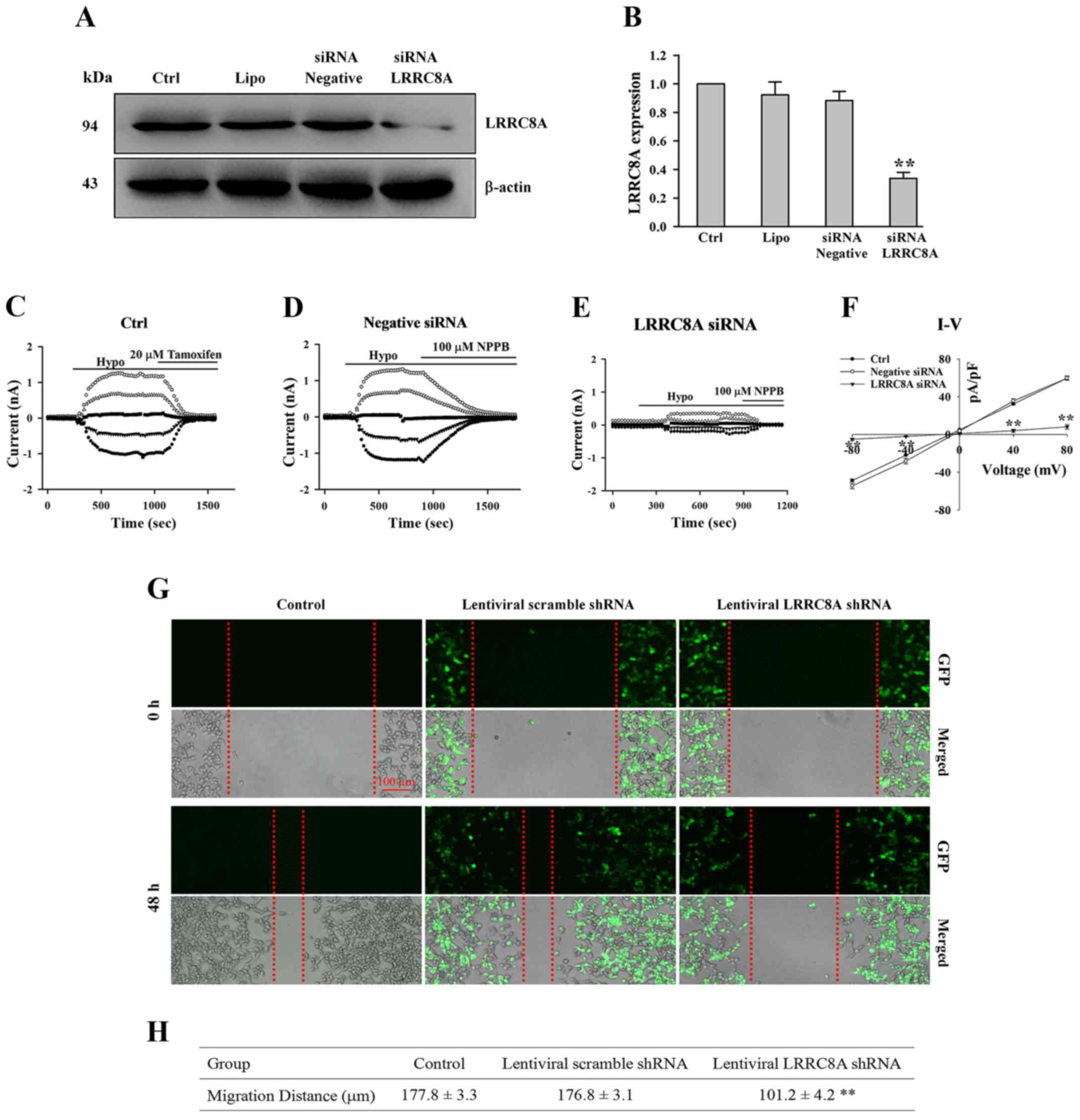
Knockdown of LRRC8A inhibits
EGF-induced migration of HCT116 cells
The previous results suggest that high expression of
LRRC8A is associated with a shorter survival time in colon cancer
patients who have positive lymph nodes. This implies that LRRC8A
proteins may facilitate the migration of colon cancer cells. We
therefore assessed the effect of LRRC8A on cell migratory potential
using ex vivo wound-scratch experiments in human colon
cancer HCT116 cells.
As illustrated in Fig.
3A and B, after HCT116 cells were transfected with LRRC8A siRNA
for 72 h, the expression of LRRC8A protein was substantially
reduced (n=3 experiments, P<0.01 vs. all control groups). A
current was induced by a hypotonic solution in HCT116 cells, which
could be almost inhibited by chloride channel blockers (20 µM
tamoxifen and 100 µM NPPB; Fig. 3C and
D). The hypotonicity-activated chloride current was clearly
attenuated by the treatment of LRRC8A siRNA; mean current density
in the LRRC8A siRNA group was 8.1±2.6 at + 80 mV, which is
significantly lower than that in the negative siRNA group (59.7±2.3
at + 80 mV; n=6–8 cells, P<0.01, Fig. 3C-F). These data suggest that LRRC8A
was the main determinant of VRAC current in HCT116 cells, which was
consistent with a previous study (6), and that this specific siRNA for LRRC8A
could efficiently interfere with the expression of LRRC8A
protein.
According to the sequence of this specific siRNA,
the LRRC8A shRNA vector was constructed and packaged as an
infectious lentivirus, which was used to interfere with LRRC8A
expression. One week after the lentivirus infection, wounds on the
monolayer cells were scratched and observed to repair for 48 h. As
shown in Fig. 3G and H, in the
control and lentiviral scramble shRNA groups, the wounds were
mostly recovered by cells migrating from the wound edges; the
average distances of migration in the control and scramble shRNA
groups were 177.8±3.3 and 176.8±3.1 µm, respectively. In the LRRC8A
sRNA group, an obvious gap remained in the wound; the migration
distance was 101.2±4.2 µm, significantly shorter than that in the
control and scramble shRNA groups (n=3 experiments, P<0.01).
These results suggest that LRRC8A may have an
important role in modulating the migration of colon cancer HCT116
cells, and that high expression of LRRC8A may facilitate the
metastasis of colon cancer in patients.
Downregulation of LRRC8A inhibits the
tumorigenesis of HCT116 cells in the nude mouse
Cancerous cells have strong capabilities of
adaptation and proliferation, which always are required for cells
to metastasize and survive in regions distant from the primary
locus. Therefore, the role of LRRC8A in cancer cell growth was
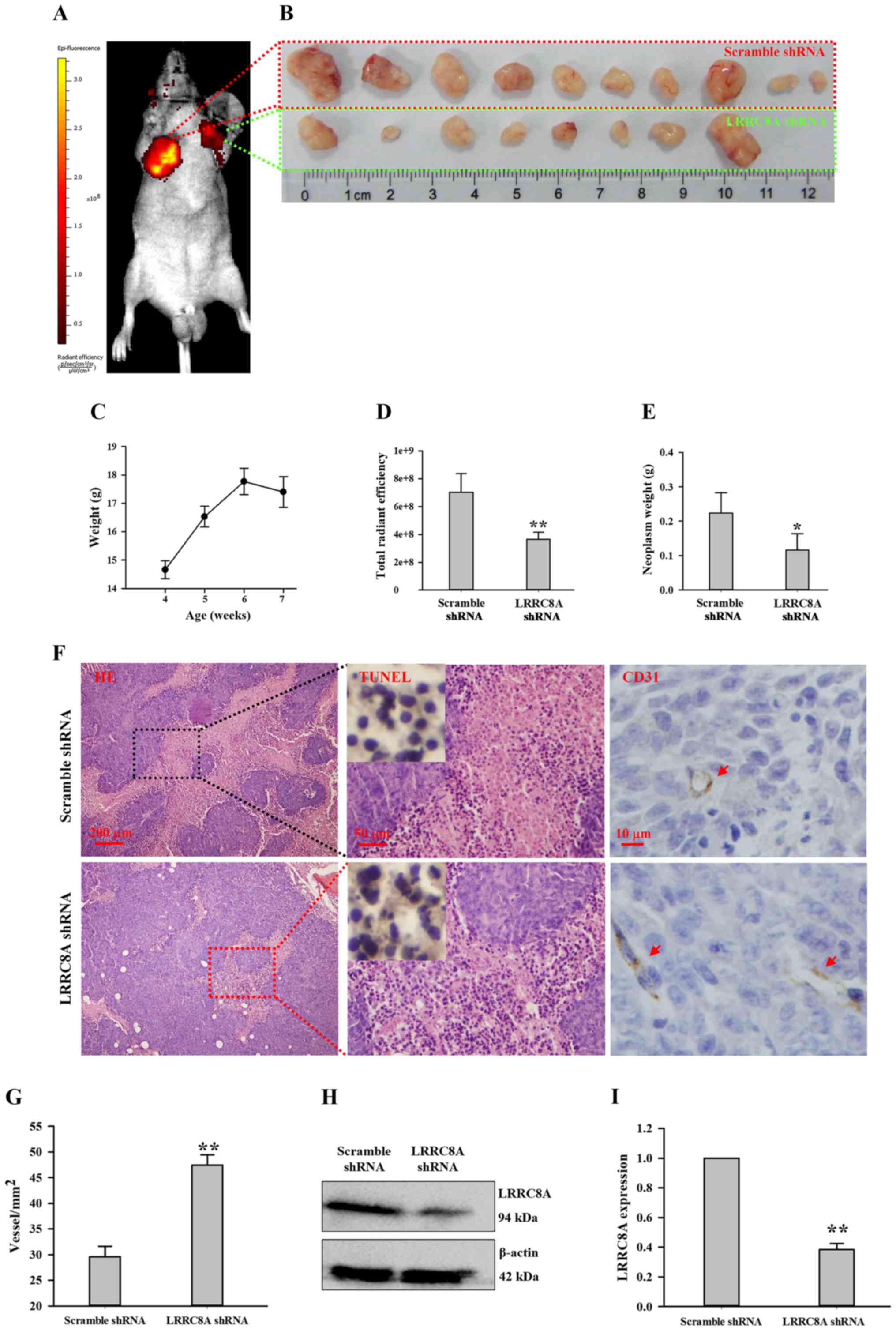
investigated via an in vivo method.
The scramble and LRRC8A shRNA lentivirus-infected
HCT116 cells were subcutaneously injected at the armpits (right and
left forelimb, respectively) of the nude mice. After 1 week, a very
small nodule was observed at the armpit of the right forelimb; the
nude mice were fed for another 2 weeks, and the nodules gradually
became large. When body weights began to decrease at the third week
(Fig. 4C), the total radiant
efficiency of GFP indicating neoplasm growth was captured by the
in vivo non-invasive small animal molecular imaging system.
As illustrated in Fig. 4A and D,
the intensity of radiance at the right forelimb armpit was much
higher than that at the left forelimb armpit (n=10, P<0.01),
suggesting downregulation of LRRC8A may inhibit HCT116 cell growth
in the nude mouse. This finding was further verified by dissecting
and weighing these nodules. In the scramble shRNA lentivirus group,
the neoplasm always had a larger volume compared with the paired
cancer tissue (Fig. 4B); the
average weight of neoplasms in the scramble shRNA group was more
than that in the LRRC8A shRNA group (Fig. 4E; n=8–10, P<0.05). In addition,
there were two mice at whose left forelimb armpits no radiance was
detected; also, there were no solid tissues to be found and
dissected (Fig. 4B).
The tissue slices of the neoplasms were stained with
H&E. As shown in Fig. 4F, in
solid tumor being formed with HCT116 cells, the organizational
structures were compact and unorganized; the cell nuclei were
large, atypical and deeply stained by hematoxylin (blue); the
cytoplasm were lightly labeled with eosin (pink). In the scramble
shRNA group, there appeared a large area of necrosis-like cells,
having characteristics of karyopyknosis, karyolysis and intensely
eosinophilic cytoplasm. The negative TUNEL staining suggested that
most of these cells were not apoptotic cells, by which these
necrotic cells were further confirmed. In the LRRC8A shRNA group,
necrosis appeared, but not widely.
A wide range of necrosis occurred in the tissue
slices of the scramble shRNA group; one possible mechanism may have
been that the speed of angiogenesis did not meet the requirement of
nutrition for HCT116 cell proliferation. In the LRRC8A shRNA group,
the speed of angiogenesis may have met the requirement of nutrition
for cell growth, which may have been inhibited by the
downregulation of LRRC8A proteins (Fig.
4H and I). Thus, vessels in these cancer tissues were detected
by CD31, a marker of endothelial cells. As illustrated in Fig. 4F and G, the vessel density of cancer
tissue in the LRRC8A shRNA group was higher than that in the
scramble shRNA group (P<0.01), which may relatively supply more
nutrition for cells.
Discussion
The cancer burden is growing at an alarming pace,
and implementation of efficient prevention strategies to curb the
disease are urgently needed.
In the growth/proliferation, migration, and/or
invasion of cancer cells, abnormal expression and/or activity of a
number of ion channels have been confirmed to be involved, such as
voltage-gated K+, Na+, Ca2+ and
TRP channels and epithelial Na+/degenerin family of ion
channels (15). It was proposed
that ion channels may become potential targets for the diagnosis,
therapy and prognosis of cancer (25–28).
It is well known that volume-regulated anion channel
(VRAC) plays key roles in all types of cellular functions; however
there have always been controversies concerning the molecular
entity for VRAC. ClC-3, a member of the ClC superfamily of
voltage-gated chloride channels, is widely expressed and
hypothesized to be VRAC. As previously reported by us and others,
the cell cycle-dependent expression and distribution of ClC-3 is
closely related to cell proliferation, migration and apoptosis,
particularly in cancerous cells (15,16,29–33).
This suggests that ClC-3 may be a promising target of anticancer
drugs or a novel prognostic biomarker for survival in tumor
patients.
In 2003, a novel gene, leucine-rich
repeat-containing 8 (LRRC8), was isolated from a girl with
congenital agammaglobulinemia in peripheral blood, which may be
required for B-cell development (18). One year later, the authors
identified another 4 LRRC8-like genes, named TA-LRRP, AD158, LRRC5
and FLJ23420 (20). All 5 genes,
designated as the LRRC8 family, encode proteins consisting of 17
extracellular leucine-rich repeats (LRRs), all of which have 4
transmembrane helices except for FLJ23420 (20,34);
later, these genes were called LRRC8A-E. The LRRC8 family was
nearly forgotten, and few investigations were reported in the
following 10 years. The recent discovery of LRRC8A as an essential
component of VRAC (6,7) profoundly motivated us to investigate
the physiological and pathophysiological functions of LRRC8
proteins, including cell volume regulation, inflammation, T-cell
development, small molecule transporting, neurotransmitter release
and anticancer drug resistance (11,14,19,21–23,35,36).
There is little knowledge concerning the biological functions of
LRRC8A in cancers, particularly cancers of the digestive system. We
therefore investigated the roles of LRRC8A in cancers of the
digestive system.
As VRAC is widely distributed in the tissues of
mammals, LRRC8A proteins were found to be widely and abundantly
expressed in the tissues of the digestive system. Our previous
study indicated that there were >50% of patients with gastric
adenocarcinoma, colorectal adenocarcinoma, and esophageal squamous
cell carcinoma who had high-expressed ClC-3 proteins (16). Among 277 cancer patients (including
cancers of the esophagus, stomach, duodenum, colon, rectum, liver
and pancreas), we found that patients with elevated expression of
LRRC8A proteins in the primary cancer tissue >50% occurred only
in colorectal cancer patients (colon cancer, 56.7%, and rectal
cancer, 57.8%; Table I). It has
been previously reported that reduced expression of LRRC8A
contributes to the acquisition of cisplatin resistance in human
ovarian and alveolar cancer cells (23,24),
suggesting that patients with high LRRC8A expression may benefit
from platinum drug treatment. Although colorectal patients are not
usually treated with platinum drugs, it may be a clinically
relevant question to investigate whether patients with high LRRC8A
expression could be treated with platinum agents. Moreover, in
cancers of the esophagus, stomach, duodenum, liver and pancreas,
whether patients with low LRRC8A expression obtain benefits from
the combined application of platinum agents and targeted
upregulation of LRRC8A may be an important strategy to explore.
In cancer patients, elevated expression of ClC-3
proteins was closely associated with poor survival (16). Thus, the effects of high-expressed
LRRC8A on cancer-related survival in cancer patients of the colon
(n=70) and rectum (n=70) were respectively analyzed. Surprisingly,
it was found that high-expressed LRRC8A was related to shorter
survival time in colon cancer patients, but not in rectal cancer
patients. The high-expressed LRRC8A had no relationship with
survival time in rectal cancer patients, which could be explained
by the following mechanisms. i) Carcinoma in the colon and rectum
may originate from tumor-initiating cells with various directions
of differentiation, which may decide the features of colon and
rectal adenocarcinoma. ii) Difference in post-translational
modification for LRRC8A may exist in colon and rectal cancer, such
as phosphorylation-dephosphorylation, acetylation-deacetylation and
oxidation-reduction. iii) Difference in the proteins interacting
with LRRC8A may exist in colon and rectal carcinoma, which remain
unclarified to help us better understand why high expression of
LRRC8A has no association with survival time in rectal cancer
patients. Furthermore, we observed that colon cancer patients who
had positive lymph node status and high expression of LRRC8A had
the highest mortality rate, ~80%. These data disclose that colon
cancer patients may be prone to high expression of LRRC8A, which is
related with poor survival. Therefore, we propose that LRRC8A may
be used as a novel prognostic biomarker to evaluate survival in
colon cancer patients. Due to the small sample size in this study,
this potential prognostic biomarker may be confirmed by a
multicenter study.
LRRC8A may become a potential target by which to
cure or curb colon cancer in patients with high expression of
LRRC8A proteins. This hypothesis was supported by our following
findings. Human colon cancer HCT116 cells with downregulated LRRC8A
were used to assay cell migration ex vivo and cell growth
in vivo, respectively. The high expression of LRRC8A may be
associated with the poorest survival in colon cancer patients who
had positive lymph nodes, which implied that LRRC8A proteins may
contribute to colon cancer metastasis via facilitating cell
migration. The EGF-induced migration in HCT116 cells with
downregulated LRRC8A proteins was significantly inhibited (Fig. 3), suggesting that LRRC8A may play an
important role in cell migration. The in vivo experiment of
tumorigenesis in nude mice demonstrated that HCT116 cells with
downregulated LRRC8A proteins grew slowly and formed smaller tumor
nodules (Fig. 4), suggesting that
LRRC8A may be closely related with cell growth. LRRC8A, as one of
the molecular determinants for VRAC, is responsible for cell volume
regulation in HCT116 cells. During migration and proliferation, the
dysfunction of cell volume regulation may be one of the possible
mechanisms with which the downregulation of LRRC8A inhibits
migration and growth. Although the detailed molecular mechanisms of
LRRC8A in modulating cell migration and growth are poorly
understood, the novel findings herein may provide a promising
strategy to curb the development of colon cancer by inhibiting the
function or expression of LRRC8A protein. It should be further
investigated whether LRRC8A could be a novel and potential target
for curing patients with colon cancer.
Although the molecular determinant for VRAC is still
being debated and the biological function of LRRC8A is still not
completely understood, we present an important and interesting
finding: poor survival in more than half of colon cancer patients
may be closely related with the high expression of LRRC8A protein,
which facilitates cell migration and growth. In conclusion, LRRC8A
may be used as a novel prognostic biomarker for survival and a
potential target for therapy in colon cancer patients.
Acknowledgements
Not applicable.
Funding
The present study was supported by the National
Natural Science Foundation of China (no. 81402312) and the China
Postdoctoral Science Foundation (no. 2014M552462).
Availability of data and materials
The datasets used during the present study are
available from the corresponding author upon reasonable
request.
Authors' contributions
HZ and JS conceived and designed the study. ZD, DZ,
HL, LZ and JN performed the experiments. HZ wrote the manuscript.
WZ, RF, LF and JHY edited the manuscript and were also involved in
the conception of the study. All authors read and approved the
manuscript and agree to be accountable for all aspects of the
research in ensuring that the accuracy or integrity of any part of
the work are appropriately investigated and resolved.
Ethics approval and consent to
participate
The animal experimental protocol was approved by the
Laboratory Animal Administration Committee of Xi'an Jiaotong
University and was carried out in accordance with the Guidelines
for Animal Experimentation of Xi'an Jiaotong University and the
Guide for the Care and Use of Laboratory Animals published by the
US National Institutes of Health (NIH Publication no. 85–23,
revised 2011). Ethics approval for research using human tissue was
obtained from the Ethics Committee in Taizhou Hospital, Zhejiang
Province, and included a waiver for consent. The Outdo Biotech Co.,
Ltd. obtained the ethical approval from the Taizhou Hospital for
research using human tissue. This institution used tumor tissues of
Taizhou Hospital to make microarray; we purchased these microarrays
for our research, which be approved by Ethics Committee of Health
Science Center, Xi'an Jiaotong University.
Patient consent for publication
Not applicable.
Competing interests
The authors state that they have no competing
interests.
References
|
1
|
McGuire S: World cancer report 2014.
Geneva, Switzerland: World Health Organization, International
agency for research on cancer, WHO press, 2015. Adv Nutr.
7:418–419. 2016. View Article : Google Scholar : PubMed/NCBI
|
|
2
|
Lang F, Busch GL, Ritter M, Völkl H,
Waldegger S, Gulbins E and Häussinger D: Functional significance of
cell volume regulatory mechanisms. Physiolo Rev. 78:247–306. 1998.
View Article : Google Scholar
|
|
3
|
Hoffmann EK, Lambert IH and Pedersen SF:
Physiology of cell volume regulation in vertebrates. Physiol Rev.
89:193–277. 2009. View Article : Google Scholar : PubMed/NCBI
|
|
4
|
Pedersen SF, Hoffmann EK and Novak I: Cell
volume regulation in epithelial physiology and cancer. Front
Physiol. 4:2332013. View Article : Google Scholar : PubMed/NCBI
|
|
5
|
Pedersen SF, Klausen TK and Nilius B: The
identification of a volume-regulated anion channel: An amazing
Odyssey. Acta Physiol (Oxf). 213:868–881. 2015. View Article : Google Scholar : PubMed/NCBI
|
|
6
|
Voss FK, Ullrich F, Münch J, Lazarow K,
Lutter D, Mah N, Andrade-Navarro MA, von Kries JP, Stauber T and
Jentsch TJ: Identification of LRRC8 heteromers as an essential
component of the volume-regulated anion channel VRAC. Science.
344:634–638. 2014. View Article : Google Scholar : PubMed/NCBI
|
|
7
|
Qiu Z, Dubin AE, Mathur J, Tu B, Reddy K,
Miraglia LJ, Reinhardt J, Orth AP and Patapoutian A: SWELL1, a
plasma membrane protein, is an essential component of
volume-regulated anion channel. Cell. 157:447–458. 2014. View Article : Google Scholar : PubMed/NCBI
|
|
8
|
Yamada T, Wondergem R, Morrison R, Yin VP
and Strange K: Leucine-rich repeat containing protein LRRC8A is
essential for swelling-activated Cl-currents and embryonic
development in zebrafish. Physiological Rep. 4:e129402016.
View Article : Google Scholar
|
|
9
|
Gaitán-Peñas H, Gradogna A, Laparra-Cuervo
L, Solsona C, Fernández-Dueñas V, Barrallo-Gimeno A, Ciruela F,
Lakadamyali M, Pusch M and Estévez R: Investigation of
LRRC8-mediated volume-regulated anion currents in xenopus oocytes.
Biophys J. 111:1429–1443. 2016. View Article : Google Scholar : PubMed/NCBI
|
|
10
|
Syeda R, Qiu Z, Dubin AE, Murthy SE,
Florendo MN, Mason DE, Mathur J, Cahalan SM, Peters EC, Montal M
and Patapoutian A: LRRC8 proteins form volume-regulated anion
channels that sense ionic strength. Cell. 164:499–511. 2016.
View Article : Google Scholar : PubMed/NCBI
|
|
11
|
Hyzinski-Garcia MC, Rudkouskaya A and
Mongin AA: LRRC8A protein is indispensable for swelling-activated
and ATP-induced release of excitatory amino acids in rat
astrocytes. J Physiol. 592:4855–4862. 2014. View Article : Google Scholar : PubMed/NCBI
|
|
12
|
Milenkovic A, Brandl C, Milenkovic VM,
Jendryke T, Sirianant L, Wanitchakool P, Zimmermann S, Reiff CM,
Horling F, Schrewe H, et al: Bestrophin 1 is indispensable for
volume regulation in human retinal pigment epithelium cells. Proc
Natl Acad Sci USA. 112:E2630–E2639. 2015. View Article : Google Scholar : PubMed/NCBI
|
|
13
|
Sirianant L, Wanitchakool P, Ousingsawat
J, Benedetto R, Zormpa A, Cabrita I, Schreiber R and Kunzelmann K:
Non-essential contribution of LRRC8A to volume regulation. Pflugers
Arch. 468:805–816. 2016. View Article : Google Scholar : PubMed/NCBI
|
|
14
|
Okada T, Islam MR, Tsiferova NA, Okada Y
and Sabirov RZ: Specific and essential but not sufficient roles of
LRRC8A in the activity of volume-sensitive outwardly rectifying
anion channel (VSOR). Channels. 11:109–120. 2017. View Article : Google Scholar : PubMed/NCBI
|
|
15
|
Zhang H, Li H, Yang L, Deng Z, Luo H, Ye
D, Bai Z, Zhu L, Ye W, Wang L and Chen L: The ClC-3 chloride
channel associated with microtubules is a target of paclitaxel in
its induced-apoptosis. Sci Rep. 3:26152013. View Article : Google Scholar : PubMed/NCBI
|
|
16
|
Xu B, Jin X, Min L, Li Q, Deng L, Wu H,
Lin G, Chen L, Zhang H, Li C, et al: Chloride channel-3 promotes
tumor metastasis by regulating membrane ruffling and is associated
with poor survival. Oncotarget. 6:2434–2450. 2015.PubMed/NCBI
|
|
17
|
Zhang H, Deng Z, Yang L, Luo H, Liu S, Li
Y, Wei Y, Peng S, Zhu L, Wang L and Chen L: The AQP-3 water channel
is a pivotal modulator of glycerol-induced chloride channel
activation in nasopharyngeal carcinoma cells. Int J Biochem Cell
Biol. 72:89–99. 2016. View Article : Google Scholar : PubMed/NCBI
|
|
18
|
Sawada A, Takihara Y, Kim JY,
Matsuda-Hashii Y, Tokimasa S, Fujisaki H, Kubota K, Endo H, Onodera
T, Ohta H, et al: A congenital mutation of the novel gene LRRC8
causes agammaglobulinemia in humans. J Clin Invest. 112:1707–1713.
2003. View
Article : Google Scholar : PubMed/NCBI
|
|
19
|
Zúñiga-Pflücker JC: New role identified
for LRR-containing proteins in T cell development. J Exp Med.
211:746–747. 2014. View Article : Google Scholar
|
|
20
|
Kubota K, Kim JY, Sawada A, Tokimasa S,
Fujisaki H, Matsuda-Hashii Y, Ozono K and Hara J: LRRC8 involved in
B cell development belongs to a novel family of leucine-rich repeat
proteins. FEBS Lett. 564:147–152. 2004. View Article : Google Scholar : PubMed/NCBI
|
|
21
|
Kumar L, Chou J, Yee CS, Borzutzky A,
Vollmann EH, von Andrian UH, Park SY, Hollander G, Manis JP,
Poliani PL and Geha RS: Leucine-rich repeat containing 8A (LRRC8A)
is essential for T lymphocyte development and function. J Exp Med.
211:929–942. 2014. View Article : Google Scholar : PubMed/NCBI
|
|
22
|
Choi H, Ettinger N, Rohrbough J, Dikalova
A, Nguyen HN and Lamb FS: LRRC8A channels support TNFalpha-induced
superoxide production by Nox1 which is required for receptor
endocytosis. Free Radic Biol Med. 101:413–423. 2016. View Article : Google Scholar : PubMed/NCBI
|
|
23
|
Sørensen BH, Nielsen D, Thorsteinsdottir
UA, Hoffmann EK and Lambert IH: Downregulation of LRRC8A protects
human ovarian and alveolar carcinoma cells against
Cisplatin-induced expression of p53, MDM2, p21Waf1/Cip1, and
Caspase-9/-3 activation. Am J Physiol Cell Physiol. 310:C857–C873.
2016. View Article : Google Scholar : PubMed/NCBI
|
|
24
|
Sørensen BH, Dam CS, Stürup S and Lambert
IH: Dual role of LRRC8A-containing transporters on cisplatin
resistance in human ovarian cancer cells. J Inorg Biochem.
160:287–295. 2016. View Article : Google Scholar : PubMed/NCBI
|
|
25
|
Li M and Xiong ZG: Ion channels as targets
for cancer therapy. Int J Physiol Pathophysiol Pharmacol.
3:156–166. 2011.PubMed/NCBI
|
|
26
|
Fraser SP and Pardo LA: Ion channels:
Functional expression and therapeutic potential in cancer.
Colloquium on ion channels and cancer. EMBO Rep. 9:512–515. 2008.
View Article : Google Scholar : PubMed/NCBI
|
|
27
|
Arcangeli A, Pillozzi S and Becchetti A:
Targeting ion channels in leukemias: A new challenge for treatment.
Curr Med Chem. 19:683–696. 2012. View Article : Google Scholar : PubMed/NCBI
|
|
28
|
Arcangeli A, Crociani O, Lastraioli E,
Masi A, Pillozzi S and Becchetti A: Targeting ion channels in
cancer: A novel frontier in antineoplastic therapy. Curr Med Chem.
16:66–93. 2009. View Article : Google Scholar : PubMed/NCBI
|
|
29
|
Mao J, Li X, Chen W, Xu B, Zhang H, Li H,
Wang L, Jin X, Zhu J, Lin G, et al: Cell cycle-dependent
subcellular distribution of ClC-3 in HeLa cells. Histochem Cell
Biol. 137:763–776. 2012. View Article : Google Scholar : PubMed/NCBI
|
|
30
|
Zhang H, Zhu L, Zuo W, Luo H, Mao J, Ye D,
Li Y, Liu S, Wei Y, Ye W, et al: The ClC-3 chloride channel protein
is a downstream target of cyclin D1 in nasopharyngeal carcinoma
cells. Int J Biochem Cell Biol. 45:672–683. 2013. View Article : Google Scholar : PubMed/NCBI
|
|
31
|
Zhang H, Li H, Liu E, Guang Y, Yang L, Mao
J, Zhu L, Chen L and Wang L: The AQP-3 water channel and the ClC-3
chloride channel coordinate the hypotonicity-induced swelling
volume in nasopharyngeal carcinoma cells. Int J Biochem Cell Biol.
57:96–107. 2014. View Article : Google Scholar : PubMed/NCBI
|
|
32
|
Ye D, Luo H, Lai Z, Zou L, Zhu L, Mao J,
Jacob T, Ye W, Wang L and Chen L: ClC-3 chloride channel proteins
regulate the cell cycle by up-regulating cyclin D1-CDK4/6 through
suppressing p21/p27 expression in nasopharyngeal carcinoma cells.
Sci Rep. 6:302762016. View Article : Google Scholar : PubMed/NCBI
|
|
33
|
Wang LW, Chen LX and Jacob T: ClC-3
expression in the cell cycle of nasopharyngeal carcinoma cells.
Acta Physiol Sin. 56:230–236. 2004.
|
|
34
|
Smits G and Kajava AV: LRRC8 extracellular
domain is composed of 17 leucine-rich repeats. Mol Immunol.
41:561–562. 2004. View Article : Google Scholar : PubMed/NCBI
|
|
35
|
Lee CC, Freinkman E, Sabatini DM and
Ploegh HL: The protein synthesis inhibitor blasticidin s enters
mammalian cells via leucine-rich repeat-containing protein 8D. J
Biol Chem. 289:17124–17131. 2014. View Article : Google Scholar : PubMed/NCBI
|
|
36
|
Planells-Cases R, Lutter D, Guyader C,
Gerhards NM, Ullrich F, Elger DA, Kucukosmanoglu A, Xu G, Voss FK,
Reincke SM, et al: Subunit composition of VRAC channels determines
substrate specificity and cellular resistance to Pt-based
anticancer drugs. EMBO J. 34:2993–3008. 2015. View Article : Google Scholar : PubMed/NCBI
|