Introduction
Esophageal cancer is a dangerous disease, based on
both morbidity and mortality. Worldwide, esophageal cancer is
currently the eighth most prevalent cancer and the sixth leading
cause of cancer-related deaths (1).
The latest epidemiological survey of malignant tumors showed that
the incidence of esophageal cancer in China is 477,900 (out of 1,37
billion), with 375,000 deaths/year (2). It is estimated that the number of new
cases of esophageal cancer will reach 2,110,000 by 2025 worldwide.
Although the incidence of many other types of cancer will decrease
during this time, the prevalence of esophageal cancer is expected
to increase by 140% (3). This
cancer includes two pathological types: Squamous cell carcinoma
(ESCC) and adenocarcinoma (EAC). The incidence of the latter has
significantly increased over the last 40 years; however, squamous
cell carcinoma (ESCC) remains the principal type of esophageal
cancer (1). In the
highest-prevalence areas for esophageal cancer, often called the
‘esophageal cancer belt’ (including China) 90% of these tumors are
ESCC (4). Endoscopic management and
new diagnostic imaging technologies may aid the early diagnosis and
improve the overall survival. Nevertheless, the 5-year survival
rate remains only 18% (5). Because
of the atypical early symptoms of esophageal cancer and the absence
of specific tumor markers for early diagnosis, most patients are
diagnosed at advanced stages, resulting in poor prognosis (6). There are no reliable biomarkers for
the early clinical diagnosis of esophageal cancer; therefore, this
area requires much work.
Deregulating cellular energetics is an important
hallmark of cancer. This concept can be thought of as equivalent to
the concept of sustaining proliferative signaling, activating
invasion and metastasis, angiogenesis and evasion of apoptosis.
Through a study by Hanahan and Weinberg, it was reported that most
cancer cells are powered by aerobic glycolysis (the so-called
Warburg effect) (7,8). Cancer cells rely on glycolysis to
provide energy, producing a large amount of lactate. When lactate
is transported into the extracellular milieu to avoid intracellular
acidification and apoptosis, an acidic microenvironment is formed.
Monocarboxylate transporters 1–4 (MCT1-4) are involved in this
process (9).
MCTs belong to the SLC16 family of genes, including
at least 14 members. MCT1-MCT4 are thought to be proton
transporters that regulate the transmembrane transport of lactate,
pyruvate and ketone bodies (9). The
function of MCTs in maintaining the homeostasis of cells in normal
tissues has been studied in detail, but there is little insight
regarding their role in cancer tissues. In acidic tumor
microenvironments, MCTs not only play roles in maintaining the
hyper-glycolytic acid-resistant phenotype of cancer cells, but also
play an important role in maintaining high glycolytic rates that
depend on mediating lactate efflux (9). MCT1 (solute carrier family 16 member;
SLC16A1) and MCT4 (SLC16A4) mediate the extrusion of large amounts
of lactic acid from malignant tumor cells, forming an acidic tumor
microenvironment, thereby increasing the invasiveness and mobility
of malignant cells (10).
Deregulating cellular energetics of cancer cells is
an emerging field of research, and research regarding the
expression and functional role of MCT1-4 recently have been
conducted. Several studies of the mechanisms of their effects have
been reported for several malignancies, including non-small cell
lung cancer (NSCLC), bladder cancer, osteosarcoma and breast cancer
(11–14). Based on these studies, we believe
that MCT1-4 may play a crucial role in tumor biological behavior
and be a potential target for cancer diagnosis and therapeutics.
The objective of the present study was to evaluate the expression
of MCT1 in ESCC as well as its prognostic implications.
Materials and methods
Ethics statement
The research protocol of the present study was
approved by the Ethics Committee of Qilu Hospital, and all patients
signed written informed consents before enrollment. Patient
information was anonymized and unidentifiable prior to
analysis.
Patients and tissue samples
The primary tumor tissues and the corresponding
para-carcinoma tissues were obtained from 103 patients, who had
confirmed diagnoses of ESCC at the Qilu Hospital of Shandong
University from February 2010 to December 2011 and had not received
any neoadjuvant-therapy. All samples were verified by pathological
assessment. All 103 patients were followed up for at least five
years. Other information was collected from clinical and
pathological records. Staging of the tumors was according to the
American Joint Committee on Cancer (AJCC, 7th edition).
Cell lines and culture
Human esophageal squamous cell carcinoma (ESCC) cell
strains KYSE-150 and Eca-109 were purchased from the China Center
for Type Culture Collection (Beijing, China) in 2017. All cells
were verified by short tandem repeat analysis. Cells were cultured
in RPMI-1640 medium (Gibco; Thermo Fisher Scientific, Inc.,
Waltham, MA, USA), supplemented with 10% fetal bovine serum
(Invitrogen; Thermo Fisher Scientific, Inc.), 100 U/ml penicillin
and 100 mg/ml streptomycin. All cells were cultivated in a 37°C
humid incubator containing 5% CO2.
Cell transfection
Eca109 and KYSE-150 cell lines were transfected with
plasmids purchased from GeneCopoeia, with shRNA targeting the MCT1
gene and shRNA as control using Lipofectamine 2000 reagent
(Invitrogen; Thermo Fisher Scientific, Inc.). The transfection
procedure was carried out in accordance with the manufacturer's
instructions. After transfection, the cells were cultured and were
harvested within 48–72 h for subsequent analysis.
Cell Counting Kit-8 (CCK-8) assay
According to the instructions of the CCK-8 kit
(BestBio, Shanghai, China), the cells in logarithmic growth phase
were digested to construct a single-cell suspension and the cells
were counted, after which they were seeded at a density of 1,000
cells/well on 96-well plates. In each 96-well, 100 µl of the
suspension was placed. Subsequently, the multiplication capacity of
Eca109 and KYSE-150 cells was assessed at 24, 48, 72, 96 and 120 h.
We added 10 µl CCK-8 solution into 100 µl fresh medium to each
well, followed by additional incubation for 2 h, and then measured
the absorbance at 450 nm with a Thermo Scientific Varioskan Flash
spectrophotometer (Thermo Scientific, Inc., Vantaa, Finland).
Colony formation assay
Cells that were digested by proteases were made into
a single-cell suspension, and 500 cells were seeded per well of a
6-well plate. Cells were fixed with methanol, stained with crystal
violet, and counted after culture for 2 weeks. We then counted the
number of colonies containing >50 cells, defined as a clone
(ImageJ 1.47v software; NIH; National Institutes of Health,
Bethesda, MD, USA).
Western blotting
Cells were lysed with cold RIPA lysis buffer
(Beijing Solarbio Science & Technology Co., Ltd., Beijing,
China) and phenylmethylsulfonyl fluoride (PMSF) (dilution 1:100)
for 30 min on ice after washing three times with PBS. Cell debris
was discarded after centrifugation (12,000 × g, for 15 min) at 4°C.
The supernatant was stored in a new EP tube. Protein concentrations
in the supernatant were determined with the BCA protein assay kit
(Beyotime Institute of Biotechnology, Shanghai, China). Boiled
mixtures of protein and sample buffer were electrophoresed on 10%
SDS-PAGE gels at 80 V for 120 min. The proteins were transferred to
PVDF membranes on ice for 2 h and blocked with 5% dried skimmed
milk at room temperature for 1.5 h. The membranes were washed with
TBST (pH 7.4) three times and were incubated with primary
antibodies at 4°C overnight. Secondary antibodies
[peroxidase-conjugated goat anti-rabbit IgG (H+L); dilution
1:5,000; cat. no. ZB-2301; peroxidase-conjugated goat anti-mouse
IgG (H+L); dilution 1:5,000; cat. no. ZB-2305; both from ZSBIO,
Beijing, China] were added to the membranes and incubated at room
temperature for 1 h. Subsequently, the membranes were exposed with
an enhanced chemiluminescence reaction (ECL) kit, and the
gray-levels of the protein bands were analyzed using ImageJ 1.47 v
software (NIH; National Institutes of Health, Bethesda, MD, USA).
The primary antibodies included: Rabbit anti-MCT1 polyclonal
antibody (dilution 1:1,000; cat. no. 20139-1-AP; ProteinTech Group
Inc., Chicago, IL, USA), rabbit anti-ACTB polyclonal antibody
(dilution 1:1,000; cat. no. ab8227; Abcam, Cambridge, UK) and mouse
anti-VEGF monoclonal antibody (dilution 1:500; cat. no. sc-7269;
Santa Cruz Biotechnology, Santa Cruz, CA, USA).
RNA extraction and qRT-PCR
Total cellular RNA in KYSE-150 and Eca109 cell lines
was extracted using TRIzol reagent (Invitrogen; Thermo Fisher
Scientific, Inc.). One microgram of RNA was reverse-transcribed by
SYBR-Green Real-Time PCR Master Mix (Toyobo Co., Ltd., Osaka,
Japan). All operations were carried out on a Bio-Rad Single Color
Real-Time PCR system (Bio-Rad Laboratories, Hercules, CA, USA). The
forward primer: 5′-CACCACCAGCGAAGTGTCAT-3′ was used to prepare the
first strand synthesis. The reverse primer:
5′-ATCAAGCCACAGCCTGACAA-3′ was applied for DNA amplification.
Immunohistochemistry (IHC)
Samples were stained for immunohistochemistry for
MCT1. All applied antibodies were validated in paraffin-embedded
tissue for immunohistochemistry (IHC) by the manufacturer.
Paraffin-embedded slides were dewaxed with xylene and rehydrated
with graded ethanol, and then antigen retrieval was conducted using
the microwave heating technique. Sections were then incubated with
a rabbit anti-MCT1 polyclonal antibody (dilution 1:100; cat. no.
20139-1-AP; ProteinTech Group Inc.) overnight at 4°C. Subsequently,
after swilling with PBS three times, we added the secondary
antibody [peroxidase-conjugated goat anti-rabbit IgG (H+L);
dilution 1:5,000; cat. no. ZB-2301; ZSBIO] to the slices and
incubation was carried out at 37°C for 30 min. The immunological
reaction was visualized with diaminobenzidine (DAB) as a
chromogenic agent and re-dyed with hematoxylin. The stain intensity
of the stained slides was evaluated by two pathologists in a
blinded manner, without prior knowledge of the basic data or
clinical features of the patients. Five fields were selected at
high magnification randomly, and the number of positive cells was
counted and averaged. The staining intensity of the sections and
the number of positive cells were both used to calculate the IHC
score. The degree of staining was graded as: 0 (no staining), 1
(weak), 2 (moderate) and 3 (intense); the number of positive cells
was classified as: 0 (<5%), 1 (5–25%), 2 (26–50%), 3 (51–75%)
and 4 (>75%). The arithmetic product of these two scores was
designated as the final score: Score 0–1 (−), score 2–4 (+), score
5–8 (++) and score 9–12 (+++). Samples with scores >8 (+++) were
regarded as having overexpression.
Statistical analysis
All statistical analyses were conducted using SPSS
24.0 software (IBM Corp., Armonk, NY, USA). The bilateral
χ2 test was used to analyze the correlation of MCT1
expression and clinicopathological characteristics. The
Kaplan-Meier method and log-rank analysis were used to draw the
survival curves and explore the differences in overall survival
(OS) and progression-free survival (PFS) between
MCT1-overexpressing and MCT1-low-expressing groups. Receiver
operating characteristic (ROC) curve analysis and the area under
the curve (AUC) were used to ascertain the predictive value of
MCT1. Univariable and multivariate Cox regression analyses were
used to determine the significant factors and for calculating the
hazard ratios (HRs). The various mRNA expression levels between
sh-NC and sh-MCT1, the CCK-8 proliferation assay data and the
colony formation results were analyzed using a paired Student's
t-test. P<0.05 was considered to indicate a statistically
significant result.
Results
Association between MCT1 expression
and clinicopathologic features
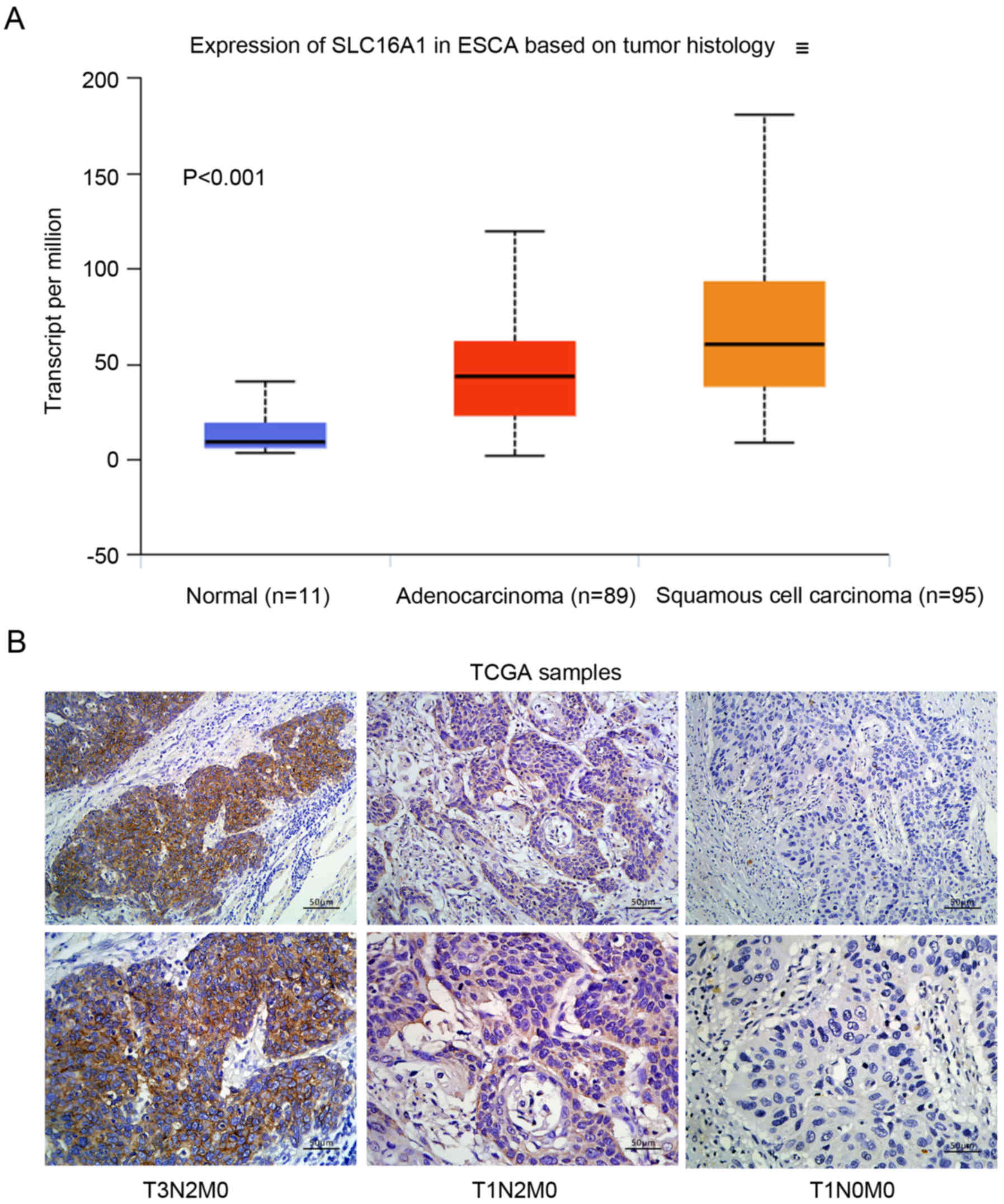
According to the TCGA database (http://ualcan.path.uab.edu/cgi-bin/TCGAExResultNew2.pl?genenam=SLC16A1&ctype=ESCA),
MCT1 (SLC16A1) was upregulated in ESCC tissues
compared to normal tissues (Fig.
1A; P<0.001). MCT1 was assessed on the cell membranes of
ESCC tumor cells. Representative images of MCT1
immunohistochemistry are shown in Fig.
1B.
In total, the study enrolled 103 ESCC patients, 35
(33.98%) of whom were female, 68 (66.02%) were males, 46 (44.7%)
were younger than 60 years, and 57 (55.3%) were older than 60
years. Among the 103 ESCC patients, 61 were in an MCT1
high-expression group (score >8) and 42 were in the
low-expression group (score ≤8). In terms of survival, 37 (35.9%)
patients were still alive at the end of the five-year follow-up
period, while 66 (64.1%) patients died during this period. The
survival time of all patients ranged from 6–80 months, and the
median survival time was 40 months. The relationship between MCT1
expression and clinicopathologic features are shown in Table I. We found that there was a
significant correlation between MCT1 expression and T stage
(P=0.005), N stage (P=0.036) and TNM stage (P=0.035). Other
clinicopathologic features had no significant correlation with MCT1
expression, including sex, age, drinking and smoking history and
tumor differentiation.
 | Table I.Correlation of MCT1 expression with
the clinicopathological features of ESCC in the FFPE cancer
tissues. |
Table I.
Correlation of MCT1 expression with
the clinicopathological features of ESCC in the FFPE cancer
tissues.
|
| MCT1
overexpression |
|
|---|
|
|
|
|
|---|
| Clinicopathological
features | No (scores ≤8)
(n=42) | Yes (scores >8)
(n=1) |
P-valuea |
|---|
| Sex |
|
| 0.071 |
|
Female | 10 | 25 |
|
|
Male | 32 | 36 |
|
| Age (years) |
|
| 0.922 |
|
<60 | 19 | 27 |
|
|
≥60 | 23 | 34 |
|
| History of
drinking |
|
| 0.054 |
| No | 16 | 35 |
|
|
Yes | 26 | 26 |
|
| History of
smoking |
|
| 0.703 |
| No | 17 | 27 |
|
|
Yes | 25 | 34 |
|
| T stage |
|
|
0.005b |
| T1 | 4 | 7 |
|
| T2 | 25 | 16 |
|
| T3 | 10 | 23 |
|
| T4 | 3 | 15 |
|
| N stage |
|
|
0.036b |
| N0 | 24 | 19 |
|
| N1 | 4 | 17 |
|
| N2 | 9 | 17 |
|
| N3 | 5 | 8 |
|
| TNM stage |
|
|
0.035b |
| I | 11 | 11 |
|
| II | 16 | 16 |
|
|
III | 15 | 34 |
|
|
Differentiation |
|
| 0.527 |
|
Well | 20 | 23 |
|
|
Moderate | 10 | 20 |
|
|
Poor | 12 | 18 |
|
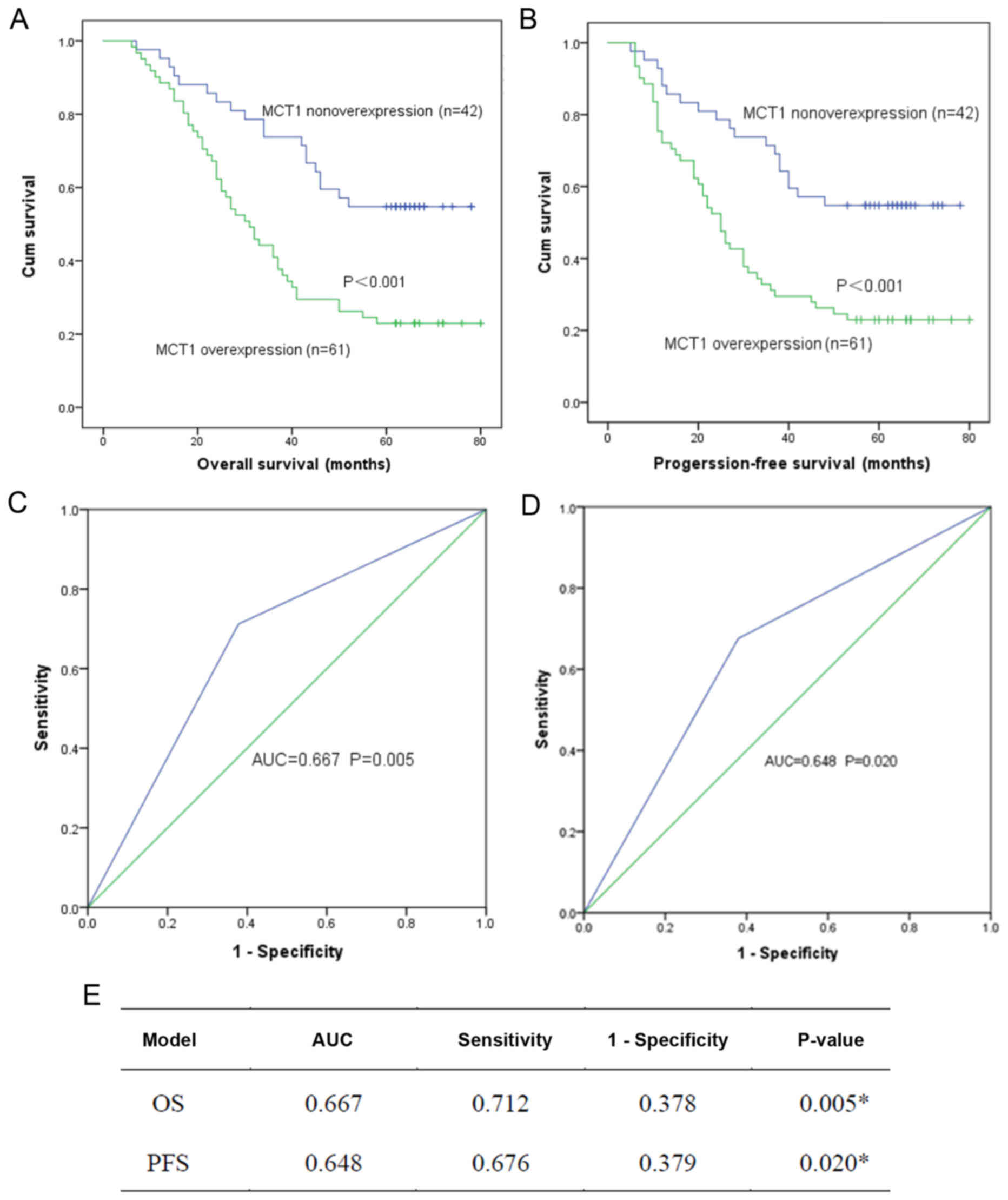
Prognostic value of MCT1
Overall survival (OS) and progression-free survival
(PFS) impacted by MCT1 were determined by the Kaplan-Meier method
and log-rank analysis (Fig. 2A and
B). A lower 5-year OS was found in the MCT1 high-expression
group (P<0.001). The same situation was observed for PFS
(P<0.001). The ROC curve was drawn and the AUC values were used
to determine the predictive efficiency of MCT1. AUC values for
death and progression were 0.667 (P=0.005) and 0.648 (P=0.020),
respectively (Fig. 2C and D).
Sensitivity and specificity are documented in Fig. 2E.
The results of the univariate and multivariate Cox
regression analyses are displayed in Table II. Upon univariate analysis, the
factors significantly related to OS were T stage (P<0.001), N
stage (P<0.001) and MCT1 expression (P=0.001). T stage
(P<0.001), N stage (P=0.003) and MCT1 expression (P=0.001) were
verified to be significantly associated with PFS. Upon multivariate
analysis, T stage was also found to be a significant prognostic
marker for ESCC patients (OS: P=0.012, PFS: P=0.013). Multivariate
analysis further confirmed that MCT1 may be a significant
prognostic marker for ESCC patients in terms of OS (P=0.01) and PFS
(P=0.012).
| Table II.Univariate and multivariate analyses
of prognostic variables based on clinical features and IHC scores
of the ESCC cases. |
Table II.
Univariate and multivariate analyses
of prognostic variables based on clinical features and IHC scores
of the ESCC cases.
|
| OS | OS | PFS | PFS |
|---|
|
|
|
|
|
|
|---|
|
| Univariate
analysis | Multivariate
analysis | Univariate
analysis | Multivariate
analysis |
|---|
|
|
|
|
|
|
|---|
| Variable | P-value | P-value | HR | 95% CI | P-value | P-value | HR | 95% CI |
|---|
| Sex (female vs.
male) | 0.18 | 0.242 | 0.696 | 0.380–1.276 | 0.12 | 0.226 | 1.451 | 0.794–2.650 |
| Age in years
(<65 vs. ≥65) | 0.84 | 0.59 | 1.167 | 0.665–2.050 | 0.952 | 0.629 | 0.871 | 0.499–1.522 |
| Drinking (yes vs.
no) | 0.341 | 0.25 | 0.695 | 0.347–1.292 | 0.318 | 0.27 | 1.418 | 0.763–2.634 |
| Smoking (yes vs.
no) | 0.845 | 0.345 | 1.346 | 0.726–2.495 | 0.827 | 0.353 | 0.745 | 0.401–1.386 |
| T stage |
<0.001a |
0.012a |
|
|
<0.001a |
0.013a |
|
|
| T1 |
|
| 1 | Ref |
|
| 1 | Ref |
| T2 |
| 0.031a | 4.281 | 1.143–16.037 |
| 0.032a | 4.214 | 1.130–15.718 |
| T3 |
| 0.006a | 5.956 | 1.680–21.115 |
| 0.006a | 5.858 | 1.659–20.692 |
| T4 |
| 0.002a | 8.023 | 2.196–29.309 |
| 0.002a | 7.781 | 2.138–28.314 |
| N stage |
<0.001a | 0.151 |
|
|
0.003a | 0.179 |
|
|
| N0 |
|
| 1 | Ref |
|
| 1 | Ref |
| N1 |
| 0.386 | 1.382 | 0.665–2.871 |
| 0.382 | 1.386 | 0.667–2.878 |
| N2 |
| 0.025a | 2.197 | 1.104–4.370 |
| 0.03a | 2.138 | 1.077–4.247 |
| N3 |
| 0.678 | 1.199 | 0.510–2.818 |
| 0.631 | 1.233 | 0.525–2.895 |
|
Differentiation | 0.395 | 0.357 |
|
| 0.414 | 0.386 |
|
|
|
Well |
|
| 1 | Ref |
|
| 1 | Ref |
|
Moderate |
| 0.96 | 1.016 | 0.536–1.926 |
| 0.93 | 1.029 | 0.546–1.939 |
|
Poor |
| 0.217 | 0.635 | 0.308–1.307 |
| 0.243 | 0.651 | 0.317–1.338 |
| MCT1 overexpression
(scores >8) |
0.001a |
0.01a | 2.273 | 1.213–4.258 |
0.001a |
0.012a | 0.45 | 0.240–0.841 |
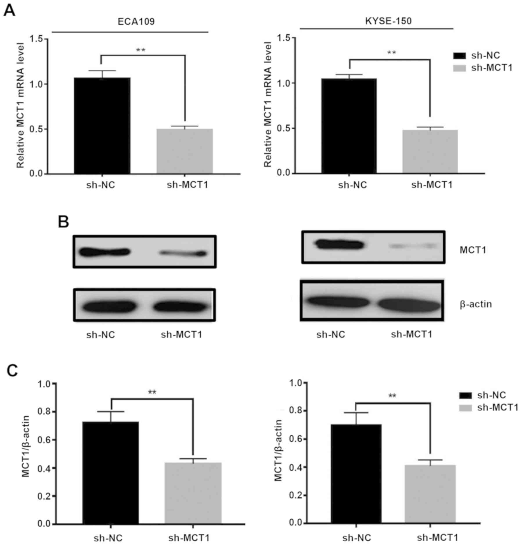
Downregulation of MCT1 inhibits the
proliferation of ESCC Eca109 and KYSE-150 cells
To determine the transfection efficiency of the
sh-MCT1 plasmids, the expression of MCT1 was verified by qRT- PCR
(Fig. 3A) and western blotting
(Fig. 3B and C) at the mRNA and
protein levels, in KYSE-150 and Eca109 cells. The mRNA of
MCT1 was significantly lower following transfection of the
sh-MCT1 plasmids in both cell lines (Eca109: 1.06±0.08 vs.
0.49±0.04, P<0.001; KYSE-150: 0.96±0.05 vs. 0.50±0.05,
P<0.001, respectively). MCT1 protein expression was lower in the
sh-MCT1 groups than levels in the sh-NC groups (MCT1/ACTB: Eca109:
0.72±0.08 vs. 0.43±0.04, P<0.001; KYSE-150: 0.7±0.08 vs.
0.41±0.04, P<0.001, respectively).
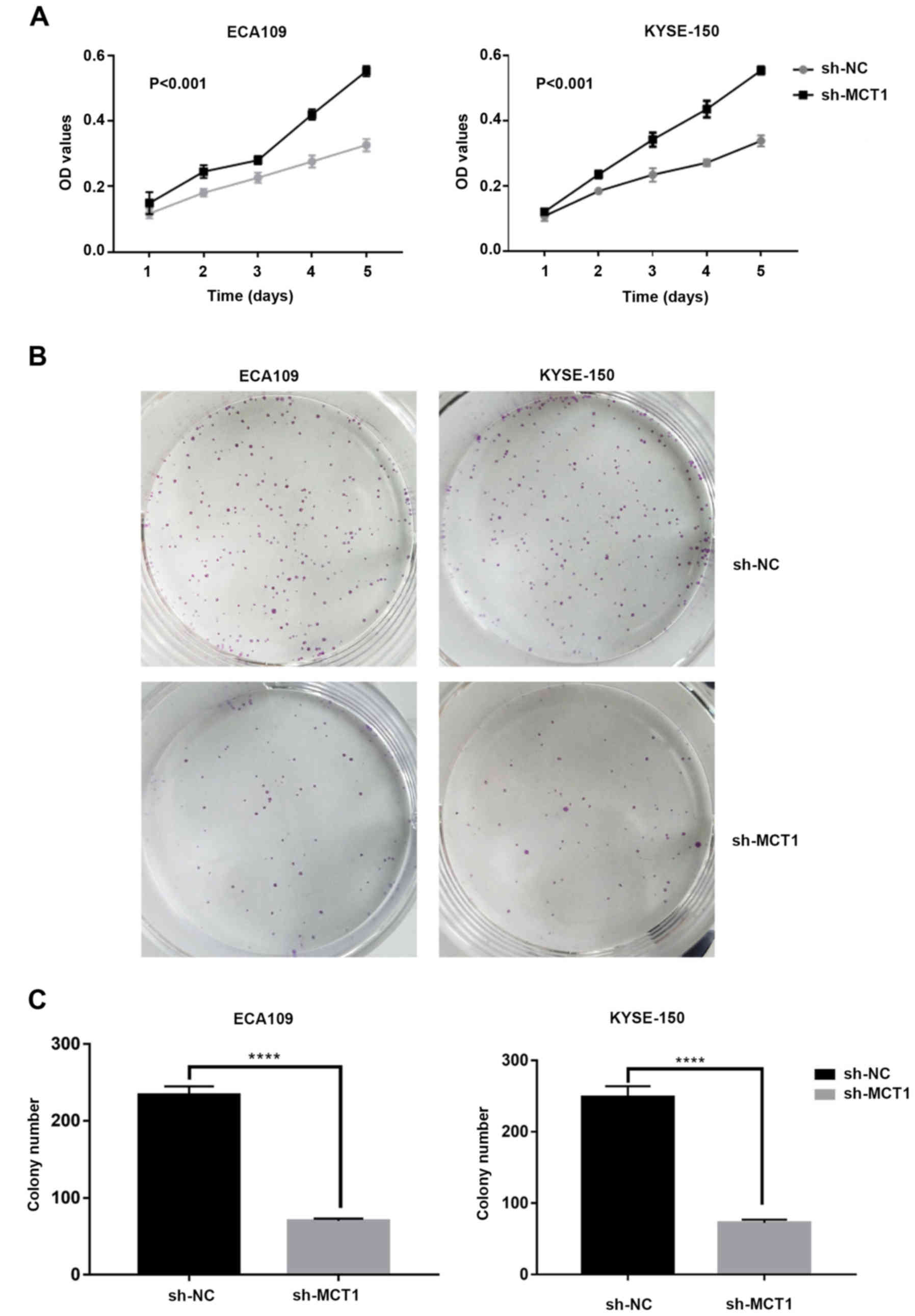
To explore the role of MCT1 in the proliferation of
ESCC cells, we conducted a CCK-8 and colony formation assays in
KYSE-150 and Eca109 cell lines. The results of the CCK-8 assay
showed that the OD values (Fig. 4A)
of the sh-MCT1 groups were significantly lower than those of the
sh-NC groups at 24, 48, 72, 96 and 120 h in both cell lines
(P<0.001). Consistent with this finding, the number of clones in
the Sh-MCT1 groups (Fig. 4B and C)
were significantly lower (Eca109: 233±11 vs. 70±3, P<0.0001;
KYSE-150: 249±15 vs. 72±4, P<0.0001, respectively). To find
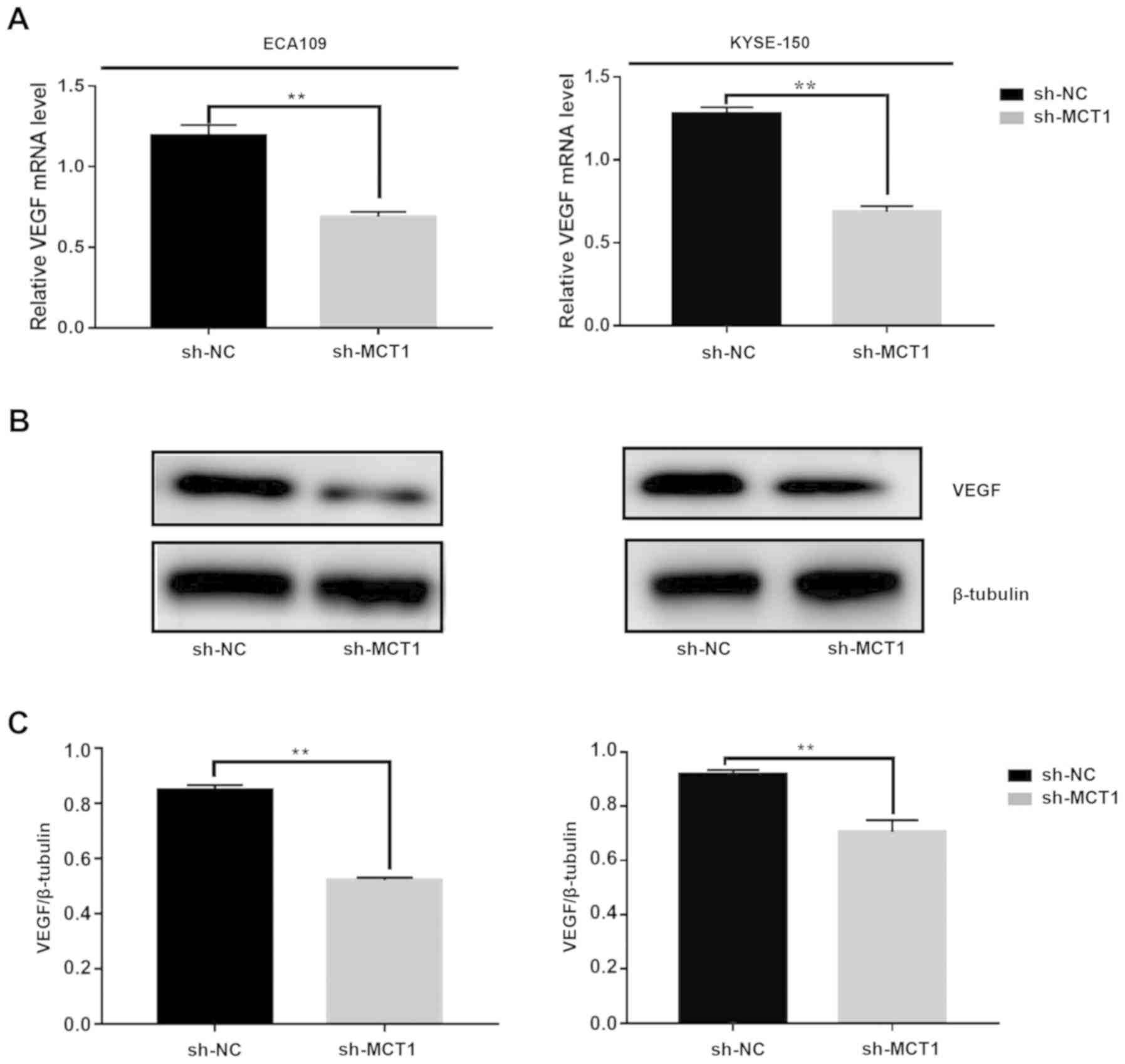
biochemical markers modified by MCT1 downregulation, we examined
the expression of VEGF in the MCT 1-silenced samples at the mRNA
(Fig. 5A) and protein levels
(Fig. 5B and C). The mRNA of VEGF
was decreased significantly with sh-MCT1 plasmid transfection in
both cell lines (Eca109: 1.193±0.07 vs. 0.69±0.03, P<0.001;
KYSE-150: 1.28±0.04 vs. 0.68±0.03, P<0.001, respectively). The
expression of VEGF protein was lower in the sh-MCT1 groups than
that in the sh-NC groups (VEGF/β-tubulin: Eca109: 0.85±0.02 vs.
0.52±0.01, P<0.001; KYSE-150: 0.92±0.02 vs. 0.71±0.04,
P<0.001, respectively). The results showed that the expression
of VEGF was significantly decreased after MCT1 downregulation. In
summary, the data suggested that MCT1 is a contributing factor to
the proliferation of ESCC cells by modifying the expression of
vascular endothelial growth factor (VEGF).
Discussion
MCT1 expression is a poor prognostic
marker
We initially explored the expression of MCT1 (solute
carrier family 16 member; SLC16A1) in esophageal cancer and its
clinical significance. All 103 patients were followed up for at
least five years to gather the follow-up data, and
clinicopathologic data were obtained for survival analysis. This is
the first study to carry out survival analysis on MCT1. We found
that MCT1 was highly expressed in certain ESCC patients. Previous
studies have shown that TNM stage plays an integral part in guiding
stage-specific treatment protocols and has a major impact on
overall survival (OS) (1). Tumor
length and the number of lymph node metastases are indicative of a
poor prognosis of esophageal cancer (15,16).
In the present study, T staging (P=0.005), N staging (P=0.036) and
TNM staging (P=0.035) were strongly correlated with the expression
of MCT1. This suggests that MCT1 may be associated with unfavorable
prognosis in esophageal cancer.
High MCT1 expression is associated
with poor outcomes
In previous studies, the expression and role of MCT1
have been adequately validated and explained in detail, especially
for breast cancer, hepatocellular carcinoma, bladder cancer and
glioblastoma; MCT1 was found to act as a biomarker of poor outcome,
while in non-small cell lung cancer (NSCLC) it exhibited an
opposite effect (11,12,14,17,18).
By univariate analysis, we found that high expression of MCT1was
associated with lower OS (P=0.001) and PFS (P=0.001). In addition,
based on CCK-8 and colony formation assays, the expression of MCT1
and the proliferation of ESCC cells were positively correlated.
This suggests that MCT1 may be useful as a prognostic biomarker,
valuable for designing clinical trials using MCT1 inhibitors.
Possible mechanism underlying the
unfavorable prognosis caused by MCT1
The present study is consistent with previous
studies that revealed a strong association between MCT1 and
unfavorable prognosis (19–21). High expression of MCT1 was
accompanied by enhancement of proliferation, migration and
invasiveness, revealing more aggressive tumor characteristics and
worse prognosis based on the present and previous results (13,22–25).
Mechanistically, the function of MCT1 as a lactic
acid transporter has been demonstrated to be closely related to
tumor progression (14,17,21,26).
Malignant cancers have been shown to exhibit characteristic
alterations of metabolism, including the ‘Warburg effect’ and
increased dependence on amino acid metabolism (7,27).
First, the anaerobic glycolysis of tumor cells under normoxic
conditions results in excess lactic acid in cells. In the present
study, we found a high expression of MCT1 in ESCC, and there was
observable MCT1 expression on the plasma membranes of KYSE-150 and
Eca109 cells (Fig. 1B). As MCT1 is
an ion transport-related molecule that releases protons to the
extracellular medium, high expression of MCT1 may result in both a
weakly alkaline intracellular pH (pHi) and an acidic extracellular
pH (pHe). The function of tumor cells depends on the maintenance of
an intracellular weak alkaline pH (23). Furthermore, previous studies have
shown that an acidic extracellular pH (pHe) increased the
expression of vascular endothelial growth factor (VEGF), cathepsin
B (CB), matrix metalloproteinase-2 and −9 (MMP-2 and −9), carbonic
anhydrase 9 and interleukin-8 (IL-8), all of which have been found
to be associated with enhanced tumor cell survival, migration and
invasion (28–31). We found that downregulation of MCT1
resulted in decreased expression of VEGF (Fig. 5), which is an important factor in
angiogenesis and tumor cell growth regulated by the VEGF-AKT-NF-κB
signaling pathway (32). This may
be a potential mechanism underlyling the suppression of ESCC cell
proliferation caused by downregulation of MCT1, but more convincing
experiments need to be carried out.
Therefore, a large amount of the available data on
the correlation between MCT1 and cancer are focused on the
contribution of MCT1 as a transporter. Notably, apart from the
primary transporting function, there is little evidence of
transporters exhibiting tumor-promoting activities relying on other
features. Nonetheless, a study conducted by Gray et al
revealed a novel function of MCT1 independent of transporter
activity. They explained that MCT1 regulated tumor migration by
activating the HGF/c-Met pathways apart from its function as a
proton transporter (33). The
HGF/c-met pathway is believed to induce tumor cells to undergo
epithelial-mesenchymal transformation (EMT), characterized by
absence of cell-cell adhesion, leading to more active tumor cell
motility, invasion and metastasis (34). EMT is an important feature of
radioresistance phenotypes in tumor cells and our finding that MCT1
activates the HGF/c-Met pathways may be a novel concept for future
study.
Limitations and future directions
Normal esophageal tissues were not included in this
study. All tissue specimens were paraffin-embedded esophageal
cancer tissues preserved in the Pathology Department of Qilu
Hospital, which were removed during esophagectomy. Whether the
margin of the tumor resection was normal tissue was not verified,
and the normal esophageal tissue that was determined could not be
removed during surgery. Therefore, normal esophageal tissues were
more difficult to obtain.
The content of lactic acid in the cell fragment and
culture supernatant of the sh-MCT1 group was determined and
compared with that of the sh-NC group, but the results were
disappointing. Different groups tended to have equal amounts of
lactic acid as shown by the results (data not shown). We should
have determined the pH values of both groups rather than the
content of certain acids, but the pH meter in our laboratory was
not available.
Surgical procedures are an important treatment means
for early stage patients. Chemotherapy and radiotherapy are
critical for patients who cannot tolerate surgery or who are at
advanced stages (1). Concurrent
radio-chemotherapy (CRT) brings significant benefits to
non-surgical patients. Chemotherapy resistance and radioresistance
are principal challenges for malignant tumor treatment. MCT1 has
been investigated in studies of cisplatin resistance in ovarian
cancer and radiosensitivity in small cell lung cancer (SCLC)
(35,36). Since the research direction of our
team is the radioresistance and radiosensitization of esophageal
cancer, the latent role of MCT1 in the radiotherapy of ESCC will be
an active area of research for us. We plan to devote further
research to this topic.
Acknowledgements
Not applicable.
Funding
The present study was supported by the National
Natural Science Foundation of China (no. 8157110923) and the
Science and Technology Plan Project of Shandong Province (no.
2017GSF18153).
Availability of data and materials
The datasets used during the present study are
available from the corresponding author upon reasonable
request.
Authors' contributions
XueC and XuanC had equal contribution in conducting
the experiments and writing the manuscript. YC was responsible for
the design of the study and served as academic advisor throughout
the process. FL was responsible for the acquisition and scoring of
the IHC slides. QY, KZ, WZ, SG, YW and SM acquired the follow-up
data and calculated the OS (overall survival) and PFS
(progression-free survival) of 103 patients. All authors read and
approved the manuscript and agree to be accountable for all aspects
of the research in ensuring that the accuracy or integrity of any
part of the work are appropriately investigated and resolved.
Ethics approval and consent to
participate
The research protocol of the present study was
approved by the Ethics Committee of Qilu Hospital, and all patients
signed written informed consents before enrollment. Patient
information was anonymized and unidentifiable prior to
analysis.
Patient consent for publication
Not applicable.
Competing interests
The authors declare that they have no competing
interests.
References
|
1
|
Napier KJ, Scheerer M and Misra S:
Esophageal cancer: A Review of epidemiology, pathogenesis, staging
workup and treatment modalities. World J Gastrointest Oncol.
6:112–120. 2014. View Article : Google Scholar : PubMed/NCBI
|
|
2
|
Chen W, Zheng R, Baade PD, Zhang S, Zeng
H, Bray F, Jemal A, Yu XQ and He J: Cancer statistics in China,
2015. CA Cancer J Clin. 66:115–132. 2016. View Article : Google Scholar : PubMed/NCBI
|
|
3
|
Lambert R and Hainaut P: The
multidisciplinary management of gastrointestinal cancer.
Epidemiology of oesophagogastric cancer. Best Pract Res Clin
Gastroenterol. 21:921–945. 2007. View Article : Google Scholar : PubMed/NCBI
|
|
4
|
Torre LA, Bray F, Siegel RL, Ferlay J,
Lortet-Tieulent J and Jemal A: Global cancer statistics, 2012. CA
Cancer J Clin. 65:87–108. 2015. View Article : Google Scholar : PubMed/NCBI
|
|
5
|
Alsop BR and Sharma P: Esophageal Cancer.
Gastroenterol Clin North Am. 45:399–412. 2016. View Article : Google Scholar : PubMed/NCBI
|
|
6
|
Pennathur A, Gibson MK, Jobe BA and
Luketich JD: Oesophageal carcinoma. Lancet. 381:400–412. 2013.
View Article : Google Scholar : PubMed/NCBI
|
|
7
|
Hanahan D and Weinberg RA: Hallmarks of
cancer: The next generation. Cell. 144:646–674. 2011. View Article : Google Scholar : PubMed/NCBI
|
|
8
|
Cairns RA, Harris IS and Mak TW:
Regulation of cancer cell metabolism. Nat Rev Cancer. 11:85–95.
2011. View
Article : Google Scholar : PubMed/NCBI
|
|
9
|
Pinheiro C, Longatto-Filho A,
Azevedo-Silva J, Casal M, Schmitt FC and Baltazar F: Role of
monocarboxylate transporters in human cancers: State of the art. J
Bioenerg Biomembr. 44:127–139. 2012. View Article : Google Scholar : PubMed/NCBI
|
|
10
|
Brahimi-Horn MC, Bellot G and Pouyssegur
J: Hypoxia and energetic tumour metabolism. Curr Opin Genet Dev.
21:67–72. 2011. View Article : Google Scholar : PubMed/NCBI
|
|
11
|
Eilertsen M, Andersen S, Al-Saad S,
Kiselev Y, Donnem T, Stenvold H, Pettersen I, Al-Shibli K,
Richardsen E, Busund LT, et al: Monocarboxylate transporters 1–4 in
NSCLC: MCT1 is an independent prognostic marker for survival. PLoS
One. 9:e1050382014. View Article : Google Scholar : PubMed/NCBI
|
|
12
|
Afonso J, Santos LL, Miranda-Goncalves V,
Morais A, Amaro T, Longatto-Filho A and Baltazar F: CD147 and
MCT1-potential partners in bladder cancer aggressiveness and
cisplatin resistance. Mol Carcinog. 54:1451–1466. 2015. View Article : Google Scholar : PubMed/NCBI
|
|
13
|
Zhao Z, Wu MS, Zou C, Tang Q, Lu J, Liu D,
Wu Y, Yin J, Xie X, Shen J, et al: Downregulation of MCT1 inhibits
tumor growth, metastasis and enhances chemotherapeutic efficacy in
osteosarcoma through regulation of the NF-kappaB pathway. Cancer
Lett. 342:150–158. 2014. View Article : Google Scholar : PubMed/NCBI
|
|
14
|
Johnson JM, Cotzia P, Fratamico R,
Mikkilineni L, Chen J, Colombo D, Mollaee M, Whitaker-Menezes D,
Domingo-Vidal M, Lin Z, et al: MCT1 in invasive ductal carcinoma:
Monocarboxylate metabolism and aggressive breast cancer. Front Cell
Dev Biol. 5:272017. View Article : Google Scholar : PubMed/NCBI
|
|
15
|
Kawahara K, Maekawa T, Okabayashi K,
Shiraishi T, Yoshinaga Y, Yoneda S, Hideshima T and Shirakusa T:
The number of lymph node metastases influences survival in
esophageal cancer. J Surg Oncol. 67:160–163. 1998. View Article : Google Scholar : PubMed/NCBI
|
|
16
|
Feng JF, Huang Y and Zhao Q: Tumor length
in elderly patients with esophageal squamous cell carcinoma: Is it
a prognostic factor? Ups J Med Sci. 118:145–152. 2013. View Article : Google Scholar : PubMed/NCBI
|
|
17
|
Jeon JY, Lee M, Whang SH, Kim JW, Cho A
and Yun M: Regulation of acetate utilization by monocarboxylate
transporter 1 (MCT1) in hepatocellular carcinoma (HCC). Oncol Res.
26:71–81. 2018. View Article : Google Scholar : PubMed/NCBI
|
|
18
|
Silva LS, Poschet G, Nonnenmacher Y,
Becker HM, Sapcariu S, Gaupel AC, Schlotter M, Wu Y, Kneisel N,
Seiffert M, et al: Branched-chain ketoacids secreted by
glioblastoma cells via MCT1 modulate macrophage phenotype. EMBO
Rep. 18:2172–2185. 2017. View Article : Google Scholar : PubMed/NCBI
|
|
19
|
Choi JW, Kim Y, Lee JH and Kim YS:
Prognostic significance of lactate/proton symporters MCT1, MCT4,
and their chaperone CD147 expressions in urothelial carcinoma of
the bladder. Urology. 84:245 e249–215. 2014. View Article : Google Scholar
|
|
20
|
Ambrosetti D, Dufies M, Dadone B, Durand
M, Borchiellini D, Amiel J, Pouyssegur J, Rioux-Leclercq N, Pages
G, Burel- Vandenbos F and Mazure NM: The two glycolytic markers
GLUT1 and MCT1 correlate with tumor grade and survival in
clear-cell renal cell carcinoma. PLoS One. 13:e01934772018.
View Article : Google Scholar : PubMed/NCBI
|
|
21
|
Fang J, Quinones QJ, Holman TL, Morowitz
MJ, Wang Q, Zhao H, Sivo F, Maris JM and Wahl ML: The
H+-linked monocarboxylate transporter
(MCT1/SLC16A1): A potential therapeutic target for high-risk
neuroblastoma. Mol Pharmacol. 70:2108–2115. 2006. View Article : Google Scholar : PubMed/NCBI
|
|
22
|
Kim Y, Choi JW, Lee JH and Kim YS:
Expression of lactate/H+ symporters MCT1 and MCT4 and
their chaperone CD147 predicts tumor progression in clear cell
renal cell carcinoma: Immunohistochemical and The Cancer Genome
Atlas data analyses. Hum Pathol. 46:104–112. 2015. View Article : Google Scholar : PubMed/NCBI
|
|
23
|
Takada T, Takata K and Ashihara E:
Inhibition of monocarboxylate transporter 1 suppresses the
proliferation of glioblastoma stem cells. J Physiol Sci.
66:387–396. 2016. View Article : Google Scholar : PubMed/NCBI
|
|
24
|
Miranda-Goncalves V, Honavar M, Pinheiro
C, Martinho O, Pires MM, Pinheiro C, Cordeiro M, Bebiano G, Costa
P, Palmeirim I, et al: Monocarboxylate transporters (MCTs) in
gliomas: Expression and exploitation as therapeutic targets. Neuro
Oncol. 15:172–188. 2013. View Article : Google Scholar : PubMed/NCBI
|
|
25
|
Payen VL, Hsu MY, Radecke KS, Wyart E,
Vazeille T, Bouzin C, Porporato PE and Sonveaux P: Monocarboxylate
transporter MCT1 promotes tumor metastasis independently of its
activity as a lactate transporter. Cancer Res. 77:5591–5601. 2017.
View Article : Google Scholar : PubMed/NCBI
|
|
26
|
Sonveaux P, Copetti T, De Saedeleer CJ,
Végran F, Verrax J, Kennedy KM, Moon EJ, Dhup S, Danhier P, Frérart
F, et al: Targeting the lactate transporter MCT1 in endothelial
cells inhibits lactate-induced HIF-1 activation and tumor
angiogenesis. PLoS One. 7:e334182012. View Article : Google Scholar : PubMed/NCBI
|
|
27
|
Koppenol WH, Bounds PL and Dang CV: Otto
Warburg's contributions to current concepts of cancer metabolism.
Nat Rev Cancer. 11:325–337. 2011. View
Article : Google Scholar : PubMed/NCBI
|
|
28
|
Shi Q, Le X, Wang B, Abbruzzese JL, Xiong
Q, He Y and Xie K: Regulation of vascular endothelial growth factor
expression by acidosis in human cancer cells. Oncogene.
20:3751–3756. 2001. View Article : Google Scholar : PubMed/NCBI
|
|
29
|
Rozhin J, Sameni M, Ziegler G and Sloane
BF: Pericellular pH affects distribution and secretion of cathepsin
B in malignant cells. Cancer Res. 54:6517–6525. 1994.PubMed/NCBI
|
|
30
|
Swietach P, Vaughan-Jones RD and Harris
AL: Regulation of tumor pH and the role of carbonic anhydrase 9.
Cancer Metastasis Rev. 26:299–310. 2007. View Article : Google Scholar : PubMed/NCBI
|
|
31
|
Xu L and Fidler IJ: Acidic pH-induced
elevation in interleukin 8 expression by human ovarian carcinoma
cells. Cancer Res. 60:46102000.PubMed/NCBI
|
|
32
|
Yu P, Liu Q, Liu K, Yagasaki K, Wu E and
Zhang G: Matrine suppresses breast cancer cell proliferation and
invasion via VEGF-Akt-NF-kappaB signaling. Cytotechnology.
59:219–229. 2009. View Article : Google Scholar : PubMed/NCBI
|
|
33
|
Gray AL, Coleman DT, Shi R and Cardelli
JA: Monocarboxylate transporter 1 contributes to growth
factor-induced tumor cell migration independent of transporter
activity. Oncotarget. 7:32695–32706. 2016. View Article : Google Scholar : PubMed/NCBI
|
|
34
|
Kalluri R and Weinberg RA: The basics of
epithelial-mesenchymal transition. J Clin Invest. 119:1420–1428.
2009. View Article : Google Scholar : PubMed/NCBI
|
|
35
|
Yan C, Yang F, Zhou C, Chen X, Han X, Liu
X, Ma H and Zheng W: MCT1 promotes the cisplatin-resistance by
antagonizing fas in epithelial ovarian cancer. Int J Clin Exp
Pathol. 8:2710–2718. 2015.PubMed/NCBI
|
|
36
|
Bola BM, Chadwick AL, Michopoulos F,
Blount KG, Telfer BA, Williams KJ, Smith PD, Critchlow SE and
Stratford IJ: Inhibition of monocarboxylate transporter-1 (MCT1) by
AZD3965 enhances radiosensitivity by reducing lactate transport.
Mol Cancer Ther. 13:2805–2816. 2014. View Article : Google Scholar : PubMed/NCBI
|