Introduction
Gastric cancer is one of the most frequent causes of
cancer-related deaths worldwide (1)
and is the most commonly diagnosed cancer, ranking third in cancer
mortality rates in Korea (2). Early
diagnosis, improved nutritional care, and new chemotherapies have
led to better outcomes over the past 20 years (3). However, chemotherapy has limited
effects (4), and targeted therapy
is used only for a small subset of patients due to restricted
availability of biomarkers (5,6). Thus,
studies of mechanisms contributing to gastric cancer are essential
for development of new therapeutic strategies.
HOXC10 is a member of the HOX genes
and contributes to hind limb development (7). In addition to its role as a
developmental regulator, recent studies revealed additional roles
of HOXC10, such as controlling the browning of white adipose
tissues and regulation of the cell cycle (8,9). In
addition, recent studies have reported aberrant expression of
HOXC10 and its effects in diverse cancers. Studies in
glioma, lung adenocarcinoma, osteosarcoma, and thyroid cancer
demonstrated that aberrant HOXC10 expression was correlated
with poor survival outcome (10–13).
HOXC10 knockdown enhanced apoptosis and attenuated
proliferation, metastasis, and expression of immunosuppressive
genes in glioma (12). In thyroid
cancer, HOXC10 knockdown was associated with cell cycle
arrest and repression of metastasis (10). In breast cancer, HOXC10 was
upregulated by estrogen, which recruits MLL3 and MLL4 to the
estrogen response element in the HOXC10 promoter region
(14). However, in breast cancer
treated with aromatase inhibitors, resistance to the inhibitors
occurred through downregulation of HOXC10 expression
mediated by hypermethylation of HOXC10 promoter regions
(15). Another study revealed that
HOXC10 contributed to chemotherapy resistance through DNA
repair by binding with cyclin-dependent kinase 7 and activating the
NF-κB pathway (16). Collectively,
HOXC10 plays roles as a transcription factor in the
development and in cancer progression. Moreover, HOXC10 mediates
additional functions by binding to other proteins.
Epigenetic alterations, including DNA methylation,
histone modification and non-coding RNAs, are as important as
genetic mutations in cancer progression and metastasis (17). DNA methylation of promoter CpG
islands interrupts binding of transcription factors, thus,
repressing gene expression (18).
In cancer, numerous tumor suppressor genes are downregulated by
hypermethylation, while oncogenes are upregulated by
hypomethylation, at their CpG promoter sites (17,18).
Recent studies have reported that HOXC10 is
upregulated in gastric cancer and promotes cell growth and
metastasis through the MAPK (19)
or NF-κB pathway (20). However,
genetic or epigenetic changes associated with HOXC10
overexpression in gastric cancer have yet to be identified. In
addition, the target genes transcriptionally regulated by
HOXC10 overexpression are not fully understood. The aim of
the present study was to investigate the epigenetic and
transcriptomic alterations associated with HOXC10
overexpression. HOXC10 expression in gastric cancer tissues
and tumorigenicity of HOXC10 in vitro were examined.
Furthermore, it was revealed that the upregulation of HOXC10
was regulated by DNA methylation of its promoter region. Several
genes transcriptionally regulated by HOXC10 were also
identified.
Materials and methods
Public data analysis
Gene expression and DNA methylation data for gastric
cancer patients were obtained from the GDC data portal (https://portal.gdc.cancer.gov/). Gene expression
data for gastric cancer patients with survival information was
downloaded from the GEO database (GSE26253) (21).
Clinical samples
Paired gastric tumor and normal tissue samples
(n=242) were collected from Chungnam National University Hospital
(CNUH; Daejeon, Korea) with informed consent obtained from all
patients and among them 171 samples have clinicopathological
information. The present study was approved by the Internal Review
Board of CNUH.
Cell culture, transfection and
5-aza-2′-deoxycitidine (5-aza-dC) treatment
Gastric cancer cell lines (SNU-001, SNU-005,
SNU-216, SNU-016, SNU-484, SNU-520, SNU-601, SNU-620, SNU-638,
SNU-668, SNU-719, AGS, KATOIII, MKN1, MKN45 and MKN74) were
obtained from the Korean Cell Line Bank (http://cellbank.snu.ac.kr/main/index.html) and were
maintained in complete RPMI-1640 and DMEM medium (Welgene, Inc.,
Gyeongsan-si, Korea) at 37°C in a humidified 5% CO2
incubator. Complete media were supplemented with 10% fetal bovine
serum (FBS; Welgene, Inc.) and 1% antibiotic-antimycotic solution
(Gibco; Thermo Fisher Scientific, Inc., Waltham, MA, USA). The
HOXC10 full cDNA clone was amplified by RT-PCR (primer
sequences are listed in Table SI)
and inserted into the pCDH-CMV-MCS-EF1-Puro (CD510B-1) lentiviral
vector. An empty vector was used as a control. For viral particle
generation, cloned DNAs and lentiviral packaging Mix
(Sigma-Aldrich; Merck KGaA, Darmstadt, Germany) were transfected
into 293T cells using X-tremeGENE™ HP DNA Transfection reagent
(Roche Diagnostics, Basel, Switzerland) in Opti-MEM media (Gibco;
Thermo Fisher Scientific, Inc.). After 2 h of incubation, the media
was changed to complete media. After 48 h of incubation, virus
particles were collected from the supernatant. For viral infection,
2×10⁵ gastric cancer cells were added to each well of a
6-well plate and incubated overnight at 37°C. Viral supernatant was
added to plated cells, and after incubation for overnight at 37°C,
the media was changed to complete media. After 24 h, infected cells
were selected using puromycin treatment. HOXC10 transfection
was confirmed by RT-PCR and western blot analysis. The siRNA in
this study was purchased from Bioneer Corp. (Daejeon, Korea). The
antisense and sense sequences of the siRNA are the following.
siCST1: CUAUAACGCAGACCUCAAU and AUUGAGGUCUGCGUUAUAG. A total of 100
nM siRNA was transfected with the Lipofectamine RNAiMAX reagent
(Invitrogen; Thermo Fisher Scientific, Inc.) according to the
manufacturer's instructions. One million cells were treated with 10
µM 5-aza-dC (Sigma-Aldrich; Merck KGaA) in a 100-mm dish for three
days with media changes every 24 h following by harvesting.
Reverse transcription PCR,
quantitative real-time PCR and western blot analysis
Total RNA was extracted using the RNeasy Plus Mini
kit (Qiagen, Venlo, The Netherlands) according to the
manufacturer's instructions. Reverse transcription was performed
using 1 µg total RNA as the template and iScript cDNA Synthesis kit
(Bio-Rad Laboratories, Hercules, CA, USA). RT-PCR assays were
performed using a PCR Master Mix (MGmed, Inc., Seoul, Korea) for 5
min at 25°C, for 30 min at 42°C, for 5 min at 85°C. Quantitative
real-time PCR (qPCR) reactions were performed in triplicate on a
CFX96™ Real-Time PCR Detection System (Bio-Rad Laboratories) using
SsoAdvanced™ Universal Inhibitor-Tolerant SYBR-Green Supermix
(Bio-Rad Laboratories) for 3 min at 95°C, and 40 cycles for 20 sec
at 95°C, for 20 sec at 60°C, 20 sec 72°C. The relative expression
of cDNA was normalized to β-actin, quantified using the
2−ΔΔCq cycle threshold method (22), and at least three independent
biological replicates were included for each reaction. Primers used
for PCR were designed either manually or using the Primer3 program
(http://bioinfo.ut.ee/primer3-0.4.0/).
All primer sequences are listed in Table SI. Cells were lysed using 2X
Laemmli sample buffer [4% SDS, 20% glycerol, 120 mM Tris-Cl (pH
6.8)] with a protease inhibitor cocktail (Roche Diagnostics).
Supernatants were collected and quantified using bovine serum
albumin (BSA) (Thermo Fisher Scientific, Inc.) by Bradford assay. A
total of 30 µg proteins were loaded onto 12% polyacrylamide gels,
electrophoresed and transferred to a polyvinylidene difluoride
(PVDF) membrane (Roche Diagnostics) and probed with the HOXC10
antibody (dilution 1:1,000; rabbit polyclonal; cat. no. ab153904;
Abcam, Cambridge, MA, USA) and β-actin antibody (dilution 1:1,000;
mouse monoclonal; cat. no. 061M4808; Sigma-Aldrich) at 4°C
overnight. HRP-conjugated anti-rabbit and anti-mouse IgG antibodies
(dilution 1:5,000; cat. nos. sc-2357 and sc-516102; Santa Cruz
Biotechnology, Inc., Dallas, TX, USA) were used as the secondary
antibody at room temperature for 2 h. Bands were detected with ECL
reagent (Amersham Biosciences, Buckinghamshire, UK) and scanned
with a Fujifilm LAS-4000 Imaging System and analyzed using
MultiGauge Software (Fujifilm, Brookvale, Australia).
Cell proliferation, colony formation,
migration and wound healing assays
Suspensions of 1.0×10³ cells were seeded
into 96-well plates. After 24, 48, 72 or 96 h of incubation at
37°C, EZ-cytox Cell Viability Assay kit mixture (DoGen, Seoul,
Korea) was added. After 1 h of incubation, the fluorescence
intensity ratio from 650 to 450 nm was measured. Suspended AGS
(1×10³) cells were added to each well of a 6-well plate
and incubated at 37°C for seven days. Colony forming cells were
stained using 0.5% crystal violet staining solution (Sigma-Aldrich;
Merck KGaA) and counted. Transwell chambers (Corning Inc., Corning,
NY, USA) were coated with fibronectin (Sigma-Aldrich; Merck KGaA).
Cells were suspended in serum-free media and seeded into the upper
chamber at a density of 1×10⁵ cells (MKN74) and
1.5×10⁴ cells (AGS) per well, and serum-containing media
was placed into the lower chamber. After incubation at 37°C for
18–20 h, cells penetrating the pores were stained using 0.5%
crystal violet staining solution (Sigma-Aldrich; Merck KGaA) and
observed under a light microscope. Suspended MKN74 cells
(5×10⁴) were plated into each well of Culture-Insert 2
Well in µ-Dish 35 mm (ibidi Gmbh, Martinsried, Germany). After 24 h
of incubation at 37°C, cells were gently removed from the
Culture-Insert using sterile tweezers. The used dish was filled
with cell-free medium and observed after 24, 48 and 72 h under a
light microscope.
Bisulfite sequencing
Genomic DNA was bisulfite modified using the EZ DNA
methylation kit (Zymo Research, Irvine, CA, USA) and PCR amplified.
PCR products were cloned into a pGEM-T Easy vector (Promega,
Madison, WI, USA) and a few clones were randomly selected for
sequencing. Bisulfite-modified DNA was amplified using a primer set
designed to amplify a site between +832 and +1176 relative to the
transcription start site using MethPrimer (http://www.urogene.org/cgi-bin/methprimer/meth-primer.cgi).
Primer sequences were 5′-GGGTAAAGTGAGTTTTTTTGAGATTTTTAA-3′
(forward) and 5′-CTTCCCTAATCCCTAACCCAAAATC-3′ (reverse).
Methylation levels are indicated as the percentage of CpG
methylated sites among all CpG sites.
RNA sequencing and data access
The RNA sequencing library was prepared using the
TruSeq RNA Sample Prep kit (Illumina, San Diego, CA, USA;
http://www.illumina.com), and sequencing was
performed using the Illumina HiSeq 2000 platform to generate 100-bp
paired-end reads. Sequenced reads were mapped to the human genome
(hg19) using STAR (v.2.5.1), and gene expression levels were
quantified with the count module in STAR (23). The TMM (trimmed mean of
M-values)-normalized CPM (counts per million) value of each gene
was set to a baseline of 1 and log2-transformed for
further analysis (i.e., clustering, heat map drawing and
correlation analysis). NGS data were deposited in the NCBI Gene
Expression Omnibus (GEO) under the accession no. GSE119196. Raw
sequence tags were deposited in the NCBI Short Read Archive (SRA)
under the accession no. SRP159087.
Chromatin immunoprecipitation
assay
Chromatin immunoprecipitation (ChIP) was performed
using Dynabeads Protein A and G (Thermo Fisher Scientific, Inc.).
For ChIP-PCR/qPCR, 2.0×10⁷ cells were cross-linked with
1% formaldehyde for 10 min at 25°C. Cells were then resuspended and
lysed for 10 min at 4°C with SDS lysis buffer. The lysate was
aliquoted into a milliTUBE 1 ml AFA Fiber (Covaris, Inc., Woburn,
MA, USA) and sonicated using a M220 Focused-ultrasonicator
(Covaris, Inc.). Fragment sizes were ~200–500 bp. Samples were
diluted with low-salt RIPA buffer and pre-cleared with 50 µl of
Dynabeads Protein A and G for 1 h at 4°C. Primary antibodies were
added to pre-cleared supernatants, and the mixtures were incubated
overnight at 4°C. Antibodies used for the ChIP assay include HOXC10
(dilution 1:400; rabbit polyclonal; cat. no. ab153904; Abcam),
phosphorylated RNA polymerase II (dilution 1:400; mouse monoclonal;
cat. no. ab5408; Abcam), and IgG antibodies (dilution 1:1,000; cat.
nos. sc-2357 and sc-516102; Santa Cruz Biotechnology, Inc.). Next,
50 µl Dynabeads Protein A and G were added to the samples, and the
mixtures were incubated for 2 h at 4°C. The beads were subsequently
washed with wash buffer (low-salt RIPA, high-salt RIPA, LiCl and
TE). Precipitated chromatin was eluted in 200 µl elution buffer
(0.1 M NaHCO3 and 1% SDS). Reverse cross-linking was performed
overnight at 65°C, and chromatin was then treated with RNase A and
5M NaCl for 30 min at 37°C and proteinase K overnight at 65°C. DNA
was purified using a QIAquick PCR Purification kit (Qiagen).
ChIP-PCR assays were performed using PCR Master Mix and
electrophoresis 1% agarose gel. ChIP-qPCR reactions were
subsequently performed.
Statistical analysis
All experiments were triplicated and data are
indicated as mean ± standard deviation. Statistical analysis was
performed using Student's t-test. A P-value <0.05 was considered
significant. The following parameters were obtained from the
medical records of the 171 patients included in the study: Age,
sex, histology, lymph node metastasis, tumor stage, and
Helicobacter pylori infection status, and these parameters
were compared using a Chi-square test. Statistical analysis of
correlation between expression and methylation was performed using
Pearson's correlation coefficient. All statistical analysis were
performed using R statistical programming language (version 3.4.2;
http://www.r-project.org/).
Results
HOXC10 is overexpressed and associated
with poor prognosis in gastric cancer tissues
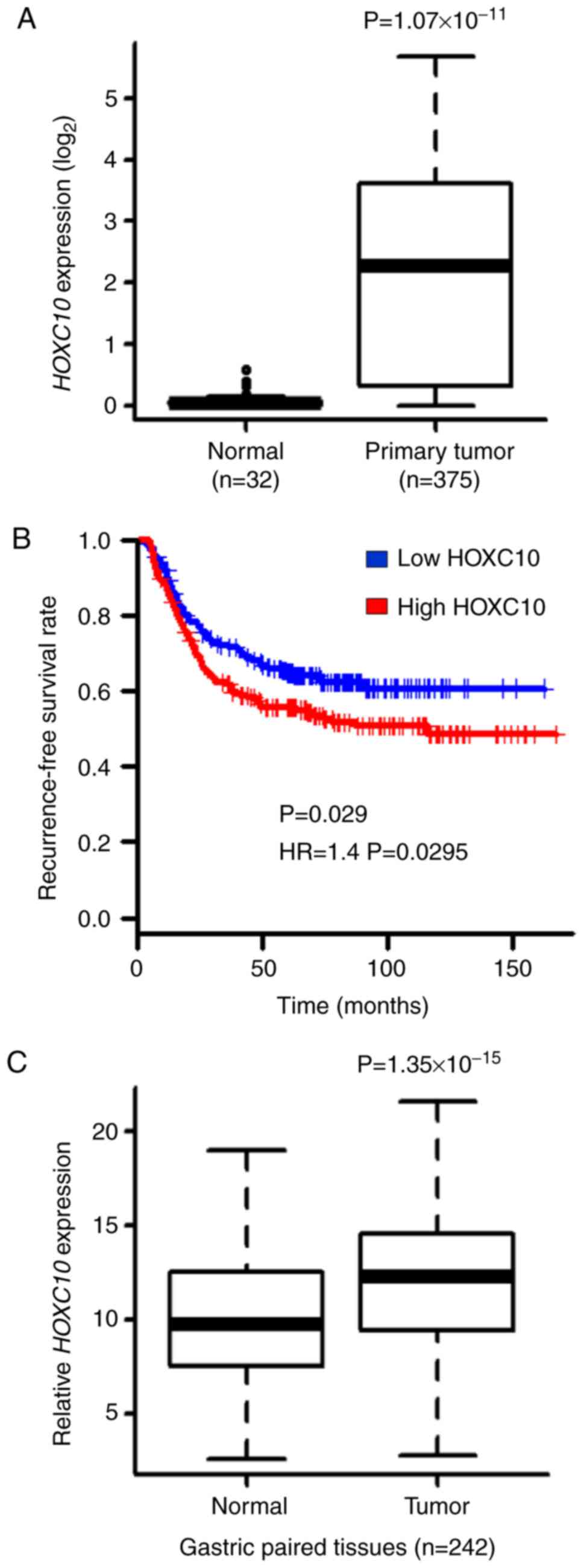
HOXC10 is upregulated in many types of
cancer. To investigate whether HOXC10 is overexpressed in
gastric cancer tissues, we used TCGA-STAD RNA-Sequencing expression
dataset. HOXC10 expression was revealed to be higher in
gastric cancer tissues than in normal tissues in TCGA dataset
(Fig. 1A). The relationship between
HOXC10 expression and recurrence-free survival was then
investigated in gastric cancer patients from the GEO dataset
(GSE26253), demonstrating that the HOXC10 high-expression
group exhibited decreased recurrence-free survival compared to the
HOXC10 low-expression group from the GEO dataset (Fig. 1B). Overexpression of HOXC10
was validated by performing RT-qPCR on paired normal and tumor
tissue samples collected from CNUH (Fig. 1C). Clinicopathological
characteristics with respect to HOXC10 expression from the
CNUH dataset are summarized in Table
I. A significant difference in HOXC10 expression was
observed between diffuse and intestinal cancer types (P=0.039), but
there was no significant difference in other parameters.
 | Table I.Clinicopathological characteristics
by HOXC10 expression in gastric cancer patients. |
Table I.
Clinicopathological characteristics
by HOXC10 expression in gastric cancer patients.
|
| Gastric tumors with
increased relative HOXC10 expression |
|
|---|
|
|
|
|
|---|
| Clinicopathological
parameters | >2-Fold increase
(n=98) | ≤2-Fold increase
(n=73) | P-value |
|---|
| Mean patient age
(in years ± SD) | 60±12 | 59±14 | 0.9270 |
| Sex |
|
| 0.4769 |
|
Male | 62 | 50 |
|
|
Female | 36 | 23 |
|
| Lauren's
classification |
|
| 0.0386a |
|
Intestinal | 23 | 26 |
|
|
Diffuse | 69 | 38 |
|
|
Mixed | 6 | 8 |
|
| No
information | 0 | 1 |
|
| Tumor
progression |
|
| 0.8769 |
|
EGC | 10 | 7 |
|
|
AGC | 87 | 66 |
|
| No
information | 1 | 0 |
|
| Stage |
|
| 0.4522 |
| I | 17 | 15 |
|
| II | 23 | 19 |
|
|
III | 27 | 21 |
|
| IV | 31 | 18 |
|
| Helicobacter
pylori infection |
|
| 0.7343 |
|
Positive | 59 | 43 |
|
|
Negative | 23 | 19 |
|
| No
information | 16 | 18 |
|
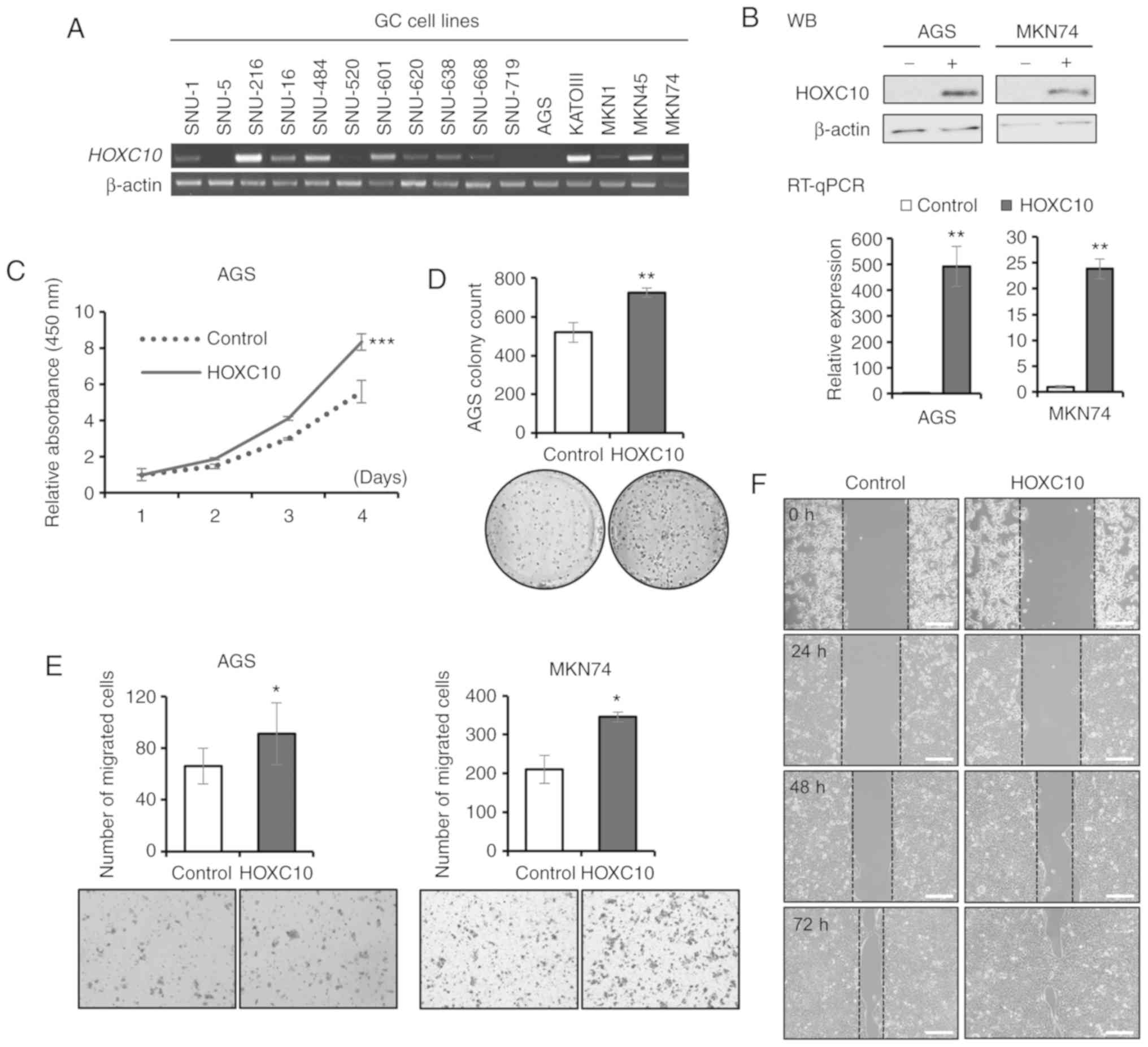
HOXC10 promotes cell proliferation and
cell migration in MKN74 and AGS cell lines
The functional effects of HOXC10
overexpression were first investigated in gastric cancer using
in vitro assays. RT-PCR of HOXC10 among 16 gastric
cancer cell lines illustrated that AGS and MKN74 cells expressed
HOXC10 at low levels, while SNU-216, SNU-484, KATOIII and
MKN45 cells expressed HOXC10 at high levels (Fig. 2A). Thus, AGS and MKN74 cells were
selected for infection with lentiviruses ectopically expressing
HOXC10. Overexpression of HOXC10 in AGS and MNK74
cells after lentivirus infection was confirmed by RT-qPCR and
western blot analysis (Fig. 2B).
HOXC10 overexpression increased proliferation (Fig. 2C) and colony formation (Fig. 2D) in AGS cells and increased
migration in both AGS and MKN74 cells (Fig. 2E and F). Recent studies have
demonstrated that overexpression of HOXC10 promotes cell
proliferation and migration in gastric cancer GC-9811P, AGS and
SGC7901 cells, and HOXC10 knockdown suppresses progression
of gastric cancer in GC-9811P, SNU638 and SGC7901 cells (19,20,24). A
mouse model injected with AGS cells containing upregulated
HOXC10 exhibited increased tumor size (19). Collectively, it was concluded that
HOXC10 overexpression promotes proliferation, colony
formation and progression in gastric cancer.
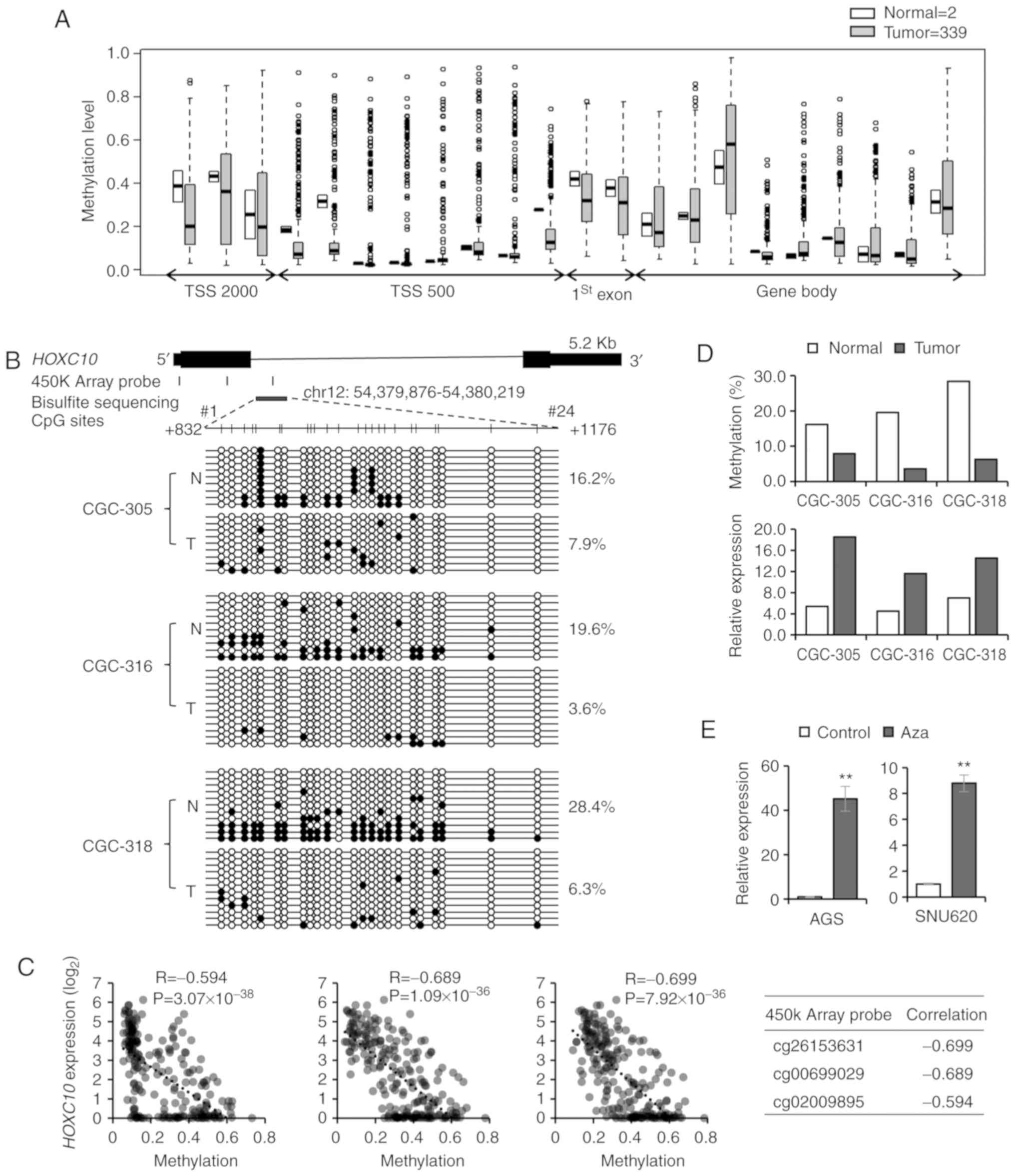
HOXC10 expression is regulated by DNA
methylation in gastric cancer tissues and cells
To reveal the mechanism of HOXC10
overexpression in gastric cancer, publicly available TCGA STAD
datasets were analyzed for DNA methylation and copy number
alterations. For copy number alteration, the HOXC10 gene was
altered in 1.69% of 295 cases (amplification, 1.02%; deep deletion,
0.68%) using cBioPortal (http://www.cbioportal.org/index.do). To determine
whether methylation of HOXC10 CpG sites affected
HOXC10 expression, we analyzed TCGA STAD 450K array dataset.
It was observed that many CpG sites in the HOXC10 gene were
hypomethylated in gastric cancer compared to normal tissues
(Fig. 3A). We next attempted
bisulfite sequencing to determine methylation levels of
HOXC10 CpG sites. In fact, reduced methylation at 24 CpG
sites of the first intronic region of HOXC10 (Chr12:
54,379,876-54,380,219; +832 to +1176) was confirmed in three
gastric cancers compared to normal tissues (Fig. 3B). The region contains the promoter
region (chr12:54,380,069-54,380,128; promoter ID: HOXC10_4)
obtained from a Eukaryotic promoter database (https://epd.vital-it.ch/index.php). Specifically,
CpG residues 3, 7, 8, 13 and 16 were significantly hypomethylated
in tumor tissues (P<0.05, Student's t-test). By correlation
analysis of DNA methylation (TCGASTAD 450K array) and gene
expression (TCGA STAD RNA-seq) data, it was revealed that three CpG
sites (indicated in Fig. 3B)
exhibited significant negative correlations between methylation of
HOXC10 CpG sites and HOXC10 expression (Pearson's
correlation coefficient r=−0.594, −0.689 and −0.699) (Fig. 3C). The methylation levels and
HOXC10 expression levels were examined in three paired
gastric tumor and normal tissues (Fig.
3D). When two gastric cancer cell lines (AGS and SNU-620 with
low HOXC10 expression) were treated with 5-aza-dC, an
inhibitor of DNA methyltransferase, HOXC10 expression
increased 45.24-fold (AGS) and 8.79-fold (SNU-620) (Fig. 3E). These results indicate that the
methylation status of CpG sites in the HOXC10 first intronic
region was important for the regulation of HOXC10 expression
in gastric cancer.
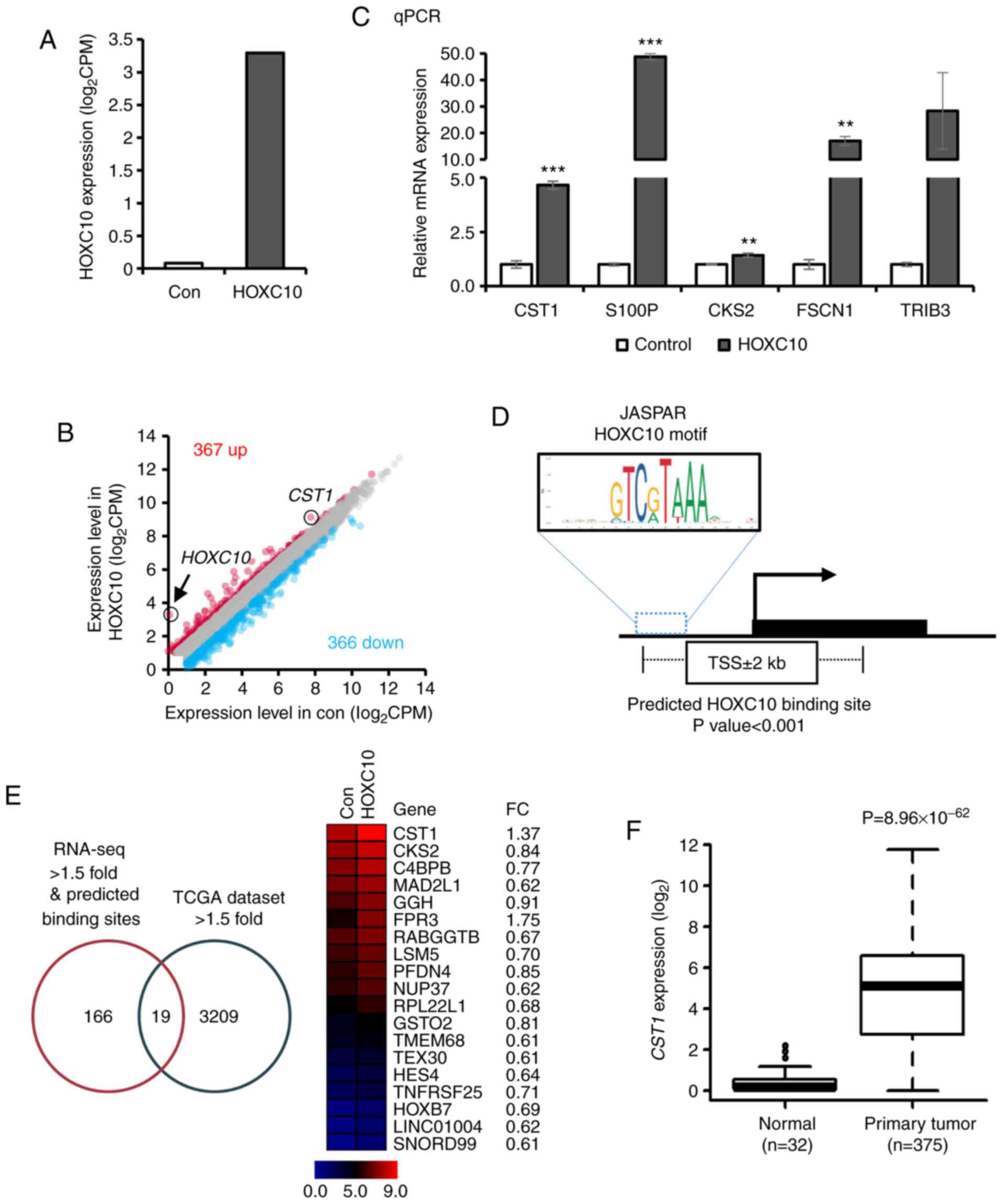
Transcription factor HOXC10 promotes
CST1 transcription
It was demonstrated that HOXC10 was
upregulated by hypomethylation of its CpG sites, and HOXC10
overexpression contributed to the progression of gastric cancer.
Since HOXC10 is a transcription factor, the functional roles of
HOXC10 overexpression were further explored in gastric
cancer by identifying its target genes. Hence, RNA sequencing of
ectopic HOXC10-overexpressing AGS cells was performed to
identify target genes regulated by HOXC10. RNA-sequencing analysis
confirmed overexpression of HOXC10 in AGS cells (3.21-fold
increase compared to control vector-infected cells; Fig. 4A). Three hundred and sixty-seven
(upregulated) and 366 (downregulated) genes were differentially
expressed in response to ectopic HOXC10 expression
[|log2 (fold-change)| >1.5; Fig. 4B and Table SII]. Five of the highly upregulated
genes (CST1, S100P, CKS2, FSCN1 and TRIB3) were
validated in HOXC10-overexpressing cells compared to
controls by RT-qPCR (Fig. 4C). The
HOXC10 binding motif sequence predicted by the JASPAR database
(http://jaspar.genereg.net/) was mapped
to predicted promoter regions (TSS±2 kb) of RNA-seq fold-change
>1.5 genes, and 185 genes were selected (P<0.001) (Fig. 4D and Table SIII). By combining two different
data sets (RNA-seq fold-change >1.5 with binding motif of HOXC10
predicted by JASPAR database, and gene expression increased
>1.5-fold in TCGA dataset), 19 genes were selected as potential
target genes regulated by HOXC10 (Fig.
4E). For example, CST1 expression was significantly
overexpressed in TCGA dataset (Fig.
4F). A recent study reported that CST1 is upregulated
and promotes cell proliferation in gastric cancer (25).
Chromatin immunoprecipitation (ChIP) was then
performed in cells with ectopic HOXC10 expression. First, a
ChIP assay was conducted at the CST1 promoter region
(Chr20:23,732,257-23,732,507) using the HOXC10 antibody. ChIP-PCR
and ChIP-qPCR analysis demonstrated HOXC10 bound to CST1
promoter region (Fig. 5A).
Additionally, to indicate whether transcriptional machinery
operated by HOXC10 binding to the CST1 promoter region, a
ChIP assay was designed at the transcription start site region of
CST1 (Chr20:23,731,473-23,731,614) with phosphorylated RNA
pol II antibody. Phosphorylated RNA pol II did bind to the
CST1 transcription start site region (Fig. 5B). A ChIP assay was also performed
on S100P (another predicted target) promoter regions and TSS
regions (Fig. 5C and D). In
addition, it was observed that gastric patients with high
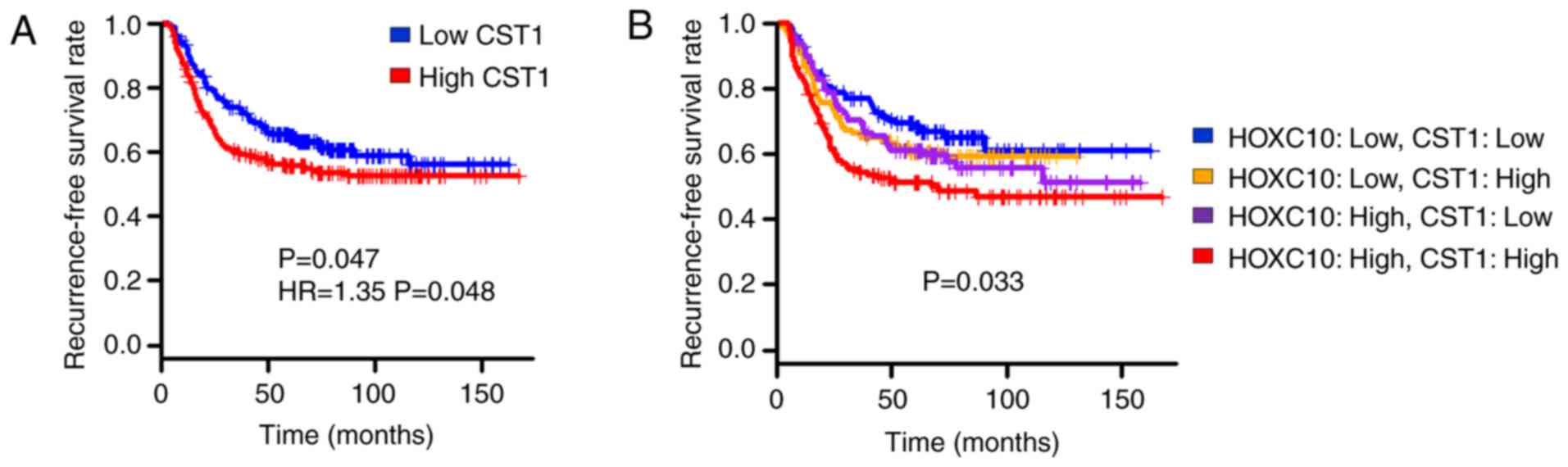
CST1 expression exhibited decreased recurrence-free survival
compared to those with low CST1 expression (Fig. 6A). Combined analysis of CST1
and HOXC10 expression again confirmed that the
CST1/HOXC10 high-expression group exhibited worse
recurrence-free survival than the CST1/HOXC10 low-expression
group (Fig. 6B). These results
indicated that overexpression of CST1 mediated by HOXC10
contributes to worse prognosis in gastric cancer patients.
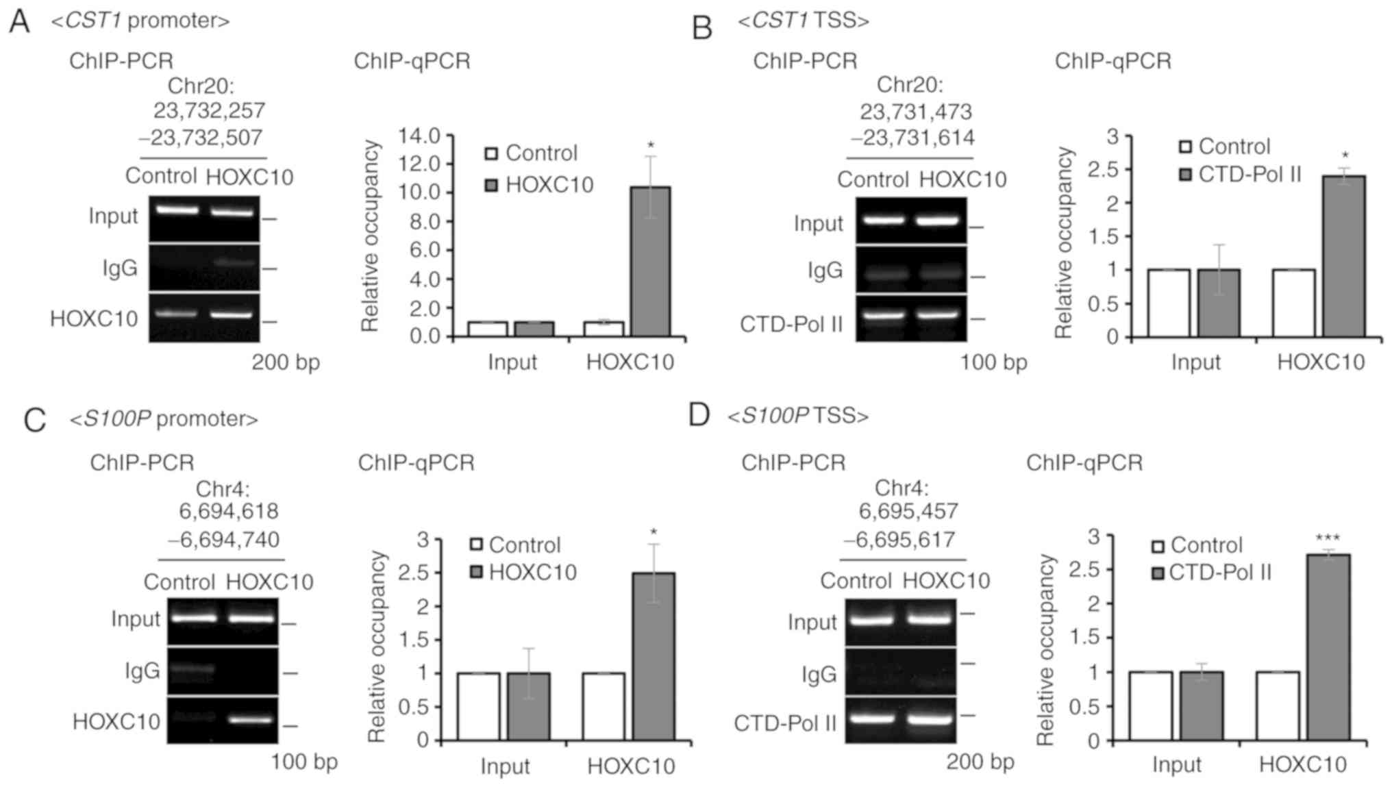 | Figure 5.Validation of CST1 and
S100P as direct target genes of HOXC10. (A) Ascertainment of
HOXC10 binding to the CST1 promoter region
(Chr20:23,732,257-23,732,507) using ChIP-PCR and ChIP-qPCR. (B)
Ascertainment of phosphorylated RNA polymerase II (phospho-Ser5)
binding to the CST1 TSS region (Chr20:
23,731,473-23,731,614) using ChIP-PCR and ChIP-qPCR. (C)
Ascertainment of HOXC10 binding to the S100P promoter region
(6,694,618-6,694,740) using ChIP-PCR and ChIP-qPCR. (D)
Ascertainment of phosphorylated RNA polymerase II (phospho-Ser5)
binding to the S100P TSS region (Chr4: 6,695,457-6,695,617)
using ChIP-PCR and ChIP-qPCR. For all, *P<0.05; ***P<0.001,
Student's t-test. The error bars represent the mean ± SD.
HOXC10, homeodomain-containing gene 10. |
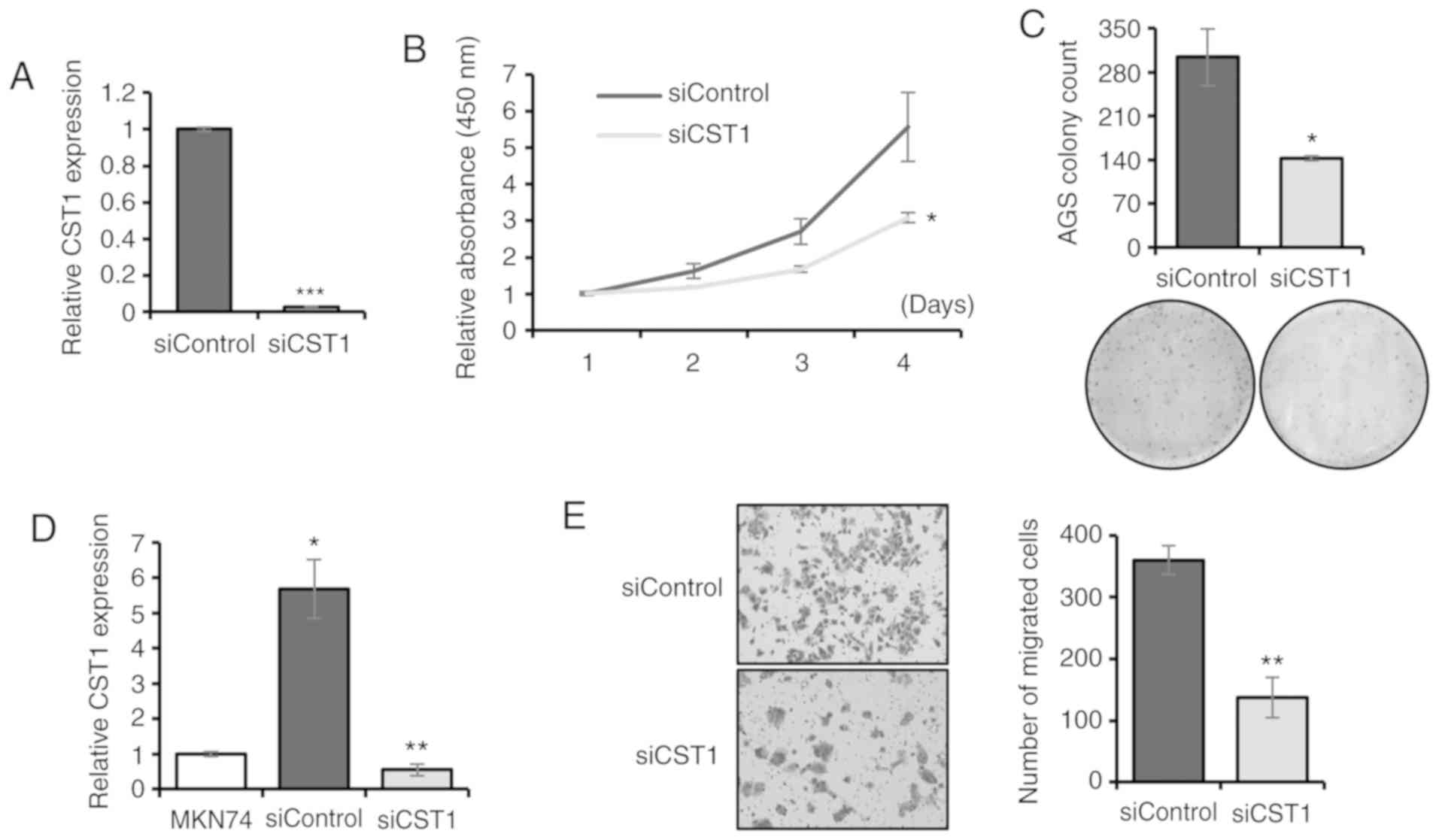
Knockdown of CST1 inhibits gastric
cancer progression
Expression of CST1 was upregulated in
HOXC10-overexpressing AGS cells (Fig. 4C) and tumor tissues of the TCGA
dataset. It was validated that HOXC10 regulated the expression of
CST1 by using ChIP-PCR. Gastric cancer patients with both
CST1 and HOXC10 overexpression exhibited the poorest survival
(Fig. 6B). Thus, the roles of
CST1 in gastric cancer progression were investigated using
transient knockdown of CST1 in HOXC10-overexpressing
cells. The silencing of CST1 was confirmed by RT-qPCR in AGS
cells (Fig. 7A). Knockdown of
CST1 inhibited cell proliferation (Fig. 7B) and colony formation (Fig. 7C) in AGS cells. CST1
expression was upregulated in HOXC10-overexpressing MKN74
cells and downregulated in CST1-silencing MKN74 cells
(Fig. 7D). Knockdown of CST1
suppressed the migration ability of MKN74 cells (Fig. 7E). As another downstream target of
HOXC10, S100P was also assessed, and it was revealed that
knockdown of S100P suppressed cell proliferation and reduced
colony size in AGS cells (Fig.
S1). CST1 and S100P were selected for further
validation based on previous studies revealing the role of those
two genes in gastric carcinogenesis. For example, CST1 was
revealed to increase cell proliferation (25) and S100P to regulate cell
proliferation (26) and colony
formation (27) in gastric cancer;
these two genes exhibited oncogenic roles in other cancers as well
(28–33). Altogether, CST1 regulated by
HOXC10 contributes to gastric cancer development, and these two
genes could be potential prognostic markers for gastric cancer.
Discussion
Aberrant overexpression of homeodomain-containing
gene 10 (HOXC10) and its role in cancer progression has been
reported in many different cancers (10–14).
Recently, a few studies have shown that HOXC10 is
overexpressed in gastric cancer and that its overexpression
promotes cell growth and migration with activation of MAPK and
NF-κB pathways (19,20). Moreover, HOXC10 expression is
correlated with EMT marker genes (SOX10 and FGBP1)
(34). We recognized that
TMEM41A is upregulated in gastric cancer, especially
metastatic and advanced tumors, and contributed to poor prognosis.
In vitro and in vivo assay showed that TMEM41A
has effects on the migration ability of gastric cancer (35). We investigated expression of
TMEM41A with our RNA-sequencing data; however, there were no
significant differences in the expression level. This study was
associated with other mechanisms in metastasis. However, mechanism
of HOXC10 overexpression in gastric cancer are not yet
known. In the present study, we demonstrated that hypomethylation
of HOXC10 CpG sites is one mechanism for aberrant
overexpression of HOXC10 in gastric cancer. The CpG sites
are located at the HOXC10 first intron and include a
promoter retrieved from a eukaryotic promoter database. Although
bisulfite sequencing was performed on a partial region of many of
the CpG sites and a few of the tissue samples, the results showed
significant correlation between the levels of both DNA methylation
and HOXC10 expression.
HOXC10 belongs to the HOX family of
transcription factors, which are major regulators of organ
development and cellular differentiation during animal development.
However, recent studies have shown non-transcriptional interactions
for Hox proteins in additional molecular processes, such as mRNA
translation, DNA repair and initiation of DNA replication (36). For example, in breast cancer,
HOXC10 induces chemotherapy resistance by binding to CDK7
and enhancing homologous recombination DNA repair processes
(16). In this regard, it is of
interest to reveal how overexpressed HOXC10 exerts its
functions in gastric cancer cells, that is, transcriptionally or
non-transcriptionally.
In this study, we primarily focused on the role of
HOXC10 as a transcriptional regulator. By analyzing RNA-seq and
ChIP assays, we identified CST1 as one of the 19 targets
genes regulated by HOXC10 in gastric cancer. CST1 is a
cysteine protease inhibitor belonging to the cystatin superfamily.
Recent studies have shown that CST1 is upregulated and
contributes to cancer progression in multiple neoplasms, including
gastric (25), breast (31), non-small cell lung cancer (NSCLC)
(30), colorectal (33) and pancreatic cancer (32). In colorectal cancer, cathepsin B is
associated with invasion of colon cancer cells by extracellular
matrix degradation. CST1 attenuates inhibition of cathepsin B by
CST3 through binding to CST3 (33),
and sustenance of cathepsin B by CST1 promotes cancer development
by inhibiting cellular senescence (37). Additionally, CST1 is a mediator of
bone metastasis (38,39). However, as mechanisms of CST1
are unclear in gastric cancer, further studies are required.
Overexpression of cytokeratin associated protein (CAP)
downregulated NF-κB activity and decreased expression of many of
its target genes, including CST1 (40). Knockdown of CST1 by miRNA
let-7d reduces cell proliferation through phosphorylation of the
NF-κB p65 subunit in colorectal cancer (41). Gene-set enrichment analysis using
our RNA-seq data revealed that the NF-κB pathway was activated
(Fig. S2A), consistent with a
previous study reporting that HOXC10 activates NF-κB in
gastric cancer and breast cancer (16,20).
We suppose that HOXC10 and CST1 promote cancer
progression through the NF-κB pathway. TLR4 is one of the
NF-κB pathway genes with HOXC10 binding motifs and is
overexpressed in cells overexpressing HOXC10
(0.82-log2FC) (Fig.
S1B).
In conclusion, HOXC10 expression is
upregulated in gastric cancer through DNA demethylation, and
HOXC10 overexpression increases proliferation and migration
of gastric cancer cells. CST1 was identified as one of the
target genes regulated by HOXC10 in gastric cancer. CST1
knockdown represses tumorigenicity of gastric cancer cells. We
propose HOXC10 and CST1 as useful prognostic markers
or therapeutic targets for gastric cancer.
Supplementary Material
Supporting Data
Acknowledgements
We thank Dr Seong-Min Park for his helpful comments
on the manuscript.
Funding
The present study was funded by grants from the
National Research Foundation of Korea (grant nos.
NRF-2017MBA9B5060884 and NRF-2014M3C9A3068554).
Availability of data and materials
All data generated or analyzed during this study
are included in this published article. Gene expression data are
available in the GEO databases under the accession no. GSE119196.
URL: https://www.ncbi.nlm.nih.gov/geo/query/acc.cgi?acc=GSE119196.
Authors' contributions
JK and DHB planned and performed the experiments.
JK and SYK wrote the manuscript. JHK collected and analyzed the
omics data. KSS selected the patients and collected the clinical
samples. YSK contributed to the tissue samples and reviewed the
manuscript. SYK supervised the study. All authors read and approved
the manuscript and agree to be accountable for all aspects of the
research in ensuring that the accuracy or integrity of any part of
the work are appropriately investigated and resolved.
Ethics approval and consent to
participate
The present study and all clinical data were
approved by the Internal Review Board of the Chungnam National
University Hospital (Daejon, Korea). Written informed consent was
provided by all the patients.
Patient consent for publication
Not applicable.
Competing interests
The authors declare that they have no competing
interests.
References
|
1
|
Bray F, Ferlay J, Soerjomataram I, Siegel
RL, Torre LA and Jemal A: Global cancer statistics 2018: GLOBOCAN
estimates of incidence and mortality worldwide for 36 cancers in
185 countries. CA Cancer J Clin. 68:394–424. 2018. View Article : Google Scholar : PubMed/NCBI
|
|
2
|
Jung KW, Won YJ, Kong HJ, Lee ES and
Community of Population-Based Regional Cancer Registries: Cancer
statistics in korea: Incidence, mortality, survival, and prevalence
in 2015. Cancer Res Treat. 50:303–316. 2018. View Article : Google Scholar : PubMed/NCBI
|
|
3
|
Ahn HS, Lee HJ, Yoo MW, Jeong SH, Park DJ,
Kim HH, Kim WH, Lee KU and Yang HK: Changes in clinicopathological
features and survival after gastrectomy for gastric cancer over a
20-year period. Br J Surg. 98:255–260. 2011. View Article : Google Scholar : PubMed/NCBI
|
|
4
|
GASTRIC (Global Advanced/Adjuvant Stomach
Tumor Research International Collaboration) Group, ; Oba K,
Paoletti X, Bang YJ, Bleiberg H, Burzykowski T, Fuse N, Michiels S,
Morita S, Ohashi Y, et al: Role of chemotherapy for
advanced/recurrent gastric cancer: An individual-patient-data
meta-analysis. Eur J Cancer. 49:1565–1577. 2013. View Article : Google Scholar : PubMed/NCBI
|
|
5
|
Kanat O, O'Neil B and Shahda S: Targeted
therapy for advanced gastric cancer: A review of current status and
future prospects. World J Gastrointest Oncol. 7:401–410. 2015.
View Article : Google Scholar : PubMed/NCBI
|
|
6
|
Yuan DD, Zhu ZX, Zhang X and Liu J:
Targeted therapy for gastric cancer: Current status and future
directions (Review). Oncol Rep. 35:1245–1254. 2016. View Article : Google Scholar : PubMed/NCBI
|
|
7
|
Jain D, Nemec S, Luxey M, Gauthier Y,
Bemmo A, Balsalobre A and Drouin J: Regulatory integration of Hox
factor activity with T-box factors in limb development.
Development. 145:1598302018. View Article : Google Scholar
|
|
8
|
Gabellini D, Colaluca IN, Vodermaier HC,
Biamonti G, Giacca M, Falaschi A, Riva S and Peverali FA: Early
mitotic degradation of the homeoprotein HOXC10 is potentially
linked to cell cycle progression. EMBO J. 22:3715–3724. 2003.
View Article : Google Scholar : PubMed/NCBI
|
|
9
|
Ng Y, Tan SX, Chia SY, Tan HY, Gun SY, Sun
L, Hong W and Han W: HOXC10 suppresses browning of white adipose
tissues. Exp Mol Med. 49:e2922017. View Article : Google Scholar : PubMed/NCBI
|
|
10
|
Feng X, Li T, Liu Z, Shi Y and Peng Y:
HOXC10 up-regulation contributes to human thyroid cancer and
indicates poor survival outcome. Mol Biosyst. 11:2946–2954. 2015.
View Article : Google Scholar : PubMed/NCBI
|
|
11
|
Tang XL, Ding BX, Hua Y, Chen H, Wu T,
Chen ZQ and Yuan CH: HOXC10 Promotes the metastasis of human lung
adenocarcinoma and indicates poor survival outcome. Front Physiol.
8:5572017. View Article : Google Scholar : PubMed/NCBI
|
|
12
|
Li S, Zhang W, Wu C, Gao H, Yu J, Wang X,
Li B, Jun Z, Zhang W, Zhou P, et al: HOXC10 promotes proliferation
and invasion and induces immunosuppressive gene expression in
glioma. FEBS J. 285:2278–2291. 2018. View Article : Google Scholar : PubMed/NCBI
|
|
13
|
Xie X, Xiao Y and Huang X: Homeobox C10
knockdown suppresses cell proliferation and promotes cell apoptosis
in osteosarcoma cells through regulating caspase 3. Onco Targets
Ther. 11:473–482. 2018. View Article : Google Scholar : PubMed/NCBI
|
|
14
|
Ansari KI, Hussain I, Kasiri S and Mandal
SS: HOXC10 is overexpressed in breast cancer and transcriptionally
regulated by estrogen via involvement of histone methylases MLL3
and MLL4. J Mol Endocrinol. 48:61–75. 2012. View Article : Google Scholar : PubMed/NCBI
|
|
15
|
Pathiraja TN, Nayak SR, Xi Y, Jiang S,
Garee JP, Edwards DP, Lee AV, Chen J, Shea MJ, Santen RJ, et al:
Epigenetic reprogramming of HOXC10 in endocrine-resistant breast
cancer. Sci Transl Med. 6:229ra2412014. View Article : Google Scholar
|
|
16
|
Sadik H, Korangath P, Nguyen NK, Gyorffy
B, Kumar R, Hedayati M, Teo WW, Park S, Panday H, Munoz TG, et al:
HOXC10 expression supports the development of chemotherapy
resistance by fine tuning DNA repair in breast cancer cells. Cancer
Res. 76:4443–4456. 2016. View Article : Google Scholar : PubMed/NCBI
|
|
17
|
Sharma S, Kelly TK and Jones PA:
Epigenetics in cancer. Carcinogenesis. 31:27–36. 2010. View Article : Google Scholar : PubMed/NCBI
|
|
18
|
Jones PA: Functions of DNA methylation:
Islands, start sites, gene bodies and beyond. Nat Rev Genet.
13:484–492. 2012. View Article : Google Scholar : PubMed/NCBI
|
|
19
|
Guo C, Hou J, Ao S, Deng X and Lyu G:
HOXC10 up-regulation promotes gastric cancer cell proliferation and
metastasis through MAPK pathway. Chin J Cancer Res. 29:572–580.
2017. View Article : Google Scholar : PubMed/NCBI
|
|
20
|
Yao S, He L, Zhang Y, Ye L, Lai Y, Huang
L, Wu L, Wu G and Zhu S: HOXC10 promotes gastric cancer cell
invasion and migration via regulation of the NF-κB pathway. Biochem
Biophys Res Commun. 501:628–635. 2018. View Article : Google Scholar : PubMed/NCBI
|
|
21
|
Lee J, Sohn I, Do IG, Kim KM, Park SH,
Park JO, Park YS, Lim HY, Sohn TS, Bae JM, et al: Nanostring-based
multigene assay to predict recurrence for gastric cancer patients
after surgery. PLoS One. 9:e901332014. View Article : Google Scholar : PubMed/NCBI
|
|
22
|
Livak KJ and Schmittgen TD: Analysis of
relative gene expression data using real-time quantitative PCR and
the 2(-Delta Delta C(T)) method. Methods. 25:402–408. 2001.
View Article : Google Scholar : PubMed/NCBI
|
|
23
|
Dobin A, Davis CA, Schlesinger F, Drenkow
J, Zaleski C, Jha S, Batut P, Chaisson M and Gingeras TR: STAR:
Ultrafast universal RNA-seq aligner. Bioinformatics. 29:15–21.
2013. View Article : Google Scholar : PubMed/NCBI
|
|
24
|
Zheng J, Ge P, Liu X, Wei J, Wu G and Li
X: MiR-136 inhibits gastric cancer-specific peritoneal metastasis
by targeting HOXC10. Tumour Biol. 39:10104283177062072017.
View Article : Google Scholar : PubMed/NCBI
|
|
25
|
Choi EH, Kim JT, Kim JH, Kim SY, Song EY,
Kim JW, Kim SY, Yeom YI, Kim IH and Lee HG: Upregulation of the
cysteine protease inhibitor, cystatin SN, contributes to cell
proliferation and cathepsin inhibition in gastric cancer. Clin Chim
Acta. 406:45–51. 2009. View Article : Google Scholar : PubMed/NCBI
|
|
26
|
Ge F, Wang C, Wang W and Wu B: S100P
predicts prognosis and drug resistance in gastric cancer. Int J
Biol Markers. 28:e387–392. 2013. View Article : Google Scholar : PubMed/NCBI
|
|
27
|
Zhang Q, Hu H, Shi X and Tang W: Knockdown
of S100P by lentiviral-mediated RNAi promotes apoptosis and
suppresses the colony-formation ability of gastric cancer cells.
Oncol Rep. 31:2344–2350. 2014. View Article : Google Scholar : PubMed/NCBI
|
|
28
|
Li Z, Chen Y, Wang X, Zhang H, Zhang Y,
Gao Y, Weng M, Wang L, Liang H, Li M, et al: LASP-1 induces
proliferation, metastasis and cell cycle arrest at the G2/M phase
in gallbladder cancer by down-regulating S100P via the PI3K/AKT
pathway. Cancer Lett. 372:239–250. 2016. View Article : Google Scholar : PubMed/NCBI
|
|
29
|
Wu TS, Tan CT, Chang CC, Lin BR, Lai WT,
Chen ST, Yen-Ping Kuo M, Rau CL, Jaw FS and Chang HH: B-cell
lymphoma/leukemia 10 promotes oral cancer progression through
STAT1/ATF4/S100P signaling pathway. Oncogene. 36:54402017.
View Article : Google Scholar : PubMed/NCBI
|
|
30
|
Cao X, Li Y, Luo RZ, Zhang L, Zhang SL,
Zeng J, Han YJ and Wen ZS: Expression of cystatin SN significantly
correlates with recurrence, metastasis, and survival duration in
surgically resected non-small cell lung cancer patients. Sci Rep.
5:82302015. View Article : Google Scholar : PubMed/NCBI
|
|
31
|
Dai DN, Li Y, Chen B, Du Y, Li SB, Lu SX,
Zhao ZP, Zhou AJ, Xue N, Xia TL, et al: Elevated expression of CST1
promotes breast cancer progression and predicts a poor prognosis. J
Mol Med (Berl). 95:873–886. 2017. View Article : Google Scholar : PubMed/NCBI
|
|
32
|
Jiang J, Liu HL, Liu ZH, Tan SW and Wu B:
Identification of cystatin SN as a novel biomarker for pancreatic
cancer. Tumour Biol. 36:3903–3910. 2015. View Article : Google Scholar : PubMed/NCBI
|
|
33
|
Kim JT, Lee SJ, Kang MA, Park JE, Kim BY,
Yoon DY, Yang Y, Lee CH, Yeom YI, Choe YK and Lee HG: Cystatin SN
neutralizes the inhibitory effect of cystatin C on cathepsin B
activity. Cell Death Dis. 4:e9742013. View Article : Google Scholar : PubMed/NCBI
|
|
34
|
Miwa T, Kanda M, Umeda S, Tanaka H, Tanaka
C, Kobayashi D, Suenaga M, Hayashi M, Yamada S, Nakayama G, et al:
Homeobox C10 influences on the malignant phenotype of gastric
cancer cell lines and its elevated expression positively correlates
with recurrence and poor survival. Ann Surg Oncol. 26:1535–1543.
2019. View Article : Google Scholar : PubMed/NCBI
|
|
35
|
Lin B, Xue Y, Qi C, Chen X and Mao W:
Expression of transmembrane protein 41A is associated with
metastasis via the modulation of ecadherin in radically resected
gastric cancer. Mol Med Rep. 18:2963–2972. 2018.PubMed/NCBI
|
|
36
|
Rezsohazy R: Non-transcriptional
interactions of hox proteins: Inventory, facts, and future
directions. Dev Dyn. 243:117–131. 2014. View Article : Google Scholar : PubMed/NCBI
|
|
37
|
Oh SS, Park S, Lee KW, Madhi H, Park SG,
Lee HG, Cho YY, Yoo J and Dong Kim K: Extracellular cystatin SN and
cathepsin B prevent cellular senescence by inhibiting abnormal
glycogen accumulation. Cell Death Dis. 8:e27292017. View Article : Google Scholar : PubMed/NCBI
|
|
38
|
Blanco MA, LeRoy G, Khan Z, Alečković M,
Zee BM, Garcia BA and Kang Y: Global secretome analysis identifies
novel mediators of bone metastasis. Cell Res. 22:1339–1355. 2012.
View Article : Google Scholar : PubMed/NCBI
|
|
39
|
Cai X, Luo J, Yang X, Deng H, Zhang J, Li
S, Wei H, Yang C, Xu L, Jin R, et al: In vivo selection for
spine-derived highly metastatic lung cancer cells is associated
with increased migration, inflammation and decreased adhesion.
Oncotarget. 6:22905–22917. 2015. View Article : Google Scholar : PubMed/NCBI
|
|
40
|
Liu XF, Xiang L, Zhang Y, Becker KG, Bera
TK and Pastan I: CAPC negatively regulates NF-κB activation and
suppresses tumor growth and metastasis. Oncogene. 31:1673–1682.
2012. View Article : Google Scholar : PubMed/NCBI
|
|
41
|
Jiang J, Liu HL, Tao L, Lin XY, Yang YD,
Tan SW and Wu B: Let7d inhibits colorectal cancer cell
proliferation through the CST1/p65 pathway. Int J Oncol.
53:781–790. 2018.PubMed/NCBI
|