Introduction
In 2018, ~458 million new cases of pancreatic cancer
were diagnosed worldwide, resulting in 432 million mortalities, the
seventh highest among all cancer-associated deaths (1). Pancreatic cancer is a lethal malignancy
with a <5% five-year survival rate, indicating a mortality rate
almost equal to its occurrence (1–4).
Pancreatic ductal adenocarcinoma (PDAC), which accounts for ~90% of
all pancreatic cancer cases (5) is
the third and sixth leading cause of cancer-associated death in the
United States and China, respectively (6–8). As 80–90%
of patients with PDAC are diagnosed at an advanced stage, when the
tumor has usually metastasized, radical resection is not possible
(6,9–11).
Conventional treatments such as chemotherapy and radiotherapy, as
well as targeted therapies, have largely failed to prolong the
overall survival (OS) time of patients with PDAC (12), and only a few novel anti-PDAC
strategies are currently in use (13,14).
Therefore, the identification of early diagnostic markers and
therapeutic targets for PDAC is critical. Taking into account the
encouraging results of immuno- and gene therapies against PDAC
(15–17), the present study aimed to identify
novel immunological targets associated with its prognosis.
Chemokines and their receptors are critical
mediators of the inflammatory and immune responses (18–20), and
have recently been implicated in tumorigenesis (21,22).
Chemokine receptors promote tumorigenesis via numerous mechanisms
(23), including inflammation, that
is known to play a significant role in the pathogenesis and
progression of pancreatic cancer (24–26). In
addition, the poor prognosis and frequent distant metastasis of
pancreatic cancer are also associated with immune surveillance
escape (12,27,28). The
chemokine receptors are classified into four subfamilies-CCR, CXCR,
XCR and CX3CR-based on variations within the cysteine motif.
Although several studies have elucidated the potential roles of
these CCRs (C-C motif chemokine receptors) in pancreatic cancer,
using cell lines or murine models (29–35), the
association of CCRs with OS remains ambiguous. Therefore, the aim
of the present study was to investigate the association between
CCRs and the prognosis of patients with PDAC.
Materials and methods
Data mining and processing
The transcriptome profiles of patients with PDAC
were obtained from TCGA database (https://cancergenome.nih.gov/, accessed at April 20,
2017), and normalized using the DESeq Package in R (36,37). The
corresponding clinical data were acquired from the University of
California, Santa Cruz Xena (UCSC Xena; http://xena.ucsc.edu/, accessed at April 20, 2017). In
order to eliminate interference from unrelated factors, the
patients were selected based on the following inclusion criteria:
i) Histological validation; ii) pathological stage I or II
according to the 7th American Joint Committee on Cancer (AJCC);
iii) availability of complete survival data; and iv) having
undergone pancreaticoduodenectomy.
Bioinformatics and correlation
analysis of CCR genes
The CCR genes were functionally annotated using Gene
Ontology (GO) terms, and the associated pathways were determined by
Kyoto Encyclopedia of Genes and Genomes (KEGG) analysis using the
Database for Annotation, Visualization and Integrated Discovery
(https://david.ncifcrf.gov/home.jsp
version 6.8, accessed at January 19, 2018) (38). The correlation between CCR genes was
analyzed by Pearson's correlation coefficient using the
corrplot package in R (version 1.2.1335; www.r-project.org). A protein-protein interaction
(PPI) map was constructed using the Search Tool for the Retrieval
of Interacting Genes/Proteins (https://string-db.org/, version 11, accessed at march
30, 2019) (39). All CCR gene symbols
(CCR1-CCR10) were entered into the platform for Homo sapiens
and an interaction score >0.4 was considered to be significant.
Finally, a gene-gene interaction network was constructed using
GeneMANIA (http://genemania.org/, accessed at March
30, 2019) (40).
Survival analysis
The association between different clinical factors
and the prognosis of patients with early-stage PDAC was determined
using Kaplan-Meier analysis with the log-rank test; the relevant
factors were then included in the multivariate Cox proportional
risk regression model to identify the CCR genes significantly
associated with OS. Based on the results of the survival analysis
of individual genes, combined effect survival analysis was
performed and a nomogram was constructed. The CCR genes associated
with PDAC prognosis were assessed using combined effect survival
analysis (Kaplan-Meier analysis with log-rank test) and the
multivariate Cox proportional risk regression model. The nomogram
was constructed in R (version 3.5.2; www.r-project.org) using the rms package, based
on clinical variables and the expression levels of CCR genes. The
scale marked on the line indicates the value range of each
variable, and the length of the line segment reflects the
contribution of this factor to the outcome event.
Prognostic signature construction
According to the results of the survival analysis,
the CCR genes associated with PDAC prognosis were combined to
construct a prognostic model based on gene expression level. The
risk score formula was as follows: Risk score=expression of
gene1 × β1 + expression of gene2 ×
β2+… expression of Genen × βn
(41,42), where βn is the regression
coefficient derived from the result of multivariate Cox
proportional hazards regression analysis for the corresponding
gene. Based on the median risk score value, the patients were
divided into a high- and low risk group. To assess the predictive
value of the prognostic signature, a time-dependent ROC curve was
constructed using the survivalROC package in R (43). Survival analysis was performed to
compare prognoses between the high- and low-risk groups.
Gene set enrichment analysis
(GSEA)
In order to identify the pathways in which the CCR
gene is enriched, and to determine whether the CCR genes in each
gene set are enriched in the upper or lower part of the
phenotype-related sorted gene list, genome-wide expression profile
datasets and corresponding grouping files determined by the
expression of CCR genes were uploaded to GSEA (44) for enrichment analysis with database c2
and c5 of the Molecular Signatures Database (MSigDB) (45). A set of genes with both false
discovery rate (FDR) <0.25 and P<0.05 was considered to be
statistically significant.
Statistical analysis
Statistical analysis was conducted using SPSS 22.0
(IBM Corporation) or R 3.52 (https://www.r-project.org/). Hazard ratios (HRs) and
95% confidence intervals (CI) were used to indicate the relative
risk between the high-C and low-CCR expression groups. Pearson's
correlation coefficient was used to determine the correlation
between CCR genes, where P<0.05 was considered to indicate a
statistically significant result. The FDR control in GSEA was
achieved using the Benjamini-Hochberg procedure and adjusted for
multiple testing (46–48), where an FDR<0.25 was considered to
indicate a statistically significant difference.
Results
Data collection and arrangement
The expression profiles of patients with PDAC were
acquired from TCGA database. After screening based on the inclusion
criteria, patients that fell outside of these parameters were
eliminated, and the profiles of the remaining 112 patients were
further analyzed.
Functional annotation and correlation
analysis of CCR genes
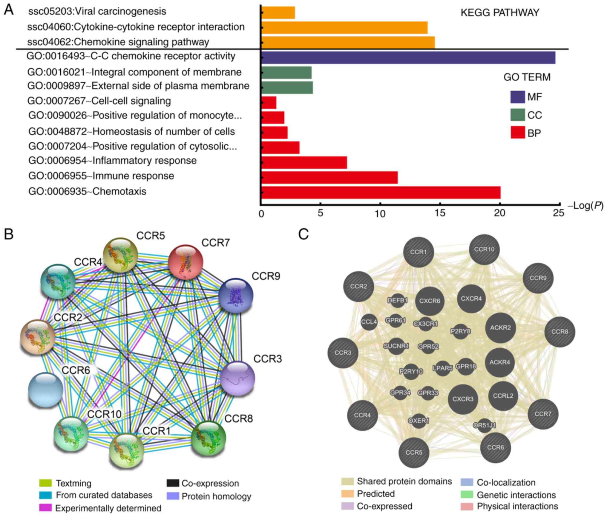
As shown in Fig. 1A
and Table SI, the results of GO and
KEGG pathway analysis indicated that CCR genes are primarily
involved in pathways related to immunity and inflammation, and that
the JAK/STAT, mitogen-activated protein kinase (MAPK) and nuclear
factor (NF)-κB signaling pathways were associated with chemokine
signaling (Fig. S1). PPI and
gene-gene interaction network analyses revealed that CCRs interact
closely with each other (Fig. 1B and
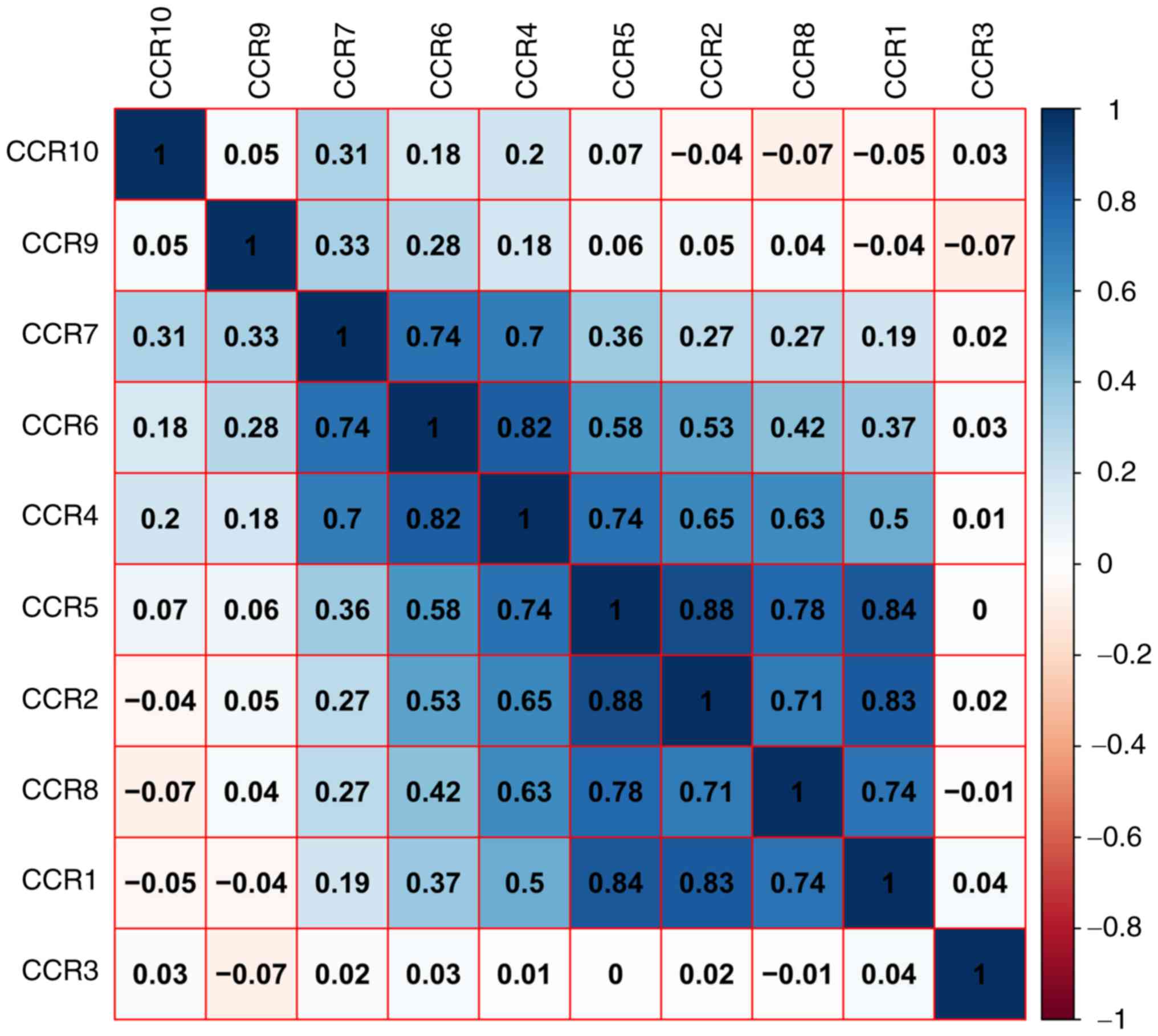
C). A matrix graph of Pearson's correlation analysis indicated
that CCR1, CCR2, CCR4, CCR5 and CCR8 are closely related to each
other with a correlation coefficient ≥0.5. In addition, a higher
degree of correlation was observed among CCR4, CCR6 and CCR7
(correlation coefficient ≥0.7) (Fig.
2). The numbers in each grid represent the correlations between
the corresponding genes.
Survival analysis of CCR genes
The clinical data of all patients are summarized in
Table SII. Histological grade,
targeted molecular therapy, radiation therapy and residual
resection were all significantly associated with OS. Patients
harboring tumors of a higher histological grade, and those who did
not receive either targeted or radiation therapies or undergo
residual resection were at a higher risk of poor prognosis. In
addition, high expression levels of CCR5 (adjusted P=0.012;
adjusted HR=0.478, 95% CI=0.269–0.852), CCR6 (adjusted P=0.026;
adjusted HR=0.527, 95% CI=0.299–0.927) and CCR9 (adjusted P=0.001;
adjusted HR=0.374, 95% CI=0.209–0.670) were significantly
associated with lower mortality rates (Table I and Fig.
3). The nomogram also indicated that CCR5, CCR6 and CCR9 may
contribute to the prognosis of PDAC, with low expression
corresponding to a high point (Fig.
4E).
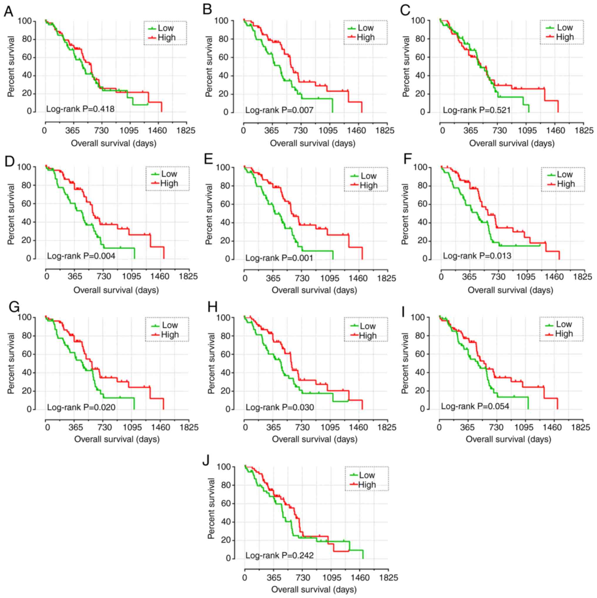 | Figure 3.Kaplan-Meier survival curve analysis
of the association between the high and low expression levels of
CCR genes and overall survival in patients with early-stage PDAC,
generated using The Cancer Genome Atlas. Overall survival curves
for (A) CCR1, (B) CCR2, (C) CCR3, (D) CCR4, (E) CCR5, (F) CCR6, (G)
CCR7, (H) CCR8, (I) CCR9 and (J) CCR10. CCR, C-C motif chemokine
receptor; PDAC, pancreatic ductal adenocarcinoma. |
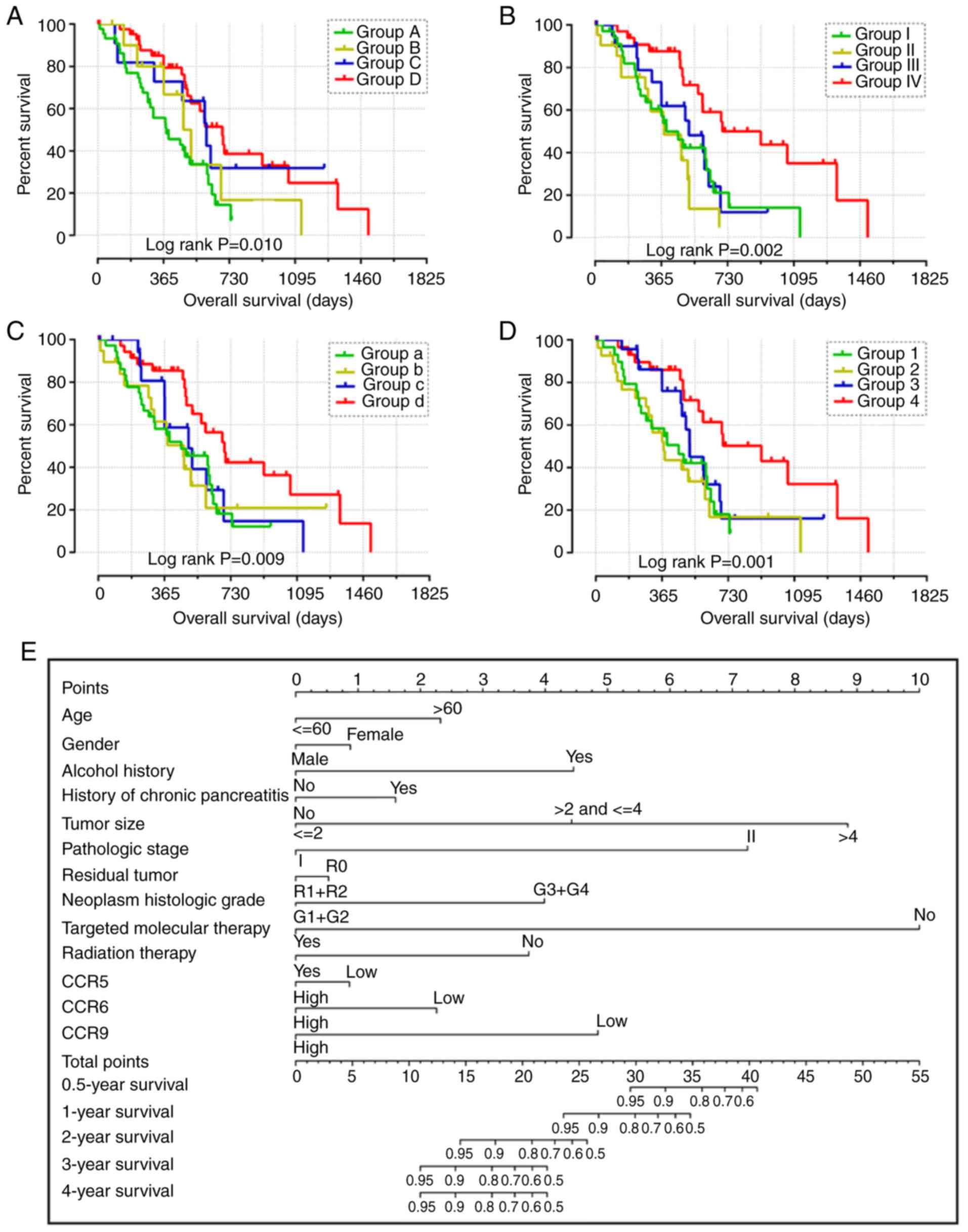 | Figure 4.Combined effect of CCR5, CCR6 and
CCR9 on the overall survival of patients with early-stage PDAC.
Nomogram for predicting 1-, 2- and 3-year events and a prognostic
model with risk score, in terms of CCR5, CCR6 and CCR9 expression
in early-stage PDAC. (A) Overall survival curves for the combined
effect of CCR5 and CCR6. Group A, Low CCR5 + Low CCR6; Group B, Low
CCR5 + High CCR6; Group C, High CCR5 + Low CCR6; Group D, High CCR5
+ High CCR6. (B) Overall survival curves for the combined effect of
CCR5 and CCR9. Group I, Low CCR5 + Low CCR9; Group II, Low CCR5 +
High CCR9; Group III, High CCR5 + Low CCR9; Group IV, High CCR5 +
High CCR9. (C) Overall survival curves for the combined effect of
CCR6 and CCR9. Group a, Low CCR5 + Low CCR9; Group b, Low CCR5 +
High CCR9; Group c, High CCR5 + Low CCR9; Group d, High CCR5 + High
CCR9. (D) Overall survival curves for the combined effect of CCR5,
CCR6 and CCR9. Group 1, Low CCR5 + Low CCR6 + Low CCR9; Group 2,
Low CCR5 + Low CCR6 + High CCR9, High CCR5 + Low CCR6 + Low CCR9,
and Low CCR5 + High CCR6 + Low CCR9; Group 3, Low CCR5 + High CCR6
+ High CCR9, High CCR5 + High CCR6 + Low CCR9, and High CCR5 + Low
CCR6 + High CCR9; Group 4, High CCR5 + High CCR6 + High CCR9. (E)
Nomogram for predicting 1-, 2- and 3-year events (mortalities) that
combine clinical data with CCR5, CCR6 and CCR7 expression. (F) From
top to bottom; risk score plot, survival status scatter plot and
heat map of the expression levels of CCR5, CCR6 and CCR9 in low-
and high-risk groups. (G) Kaplan-Meier curves for low- and
high-risk groups. (H) Receiver operating characteristic curve for
predicting 1-, 2- and 3-year survival in patients with early-stage
PDAC by risk score. CCR, C-C motif chemokine receptor; PDAC,
pancreatic ductal adenocarcinoma. |
 | Table I.Prognostic value of the expression
levels of CCR genes in early-stage pancreatic ductal adenocarcinoma
using The Cancer Genome Atlas database. |
Table I.
Prognostic value of the expression
levels of CCR genes in early-stage pancreatic ductal adenocarcinoma
using The Cancer Genome Atlas database.
|
|
| Overall
survival |
|---|
|
|
|
|
|---|
| Gene expression
level | Patients
(n=112) | Number of
events | Median survival
time (days) | Crude HR (95%
CI) | Crude Log-rank
P-value | Adjusted HR (95%
CI) | Adjusted
P-valuea |
|---|
| CCR1 |
|
|
|
|
|
|
|
|
Low | 56 | 34 | 485 | 1 |
| 1 |
|
|
High | 56 | 35 | 592 | 0.820
(0.508–1.326) | 0.418 | 0.812
(0.474–1.393) | 0.450 |
| CCR2 |
|
|
|
|
|
|
|
|
Low | 56 | 38 | 458 | 1 |
| 1 |
|
|
High | 56 | 31 | 603 | 0.518
(0.317–0.846) | 0.007 | 0.668
(0.379–1.178) | 0.163 |
| CCR3 |
|
|
|
|
|
|
|
|
Low | 56 | 33 | 511 | 1 |
| 1 |
|
|
High | 56 | 36 | 518 | 0.854
(0.527–1.385) | 0.521 | 0.947
(0.556–1.613) | 0.841 |
| CCR4 |
|
|
|
|
|
|
|
|
Low | 56 | 40 | 470 | 1 |
| 1 |
|
|
High | 56 | 29 | 607 | 0.490
(0.299–0.804) | 0.004 | 0.601
(0.343–1.054) | 0.076 |
| CCR5 |
|
|
|
|
|
|
|
|
Low | 56 | 39 | 393 | 1 |
| 1 |
|
|
High | 56 | 30 | 603 | 0.433
(0.263–0.714) | 0.001 | 0.478
(0.269–0.852) | 0.012 |
| CCR6 |
|
|
|
|
|
|
|
|
Low | 56 | 39 | 458 | 1 |
| 1 |
|
|
High | 56 | 30 | 596 | 0.539
(0.329–0.882) | 0.013 | 0.527
(0.299–0.927) | 0.026 |
| CCR7 |
|
|
|
|
|
|
|
|
Low | 56 | 38 | 473 | 1 |
| 1 |
|
|
High | 56 | 31 | 592 | 0.562
(0.344–0.918) | 0.020 | 0.630
(0.360–1.102) | 0.105 |
| CCR8 |
|
|
|
|
|
|
|
|
Low | 56 | 38 | 470 | 1 |
| 1 |
|
|
High | 56 | 31 | 603 | 0.588
(0.362–0.955) | 0.030 | 0.642
(0.372–1.107) | 0.111 |
| CCR9 |
|
|
|
|
|
|
|
|
Low | 56 | 37 | 485 | 1 |
| 1 |
|
|
High | 56 | 32 | 568 | 0.621
(0.381–1.014) | 0.054 | 0.374
(0.209–0.670) | 0.001 |
| CCR10 |
|
|
|
|
|
|
|
|
Low | 56 | 40 | 481 | 1 |
| 1 |
|
|
High | 56 | 29 | 634 | 0.750
(0.462–1.217) | 0.242 | 0.835
(0.491–1.422) | 0.507 |
Combined effect survival analysis of
CCR genes
The patients were stratified into groups based on
the expression levels of different CCR genes. The expression levels
of CCR genes in different groups are summarized in Table II. Favorable overall survival was
observed in Group D (compared with Groups A, B and C; adjusted
P=0.012; adjusted HR=0.434, 95% CI= 0.226–0.833), Group IV
(compared with Group I, II and III; adjusted P<0.001; adjusted
HR=0.236, 95% CI=0.107–0.520), Group d (compared with Group a, b
and c; adjusted P=0.001; adjusted HR=0.284, 95% CI=0.136–0.595) and
Group 4 (compared with Group 1, 2 and 3; adjusted P=0.001; adjusted
HR=0.253, 95% CI=0.112–0.574) (Table
II and Fig. 4A-D).
| Table II.Joint effects survival analysis of
CCR gene expression levels with overall survival in patients with
early-stage pancreatic ductal adenocarcinoma derived from The
Cancer Genome Atlas database. |
Table II.
Joint effects survival analysis of
CCR gene expression levels with overall survival in patients with
early-stage pancreatic ductal adenocarcinoma derived from The
Cancer Genome Atlas database.
| Group | CCR5 | CCR6 | CCR9 | Patients (n) | Number of
events | Median survival
time (days) | Crude HR (95%
CI) | Crude P-value | Adjusted HR (95%
CI) | Adjusted
P-valuea |
|---|
| A | Low | Low |
| 45 | 32 | 381 | 1 |
| 1 |
|
| B | Low | High |
| 11 | 7 | 476 | 0.703
(0.307–1.612) | 0.406 | 0.996
(0.374–2.651) | 0.994 |
| C | High | Low |
| 11 | 7 | 603 | 0.451
(0.196–1.038) | 0.061 | 0.803
(0.283–2.280) | 0.680 |
| D | High | High |
| 45 | 32 | 591 | 0.386
(0.218–0.681) | 0.010 | 0.434
(0.226–0.833) | 0.012 |
| I | Low |
| Low | 35 | 24 | 393 | 1 |
| 1 |
|
| II | Low |
| High | 21 | 15 | 381 | 1.469
(0.759–2.841) | 0.253 | 0.780
(0.367–1.657) | 0.518 |
| III | High |
| Low | 21 | 13 | 517 | 0.846
(0.430–1.667) | 0.630 | 1.158
(0.550–2.437) | 0.699 |
| IV | High |
| High | 35 | 17 | 702 | 0.345
(0.178–0.667) | 0.002 | 0.236
(0.107–0.520) | <0.001 |
| A |
| Low | Low | 37 | 26 | 458 | 1 |
| 1 |
|
| B |
| Low | High | 19 | 13 | 470 | 1.020
(0.521–1.998) | 0.954 | 0.568
(0.256–1.261) | 0.165 |
| C |
| High | Low | 19 | 11 | 695 | 0.828
(0.412–1.702) | 0.624 | 0.931
(0.446–1.943) | 0.849 |
| D |
| High | High | 37 | 19 | 518 | 0.438
(0.235–0.817) | 0.009 | 0.284
(0.136–0.595) | 0.001 |
| 1 | Low | Low | Low | 30 | 21 | 458 | 1 |
| 1 |
|
| 2 | Low | Low | High | 27 | 19 | 375 | 1.094
(0.585–2.047) | 0.778 | 0.874
(0.451–1.692) | 0.689 |
|
| High | Low | Low | – | – | – |
|
|
|
|
|
| Low | High | Low | – | – | – |
|
|
|
|
| 3 | Low | High | High | 24 | 14 | 518 |
0.667(0.338–1.316) | 0.243 |
0.747(0.347–1.609) | 0.457 |
|
| High | High | Low | – | – | – |
|
|
|
|
|
| High | Low | High | – | – | – |
|
|
|
|
| 4 | High | High | High | 31 | 15 | 913 |
0.350(0.172–0.711) | 0.004 |
0.253(0.112–0.574) | 0.001 |
Prognostic signature construction
Using both single gene survival analysis and
combined effect survival analysis, CCR5, CCR6 and CCR9 were
demonstrated to be associated the prognosis of patients with
early-stage PDAC; therefore, these three genes were selected for
the construction of the prognostic signature. The regression
coefficient of CCR5, CCR6 and CCR9 from the multivariate Cox
proportional hazards regression model was −0.836, −0.618 and −0.476
respectively. Because all β-values in this investigation were
<0, and to make the result easier to interpret, a constant was
added to the end of the following risk score formula: Risk
score=expression of CCR5 × −0.836 + expression of CCR × −0.618 +
expression of CCR × −0.476 + 4[constant]. The effect of the
constant is to ensure that the risk score output is >0. Survival
analysis between the high and low risk score groups indicated that
a high risk score was significantly associated with the poor
outcome of patients with early-stage PDAC (adjusted P=0.018;
adjusted HR=1.988, 95% CI=1.125–3.513) (Fig. 4F and G). Time-dependent ROC analysis
demonstrated that the prognostic signature effectively predicted
the outcome of patients with early-stage PDAC (1-year AUC=0.674;
2-year AUC=0.649; 3-year AUC=0.673; Fig.
4H).
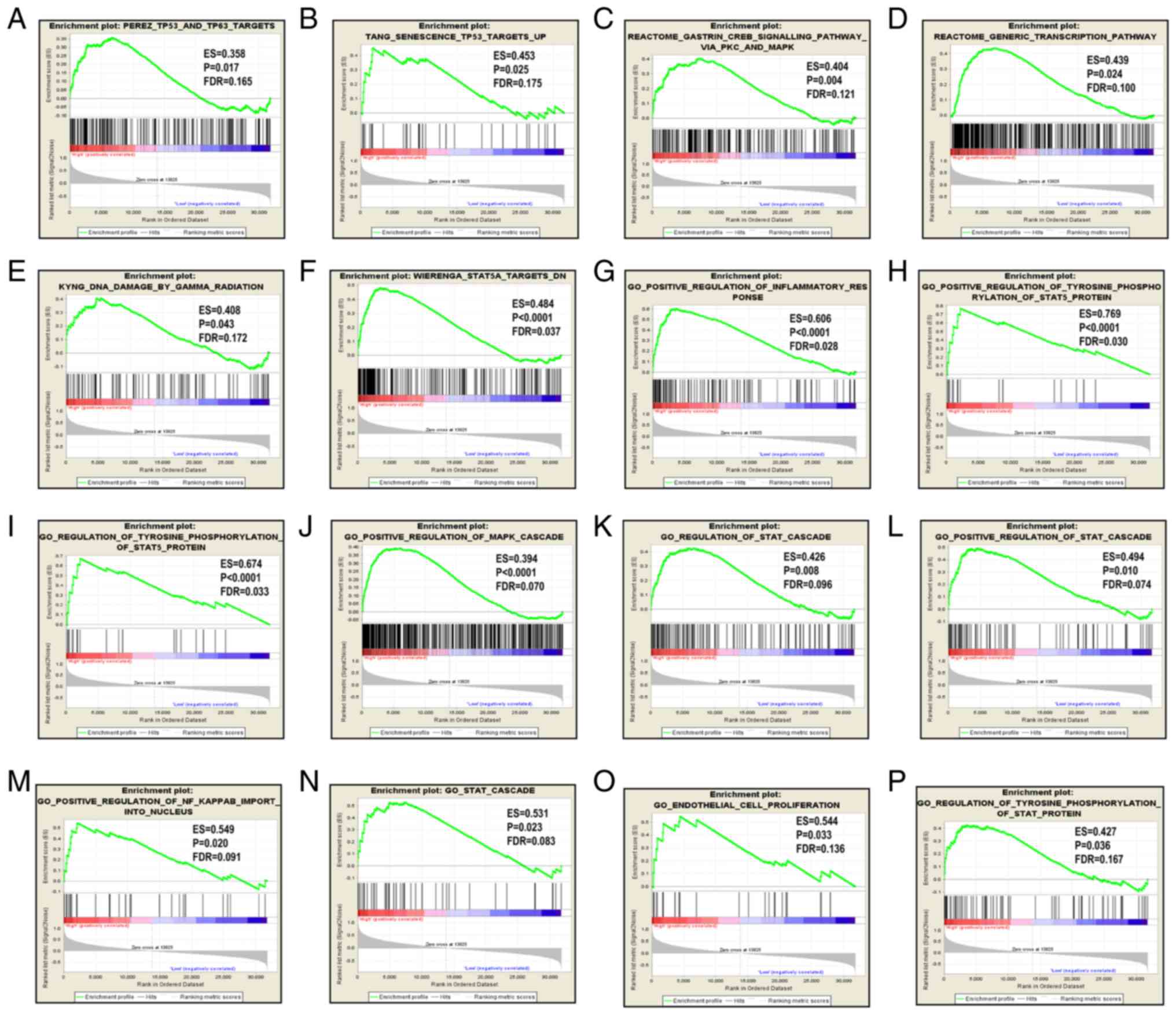
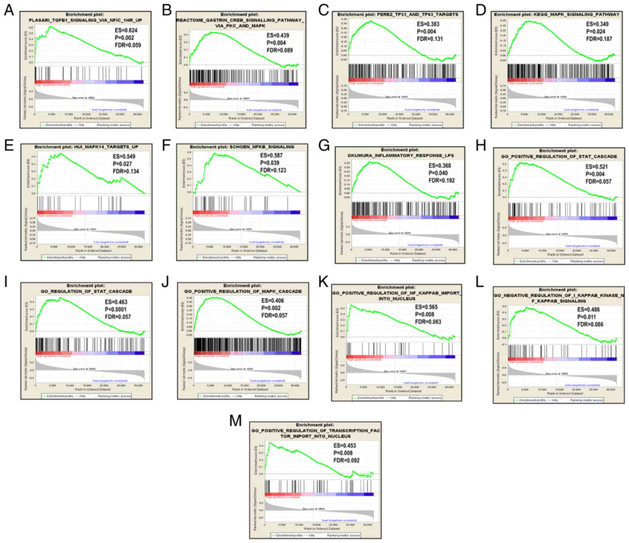
GSEA
Since CCR5, CCR6 and CCR9 were favorably associated
with OS, the patients were stratified according to their respective
median expression values. GSEA results are displayed in Figs. 5–7.
Analysis of the C2 (curated) gene sets revealed that CCR5 was
enriched in TP53 target, TP63 target, MAPK signaling pathway,
generic transcription pathway, DNA damage and STAT5A target
(Table SIII, Fig. 5A-F); in the C5 (GO) gene sets, CCR5
was enriched in inflammatory response, STAT cascade, MAPK cascade,
regulation of NF-κB and endothelial proliferation (Table SIII, Fig.
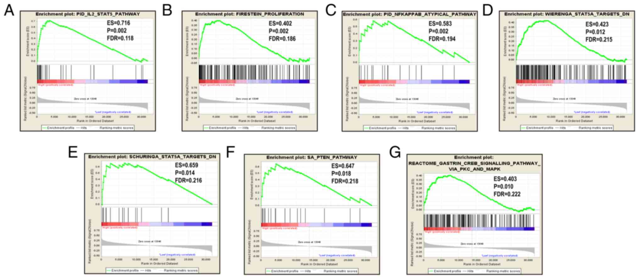
5G-P); CCR6 was enriched in TGF-β1 signaling pathway, TP53 and
TP63 targets, KEGG MAPK signaling pathway, MAPK14 targets and NF-κB
signaling in the C2 set (Table SIV
and Fig. 6A-G), and in STAT cascade,
MAPK cascade, NF-κB import into nucleus, NF-κB signaling and
transcription factor importing into nucleus in the C5 set (Table SV, Fig.
6H-M); CCR9 was enriched in the IL-2/STAT5 pathway,
proliferation, NF-κB atypical pathway, STAT 5 targets and PTEN
pathway in the C2 set (Table SVI and
Fig. 7A-G).
Discussion
Chemokines and chemokine receptors serve critical
roles in oncogenesis and cancer progression via a number of complex
mechanisms. It was reported that the chemokines secreted by the
tumor, immune and stromal cells were able to initiate the
uncontrolled proliferation and metastasis of tumor cells in an
autocrine and paracrine manner, by binding to their cognate
receptors (49). To date, CXCR4 is
the most widely studied and clearly understood chemokine receptor
associated with cancer, and was revealed to be involved in the
development, growth, invasion, angiogenesis and metastasis of
pancreatic cancer in a number of previous studies (50–57).
However, a limited number of studies have investigated the role of
the CCR gene in PDAC; therefore, the present study primarily
focused on the CCR gene family in PDAC.
Chemokine receptors have been reported to impact
tumor progression by regulating the MAPK/ERK, JAK/STAT and NF-κB
signaling pathways (49,58–60); CCR5
upregulated c-Fos in tumor cells by stimulating the JAK/STAT
pathway (60), and CCR5 stimulation
by CCL5 restricted the proliferation of breast cancer cells by
increasing p53 transcription via the JAK2 and p38-MAPK pathways
(61). Further studies have also
illustrated the anti-invasive and anti-metastatic roles of CCR5 in
mouse models of breast cancer (62,63).
However, a contradictory study revealed that the CCR5-Δ53
polymorphism was associated with a greater risk of developing
gallbladder cancer (64),
highlighting that the role of CCR5 may be cancer type-dependent. In
the present study, bioinformatics analysis suggested that CCR genes
were primarily involved in immune and inflammatory responses, and
also revealed that the JAK/STAT, MAPK and NF-κB signaling pathways
are involved in chemokine signaling. These pathways are consistent
with the downstream pathways regulated by p53, therefore, it was
hypothesized that CCR5 may also serve a role in PDAC by activating
p53. Furthermore, CCR3, CCR4, CCR5 and CCR8 were also found to be
associated with viral carcinogenesis, a greater number of CCR genes
than those identified to be concerned with carcinogenesis in
previous studies (23,65–68). Since
various studies have shown its close association with inflammation
and immunity (69–75), this is highly relevant to PDAC. Also,
considering the function of CCR genes in mediating inflammation,
and that CCR5, CCR6 and CCR9 were significantly associated with the
overall survival of patients with early-stage PDAC (as indicated by
the results of survival analysis), it was concluded that CCR5, CCR6
and CCR9 serve important roles in the development and progression
of PDAC. Furthermore, the results of the bioinformatics and
survival analysis of CCR genes in PDAC also verified previous
findings of the role of CCR genes in cancer (23,35,61,76,77).
The present study is believed to be the first to
show that high expression levels of CCR5, CCR6 and CCR9 are
associated with prolonged overall survival in patients with
early-stage PDAC. The role of CCR5 as a protective factor in PDAC
is in agreement with previous studies; it was reported that
knocking out CCR5 in pancreatic tumor-bearing mice reduced the
infiltration and subsequent cytotoxicity of NK cell in tumors
(78). Furthermore, smokers carrying
a CCR5 mutant allele have a significantly higher risk of developing
pancreatic cancer (76). Moreover,
the protective role of CCR5 has also been reported in other
malignancies. The CCR5 superagonist 1P7 was found to act as an
adjuvant to anti-tumor DNA vaccination by inducing specific CD8+
T-cell responses (77), and the
CCR5-Δ53 polymorphism was discovered to be associated with
susceptibility to breast cancer in the Indian population (35). It has also been noted that in breast
cancer, the absence of CCR5 on the tumor cell surface may promote
the proliferation of tumor cells which carry wild-type p53, but not
those with mutated p53 (61). CCR5
may also serve a role in PDAC via its indirect impact on tumor
cells, such as regulating the anti-tumor immune response. CCR5 is
also involved in the chemotaxis of activated naive T cells and
T-cells homing (79,80).
Existing studies of CCR6 expression in pancreatic
cancer are ambiguous; while one study reported higher levels of
CCR6 expression in the pancreatic tumor relative to the adjacent
healthy tissues (81), another showed
lower expression levels in pancreatic cancer cell lines than normal
pancreatic cells (82). Due to the
limited number of studies surrounding CCR6 and CCR9 in pancreatic
cancer, it was not possible to support the present findings of
these CCRs. Therefore, it was surmised that CCR6 and CCR9 may
modulate other tumor suppressor genes to inhibit tumor progression,
in the same manner as the CCR5-mediated activation of TP53
(61,83,84). This
was supported by the GSEA results of the present study, which
suggested that CCR6 was enriched in the p53 and STAT cascade, and
that CCR9 was enriched in the STAT cascade and NF-κB signaling
pathway. However, further studies are required to elucidate the
exact mechanisms involved.
The present study possessed various limitations.
Firstly, the sample size was relatively small, which may have led
to false negative results. Secondly, since the clinical information
of a number of patients was incomplete, the clinical variables used
for adjustment were not comprehensive. Thirdly, the relationship
between CCR and prognosis was only explored at the transcriptional
level. Nevertheless, not only was a novel association between the
CCR genes and the prognosis of early-stage PDAC discovered, but
also the potential molecular mechanisms. Further studies are
required to validate these findings and to establish CCRs as
therapeutic targets for PDAC.
Though there were several limitations to this
investigation, the present study was the first to reveal the
association between the CCR genes and the prognosis of early-stage
PDAC. In addition, GSEA was used to identify the potential
molecular mechanisms of CCR genes that may impact the prognosis of
patients with early-stage PDAC. With subsequent studies to verify
these findings, CCR genes may become novel targets for the
treatment of PDAC.
In conclusion, CCR5, CCR6 and CCR9 represent
potential prognostic biomarkers for patients with early-stage PDAC,
and are involved in signaling pathways such as those of p53, NF-κB,
generic transcription, MAPK and the STAT cascade.
Supplementary Material
Supporting Data
Supporting Data
Supporting Data
Supporting Data
Supporting Data
Supporting Data
Supporting Data
Supporting Data
Acknowledgements
Not applicable.
Funding
The present study was partly supported by the
National Natural Science Foundation of China (grant no. 81560535,
81802874, 81072321, 30760243, 30460143 and 30560133), the Natural
Science Foundation of Guangxi Province of China (grant no.
2018GXNSFBA138013 and 2018GXNSFAA050119), the 2009 Program for New
Century Excellent Talents in University, Guangxi Natural Sciences
Foundation (grant no. GuiKeGong 1104003A-7) and the Guangxi Health
Ministry Medicine Grant (Key-Scientific Research-grant no.
Z201018). The present study was also supported by the Scientific
Research Fund of the Health and Family Planning Commission of
Guangxi Zhuang Autonomous Region (grant no. Z2016318), the Key
laboratory of High-Incidence-Tumor Prevention and Treatment
(Guangxi Medical University), Ministry of Education (grant no.
GKE2018-01), the Guangxi Key R and D Program (grant no.
GKEAB18221019), the Basic Ability Improvement Project for
Middle-aged and Young Teachers in Colleges and Universities in
Guangxi (grant no. 2018KY0110), the Innovation Project of Guangxi
Graduate Education (grant no. JGY2018037) and the 2018 Innovation
Project of Guangxi Graduate Education (grant no. YCBZ2018036).
Availability of data and materials
The datasets used and/or analyzed during the present
study are available from the corresponding author on reasonable
request.
Authors' contributions
XZ and TP conceived and designed the study; XZ, XL,
XW and KH acquired and processed the raw data. CY, TY, JL, CH, GZ,
HS, WQ, QH, ZL, JH, YG, XY, ZC and TP performed the data analysis.
XZ wrote the manuscript, and Tp guided and supervised the
manuscript writing. All authors read and approved the final
manuscript.
Ethics approval and consent to
participate
Not applicable.
Patient consent for publication
Not applicable.
Competing interests
The authors declare that they have no competing
interests.
References
|
1
|
Bray F, Ferlay J, Soerjomataram I, Siegel
RL, Torre LA and Jemal A: Global cancer statistics 2018: GLOBOCAN
estimates of incidence and mortality worldwide for 36 cancers in
185 countries. CA Cancer J Clin. 68:394–424. 2018. View Article : Google Scholar : PubMed/NCBI
|
|
2
|
Ferlay J, Soerjomataram I, Dikshit R, Eser
S, Mathers C, Rebelo M, Parkin DM, Forman D and Bray F: Cancer
incidence and mortality worldwide: Sources, methods and major
patterns in GLOBOCAN 2012. Int J Cancer. 136:E359–E386. 2015.
View Article : Google Scholar : PubMed/NCBI
|
|
3
|
Quaresma M, Coleman MP and Rachet B:
40-year trends in an index of survival for all cancers combined and
survival adjusted for age and sex for each cancer in England and
Wales, 1971–2011: A population-based study. Lancet. 385:1206–1218.
2015. View Article : Google Scholar : PubMed/NCBI
|
|
4
|
Miller KD, Siegel RL, Lin CC, Mariotto AB,
Kramer JL, Rowland JH, Stein KD, Alteri R and Jemal A: Cancer
treatment and survivorship statistics, 2016. CA Cancer J Clin.
66:271–289. 2016. View Article : Google Scholar : PubMed/NCBI
|
|
5
|
Hidalgo M, Cascinu S, Kleeff J, Labianca
R, Lohr JM, Neoptolemos J, Real FX, Van Laethem JL and Heinemann V:
Addressing the challenges of pancreatic cancer: Future directions
for improving outcomes. Pancreatology. 15:8–18. 2015. View Article : Google Scholar : PubMed/NCBI
|
|
6
|
Siegel RL, Miller KD and Jemal A: Cancer
statistics, 2017. CA Cancer J Clin. 67:7–30. 2017. View Article : Google Scholar : PubMed/NCBI
|
|
7
|
Lin QJ, Yang F, Jin C and Fu DL: Current
status and progress of pancreatic cancer in China. World J
Gastroentero. 21:7988–8003. 2015. View Article : Google Scholar
|
|
8
|
Chen W, Zheng R, Baade PD, Zhang S, Zeng
H, Bray F, Jemal A, Yu XQ and He J: Cancer statistics in China,
2015. CA Cancer J Clin. 66:115–132. 2016. View Article : Google Scholar : PubMed/NCBI
|
|
9
|
Kamisawa T, Wood LD, Itoi T and Takaori K:
Pancreatic cancer. Lancet. 388:73–85. 2016. View Article : Google Scholar : PubMed/NCBI
|
|
10
|
Heinemann V, Boeck S, Hinke A, Labianca R
and Louvet C: Meta-analysis of randomized trials: Evaluation of
benefit from gemcitabine-based combination chemotherapy applied in
advanced pancreatic cancer. BMC Cancer. 8:822008. View Article : Google Scholar : PubMed/NCBI
|
|
11
|
Jemal A, Siegel R, Ward E, Hao Y, Xu J and
Thun MJ: Cancer statistics, 2009. CA Cancer J Clin. 59:225–249.
2009. View Article : Google Scholar : PubMed/NCBI
|
|
12
|
Vincent A, Herman J, Schulick R, Hruban RH
and Goggins M: Pancreatic cancer. Lancet. 378:607–620. 2011.
View Article : Google Scholar : PubMed/NCBI
|
|
13
|
Conroy T, Desseigne F, Ychou M, Bouche O,
Guimbaud R, Becouarn Y, Adenis A, Raoul JL, Gourgou-Bourgade S, de
la Fouchardiere C, et al: FOLFIRINOX versus gemcitabine for
metastatic pancreatic cancer. N Engl J Med. 364:1817–1825. 2011.
View Article : Google Scholar : PubMed/NCBI
|
|
14
|
Mohammed S, Van Buren G II and Fisher WE:
Pancreatic cancer: Advances in treatment. World J Gastroenterol.
20:9354–9360. 2014.PubMed/NCBI
|
|
15
|
Okazaki T, Chikuma S, Iwai Y, Fagarasan S
and Honjo T: A rheostat for immune responses: The unique properties
of PD-1 and their advantages for clinical application. Nat Immunol.
14:1212–1218. 2013. View
Article : Google Scholar : PubMed/NCBI
|
|
16
|
Wei SC, Duffy CR and Allison JP:
Fundamental mechanisms of immune checkpoint blockade therapy.
Cancer Discov. 8:1069–1086. 2018. View Article : Google Scholar : PubMed/NCBI
|
|
17
|
Vassaux G, Angelova A, Baril P, Midoux P,
Rommelaere J and Cordelier P: The promise of gene therapy for
pancreatic cancer. Hum Gene Ther. 27:127–133. 2016. View Article : Google Scholar : PubMed/NCBI
|
|
18
|
Mantovani A: The chemokine system:
Redundancy for robust outputs. Immunol Today. 20:254–257. 1999.
View Article : Google Scholar : PubMed/NCBI
|
|
19
|
Moser B and Willimann K: Chemokines: Role
in inflammation and immune surveillance. Ann Rheum Dis. 63 (Suppl
2):ii84–ii89. 2004. View Article : Google Scholar : PubMed/NCBI
|
|
20
|
Zabel BA, Zuniga L, Ohyama T, Allen SJ,
Cichy J, Handel TM and Butcher EC: Chemoattractants, extracellular
proteases, and the integrated host defense response. Exp Hematol.
34:1021–1032. 2006. View Article : Google Scholar : PubMed/NCBI
|
|
21
|
Van Damme J, Proost P, Lenaerts JP and
Opdenakker G: Structural and functional identification of two
human, tumor-derived monocyte chemotactic proteins (MCP-2 and
MCP-3) belonging to the chemokine family. J Exp Med. 176:59–65.
1992. View Article : Google Scholar : PubMed/NCBI
|
|
22
|
Fujiwara H and Hamaoka T: Coordination of
chemokine and adhesion systems in intratumoral T cell migration
responsible for the induction of tumor regression. Int
Immunopharmacol. 1:613–623. 2001. View Article : Google Scholar : PubMed/NCBI
|
|
23
|
Lacalle RA, Blanco R, Carmona-Rodriguez L,
Martin-Leal A, Mira E and Manes S: Chemokine receptor signaling and
the hallmarks of cancer. Int Rev Cell Mol Biol. 331:181–244. 2017.
View Article : Google Scholar : PubMed/NCBI
|
|
24
|
Dite P, Hermanova M, Trna J, Novotny I,
Ruzicka M, Liberda M and Bartkova A: The role of chronic
inflammation: Chronic pancreatitis as a risk factor of pancreatic
cancer. Dig Dis. 30:277–283. 2012. View Article : Google Scholar : PubMed/NCBI
|
|
25
|
Ibrahimi S, Mukherjee S, Alhyari L, Rubin
E and Aljumaily R: Spontaneous regression of metastatic pancreatic
cancer: A role for recurrent inflammation. Pancreas. 48:e4–e6.
2019. View Article : Google Scholar : PubMed/NCBI
|
|
26
|
Incio J, Liu H, Suboj P, Chin SM, Chen IX,
Pinter M, Ng MR, Nia HT, Grahovac J, Kao S, et al: Obesity-Induced
inflammation and desmoplasia promote pancreatic cancer progression
and resistance to chemotherapy. Cancer Discov. 6:852–869. 2016.
View Article : Google Scholar : PubMed/NCBI
|
|
27
|
Hayes JB, Sircy LM, Heusinkveld LE, Ding
W, Leander RN, McClelland EE and Nelson DE: Modulation of
macrophage inflammatory nuclear Factor κB (NF-κB) signaling by
intracellular cryptococcus neoformans. J Biol Chem.
291:15614–15627. 2016. View Article : Google Scholar : PubMed/NCBI
|
|
28
|
Lu C, Paschall AV, Shi H, Savage N, Waller
JL, Sabbatini ME, Oberlies NH, Pearce C and Liu K: The MLL1-H3K4me3
Axis-Mediated PD-L1 expression and pancreatic cancer immune
evasion. J Natl Cancer Inst. 1092017.doi: 10.1093/jnci/djw283.
|
|
29
|
Cui K, Zou H, Shi M, Ou Y, Han L, Zhang B,
Hu D and Li S: Gene expression profiles in chemokine (C-C Motif)
Ligand 21-Overexpressing pancreatic cancer cells. Pathol Oncol Res.
Apr 23–2018.doi: 10.1007/s12253-018-0390-z (Epub ahead of print).
View Article : Google Scholar
|
|
30
|
Hutchinson L: Pancreatic cancer:
Disrupting the chemokine axis in PDAC. Nat Rev Clin Oncol.
13:3302016. View Article : Google Scholar : PubMed/NCBI
|
|
31
|
Marchesi F, Grizzi F, Laghi L, Mantovani A
and Allavena P: Molecular mechanisms of pancreatic cancer
dissemination: The role of the chemokine system. Curr Pharm Des.
18:2432–2438. 2012. View Article : Google Scholar : PubMed/NCBI
|
|
32
|
Nakata B, Fukunaga S, Noda E, Amano R,
Yamada N and Hirakawa K: Chemokine receptor CCR7 expression
correlates with lymph node metastasis in pancreatic cancer.
Oncology. 74:69–75. 2008. View Article : Google Scholar : PubMed/NCBI
|
|
33
|
Feig C, Jones JO, Kraman M, Wells RJ,
Deonarine A, Chan DS, Connell CM, Roberts EW, Zhao Q, Caballero OL,
et al: Targeting CXCL12 from FAP-expressing carcinoma-associated
fibroblasts synergizes with anti-PD-L1 immunotherapy in pancreatic
cancer. Proc Natl Acad Sci USA. 110:20212–20217. 2013. View Article : Google Scholar : PubMed/NCBI
|
|
34
|
Gebauer F, Tachezy M, Effenberger K, von
Loga K, Zander H, Marx A, Kaifi JT, Sauter G, Izbicki JR and
Bockhorn M: Prognostic impact of CXCR4 and CXCR7 expression in
pancreatic adenocarcinoma. J Surg Oncol. 104:140–145. 2011.
View Article : Google Scholar : PubMed/NCBI
|
|
35
|
Lee YH and Song GG: Association between
chemokine receptor 5 delta32 polymorphism and susceptibility to
cancer: A meta-analysis. J Recept Signal Transduct Res. 35:509–515.
2015. View Article : Google Scholar : PubMed/NCBI
|
|
36
|
Anders S and Huber W: Differential
expression analysis for sequence count data. Genome Biol.
11:R1062010. View Article : Google Scholar : PubMed/NCBI
|
|
37
|
Yip SH, Wang P, Kocher JA, Sham PC and
Wang J: Linnorm: Improved statistical analysis for single cell
RNA-seq expression data. Nucleic Acids Res. 45:e1792017. View Article : Google Scholar : PubMed/NCBI
|
|
38
|
Huang DW, Sherman BT, Tan Q, Kir J, Liu D,
Bryant D, Guo Y, Stephens R, Baseler MW, Lane HC and Lempicki RA:
DAVID Bioinformatics Resources: Expanded annotation database and
novel algorithms to better extract biology from large gene lists.
Nucleic Acids Res. 35:W169–W175. 2007. View Article : Google Scholar : PubMed/NCBI
|
|
39
|
Szklarczyk D, Franceschini A, Wyder S,
Forslund K, Heller D, Huerta-Cepas J, Simonovic M, Roth A, Santos
A, Tsafou KP, et al: STRING v10: Protein-protein interaction
networks, integrated over the tree of life. Nucleic Acids Res 43
(Database Issue). D447–D452. 2015. View Article : Google Scholar
|
|
40
|
Montojo J, Zuberi K, Rodriguez H, Bader GD
and Morris Q: GeneMANIA: Fast gene network construction and
function prediction for Cytoscape. F1000Res. 3:1532014. View Article : Google Scholar : PubMed/NCBI
|
|
41
|
Liao X, Huang K, Huang R, Liu X, Han C, Yu
L, Yu T, Yang C, Wang X and Peng T: Genome-scale analysis to
identify prognostic markers in patients with early-stage pancreatic
ductal adenocarcinoma after pancreaticoduodenectomy. Onco Targets
Ther. 10:4493–4506. 2017. View Article : Google Scholar : PubMed/NCBI
|
|
42
|
Liao X, Zhu G, Huang R, Yang C, Wang X,
Huang K, Yu T, Han C, Su H and Peng T: Identification of potential
prognostic microRNA biomarkers for predicting survival in patients
with hepatocellular carcinoma. Cancer Manag Res. 10:787–803. 2018.
View Article : Google Scholar : PubMed/NCBI
|
|
43
|
Heagerty PJ and Zheng Y: Survival model
predictive accuracy and ROC curves. Biometrics. 61:92–105. 2005.
View Article : Google Scholar : PubMed/NCBI
|
|
44
|
Subramanian A, Tamayo P, Mootha VK,
Mukherjee S, Ebert BL, Gillette MA, Paulovich A, Pomeroy SL, Golub
TR, Lander ES and Mesirov JP: Gene set enrichment analysis: A
knowledge-based approach for interpreting genome-wide expression
profiles. Proc Natl Acad Sci USA. 102:15545–15550. 2005. View Article : Google Scholar : PubMed/NCBI
|
|
45
|
Liberzon A, Birger C, Thorvaldsdottir H,
Ghandi M, Mesirov JP and Tamayo P: The molecular signatures
database (MSigDB) hallmark gene set collection. Cell Syst.
1:417–425. 2015. View Article : Google Scholar : PubMed/NCBI
|
|
46
|
Francois O, Martins H, Caye K and
Schoville SD: Controlling false discoveries in genome scans for
selection. Mol Ecol. 25:454–469. 2016. View Article : Google Scholar : PubMed/NCBI
|
|
47
|
Glickman ME, Rao SR and Schultz MR: False
discovery rate control is a recommended alternative to
Bonferroni-type adjustments in health studies. J Clin Epidemiol.
67:850–857. 2014. View Article : Google Scholar : PubMed/NCBI
|
|
48
|
Reiner A, Yekutieli D and Benjamini Y:
Identifying differentially expressed genes using false discovery
rate controlling procedures. Bioinformatics. 19:368–375. 2003.
View Article : Google Scholar : PubMed/NCBI
|
|
49
|
Sarvaiya PJ, Guo D, Ulasov I, Gabikian P
and Lesniak MS: Chemokines in tumor progression and metastasis.
Oncotarget. 4:2171–2185. 2013. View Article : Google Scholar : PubMed/NCBI
|
|
50
|
Xiao G, Wang X and Yu Y: CXCR4/Let-7a Axis
regulates metastasis and chemoresistance of pancreatic cancer cells
through targeting HMGA2. Cell Physiol Biochem. 43:840–851. 2017.
View Article : Google Scholar : PubMed/NCBI
|
|
51
|
Gao Z, Wang X, Wu K, Zhao Y and Hu G:
Pancreatic stellate cells increase the invasion of human pancreatic
cancer cells through the stromal cell-derived factor-1/CXCR4 axis.
Pancreatology. 10:186–193. 2010. View Article : Google Scholar : PubMed/NCBI
|
|
52
|
Shakir M, Tang D, Zeh HJ, Tang SW,
Anderson CJ, Bahary N and Lotze MT: The chemokine receptors
CXCR4/CXCR7 and their primary heterodimeric ligands CXCL12 and
CXCL12/high mobility group box 1 in pancreatic cancer growth and
development: Finding flow. Pancreas. 44:528–534. 2015. View Article : Google Scholar : PubMed/NCBI
|
|
53
|
Zhang J, Liu C, Mo X, Shi H and Li S:
Mechanisms by which CXCR4/CXCL12 cause metastatic behavior in
pancreatic cancer. Oncol Lett. 15:1771–1776. 2018.PubMed/NCBI
|
|
54
|
Wang J, Wang H, Cai J, Du S, Xin B, Wei W,
Zhang T and Shen X: Artemin regulates CXCR4 expression to induce
migration and invasion in pancreatic cancer cells through
activation of NF-κB signaling. Exp Cell Res. 365:12–23. 2018.
View Article : Google Scholar : PubMed/NCBI
|
|
55
|
Sleightholm RL, Neilsen BK, Li J, Steele
MM, Singh RK, Hollingsworth MA and Oupicky D: Emerging roles of the
CXCL12/CXCR4 axis in pancreatic cancer progression and therapy.
Pharmacol Ther. 179:158–170. 2017. View Article : Google Scholar : PubMed/NCBI
|
|
56
|
Little EC, Kubic JD, Salgia R, Grippo PJ
and Lang D: Canonical and alternative transcript expression of PAX6
and CXCR4 in pancreatic cancer. Oncol Lett. 13:4027–4034. 2017.
View Article : Google Scholar : PubMed/NCBI
|
|
57
|
Aravindan S, Ramraj S, Kandasamy K,
Thirugnanasambandan SS, Somasundaram DB, Herman TS and Aravindan N:
Hormophysa triquerta polyphenol, an elixir that deters CXCR4- and
COX2-dependent dissemination destiny of treatment-resistant
pancreatic cancer cells. Oncotarget. 8:5717–5734. 2017. View Article : Google Scholar : PubMed/NCBI
|
|
58
|
Chakraborty K, Bose A, Chakraborty T,
Sarkar K, Goswami S, Pal S and Baral R: Restoration of dysregulated
CC chemokine signaling for monocyte/macrophage chemotaxis in head
and neck squamous cell carcinoma patients by neem leaf glycoprotein
maximizes tumor cell cytotoxicity. Cell Mol Immunol. 7:396–408.
2010. View Article : Google Scholar : PubMed/NCBI
|
|
59
|
Jo H, Zhang R, Zhang H, McKinsey TA, Shao
J, Beauchamp RD, Ballard DW and Liang P: NF-kappa B is required for
H-ras oncogene induced abnormal cell proliferation and
tumorigenesis. Oncogene. 19:841–849. 2000. View Article : Google Scholar : PubMed/NCBI
|
|
60
|
Wong M and Fish EN: RANTES and MIP-1alpha
activate stats in T cells. J Biol Chem. 273:309–314. 1998.
View Article : Google Scholar : PubMed/NCBI
|
|
61
|
Manes S, Mira E, Colomer R, Montero S,
Real LM, Gomez-Mouton C, Jimenez-Baranda S, Garzon A, Lacalle RA,
Harshman K, et al: CCR5 expression influences the progression of
human breast cancer in a p53-dependent manner. J Exp Med.
198:1381–1389. 2003. View Article : Google Scholar : PubMed/NCBI
|
|
62
|
Datar I, Qiu X, Ma HZ, Yeung M, Aras S, de
la Serna I, Al-Mulla F, Tan TZ, Thiery JP, Trumbly R, et al:
Correction: RKIP regulates CCL5 expression to inhibit breast cancer
invasion and metastasis by controlling macrophage infiltration.
Oncotarget. 7:269252016. View Article : Google Scholar : PubMed/NCBI
|
|
63
|
Velasco-Velazquez M and Pestell RG: The
CCL5/CCR5 axis promotes metastasis in basal breast cancer.
Oncoimmunology. 2:e236602013. View Article : Google Scholar : PubMed/NCBI
|
|
64
|
Srivastava A, Pandey SN, Choudhuri G and
Mittal B: CCR5 Delta32 polymorphism: Associated with gallbladder
cancer susceptibility. Scand J Immunol. 67:516–522. 2008.
View Article : Google Scholar : PubMed/NCBI
|
|
65
|
Li K, Xu B, Xu G and Liu R: CCR7 regulates
Twist to induce the epithelial-mesenchymal transition in pancreatic
ductal adenocarcinoma. Tumour Biol. 37:419–424. 2016. View Article : Google Scholar : PubMed/NCBI
|
|
66
|
Feng R, Morine Y, Ikemoto T, Imura S,
Iwahashi S, Saito Y and Shimada M: Nab-paclitaxel interrupts
cancer-stromal interaction through C-X-C motif chemokine
10-mediated interleukin-6 downregulation in vitro. Cancer Sci.
109:2509–2519. 2018. View Article : Google Scholar : PubMed/NCBI
|
|
67
|
Lanca T, Costa MF, Goncalves-Sousa N, Rei
M, Grosso AR, Penido C and Silva-Santos B: Protective role of the
inflammatory CCR2/CCL2 chemokine pathway through recruitment of
type 1 cytotoxic γδ T lymphocytes to tumor beds. J Immunol.
190:6673–6680. 2013. View Article : Google Scholar : PubMed/NCBI
|
|
68
|
Gonzalez-Arriagada WA, Lozano-Burgos C,
Zuniga-Moreta R, Gonzalez-Diaz P and Coletta RD:
Clinicopathological significance of chemokine receptor (CCR1, CCR3,
CCR4, CCR5, CCR7 and CXCR4) expression in head and neck squamous
cell carcinomas. J Oral Pathol Med. 47:755–763. 2018. View Article : Google Scholar : PubMed/NCBI
|
|
69
|
Farrow B and Evers BM: Inflammation and
the development of pancreatic cancer. Surg Oncol. 10:153–169. 2002.
View Article : Google Scholar : PubMed/NCBI
|
|
70
|
Greer JB and Whitcomb DC: Inflammation and
pancreatic cancer: An evidence-based review. Curr Opin Pharmacol.
9:411–418. 2009. View Article : Google Scholar : PubMed/NCBI
|
|
71
|
Hausmann S, Kong B, Michalski C, Erkan M
and Friess H: The role of inflammation in pancreatic cancer. Adv
Exp Med Biol. 816:129–151. 2014. View Article : Google Scholar : PubMed/NCBI
|
|
72
|
McKay CJ, Glen P and McMillan DC: Chronic
inflammation and pancreatic cancer. Best Pract Res Clin
Gastroenterol. 22:65–73. 2008. View Article : Google Scholar : PubMed/NCBI
|
|
73
|
Momi N, Kaur S, Krishn SR and Batra SK:
Discovering the route from inflammation to pancreatic cancer.
Minerva Gastroenterol Dietol. 58:283–297. 2012.PubMed/NCBI
|
|
74
|
Padoan A, Plebani M and Basso D:
Inflammation and pancreatic cancer: Focus on metabolism, cytokines,
and immunity. Int J Mol Sci. 20(pii): E6762019. View Article : Google Scholar : PubMed/NCBI
|
|
75
|
Shadhu K and Xi C: Inflammation and
pancreatic cancer: An updated review. Saudi J Gastroenterol.
25:3–13. 2019.PubMed/NCBI
|
|
76
|
Duell EJ, Casella DP, Burk RD, Kelsey KT
and Holly EA: Inflammation, genetic polymorphisms in
proinflammatory genes TNF-A, RANTES, and CCR5, and risk of
pancreatic adenocarcinoma. Cancer Epidemiol Biomarkers Prev.
15:726–731. 2006. View Article : Google Scholar : PubMed/NCBI
|
|
77
|
Dorgham K, Abadie V, Iga M, Hartley O,
Gorochov G and Combadiere B: Engineered CCR5 superagonist chemokine
as adjuvant in anti-tumor DNA vaccination. Vaccine. 26:3252–3260.
2008. View Article : Google Scholar : PubMed/NCBI
|
|
78
|
Song Y, Gan Y, Wang Q, Meng Z, Li G, Shen
Y, Wu Y, Li P, Yao M, Gu J and Tu H: Enriching the housing
environment for mice enhances their Nk cell antitumor immunity via
sympathetic Nerve-Dependent regulation of NKG2D and CCR5. Cancer
Res. 77:1611–1622. 2017. View Article : Google Scholar : PubMed/NCBI
|
|
79
|
Taub DD, Turcovski-Corrales SM, Key ML,
Longo DL and Murphy WJ: Chemokines and T lymphocyte activation: I.
Beta chemokines costimulate human T lymphocyte activation in vitro.
J Immunol. 156:2095–2103. 1996.PubMed/NCBI
|
|
80
|
Tan MC, Goedegebuure PS, Belt BA, Flaherty
B, Sankpal N, Gillanders WE, Eberlein TJ, Hsieh CS and Linehan DC:
Disruption of CCR5-dependent homing of regulatory T cells inhibits
tumor growth in a murine model of pancreatic cancer. J Immunol.
182:1746–1755. 2009. View Article : Google Scholar : PubMed/NCBI
|
|
81
|
Rubie C, Frick VO, Ghadjar P, Wagner M,
Grimm H, Vicinus B, Justinger C, Graeber S and Schilling MK:
CCL20/CCR6 expression profile in pancreatic cancer. J Transl Med.
8:452010. View Article : Google Scholar : PubMed/NCBI
|
|
82
|
Masai K, Iwashita Y, Tominaga M, Hirano S,
Shibata K, Matsumoto T, Sasaki A, Ohta M and Kitano S: mRNA
expression of chemokine receptors in hepatic and pancreatic tumor
cell lines. Gan To Kagaku Ryoho. 31:1261–1263. 2004.(In Japanese).
PubMed/NCBI
|
|
83
|
Mehta SA, Christopherson KW,
Bhat-Nakshatri P, Goulet RJ Jr, Broxmeyer HE, Kopelovich L and
Nakshatri H: Negative regulation of chemokine receptor CXCR4 by
tumor suppressor p53 in breast cancer cells: Implications of p53
mutation or isoform expression on breast cancer cell invasion.
Oncogene. 26:3329–3337. 2007. View Article : Google Scholar : PubMed/NCBI
|
|
84
|
Shiraishi K, Fukuda S, Mori T, Matsuda K,
Yamaguchi T, Tanikawa C, Ogawa M, Nakamura Y and Arakawa H:
Identification of fractalkine, a CX3C-type chemokine, as a direct
target of p53. Cancer Res. 60:3722–3726. 2000.PubMed/NCBI
|