1. Introduction
Dentistry has undergone substantial technological
advancement, leading to the development of innovative diagnostic
and therapeutic approaches. Biophotonics has emerged as an
interdisciplinary field that integrates optical science with
biological systems to facilitate the detection, monitoring and
treatment of disease. Biophotonics technologies enable non-invasive
imaging, molecular characterization and targeted therapeutic
interventions at cellular and tissue levels. They provide
significant advantages over conventional approaches by providing
real-time information, enhanced precision and reduced patient
morbidity (1,2). Traditionally, oral diseases such as
caries, periodontitis, peri-implantitis, oral lichen planus and
oral cancer have been viewed primarily as a function of bacterial
plaque, the host immune response, and changes in the structure of
the enamel, dentin, periodontium and mucosa (1). Endodontic assistance primarily
involves a somewhat visual-tactile examination via visual-tactile
means, radiographs and histopathology or tissue examination;
however, these methods usually only identify the pathology after it
has progressed to an advanced stage. Although therapeutic
interventions may be effective, they often involve invasive
procedures, radiation exposure or a long period of recovery once an
individual has undergone treatment. Biophotonics achieves these
goals, however, by using the properties of light-matter
interactions to provide for real-time, specific molecular
(diagnosing and/or treating) therapies without radiation (3).
Using various photochemical, photothermal,
photomechanical and photoionization effects, rapid, sensitive,
specific, low-cost and non-invasive methods have been developed.
The development of biophotonics has its roots in Einstein's theory
of photons and the first ruby laser in 1960, which was introduced
into dentistry for early hard-tissue research in 1964(4). The challenge of thermal injury from
CO2 lasers was overcome by the introduction of
short-pulsed infrared systems using Er:YAG (2.94 µm) and Er,Cr:YSGG
(2.78 µm) laser configurations in the 1980s and 1990s, which
allowed for very precise ablation and minimal collateral damage
(5,6).
Recent advances in biophotonics have expanded the
scope of dental diagnostics and therapeutics by enabling the
improved visualization of tissue changes, enhanced microbial
detection and more targeted treatment strategies. The synergistic
association emerging between biophotonics and artificial
intelligence (AI), quantum-enhanced imaging and advanced
nanomaterials also enhances the likelihood that biophotonics will
enable predictive, personalized and preventative oral healthcare
solutions (7). While there have
been some established advances in biophotonics, it continues to
face a number of the same challenges that it has historically
faced, e.g., limited penetration depth into highly scattering
tissues, high costs associated with biophotonics devices,
biophotonics device dependency and regulatory barriers regarding
newly developed biophotonics devices (8). These deficiencies in future progress
emphasize the need for increased research, standardization and
translational efforts across the spectrum of oral healthcare,
particularly in economically disadvantaged communities/areas where
equal access is a priority.
The aim of the present narrative review was to
provide a broad overview through a discussion of the core ideas,
present-day practices, diagnosis and treatment methods, and the
clinical relevance. The present review seeks to connect current
science with future prospects in terms of progress in
photonics-based precision oral healthcare that will be
non-invasive, patient-centred and accessible to all.
2. Literature search methods
A well-structured search of the existing literature
was carried out across the PubMed, Scopus, Web of Science and
Google Scholar databases. The search strategy used combinations of
the following key words: Biophotonic dentistry, optical coherence
tomography (OCT), Biophotonic laser therapy for periodontitis,
photodynamic therapy (PDT) for oral cancer, Raman spectroscopy for
dental caries, nanobioscience/mechanics in Biophotonic dentistry,
AI in dental imaging, and nanomaterials (2000-2026). The search
results included clinical trials/observational
studies/molecular-level studies/reviews/market reports;
editorials/non-peer-reviewed materials/irrelevant articles were
excluded. The titles and abstracts were independently screened by
two authors (ST. and SUN), followed by full-text assessments. Data
extraction focused on biophotonic mechanisms, diagnostic
applications, therapeutic modalities, clinical outcomes, emerging
technologies and translational implications in dentistry.
3. Evolution and classification of
biophotonics
In 1964, biophotonics made their way into the field
of dentistry with hard tissue studies (4,6).
Early CO2 lasers caused the carbonization of hard
tissues and were replaced by short-pulsed infrared systems (Er:YAG
2.94 µm, Er,Cr:YSGG 2.78 µm) in the 1980s and early 1990s due to
the improved ability to perform precise ablative procedures
(5,6).
The modern classification system categorizes
biophotonics based on their methods of interaction (photochemical,
photothermal, photomechanical and photoionization) and on their
wavelength (visible=400-700 nm; near-infrared therapeutic window)
(3).
Biophotonics can be further divided into three
categories: Bioimaging, biosensing and photonic therapy, all of
which are particularly relevant to both periodontal and restorative
treatment in dentistry (1,2).
4. Biophotonic components, light-tissue
interactions and host responses
Biophotonic systems in dentistry employ light-based
technologies, such as lasers and light-emitting diodes (LEDs)
together with optical delivery and detection units to interact with
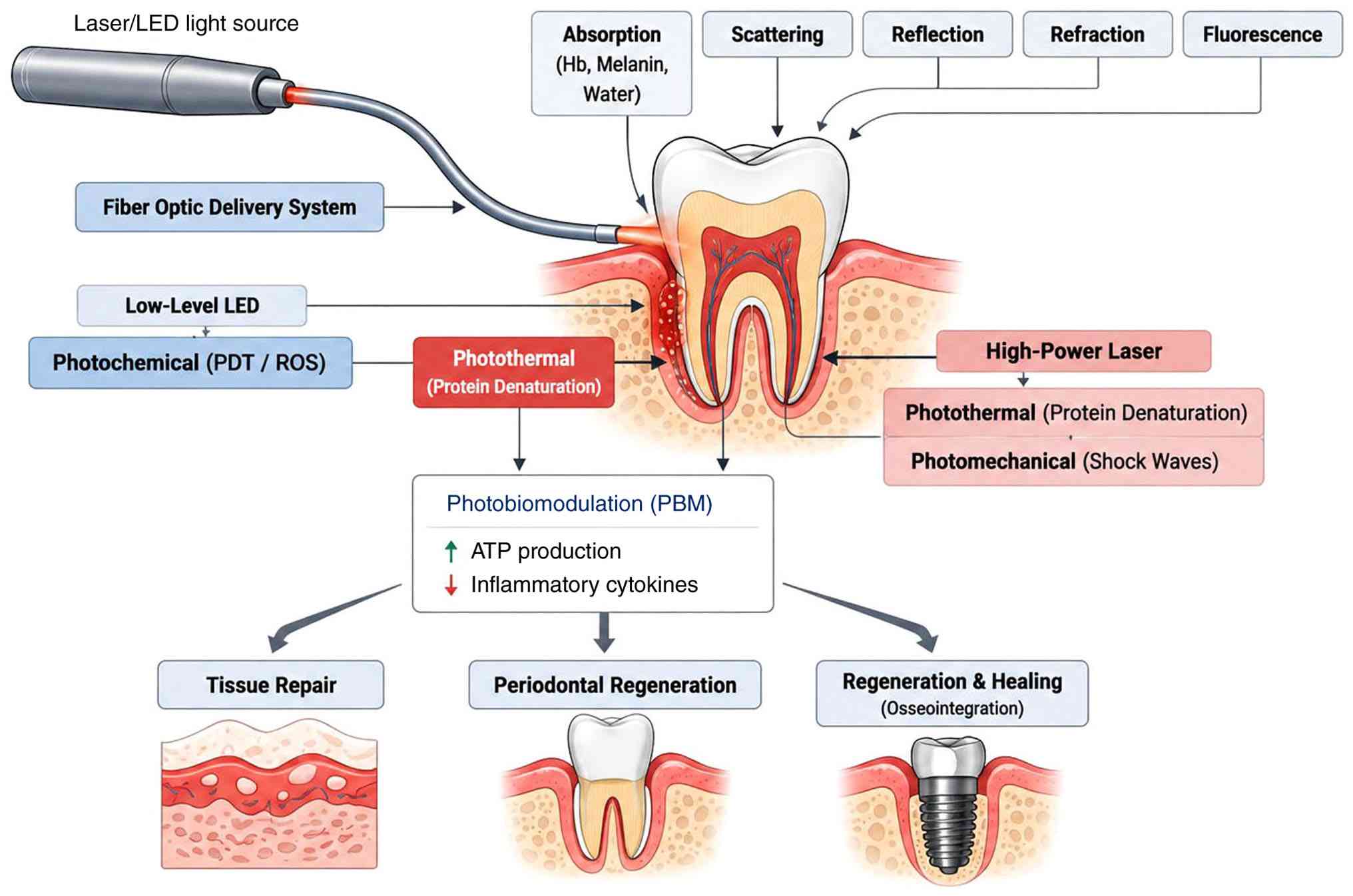
oral tissues (3). Following
exposure to biological tissues, light may undergo absorption,
scattering, reflection, or fluorescence depending on the optical
characteristics of the target site. These interactions produce
various photonic effects, including photochemical, photothermal,
photomechanical, and photoionization responses, which form the
basis of several modern diagnostic and therapeutic dental
applications (3,7).
As illustrated in Fig.
1, light-induced tissue interactions can trigger multiple
cellular and molecular responses, including reactive oxygen species
(ROS) formation, enhanced mitochondrial activity, fibroblast
proliferation, collagen remodelling and antimicrobial effects.
Collectively, these biological responses contribute to improved
wound healing, periodontal regeneration, osseointegration,
inflammation control and minimally invasive management of oral
diseases Fig. 1 illustrates the
fundamental principles of light-tissue interactions that underpin
biophotonic applications in dentistry. When light from lasers or
LEDs is transmitted to oral tissues by optical systems, it
undergoes absorption, scattering, reflection, and fluorescence
depending on composition of tissues and optical properties
(3). These interactions lead to
various biological effects, such as photochemical, photothermal,
photomechanical and photobiomodulatory, which form basis of several
diagnostic and therapeutic modalities (3,9).
Clinically, these mechanisms help in antimicrobial activity,
inflammation control, wound healing, periodontal regeneration and
enhanced osseointegration. Fig. 1
highlights how a single optical stimulus can generate distinct
biological responses depending on wavelength, energy density, and
tissue characteristics, thereby enabling diverse applications
across multiple dental specialties (9,10).
5. Biophotonic modalities: Mechanisms,
dental impli-cations and clinical translation
Clinically, dentistry is being transformed through
biophotonics by bringing cutting-edge light science into an
integrated system of detection, treatment and management of oral
diseases (7). Imaging techniques,
such as fluorescence imaging use autofluorescence to visualize
early caries, demineralization and oral cancer so that these
conditions can be managed with minimal invasiveness (11-13).
OCT provides ‘optical biopsy’ capabilities with a
resolution between 5 and 15 µm to diagnose internal defects, cracks
and bone loss (14). Molecular
specificity is achieved using Raman spectroscopy to analyse biofilm
and mineral content. Lasers (Er:YAG, diode, Nd:YAG, and
CO2) allow for precision ablation for soft tissue
surgery and decontamination with haemostasis. PDT generates ROS to
kill bacteria/viruses and to provide targeted anticancer therapy
(15).
Photobiomodulation (PBM) enhances mitochondrial
activity to accelerate healing, alleviate pain and facilitate
osseointegration (9). The majority
of these technologies will not be used in isolation; therefore,
multimodal combinations will become the norm (e.g.,
fluorescence-guided PDT and OCT-monitored laser therapy), as they
reflect care that is centred on the patient. A comparative overview
of major biophotonic modalities, including their mechanisms and
clinical implications, is presented in Table I. To contextualize the clinical
value of the modalities summarized in Tables I, II provides a comparative analysis with
traditional approaches commonly used in dentistry. This highlights
potential of biophotonics to overcome key limitations of
conventional methods.
 | Table ICore biophotonic modalities in
dentistry (10,50). |
Table I
Core biophotonic modalities in
dentistry (10,50).
| Modality | Mechanism | Primary dental
applications | Clinical
advantages | Main
limitations |
|---|
| Fluorescence
Imaging |
Autofluorescence | Early caries, oral
cancer screening | Rapid,
non-invasive |
Surface-limited |
| OCT | Interferometry | Restoration/root
canal assessment, bone loss | High-resolution,
radiation-free | Cost, equipment
size |
| Raman
Spectroscopy | Vibrational
scattering | Biofilm/mineral
analysis | Molecular
specificity | Signal weakness,
time |
| Lasers (Er:YAG,
diode, etc.) | Photothermal
ablation | Caries removal,
soft-tissue surgery, decontamination | Precision,
haemostasis | Learning curve,
thermal risk |
| PDT | ROS generation | Antimicrobial,
anticancer | Targeted,
antibiotic-sparing | Photosensitizer
side-effects |
| PBM | Mitochondrial
stimulation | Pain relief,
healing acceleration | Non-invasive,
safe | Dose optimization
needed |
| Table IIComparison between conventional
dental approaches and biophotonic techniques, including their
advantages and limitations (1,7,29). |
Table II
Comparison between conventional
dental approaches and biophotonic techniques, including their
advantages and limitations (1,7,29).
|
Application/specialty focus | Traditional
approach | Biophotonic
approach | Key advantages of
biophotonics | Persistent
challenges of biophotonics |
|---|
| Detection of early
caries | Visual-tactile +
bitewing radiographs | Fluorescence
imaging/QLF/Raman spectroscopy | Radiation-free,
detects incipient lesions, guides remineralization | Surface-limited
depth, operator variability |
|
Periodontal/peri-implant decontamination
and pocket management | Scaling and root
planing ± systemic antibiotics | PDT/Er:YAG or diode
lasers/PBM | Targeted ROS,
antibiotic-sparing, enhanced pocket reduction and regeneration | Photosensitizer
effects, parameter optimization needed |
| Root canal anatomy
and disinfection | Radiographs +
chemical irrigants | OCT ‘optical
biopsy’/laser-assisted irrigation/PDT | High-resolution
(5-15 µm), no radiation, improved tubule disinfection | Equipment
cost/size, limited deep access |
| Soft-tissue
procedures (gingivectomy, frenectomy, biopsy) |
Scalpel/electrosurgery |
Diode/Nd:YAG/Er:YAG/CO2
lasers | Precision,
haemostasis, reduced pain/swelling/bleeding, faster healing | Thermal risk if
misused, learning curve |
| Oral mucosal
lesion/cancer screening and margins | Visual + incisional
biopsy + histopathology |
Autofluorescence/OCT/Raman
spectroscopy | Non-invasive early
dysplasia detection, real-time molecular margins | Signal weakness
(Raman), penetration in thick tissues |
|
Post-surgical/post-procedure healing and
pain management | Analgesics,
anti-inflammatories | Photobiomodulation
(PBM) | Mitochondrial ATP
boost, cytokine reduction, accelerated regeneration | Dose/protocol
variability |
| Restorative cavity
preparation and bleaching | Mechanical
burs/chemical bleaching | Er:YAG/Er,Cr YSGG
lasers/laser-activated bleaching | Selective ablation,
no vibration/noise, better adaptation, reduced sensitivity | Slower speed in
bulk removal, higher device cost |
| Overall
accessibility and economics | Low initial cost,
widespread availability | High upfront device
and training cost | Potential long-term
savings (fewer complications/retreatments) | Limited adoption in
LMICs/resource poor settings |
6. Biophotonics applications across major
branches of dentistry
Management of periodontology and
peri-implant disease
Er:YAG and diode lasers provide the selective
ablation of inflamed tissue and bacterial decontamination with
excellent haemostasis (10,16).
PDT, which uses photosensitizers activated by red/near-infrared
light, generates ROS to eliminate persistent pathogens in deep
pockets (17,18). PBM enhances mitochondrial function,
reduces the levels of pro-inflammatory cytokines and accelerates
fibroblast activity to improve attachment gain and osseointegration
(19). OCT and fluorescence
imaging enable the real-time monitoring of bone loss and pocket
depth without radiation (20).
Although these technologies have demonstrated promising clinical
outcomes, variability in treatment protocols and limited long-term
evidence continue to restrict widespread standardization and
adoption.
Endodontics
OCT can be used to obtain high-resolution,
radiation-free ‘optical biopsies’ to map complex anatomy, detect
microcracks and assess apical pathology or restoration integrity
(14,21). Laser-assisted irrigation (e.g.,
Er,Cr:YSGG) and PDT improve biofilm disruption and bacterial
reduction in dentinal tubules (22,23).
PBM promotes periapical healing and reduces post-treatment pain
through enhanced cellular energy production. However, the complete
and predictable disinfection of complex root canal systems remains
challenging, and the additional clinical benefit of adjunctive
biophotonic therapies over conventional chemomechanical preparation
remains a subject of ongoing investigation (24,25).
Restorative and aesthetic
dentistry
Fluorescent imaging and Raman spectroscopy are
useful in identifying early caries, demineralization and biofilm
composition at the molecular level, which play a critical role in
the minimally invasive treatment of diseases (11,26).
Er:YAG lasers provide selective caries removal with minimum damage
to hard dental tissues while improving the marginal adaptation
(27). Bleaching activated by
laser accelerates whitening, while reducing sensitivity. OCT
provides information about the restoration interfaces and secondary
caries without intrusive probe placement. The advantages include
the comfort of the patient and the conservation of healthy tooth
structure (28). However, the
interpretation of optical signals is subject to numerous factors,
for example the type of the tooth, any discolorations present, as
well as the type of material used (28,29).
Orthodontics
Biophotonics primarily supports adjunctive pain
control and tissue remodelling during tooth movement. Low-level PBM
(using diode lasers or LEDs in the red/near-infrared range)
modulates cytokine release to accelerate alignment and intrusion,
while alleviating discomfort following arch wire changes or
separator placement (30).
Orthodontically induced root resorption may be reduced by
implementing more effective treatment techniques (e.g., 850 nm or
greater light wavelength and adequate fluence) (31). However, the reported effects of PBM
on the rate of orthodontic tooth movement remain inconsistent
across studies. While some clinical studies have reported
accelerated alignment and reduced treatment duration following PBM
application, others have found minimal or no significant effect on
tooth movement. These discrepancies have been attributed to
differences in wavelength selection, energy density, irradiation
protocols, treatment frequency, and study methodologies (28-30).
Paediatric dentistry
Child-friendly, minimally invasive techniques are
paramount, and biophotonics aligns well by reducing fear,
anaesthesia needs and trauma. PDT provides effective antimicrobial
action against caries pathogens and in pulp therapy without
generating resistance (15,32).
Lasers (e.g., Er:YAG for the removal of caries and soft-tissue
procedures, such as frenectomy) provide painless, bloodless
interventions (27,33). Fluorescence aids the screening of
early caries in uncooperative children (11,12).
PBM controls post procedure inflammation and pain. These methods
support the preservation of primary teeth and behaviour management,
with clinical reports showing good outcomes in high-caries-risk or
special-needs patients (25).
However, despite these advantages, the evidence base in paediatric
populations remains comparatively limited, and further
well-designed clinical studies are required to establish
standardized protocols and long-term safety outcomes.
Oral medicine, pathology and
oncology
Biophotonics facilitates the non-invasive screening
and targeted therapy for mucosal lesions and malignancies.
Autofluorescence and OCT enable the early visualization of
dysplasia, lichen planus, or cancer margins (13,34).
Raman spectroscopy provides molecular fingerprinting of suspicious
tissues (35). PDT results in the
selective destruction of precancerous/cancerous cells and resistant
biofilms with minimal collateral damage (36,37).
PBM is guideline-recommended for preventing radiation-induced oral
mucositis. This branch benefits from high diagnostic specificity
and antibiotic-sparing effects (38). However, despite encouraging
diagnostic performance reported in a number of studies (33-35),
histopathological examination remains the gold standard for
definitive diagnosis, and further large-scale clinical validation
is required prior to widespread clinical adoption.
7. Emerging biophotonic technologies in oral
health
The introduction of AI provides real-time decision
support for data analysis, while deep learning enables the use of
fluorescence/OCT/Raman spectra to estimate caries development and
oral cancer outer limits with >95% specificity (7,39).
Multimodal AI will allow for capable explainable
diagnostics in the oral health field (39). With the ability of AI to evaluate
hyperspectral imaging modalities, risk categorization for
periodontitis/peri-implantitis can be performed chair-side and
thereby improved for high-risk patients. Data privacy and bias are
challenges, but federated learning can help expedite the use of AI
(39,40).
The application of quantum biophotonics allows for
subshot-noise sensitivity and deeper penetration. The use of
entangled photons may allow clinicians to achieve subcellular
resolution in periodontal tissues or identify malignancies earlier
than previously possible. NIR-II quantum dots allow for
toxicity-free imaging of cm-scale bone regeneration via
non-invasive imaging; thus, preclinical studies may support the use
of optical biopsy as a diagnostic tool. Nanomaterials are expected
to improve targeted therapies (41).
MXenes and upconverting nanoparticles can be used
for PDT in deep pockets. Biomaterial scaffolds are biodegradable
and promote regenerative processes, while allowing for monitoring
(42). Graphene (hybrid)
nanomaterials can also be used to create bioactive, light-activated
(through antibacterial processes) implants. Intraoral SERS patch
devices can be used to continuously monitor the presence of
biomarkers in a patient, while also providing real-time monitoring.
In summary, dentists can use combination platforms (OCT + Raman +
photoacoustic), as well as other wearable tele-dentistry devices
(43,44).
These align with the biophotonics roadmap,
emphasizing label-free specificity and scalability, although costs
and regulation remain hurdles.
8. Future directions
Light is evolving from a diagnostic tool to a
therapeutic precision instrument, necessitating a paradigm shift.
Future research needs to prioritize the following: i) The
application of non-invasive photonic biomarkers (i.e., salivary
OCT/Raman assays) for use on the chair-side, risk assessments and
the longitudinal tracking of the progression of chronic periodontal
disease and the recurrence of oral cancer (44-46).
ii) Integrating omics (photonic, metagenomic and AI) to map the
virome-bacteriome present within periodontal lesions (1). iii) The development of novel
theranostics (quantum dots, MXene nanoparticles and CRISPR-directed
photonic activation for use in PDT and photobiomodulation) that
allow for precision PDT/PBM application in periodontal pockets
and/or peri-implant sites with the instant release of therapeutic
agents (3,43). iv) Utilizing wearable intraoral
sensors and teledentistry to provide personalized care in
real-time, especially for underserved populations (44). v) Conducting standardized clinical
trials and developing regulatory pathways for quantum and AI
biophotonic devices to be translated into routine clinical practice
expeditiously.
Proposed roadmap for clinical
translation (2030-2040)
The following roadmap represents a conceptual and
forward-looking framework based on current technological trends and
emerging research directions. It should not be interpreted as a
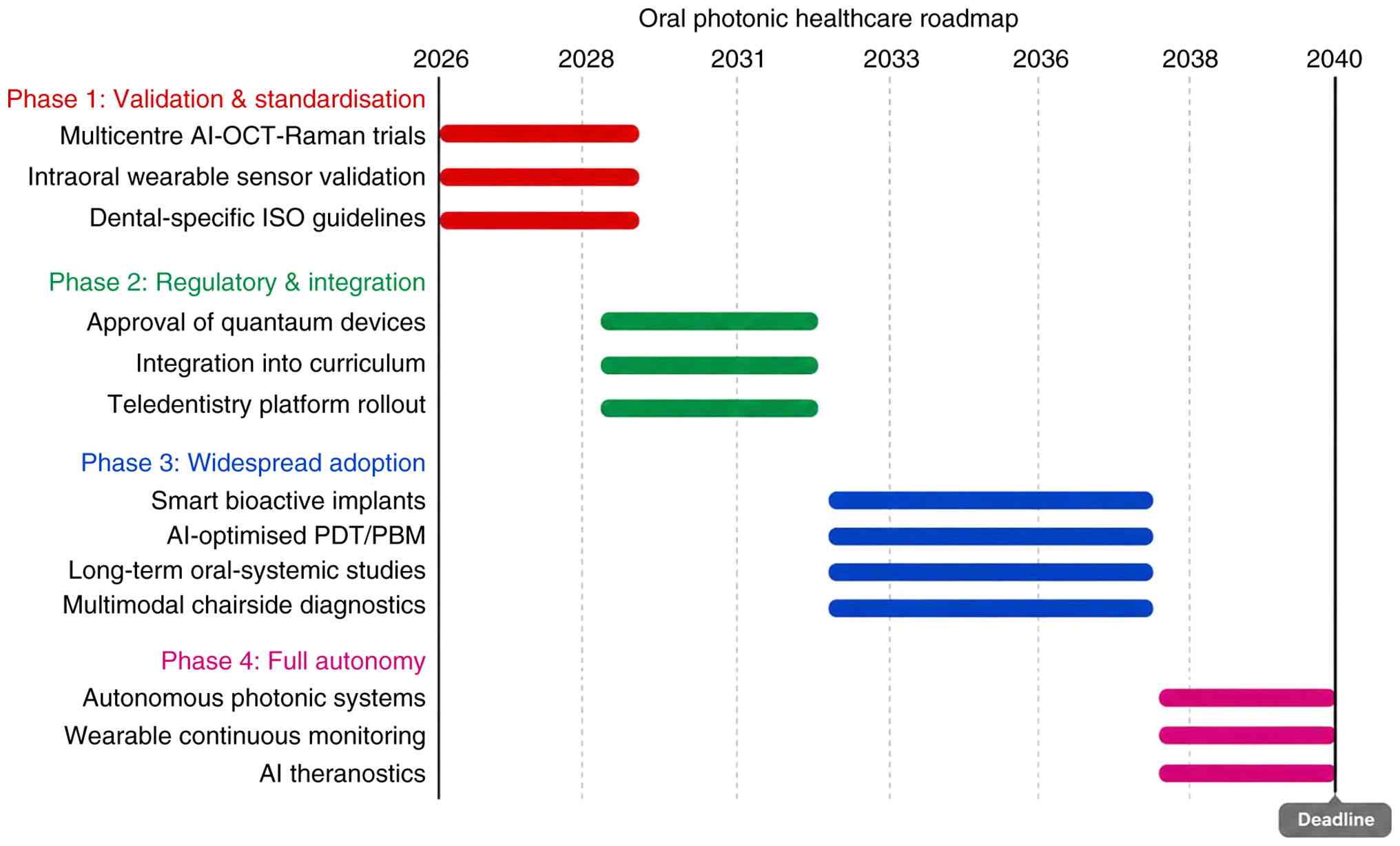
definitive prediction of future clinical adoption timelines: i)
2026-2028: Multicentre validation trials of AI-OCT-Raman systems,
intraoral wearable sensors and the development of dental-specific
standardization guidelines. ii) 2029-2032: Regulatory approval of
quantum-enhanced and nanomaterial devices; integration into dental
school and teledentistry. iii) 2033-2037: Widespread adoption of
smart implants and AI-optimized PBM/PDT along with long-term
outcome studies linking oral photonic monitoring to systemic
health. iv) 2038-2040: Fully autonomous photonic systems for
predictive oral healthcare along global equitable access
programmes.
The integration of AI, nanotechnology and advanced
multimodal imaging systems in biophotonics is illustrated in
Fig. 2. Fig. 2 presents a conceptual roadmap for
the future integration of emerging biophotonic technologies into
dental practice. The proposed framework outlines a progressive
transition from validation and standardization of advanced photonic
systems to regulatory approval, widespread clinical adoption, and
eventually autonomous photonic healthcare platforms. The roadmap
highlights the anticipated role of artificial intelligence,
multimodal imaging, nanotechnology-based therapeutics, smart
bioactive implants, and wearable biosensors in supporting precision
dentistry. These technologies have the potential to improve
diagnostic accuracy, enable personalized treatment planning, and
facilitate continuous monitoring of oral health. However, the
projected timeline should be interpreted cautiously, as successful
implementation will depend on clinical validation,
cost-effectiveness, regulatory approval, ethical considerations,
and long-term safety data.
9. Clinical integration, implementation and
challenges
The first step towards the successful clinical
development of biophotonic technologies is to demonstrate
biological plausibility and validity of diagnosis. However,
integration into practice does not depend only on technical
performance, but also on clinician training, treatment protocols,
reimbursement and regulation. Further analyses of
cost-effectiveness are warranted to justify large-scale adoption
(47).
Implementation in clinical practices continues to
face various challenges. Chairside devices should be compatible
with existing workflows so that the treatment time is not
excessively increased. Moreover, these devices demand proper
training so that the diagnostic output is well understood and
interpreted by the practitioners. Furthermore, the high acquisition
and maintenance costs of advanced modalities may restrict adoption,
mainly in resource-constrained settings.
Additional challenges exist in rural and
low-resource environments. The use of portable fluorescence-based
diagnostic systems, teledentistry and simplified diagnostic
procedures can make access easier and ensure the early detection of
diseases of concern among vulnerable groups of individuals. In
comparison with more advanced technologies, portable fluorescence
devices may be a better option when it comes to application in
low-resource environments, since they do not require too much
investment in the infrastructure, and their use is relatively easy.
The combination of portable fluorescence diagnostic systems with
community dental health systems can perform early screening and
referral actions in vulnerable groups of individuals (48).
Regulatory considerations are particularly critical
for diagnostic systems assisted by AI and emerging multimodal
imaging platforms, as they require rigorous validation of safety,
reproducibility and clinical utility prior to routine use (40).
Educational preparedness represents an additional
challenge. The use of biophotonic technologies is accompanied by
the requirement of sufficient understanding of optical diagnostics,
interpretation of images, as well as digital workflow. That is why
it is reasonable to incorporate the educational program with these
biophotonic technologies into dental studies. Moreover, variability
in clinical protocols remains an issue, which requires
standardization and multicenter validation. The effective
utilization of biophotonic technologies requires competency in
optical diagnostics, image interpretation and digital workflows,
highlighting the need for integration into dental curricula and
continuing professional education (49).
10. Conclusion
Technical advancements in biophotonics have expanded
the understanding of diagnostics and treatment in dentistry beyond
what is currently possible with standard techniques. Future
enhancements using AI, quantum technology, intelligent
nanomaterials and wearable devices will create precise preventive
and person-specific treatment protocols for dental patients that
will support their overall health and well-being. The roadmap
provided in the present review addresses a number of the challenges
that need to be overcome by the dental profession to accelerate the
translation of biophotonics into clinical practice, improve the
quality of care for patients and establish biophotonics as one of
the cornerstones of dentistry in the 21st century. Additionally,
the present review presents a timely, structured opportunity for
dental researchers and practitioners to achieve this paradigm
shift.
Acknowledgements
Not applicable.
Funding
Funding: No funding was received.
Availability of data and materials
Not applicable.
Authors' contributions
ST and SUN were involved in the conceptualization of
the present review, as well as in data curation, in the literature
search, the evaluation of studies from the literature for inclusion
in the review, project administration, validation, visualization
and in the writing of the original draft, and in the writing,
reviewing and editing of the manuscript. ST, SUN, MM and SK were
involved in the conceptualization of the present review, in data
curation, investigation, in the literature search, in project
administration, validation and study supervision. All authors have
read and approved the final manuscript. Data authentication is not
applicable.
Ethics approval and consent to
participate
Not applicable.
Patient consent for publication
Not applicable.
Competing interests
The authors declare that they have no competing
interests.
Use of artificial intelligence tools
During the preparation of this work, AI tools
(Rubriq) were used to improve the readability and language of the
manuscript or to generate images, and subsequently, the authors
revised and edited the content produced by the AI tools as
necessary, taking full responsibility for the ultimate content of
the present manuscript.
References
|
1
|
Deepyanti Verma K, Mishra I, Dixit A,
Dhanalakshmi P, Padhy K, Kamma PKG and Obulareddy VT: Biophotonics
in Dentistry-an overview. J Pharm Bioallied Sci. 16 (Suppl
1):S72–S74. 2024.PubMed/NCBI View Article : Google Scholar
|
|
2
|
Daghigh Ahmadi E, Hafeji S, Khurshid Z,
Imran E, Zafar MS, Saeinasab M and Sefat F: Biophotonics in
dentistry. Appl Sci. 12(4254)2022.
|
|
3
|
Amoozadeh M, Hariri A, Zarepour A,
Khosravi A, Iravani S and Zarrabi A: Biophotonic (nano)structures:
From fundamentals to emerging applications. RSC Adv.
15:26138–26172. 2025.PubMed/NCBI View Article : Google Scholar
|
|
4
|
Gounder R and Gounder S: Laser science and
its applications in prosthetic rehabilitation. J Lasers Med Sci.
7:209–213. 2016.PubMed/NCBI View Article : Google Scholar
|
|
5
|
Vagdouti T, Theodoridis C, Tseleki G and
Vouros I: Long-term clinical outcomes of Er:YAG or Er,Cr:YSGG
lasers utilized as monotherapy or as adjuncts to mechanical therapy
in the treatment of chronic periodontitis: A systematic review.
Lasers Dent Sci. 7:1–16. 2023.
|
|
6
|
Van As G: Erbium lasers in dentistry. Dent
Clin North Am. 48:1017–1059. 2004.PubMed/NCBI View Article : Google Scholar
|
|
7
|
Baldini F, Dholakia K, French P,
Guntinas-Lichius O, Kohler A, Mäntele W, Marcu L, Sroka R, Umapathy
S and Popp J: Shining a light on the future of biophotonics. J
Biophotonics. 18(e202500148)2025.PubMed/NCBI View Article : Google Scholar
|
|
8
|
Amoozadeh M, Hariri A, Zarepour A,
Khosravi A, Iravani S and Zarrabi A: Biophotonic (nano)structures:
From fundamentals to emerging applications. RSC Adv.
15:26138–26172. 2025.PubMed/NCBI View Article : Google Scholar
|
|
9
|
Hamblin MR: Mechanisms and applications of
the anti-inflammatory effects of photobiomodulation. AIMS Biophys.
4:337–361. 2017.PubMed/NCBI View Article : Google Scholar
|
|
10
|
Aoki A, Mizutani K, Schwarz F, Sculean A,
Yukna RA, Takasaki AA, Romanos GE, Taniguchi Y, Sasaki KM, Zeredo
JL, et al: Periodontal and peri-implant wound healing following
laser therapy. Periodontol 2000. 68:217–269. 2015.PubMed/NCBI View Article : Google Scholar
|
|
11
|
Pretty IA: Caries detection and diagnosis:
Novel technologies. J Dent. 34:727–739. 2006.PubMed/NCBI View Article : Google Scholar
|
|
12
|
Lussi A, Hibst R and Paulus R: DIAGNOdent:
An optical method for caries detection. J Dent Res. 83:C80–C83.
2004.PubMed/NCBI View Article : Google Scholar
|
|
13
|
Poh CF, Ng SP, Williams PM, Zhang L,
Laronde DM, Lane P, Macaulay C and Rosin MP: Direct fluorescence
visualization of clinically occult High-risk oral premalignant
disease using a simple hand-held device. Head Neck. 29:71–76.
2007.PubMed/NCBI View Article : Google Scholar
|
|
14
|
Rigotti P, Polizzi A, Verzì AE, Lacarrubba
F, Micali G and Isola GL: Impact of optical coherence tomography
(OCT) for periodontitis diagnostics: Current overview and advances.
Dent J (Basel). 13(305)2025.PubMed/NCBI View Article : Google Scholar
|
|
15
|
Suresh N, Joseph B, Sathyan P, Sweety VK,
Waltimo T and Anil S: Photodynamic therapy: An emerging therapeutic
modality in dentistry. Bioorg Med Chem. 114(117962)2024.PubMed/NCBI View Article : Google Scholar
|
|
16
|
Cobb CM: Lasers in periodontics: A review
of the literature. J Periodontol. 77:545–564. 2006.PubMed/NCBI View Article : Google Scholar
|
|
17
|
Soukos NS and Goodson JM: Photodynamic
therapy in the control of oral biofilms. Periodontol 2000.
55:143–166. 2011.PubMed/NCBI View Article : Google Scholar
|
|
18
|
Meisel P and Kocher T: Photodynamic
therapy for periodontal diseases: State of the art. J Photochem
Photobiol B. 79:159–170. 2005.PubMed/NCBI View Article : Google Scholar
|
|
19
|
Kuzu TE, Öztürk K, Gürgan CA, Üşümez A,
Yay A and Göktepe Ö: Effect of photobiomodulation therapy on
Peri-implant bone healing in Extra-short implants in a rabbit
model: A pilot study. Photobiomodul Photomed Laser Surg.
40:402–409. 2022.PubMed/NCBI View Article : Google Scholar
|
|
20
|
Colston BW, Everett MJ, Da Silva LB, Otis
LL, Stroeve P and Nathel H: Imaging of hard- and soft-tissue
structure in the oral cavity by optical coherence tomography. Appl
Opt. 37:3582–3585. 1998.PubMed/NCBI View Article : Google Scholar
|
|
21
|
Shemesh H, van Soest G, Wu MK and
Wesselink PR: Diagnosis of vertical root fractures with optical
coherence tomography. J Endod. 34:739–742. 2008.PubMed/NCBI View Article : Google Scholar
|
|
22
|
DiVito E, Peters OA and Olivi G:
Effectiveness of the erbium:YAG laser and new design radial and
stripped tips in removing the smear layer after root canal
instrumentation. Lasers Med Sci. 27:273–280. 2012.PubMed/NCBI View Article : Google Scholar
|
|
23
|
Garcez AS, Ribeiro MS, Tegos GP, Núñez SC,
Jorge AOC and Hamblin MR: Antimicrobial photodynamic therapy
combined with conventional endodontic treatment to eliminate root
canal biofilm infection. Lasers Surg Med. 39:59–66. 2007.PubMed/NCBI View Article : Google Scholar
|
|
24
|
Arslan H, Doğanay E, Karataş E, Ünlü MA
and Ahmed HMA: Effect of Low-level laser therapy on postoperative
pain after root canal retreatment: A Preliminary
Placebo-controlled, Triple-blind, randomized clinical trial. J
Endod. 43:1765–1769. 2017.PubMed/NCBI View Article : Google Scholar
|
|
25
|
Tanya S and Srisilapanan P: Clinical
efficacy and safety of photobiomodulation therapy for orofacial
conditions in older adults: A systematic review of randomized
controlled trials. Dent J. 14(231)2026.PubMed/NCBI View Article : Google Scholar
|
|
26
|
Ko ACT, Hewko M, Sowa MG, Dong CCS,
Cleghorn B and Choo-Smith LP: Early dental caries detection using a
Fibre-optic coupled polarization-resolved Raman spectroscopic
system. Opt Express. 16:6274–6284. 2008.PubMed/NCBI View Article : Google Scholar
|
|
27
|
Hibst R and Keller U: Experimental studies
of the application of the Er:YAG laser on dental hard substances:
I. Measurement of the ablation rate. Lasers Surg Med. 9:338–344.
1989.PubMed/NCBI View Article : Google Scholar
|
|
28
|
Shimada Y, Sadr A, Burrow MF, Tagami J,
Ozawa N and Sumi Y: Validation of swept-source optical coherence
tomography (SS-OCT) for the diagnosis of occlusal caries. J Dent.
38:655–665. 2010.PubMed/NCBI View Article : Google Scholar
|
|
29
|
Ramakrishnaiah R, Rehman G ur,
Basavarajappa S, Al Khuraif AA, Durgesh BH, Khan AS and Rehman I
ur: Applications of Raman spectroscopy in dentistry: Analysis of
tooth structure. Appl Spectrosc Rev. 50:332–350. 2015.
|
|
30
|
Kau CH, Kantarci A, Shaughnessy T,
Vachiramon A, Santiwong P, de la Fuente A, Skrenes D, Ma D and
Brawn P: Photobiomodulation accelerates orthodontic alignment in
the early phase of treatment. Prog Orthod. 14(30)2013.PubMed/NCBI View Article : Google Scholar
|
|
31
|
Bahrami R, Nikparto N, Gharibpour F,
Pourhajibagher M and Bahador A: The effect of light-emitting
diode-mediated photobiomodulation therapy on orthodontic tooth
movement: A literature review. Photodiagnosis Photodyn Ther.
52(104488)2025.PubMed/NCBI View Article : Google Scholar
|
|
32
|
Paschoal MA, Santos-Pinto L, Lin M and
Duarte S: Streptococcus mutans photoinactivation by combination of
short exposure of a Broad-spectrum visible light and low
concentrations of photosensitizers. Photomed Laser Surg.
32:175–180. 2014.PubMed/NCBI View Article : Google Scholar
|
|
33
|
Boj JR, Poirier C, Hernandez M, Espassa E
and Espanya A: Review: Laser soft tissue treatments for paediatric
dental patients. Eur Arch Paediatr Dent. 12:100–105.
2011.PubMed/NCBI View Article : Google Scholar
|
|
34
|
Wilder-Smith P, Lee K, Guo S, Zhang J,
Osann K, Chen Z and Messadi D: In vivo diagnosis of oral dysplasia
and malignancy using optical coherence tomography: Preliminary
studies in 50 patients. Lasers Surg Med. 41:353–357.
2009.PubMed/NCBI View Article : Google Scholar
|
|
35
|
Hanna K, Asiedu AL, Theurer T, Muirhead D,
Speirs V, Oweis Y and Abu-Eid R: Advances in Raman spectroscopy for
characterising oral cancer and oral potentially malignant
disorders. Expert Rev Mol Med. 26(e25)2024.PubMed/NCBI View Article : Google Scholar
|
|
36
|
Dolmans DEJGJ, Fukumura D and Jain RK:
Photodynamic therapy for cancer. Nat Rev Cancer. 3:380–387.
2003.PubMed/NCBI View Article : Google Scholar
|
|
37
|
Abdul NS: A systematic review on the
effectiveness of photodynamic therapy in the treatment of oral
precancerous lesions and oral cancers. J Pharm Bioallied Sci. 17
(Suppl 4):S2862–S866. 2025.PubMed/NCBI View Article : Google Scholar
|
|
38
|
Maghfour J, Mineroff J, Ozog DM, Jagdeo J,
Lim HW, Kohli I, Anderson R, Kelly KM, Mamalis A, Munavalli G, et
al: Evidence-based consensus on the clinical application of
photobiomodulation. J Am Acad Dermatol. 93:429–443. 2025.PubMed/NCBI View Article : Google Scholar
|
|
39
|
Sitaras S, Tsolakis IA, Gelsini M,
Tsolakis AI, Schwendicke F, Wolf TG and Perlea P: Applications of
artificial intelligence in dental medicine: A critical review. Int
Dent J. 75:474–486. 2025.PubMed/NCBI View Article : Google Scholar
|
|
40
|
Schwendicke F, Samek W and Krois J:
Artificial intelligence in dentistry: Chances and challenges. J
Dent Res. 99:769–774. 2020.PubMed/NCBI View Article : Google Scholar
|
|
41
|
Juzenas P, Chen W, Sun YP, Coelho MAN,
Generalov R, Generalova N and Christensen IL: Quantum dots and
nanoparticles for photodynamic and radiation therapies of cancer.
Adv Drug Deliv Rev. 60:1600–1614. 2008.PubMed/NCBI View Article : Google Scholar
|
|
42
|
Huang D, Swanson EA, Lin CP, Schuman JS,
Stinson WG, Chang W, Hee MR, Flotte T, Gregory K, Puliafito CA, et
al: Optical coherence tomography. Science. 254:1178–1181.
1991.PubMed/NCBI View Article : Google Scholar
|
|
43
|
Keerthiga R, Zhao Z, Pei D and Fu A:
Photodynamic nanophotosensitizers: Promising materials for tumor
theranostics. ACS Biomater Sci Eng. 6:5474–5485. 2020.PubMed/NCBI View Article : Google Scholar
|
|
44
|
Tai Y, Li Y, Mornay KM, Woodard MK, Wang
W, Yang X and Pan J: Biosensors in dental, oral and craniofacial
applications. NPJ Biosens. 3(14)2026.PubMed/NCBI View Article : Google Scholar
|
|
45
|
Gieroba B, Krysa M, Wojtowicz K, Wiater A,
Pleszczyńska M, Tomczyk M and Sroka-Bartnicka A: The FT-IR and
Raman spectroscopies as tools for biofilm characterization created
by cariogenic streptococci. Int J Mol Sci. 21(3811)2020.PubMed/NCBI View Article : Google Scholar
|
|
46
|
Zhang Y, Ren L, Wang Q, Wen Z, Liu C and
Ding Y: Raman spectroscopy: A potential diagnostic tool for oral
diseases. Front Cell Infect Microbiol. 12(775236)2022.PubMed/NCBI View Article : Google Scholar
|
|
47
|
Ohyama H, Duong ML, Yancoskie AE, Smiley
AB, Syed AZ, Reddy MS and Bencharit S: Challenges and opportunities
in implementing digital technology in dental curriculum: A review
and perspective. Cureus. 17(e83272)2025.PubMed/NCBI View Article : Google Scholar
|
|
48
|
Ashtiani GH, Sabbagh S, Moradi S, Azimi S
and Ravaghi V: Diagnostic accuracy of tele-dentistry in screening
children for dental caries by community health workers in a
lower-middle-income country. Int J Paediatr Dent. 34:567–575.
2024.PubMed/NCBI View Article : Google Scholar
|
|
49
|
Tay JRH, Bosworth HB, Fox CH, Gallagher
JE, Jakubovics NS, Schwendicke F and Nascimento GG: Implementation
research is underdeveloped in oral health: Bridging the gap. J Dent
Res. 105:570–577. 2026.PubMed/NCBI View Article : Google Scholar
|