Introduction
Hypoxic ischemic encephalopathy (HIE) is fetal or
neonatal cerebral injury caused by partial or complete anoxia and a
reduction or suspension of the cerebral blood flow resulting from
perinatal asphyxia, which has chronic sequelae on the nerve system
and is a cause of acute neonatal death (1). Early detection, diagnosis and
intervention of HIE is important for the reduction of perinatal
mortality and disability rates for newborns (2).
Nuclear factor erythroid-2-related factor 2 (Nrf2)
is a nuclear transcription factor with neuroprotective effects
against central nervous system disease (3). The Nrf2 pathway is anti-apoptotic and
has multiple functions, including resistance to oxidative stress,
regulation of inflammatory injury and relief from calcium
overloading (4). Research findings
indicate that Nrf2 regulates downstream molecules to protect
nerves, particularly against apoptosis (5).
Heme oxygenase-1 (HO-1) is an induced enzyme with a
relative molecular mass of 32 kDa, which is a major heat
shock/stress protein, mainly distributed among tissues and organs
with active metabolism of blood corpuscles, such as the spleen,
liver, reticuloendothelial system and bone marrow (6). HO-1 has low expression levels in brain
tissues; however, under stress conditions, HO-1 can be expressed at
very high levels by neurogliocytes and stellate neurons (7). Stimulating factors such as heat shock,
heavy metals, protoheme, inflammatory stimuli, hypoxia, ultraviolet
radiation, endotoxins, prostaglandin, lipopolysaccharides, nitric
oxide (NO) and stress states such as hunger, fever and mechanical
injuries may increase HO-1 activity (8).
Curcumin has been applied in traditional medicine
for thousands of years, particularly in Ayurvedic medicine
(9). It has been demonstrated to
possess a wide range of pharmacological activities with little side
effects and high safety. Extensive studies have been conducted on
curcumin, which have demonstrated that curcumin has significant
anti-infection, anti-inflammatory, antioxidative, anti-platelet
aggregation, lipid-reducing and anti-fibrotic activities, in
addition to protecting against myocardial injury and suppressing
tumor growth (9–11). Curcumin has been shown to ameliorate
myocardial ischemia-reperfusion injuries and protect the myocardium
through inhibition of the activation of inflammation-related genes
by NF-κB (12). Thus, in the present
study, the effects of curcumin on hypoxic-ischemic brain injury and
the potential underlying mechanisms were investigated.
Materials and methods
Animals and hypoxia-ischemia
induction
All animal experiments were approved by the Animal
Ethics Committee of the Second Affiliated Hospital of Shandong
University of TCM (Jinan, China). Male Sprague Dawley rats
(1-week-old; weight, 52±1 g; n=50) were purchased from the Animal
Laboratory of Shandong University (Jinan, China), and housed with a
12:12-h light-dark cycle, a temperature of 22–23°C and 55–60%
humidity, with free access to a standard laboratory chow diet and
drinking water.
The hypoxia-ischemia model was created as follows.
Briefly, rats were anesthetized with 2% isoflurane (Sigma-Aldrich;
Merck KGaA, Darmstadt, Germany) in a 1:1 mixture of nitrous oxide
and oxygen. The left common carotid arteries of rats were ligated
and the rats was allowed to recover for 1 h. After recovery, rats
were placed in an incubator perfused with 10±0.01% oxygen in
nitrogen at 37°C for 1 h. In the hemisphere ipsilateral to the left
common carotid artery, arterial ligation and hypoxia resulted in
the creation of the HIE model. In the sham group, rats were only
anesthetized and did not undergo surgery to induce HIE.
Experimental design
Rats were randomly divided into three groups: Sham
group (n=10), hypoxic-ischemic brain injury model group (n=20) and
curcumin group (n=20). In the curcumin group, rats subjected to
hypoxic-ischemic brain injury were treated with 150 mg/kg/day
curcumin (Sigma-Aldrich; Merck KGaA, Darmstadt, Germany) by gavage
for 3 days, 2 days after HIE surgery. In the sham group and the
hypoxic-ischemic brain injury model group, rats were treated with
500 µl normal saline at the same time points. Rats were sacrificed
by cervical dislocation while anesthetized with isoflurane after
treatment with curcumin.
Assessment of brain damage
After sacrifice, the brains were dissected,
paraffin-embedded and cut into 10-µm coronal sections. Myelin basic
protein (MBP) was stained, and the extent of white- and gray-matter
injury was analyzed via quantitative measurements of the injury
area using Micro Image version 4.0 (Micro-Macro AB, Gothenburg,
Sweden), with the following formula: Contralateral
hemisphere-ipsilateral hemisphere)=(contralateral hemisphere)
×100%. The sections were dipped in 1% thionin/toluidine solution,
soaked in acid fuchsin solution and dehydrated in a gradient of
ethanol.
Histopathology
Following sacrifice, the brain was immediately
removed and fixed with 10% neutral buffered formalin at room
temperature for 24 h. Tissue samples were cut into sections of 4-µm
thickness and stained with hematoxylin and eosin (H&E). Tissue
samples were observed using confocal microscope (magnification,
×20; ZEISS, LSM510; Zeiss AG, Oberkochen, Germany).
Analysis of superoxide dismutase (SOD)
activities and malondialdehyde (MDA) levels
SOD activities and MDA levels were assayed using
ELISA kits (A001-2 and A003-1; Nanjing Jiancheng Biology
Engineering Institute, Nanjing, China) and absorbance's were
measured using an ultraviolet spectrophotometer at 450 nm.
Western blot analysis
Brain samples were transferred into ice-cold RIPA
assay (Beyotime Institute of Biotechnology; Shanghai, China)
containing a cocktail of protein phosphatase and proteinase
inhibitors. Samples were collected at 15,000 × g at 4°C for 10 min.
The supernatant was assayed using a Bio-Rad Bradford Protein Assay
kit (Bio-Rad Laboratories, Inc., Hercules, CA, USA). Protein (50
µg) was separated using 8–10% SDS-PAGE and blots were blocked in
buffer solution containing 5% milk and 0.1% Tween-20 in PBS,
following transfer onto polyvinylidene fluoride membranes.
Membranes was then blotted for 2 h at room temperature with
antibodies targeting inducible nitric oxide synthase (iNOS;
1:4,000; Sigma-Aldrich; Merck KGaA), Nrf2 (1:4,000; Sigma-Aldrich;
Merck KGaA), HO-1 (1:4,000; Sigma-Aldrich; Merck KGaA) or β-actin
(1:4,000; Sigma-Aldrich; Merck KGaA) at 4°C overnight. Membranes
were washed with TBST for 15 min and incubated with goat
anti-rabbit HRP secondary antibody (1:3,000; HA-1001-100; Hangzhou
Hua'An Biotechnology Co., Ltd., Hangzhou, China) for 1 h at 37°C.
Protein expression was detected using an enhanced chemiluminescence
kit (Beyotime Institute of Biotechnology, Shanghai, China) and
quantified using Bio-Rad Image Lab 3.0 (Bio-Rad).
Caspase-3 activity assay
Brain samples were transferred into ice-cold
Tris-HCl buffer solution containing a cocktail of protein
phosphatase and proteinase inhibitors. Samples were collected at
15,000 × g at 4°C for 10 min. The supernatant was assayed using a
Bio-Rad Bradford Protein Assay kit. An equal amount of total
protein (10 µg) extract from each rat was incubated at 37°C with
Ac-LEHD-pNA for the caspase-3 assay (Beyotime Institute of
Biotechnology; Shanghai, China) for 4 h and the caspase-3 activity
was determined by measuring the absorbance at 405 nm using an
ultraviolet spectrophotometer.
Statistical analysis
All results are expressed as the mean ± standard
deviation. Analysis of variance followed by the least significant
difference post hoc test was used for the comparison of data from
more than two groups using SPSS software (17.0; SPSS, Inc.,
Chicago, IL, USA). P<0.05 was considered to indicate a
statistically significant difference.
Results
Curcumin attenuates hypoxic-ischemic
brain injury in neonatal rats
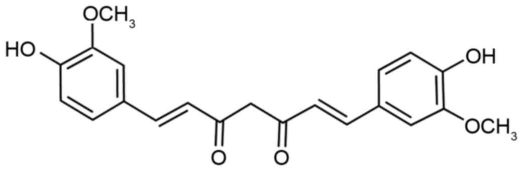
The chemical structure of curcumin is displayed in
Fig. 1. The effects of curcumin on
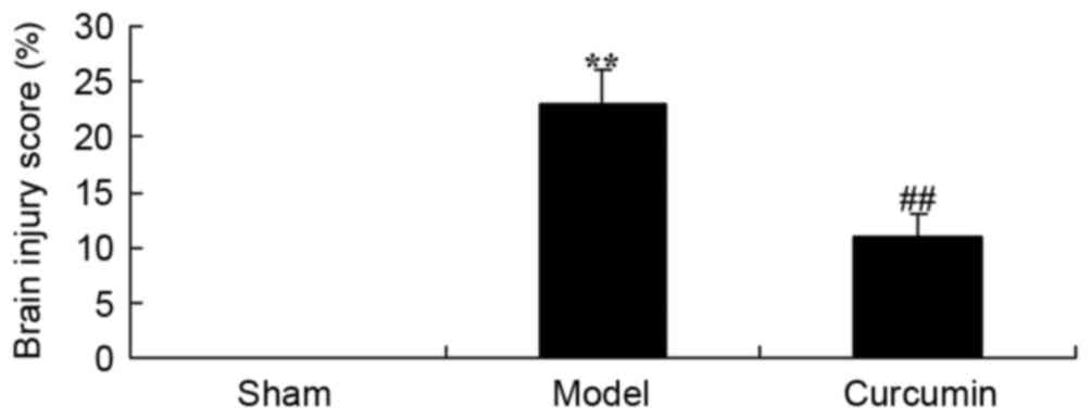
hypoxic-ischemic brain injury were examined by calculation of a
brain injury score. The brain injury score of the model group was
significantly increased compared with that of the sham control
group (Fig. 2). In the rats treated
with curcumin (150 mg/kg), the brain injury induced by
hypoxic-ischemic brain injury was reduced compared with that in the
model group (Fig. 2).
Curcumin attenuates MBP levels in
brain-injured neonatal rats
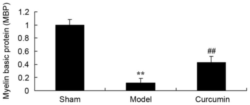
To evaluate whether curcumin attenuates changes in
MBP expression in neonatal rats, MBP levels were analyzed following
hypoxic-ischemic brain injury. As shown in Fig. 3, the level of MBP in the model group
was lower than that of the sham control group. Treatment with 150
mg/kg curcumin effectively increased the MBP levels of neonatal
rats with hypoxic-ischemic brain injury (Fig. 3).
Curcumin attenuates histopathological
changes in brain-injured neonatal rats
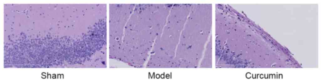
To confirm the effects of curcumin in neonatal rats,
histopathological examination was conducted using H&E staining.
The number of neuronal cells in the hypoxic-ischemic brain injury
model group was lower than that of the sham group (Fig. 4). Treatment with curcumin effectively
augmented the quantity of neuronal cells in neonatal rats with
hypoxic-ischemic brain injury (Fig.
4).
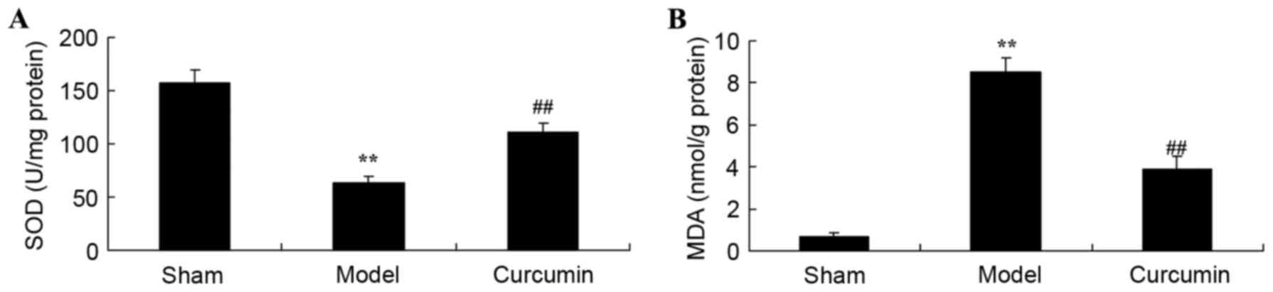
Curcumin attenuates changes in SOD
activity and MDA levels in brain-injured neonatal rats
To determine whether curcumin attenuates oxidative
stress in neonatal rats, SOD activities and MDA levels were
measured using ELISA kits. As shown in Fig. 5A, the SOD activity of the
hypoxic-ischemic brain injury model group was clearly inhibited, as
compared with that of the sham control group. In addition,
hypoxic-ischemic brain injury observably increased MDA levels in
neonatal rats, as compared with those in the sham control group
(Fig. 5B). However, treatment with
curcumin observably reversed the changes in SOD activity and MDA
levels in neonatal rats following hypoxic-ischemic brain injury
(Fig. 5).
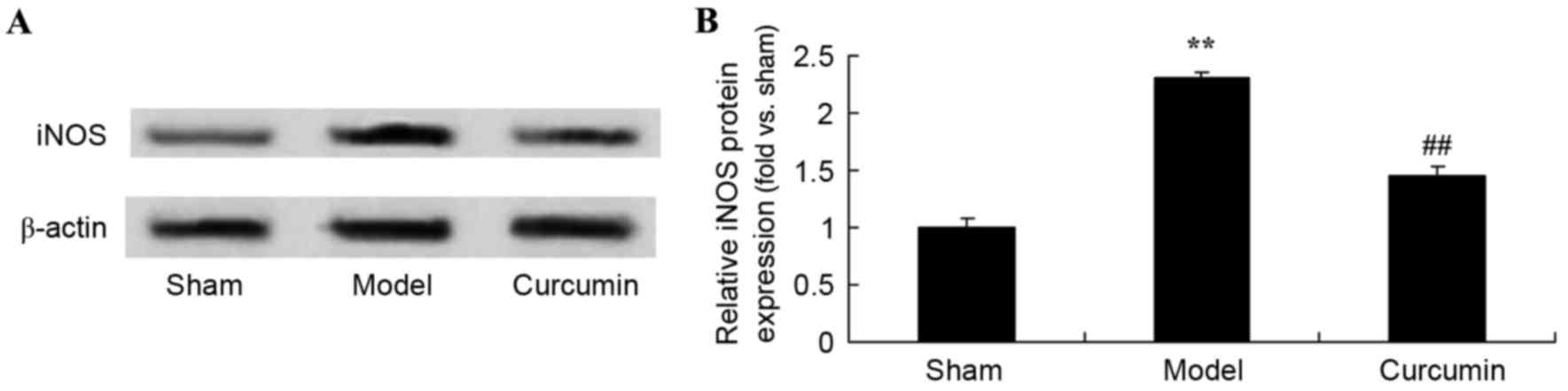
Curcumin attenuates the expression of
iNOS in brain-injured neonatal rats
To evaluate the attenuating effects of curcumin on
the expression of iNOS in neonatal rats, iNOS expression was
analyzed using western blot analysis. Following hypoxic-ischemic
brain injury, iNOS protein expression was markedly increased in the
model group, as compared with the sham control group (Fig. 6). Treatment with curcumin
significantly suppressed iNOS protein expression in neonatal rats
with hypoxic-ischemic brain injury (Fig.
6).
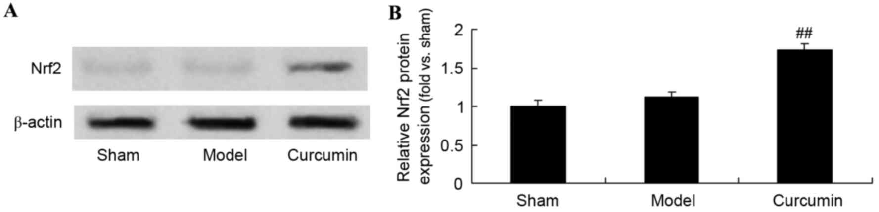
Curcumin increases the expression of
Nrf2 in brain-injured neonatal rats
To further evaluate the effects of curcumin in
neonatal rats, the expression of Nrf2 protein was analyzed using
western blot analysis. The Nrf2 protein expression level in the
sham group was similar to that of the model control group (Fig. 7). However, curcumin treatment
markedly promoted Nrf2 protein expression in neonatal rats with
hypoxic-ischemic brain injury (Fig.
7).
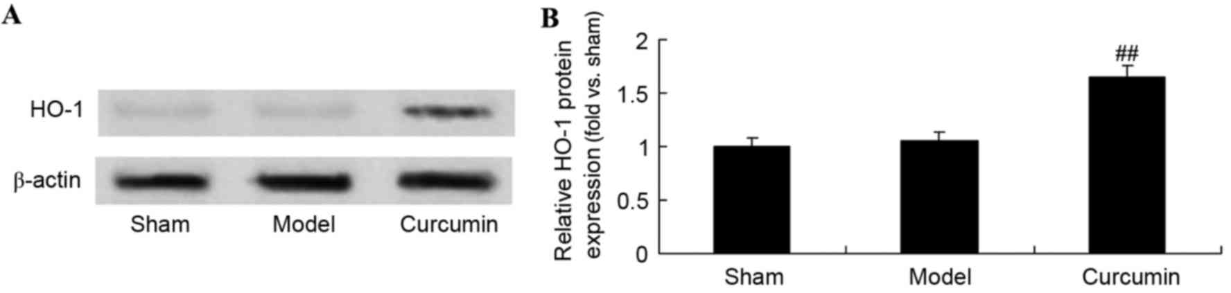
Curcumin attenuates the expression of
HO-1 in brain-injured neonatal rats
The effects of curcumin on neonatal rats with
hypoxic-ischemic brain injury were further analyzed using western
blotting analysis to determine HO-1 protein levels. As shown in
Fig. 8, the protein expression of
HO-1 in the sham group was similar to that in the model control
group. Following curcumin treatment, there was a significant
upregulation of HO-1 protein expression in the curcumin treated
group, as compared with the model group (Fig. 8).
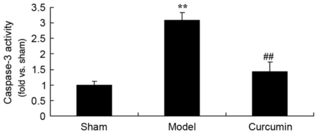
Curcumin attenuates the activity of
caspase-3 in brain-injured neonatal rats
To further investigate the effects of curcumin in
neonatal rats, the activity of caspase-3 was determined. The
caspase-3 activity was significantly increased in the
hypoxic-ischemic brain injury model group, as compared with the
sham control group (Fig. 9).
Curcumin treatment significantly inhibited the caspase-3 activity
in neonatal rats with hypoxic-ischemic brain injury (Fig. 9).
Discussion
The occurrence rate of HIE for term infants has been
reported to be 6/1,000, and 1/1,000 of such infants succumb or have
nervous system dysfunction (13).
Each year, China has 18–20 million live births and the morbidity of
asphyxia has reached as high as 13.6%, among which 15.6% of cases
are injured or disabled (14). Each
year, ~300,000 children are handicapped, which greatly threatens
their quality of life (15). In
order to better prevent injury and disability, the timely detection
and treatment of HIE is necessary, and the exploration of
indicators or methods with relatively high specificity is required
(16). In the present study,
treatment with curcumin was evaluated and was found to effectively
reduce brain injury score, increase MBP levels and augment the
quantity of neuronal cells in neonatal rats with hypoxic-ischemic
brain injury.
When HIE occurs, oxidative phosphorylation
dysfunction of mitochondria results in a reduction of ATP
production and produces a large amount of reactive oxygen species
(ROS), resulting in oxidative stress (17). With regard to cerebral ischemia
reperfusion, certain adaptive regulatory mechanisms in the HIE
process have attracted attention (18). Following HIE, the oxidation and
anti-oxidation system of the body is unbalanced. High levels of
free radicals are generated, which results in damage, i.e.,
oxidative stress (19). Free
radicals are ROS, including superoxide anion and hydrogen peroxide,
which can cause the peroxidation of lipid, proteins and nucleic
acids (20). Neurons have high
metabolic activities and oxygen consumption as well as low levels
of endogenous anti-oxidant enzymes, and are extremely sensitive to
oxidative stress (21). In addition,
abundant lipids exist in the brain and can react with ROS,
resulting in lipid peroxidation of neural membranes (22). Consequently, the central nervous
system is sensitive to oxidative stress injuries. In the present
study, it was found that treatment with curcumin observably
reversed the changes in SOD activities and MDA levels in neonatal
rats with hypoxic-ischemic brain injury. Similar mechanisms have
been indicated in the cardiovascular system; Qian et al
(9) suggested that curcumin
mitigates cardiac injury through suppression of oxidation and
inflammation in obese rats.
To exert their physiological functions, vascular
endothelial cells secrete various vasoactive substances. Free
radicals are damaging to endothelial cells. Autologous vascular
wall cells undergo stress reactions and exert resistance to
oxidative stress. NO is an endothelium-derived relaxing factor
(23). When oxidative stress occurs,
the expression of NO synthase decreases, with concurrent reduction
of its substrate and cofactors (24). A reduction in the activity of NO
synthase and the degradation of NO by active oxygen can both cause
abnormalities in NO levels (22).
The present study demonstrated that treatment with curcumin clearly
suppressed the elevation of iNOS expression induced by
hypoxic-ischemic brain injury in neonatal rats. Gao et al
(25) reported that curcumin induces
M2 macrophage polarization by iNOS.
Recent studies have shown that neuronal apoptosis
has an essential role in secondary injury following traumatic brain
injury (26,27). The Nrf2 pathway is an anti-apoptotic
pathway that has been demonstrate do function in many organs,
particularly in the central nervous system (5). With a short half-life, Nrf2 is an
important factor. Under physiological conditions, Nrf2 exists in
the cytoplasm where it binds with Keap1 protein (28). When oxidative stress from external
stimuli occurs, phosphorylation of Nrf2 separates it from the
adapter protein Keap1 and transfers it into the cell nucleus to
combine with the promoter sequence to regulate its downstream genes
(29). The Nrf2 anti-apoptotic
pathway has multiple functions relating to protection against
anti-oxidative stress, regulation of inflammatory injury and relief
from calcium overloading (30). In
the present study, curcumin treatment markedly promoted Nrf2
protein expression in neonatal rats with hypoxic-ischemic brain
injury. Zhang et al (31)
suggested that curcumin protects renal tubular epithelial cells
through the Nrf2-mediated upregulation of HO-1.
HO-1 is a member of the heat shock protein family,
which is able to stabilize tissue proteins, mitochondrial membranes
and cytomembranes and relieve neurocyte injuries (7). Following stress, heat shock proteins
are typically located perinuclearly and may combine with plasma
membranes to stabilize them (27).
The stability of brain cell functions and structures is dependent
upon plasma membranes (32). It has
been found that increased HO-1 expression provides resistance
against neuronal death mediated via glutamic acid-induced oxidative
stress, and significantly reduces membrane damage and nuclear
accumulation (28). Furthermore,
when HO-1 levels increase, the calcium ion concentration in resting
cells decreases and the production rate of oxygen radicals is
largely reduced (4). In the present
study, it was found that curcumin treatment significantly
upregulated HO-1 protein expression in neonatal rats with
hypoxic-ischemic brain injury. When considered together, these
findings suggest that the Nrf2-mediated upregulation of HO-1 may
play a detrimental role in hypoxic-ischemic brain injury.
Apoptosis is a programmed death under physiological
or pathological conditions. Three pathways of cell apoptosis have
been described, i.e., the death receptor pathway in cytomembranes,
the mitochondrial pathway in cytomembranes and stress pathways of
the endoplasmic reticulum (13,33). The
latter two pathways have been investigated via the measurement
ofcaspase-3 and caspase-12 activities in a study on cell apoptosis
in a neonatal hypoxia-ischemia rat model (34). In the present study, curcumin
treatment significantly inhibited caspase-3 activity in the brains
of neonatal rats with hypoxic-ischemic brain injury. In a previous
study, Bucak et al (35)
demonstrated that the protective effect of curcumin against
paclitaxel-induced inner ear damage in rats was also mediated via
the suppression of caspase-3 activity.
In summary, the present study provides evidence in a
1-week-old Sprague Dawley rat model confirming the protective
effects of curcumin against hypoxic-ischemic brain injury in
neonatal rats. The results of the study indicate that the Nrf2/HO-1
pathway underlies the protective effect of curcumin on
hypoxic-ischemic brain injury, and reveal the effective targeting
of Nrf2/HO-1 by curcumin.
References
|
1
|
Shankaran S, Laptook AR, Pappas A,
McDonald SA, Das A, Tyson JE, Poindexter BB, Schibler K, Bell EF,
Heyne RJ, et al: Effect of depth and duration of cooling on deaths
in the NICU among neonates with hypoxic ischemic encephalopathy: A
randomized clinical trial. JAMA. 312:2629–2639. 2014. View Article : Google Scholar : PubMed/NCBI
|
|
2
|
Kracer B, Hintz SR, Van Meurs KP and Lee
HC: Hypothermia therapy for neonatal hypoxic ischemic
encephalopathy in the state of California. J Pediatr. 165:267–273.
2014. View Article : Google Scholar : PubMed/NCBI
|
|
3
|
Re L, Martinez-Sãnchez G, Bordicchia M,
Malcangi G, Pocognoli A, Morales-Segura MA, Rothchild J and Rojas
A: Is ozone pre-conditioning effect linked to Nrf2/EpRE activation
pathway in vivo? A preliminary result. Eur J Pharmacol.
742:158–162. 2014. View Article : Google Scholar : PubMed/NCBI
|
|
4
|
Wu Z, Uchi H, Morino-Koga S, Shi W and
Furue M: Z-ligustilide ameliorated ultraviolet B-induced oxidative
stress and inflammatory cytokine production in human keratinocytes
through upregulation of Nrf2/HO-1 and suppression of NF-κB pathway.
Exp Dermatol. 24:703–708. 2015. View Article : Google Scholar : PubMed/NCBI
|
|
5
|
Sasaki H, Suzuki A, Shitara M, Hikosaka Y,
Okuda K, Moriyama S, Yano M and Fujii Y: Polymorphisms of NRF2 gene
correlated with decreased FEV1 in lung cancers of smokers. Biomed
Rep. 1:484–488. 2013.PubMed/NCBI
|
|
6
|
Barnett M, Hall S, Dixit M and Arany I:
Simvastatin attenuates oleic acid-induced oxidative stress through
CREB-dependent induction of heme oxygenase-1 in renal proximal
tubule cells. Pediatr Res. 79:243–250. 2016. View Article : Google Scholar : PubMed/NCBI
|
|
7
|
Lin SH, Song W, Cressatti M, Zukor H, Wang
E and Schipper HM: Heme oxygenase-1 modulates microRNA expression
in cultured astroglia: Implications for chronic brain disorders.
Glia. 63:1270–1284. 2015. View Article : Google Scholar : PubMed/NCBI
|
|
8
|
Morita K, Itoh M, Nishibori N, Her S and
Lee MS: Spirulina non-protein components induce BDNF gene
transcription via HO-1 activity in C6 glioma cells. Appl Biochem
Biotechnol. 175:892–901. 2015. View Article : Google Scholar : PubMed/NCBI
|
|
9
|
Qian Y, Zhong P, Liang D, Xu Z, Skibba M,
Zeng C, Li X, Wei T, Wu L and Liang G: A newly designed curcumin
analog Y20 mitigates cardiac injury via anti-inflammatory and
anti-oxidant actions in obese rats. PLoS One. 10:e01202152015.
View Article : Google Scholar : PubMed/NCBI
|
|
10
|
Patel PB, Thakkar VR and Patel JS:
Cellular effect of curcumin and citral combination on breast cancer
cells: Induction of apoptosis and cell cycle arrest. J Breast
Cancer. 18:225–234. 2015. View Article : Google Scholar : PubMed/NCBI
|
|
11
|
Jang EM, Choi MS, Jung UJ, Kim MJ, Kim HJ,
Jeon SM, Shin SK, Seong CN and Lee MK: Beneficial effects of
curcumin on hyperlipidemia and insulin resistance in high-fat-fed
hamsters. Metabolism. 57:1576–1583. 2008. View Article : Google Scholar : PubMed/NCBI
|
|
12
|
Yeh CH, Chen TP, Wu YC, Lin YM and Lin P
Jing: Inhibition of NFkappaB activation with curcumin attenuates
plasma inflammatory cytokines surge and cardiomyocytic apoptosis
following cardiac ischemia/reperfusion. J Surg Res. 125:109–116.
2005. View Article : Google Scholar : PubMed/NCBI
|
|
13
|
Liu L, Liu C, Lu Y, Liu L and Jiang Y: ER
stress related factor ATF6 and caspase-12 trigger apoptosis in
neonatal hypoxic-ischemic encephalopathy. Int J Clin Exp Pathol.
8:6960–6966. 2015.PubMed/NCBI
|
|
14
|
Kaandorp JJ, Benders MJ, Rademaker CM,
Torrance HL, Oudijk MA, de Haan TR, Bloemenkamp KW, Rijken M, van
Pampus MG, Bos AF, et al: Antenatal allopurinol for reduction of
birth asphyxia induced brain damage (ALLO-Trial); a randomized
double blind placebo controlled multicenter study. BMC Pregnancy
Childbirth. 10:82010. View Article : Google Scholar : PubMed/NCBI
|
|
15
|
Bell MJ, Adelson PD, Hutchison JS,
Kochanek PM, Tasker RC, Vavilala MS, Beers SR, Fabio A, Kelsey SF
and Wisniewski SR: Multiple Medical Therapies for Pediatric
Traumatic Brain Injury Workgroup: Differences in medical therapy
goals for children with severe traumatic brain injury-an
international study. Pediatr Crit Care Med. 14:811–818. 2013.
View Article : Google Scholar : PubMed/NCBI
|
|
16
|
Agut T, Leon M, Rebollo M, Muchart J, Arca
G and Garcia-Alix A: Early identification of brain injury in
infants with hypoxic ischemic encephalopathy at high risk for
severe impairments: Accuracy of MRI performed in the first days of
life. BMC Pediatr. 14:1772014. View Article : Google Scholar : PubMed/NCBI
|
|
17
|
Zhao M, Zhu P, Fujino M, Zhuang J, Guo H,
Sheikh I, Zhao L and Li XK: Oxidative stress in hypoxic-ischemic
encephalopathy: Molecular mechanisms and therapeutic strategies.
Int J Mol Sci. 17(pii): E20782016. View Article : Google Scholar : PubMed/NCBI
|
|
18
|
Vasiljević B, Maglajlić-Djukić S, Gojnić M
and Stanković S: The role of oxidative stress in perinatal
hypoxic-ischemic brain injury. Srp Arh Celok Lek. 140:35–41. 2012.
View Article : Google Scholar : PubMed/NCBI
|
|
19
|
Burchell SR, Dixon BJ, Tang J and Zhang
JH: Isoflurane provides neuroprotection in neonatal hypoxic
ischemic brain injury. J Investig Med. 61:1078–1083. 2013.
View Article : Google Scholar : PubMed/NCBI
|
|
20
|
Zhu C, Xu F, Fukuda A, Wang X, Fukuda H,
Korhonen L, Hagberg H, Lannering B, Nilsson M, Eriksson PS, et al:
X chromosome-linked inhibitor of apoptosis protein reduces
oxidative stress after cerebral irradiation or hypoxia-ischemia
through up-regulation of mitochondrial antioxidants. Eur J
Neurosci. 26:3402–3410. 2007. View Article : Google Scholar : PubMed/NCBI
|
|
21
|
Perrone S, Tataranno LM, Stazzoni G,
Ramenghi L and Buonocore G: Brain susceptibility to oxidative
stress in the perinatal period. J Matern Fetal Neonatal Med. 28
Suppl 1:S2291–S2295. 2015. View Article : Google Scholar
|
|
22
|
Chang CC, Wang YH, Chern CM, Liou KT, Hou
YC, Peng YT and Shen YC: Prodigiosin inhibits gp91(phox) and iNOS
expression to protect mice against the oxidative/nitrosative brain
injury induced by hypoxia-ischemia. Toxicol Appl Pharmacol.
257:137–147. 2011. View Article : Google Scholar : PubMed/NCBI
|
|
23
|
Sosunov SA, Ameer X, Niatsetskaya ZV,
Utkina-Sosunova I, Ratner VI and Ten VS: Isoflurane anesthesia
initiated at the onset of reperfusion attenuates oxidative and
hypoxic-ischemic brain injury. PLoS One. 10:e01204562015.
View Article : Google Scholar : PubMed/NCBI
|
|
24
|
Feng Y, Lu S, Wang J, Kumar P, Zhang L and
Bhatt AJ: Dexamethasone-induced neuroprotection in hypoxic-ischemic
brain injury in newborn rats is partly mediated via Akt activation.
Brain Res. 1589:68–77. 2014. View Article : Google Scholar : PubMed/NCBI
|
|
25
|
Gao S, Zhou J, Liu N, Wang L, Gao Q, Wu Y,
Zhao Q, Liu P, Wang S, Liu Y, et al: Curcumin induces M2 macrophage
polarization by secretion IL-4 and/or IL-13. J Mol Cell Cardiol.
85:131–139. 2015. View Article : Google Scholar : PubMed/NCBI
|
|
26
|
Zhao H, Mitchell S, Ciechanowicz S, Savage
S, Wang T, Ji X and Ma D: Argon protects against hypoxic-ischemic
brain injury in neonatal rats through activation of nuclear factor
(erythroid-derived 2)-like 2. Oncotarget. 7:25640–25651.
2016.PubMed/NCBI
|
|
27
|
Zhai X, Lin H, Chen Y, Chen X, Shi J, Chen
O, Li J and Sun X: Hyperbaric oxygen preconditioning ameliorates
hypoxia-ischemia brain damage by activating Nrf2 expression in vivo
and in vitro. Free Radic Res. 50:454–466. 2016. View Article : Google Scholar : PubMed/NCBI
|
|
28
|
Li B, Choi HJ, Lee DS, Oh H, Kim YC, Moon
JY, Park WH, Park SD and Kim JE: Amomum tsao-ko suppresses
lipopolysaccharide-induced inflammatory responses in RAW264.7
macrophages via Nrf2-dependent heme oxygenase-1 expression. Am J
Chin Med. 42:1229–1244. 2014. View Article : Google Scholar : PubMed/NCBI
|
|
29
|
Yang H, Xu W, Zhou Z, Liu J, Li X, Chen L,
Weng J and Yu Z: Curcumin attenuates urinary excretion of albumin
in type II diabetic patients with enhancing nuclear factor
erythroid-derived 2-like 2 (Nrf2) system and repressing
inflammatory signaling efficacies. Exp Clin Endocrinol Diabetes.
123:360–367. 2015. View Article : Google Scholar : PubMed/NCBI
|
|
30
|
Rzepecka J, Pineda MA, Al-Riyami L,
Rodgers DT, Huggan JK, Lumb FE, Khalaf AI, Meakin PJ, Corbet M,
Ashford ML, et al: Prophylactic and therapeutic treatment with a
synthetic analogue of a parasitic worm product prevents
experimental arthritis and inhibits IL-1β production via
NRF2-mediated counter-regulation of the inflammasome. J Autoimmun.
60:59–73. 2015. View Article : Google Scholar : PubMed/NCBI
|
|
31
|
Zhang X, Liang D, Guo L, Liang W, Jiang Y,
Li H, Zhao Y, Lu S and Chi ZH: Curcumin protects renal tubular
epithelial cells from high glucose-induced
epithelial-to-mesenchymal transition through Nrf2-mediated
upregulation of heme oxygenase-1. Mol Med Rep. 12:1347–1355.
2015.PubMed/NCBI
|
|
32
|
Zimmermann K, Baldinger J, Mayerhofer B,
Atanasov AG, Dirsch VM and Heiss EH: Activated AMPK boosts the
Nrf2/HO-1 signaling axis-A role for the unfolded protein response.
Free Radic Biol Med. 88:417–426. 2015. View Article : Google Scholar : PubMed/NCBI
|
|
33
|
Askalan R, Gabarin N, Armstrong EA, Liu Y
Fang, Couchman D and Yager JY: Mechanisms of neurodegeneration
after severe hypoxic-ischemic injury in the neonatal rat brain.
Brain Res. 1629:94–103. 2015. View Article : Google Scholar : PubMed/NCBI
|
|
34
|
Cai J, Kang Z, Liu WW, Luo X, Qiang S,
Zhang JH, Ohta S, Sun X, Xu W, Tao H and Li R: Hydrogen therapy
reduces apoptosis in neonatal hypoxia-ischemia rat model. Neurosci
Lett. 441:167–172. 2008. View Article : Google Scholar : PubMed/NCBI
|
|
35
|
Bucak A, Ozdemir C, Ulu S, Gonul Y,
Aycicek A, Uysal M and Cangal A: Investigation of protective role
of curcumin against paclitaxel-induced inner ear damage in rats.
Laryngoscope. 125:1175–1182. 2015. View Article : Google Scholar : PubMed/NCBI
|