Introduction
Tuberculosis is an infectious disease caused by the
pathogenic bacterium Mycobacterium tuberculosis. Besides,
the development of tuberculosis vaccine and anti-tuberculosis
drugs, tuberculosis epidemic situation around the world is quite
severe due to existence of drug-resistant strains. According to the
statistical data from global patients with tuberculosis, the number
of infected patients in Chinese ranked the second in the world
(1). Further, the mortality rate of
patients from tuberculosis is approximately 130,000 every year,
taking the first place in death toll of various infectious diseases
(2). Osteoarticular tuberculosis has
the highest morbidity, among which there are 50% of patients with
spinal tuberculosis. The morbidity of osteoarticular tuberculosis
accounts for 3–7% of total tuberculosis morbidity (3). The decreasing immunity of susceptible
population is the prime cause of the rising trend. In terms of
current research results, vitamin D deficiency is also linked with
the susceptibility to Mycobacterium tuberculosis (4). However, a specific mechanism behind
this relation is not clear (5).
Therefore, the present study evaluated the influence of vitamin D
deficiency on T cell subsets in patients with spinal tuberculosis.
Furthermore, the influence of vitamin D deficiency on the
expression of cytokines IL-1β, IL-6 and TNF-α in intervertebral
disc lesion of patients with spinal tuberculosis were also
studied.
Patients and methods
Case collection
One hundred and seventeen patients with spinal
tuberculosis who received operative treatment in the Department of
Orthopedics in Wuhan City Third Hospital (Wuhan, China) from March
2012 to March 2015 were collected. These subjects included 62 males
and 55 females, with average age of 39.74±8.97 years. The contents
of vitamin D in patients were measured by ELISA method and the
subjects were divided into control group (64 cases, with average
age of 41.58±7.27 years, vitamin D content <25 nmol/l) and
experimental group (53 cases, with average age of 39.13±8.41 years,
vitamin D content >50 nmol/l). After pathological examination,
all cases were diagnosed as spinal tuberculosis for the first time.
The nutritional states of all patients were normal. The patients
with the medical history of liver and kidney disease, acquired
immune deficiency syndrome (AIDS), neoplastic disease, rheumatism
immunological disease and thyroid disease etc were excluded. The
study was approved by the Ethics Committee of Wuhan City Third
Hospital. Written informed consents were signed by the patients
and/or guardians.
Main devices and reagents
DNYS8-VORTEX-5 vortex vibrator was from Beijing
Chinese and Western Yuanda Scientific and Technical Corporation,
Beijing, China. Ultra-cold storage freezer was from Thermo Fisher
Scientific (Waltham, MA, USA). Centrifugal machine was from
Eppendorp (Hauppauge, NY, USA), FACSCanto II flow cytometry was
from Shanghai Zhiyan Scientific Instrument Co. Ltd. (Shanghai,
China); and DK-S420 Electro-Thermostatic Water Cabinet was obtained
from Hangzhou Aipu Equipment Co. Ltd. (Zhejiang, China). The
microscope and image acquisition system and KH-Q300 paraffin
slicing machine were obtained from Hubei Kuohai Medical Treatment
Science and Technology Co. Ltd. (Hubei, China).
Vitamin D detection kit was from
Shanghai Keshun Biotechnology Ltd
(Shanghai, China), CD3/CD4/CD8 T cell staining kit
for human was obtained from Beijing Lvyuanbode Biotechnology Ltd.
(Beijing, China) and the regulatory T cell staining kit for human
was of Shanghai Yanhui Biotechnology Co. Ltd. (Shanghai, China).
Further, Leica IL-1β and Leica TNF-α were from Beijing Bioss
Biotechnology Co. Ltd. DAB color development kit was from Beijing
Zhongshan Ltd. (both, Beijing, China).
Treatment of operation lesion
A part of a tuberculosis lesion was obtained during
surgery. According to different parts of the lesion, intervertebral
discs were collected respectively, including necrotic end plate,
fibrous ring and nucleus pulposus. The specimens obtained were
washed by sterile water for injection and were then put into 10%
formalin for fixation.
Determination of vitamin D levels
Venous blood (4 ml) was collected from all selected
cases on admission, followed by serum preparation. Sera were then
sealed and saved at −80°C. Vitamin D detection kit
(Immunodiagnostic Systems Ltd., Boldon, UK) was used to test
vitamin D levels in serums of patients. Absorbance was read at 450
nm wavelength. The standard curve was drawn to find the
corresponding concentration value of vitamin D.
Determination of T lymphocyte
subsets
Before treatment, 4 ml fasting blood of all selected
cases were collected on admission, and T lymphocyte subpopulation
was detected by flow cytometry (BD Biosciences, Franklin Lakes, NJ,
USA), including CD3+, CD4+, CD8+
and CD4+/CD8+.
Hematoxylin and eosin (H&E)
staining
The sections were conducted with transparent
disposal by xylene and gradient de-waxing by alcohol. After
hematoxylin staining for 15 min, 0.5% eosin solution was applied
for re-dyeing followed by dehydration and sealing of slides.
Immumohistochemical staining
Paraffin section was used for transparent disposal
by xylene and gradient dewaxing by alcohol. At 37°C, 0.3% hydrogen
peroxide formaldehyde solution was added after digestion of smear
through pepsase. It was sealed after 15 min, washed by PBS solution
2 times and incubated for 15 min with bovine serum albumin. Rabbit
anti-human IL-1β, IL-6 and TNF-α polyclonal antibodies (1:500; cat.
nos. 16806-1-AP, 21865-1-AP, 17590-1-AP; Proteintech, Wuhan, China)
were added and saved at 4°C. The next day, antibodies were wiped
off and washed by PBS solution. Goat anti-rabbit polyclonal
antibody (1:1,000; cat. no. SA00001-2; Proteintech) was added for
incubation, and DAB was used for color development. Finally, the
section was dyed by hematoxylin and dispose after washing with
distilled water and gradient de-waxing by alcohol.
Observation and counting
The dyed sections were magnified and recorded with
image capture. Five erosion areas were selected randomly from
intervertebral disc tissue. IPP software was used to measure the
average optical value of cytokines, and the average of these five
areas were taken as the expression quantity of cytokines.
Statistical analysis
SPSS 19.0 (SPSS, Inc., Chicago, IL, USA) was used
for statistic analyses of the results. Measurement data were
expressed as mean ± standard deviation. Independent sample t-test
was used to detect the comparison among groups. Pearson's
correlation analysis was applied to the correlative analysis,
α=0.05 as test level. P<0.05 was considered to indicate a
statistically significant difference.
Results
Comparison of T cell subsets
The expression of T lymphocyte subsets in control
group (short of vitamin D) was significantly lower than that of
experimental group (vitamin D was normal) (P<0.05). At the same
time, the ratio of CD4+/CD8+ in control group
was significantly lower than that of experimental group (P<0.05)
(Table I).
 | Table I.Comparison of T cell subsets between
two groups of patients. |
Table I.
Comparison of T cell subsets between
two groups of patients.
| Groups | CD3+
(%) | CD4+
(%) | CD8+
(%) |
CD4+/CD8+ | Vitamin D
(nmol/l) |
|---|
| Control group | 65.89±10.14 | 34.75±6.13 | 32.51±5.79 | 1.04±0.41 | 23.07±1.56 |
| Experimental
group | 74.57±9.46 | 44.88±7.47 | 37.82±6.08 | 1.69±0.52 | 55.63±4.82 |
| P-value | P<0.05 | P<0.05 | P<0.05 | P<0.05 | P<0.05 |
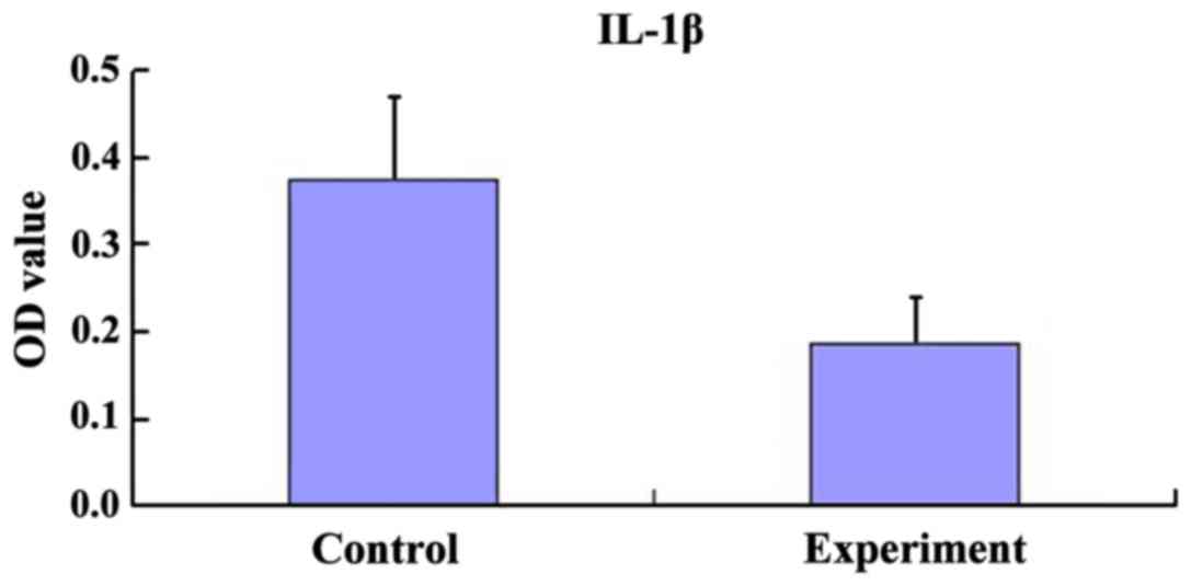
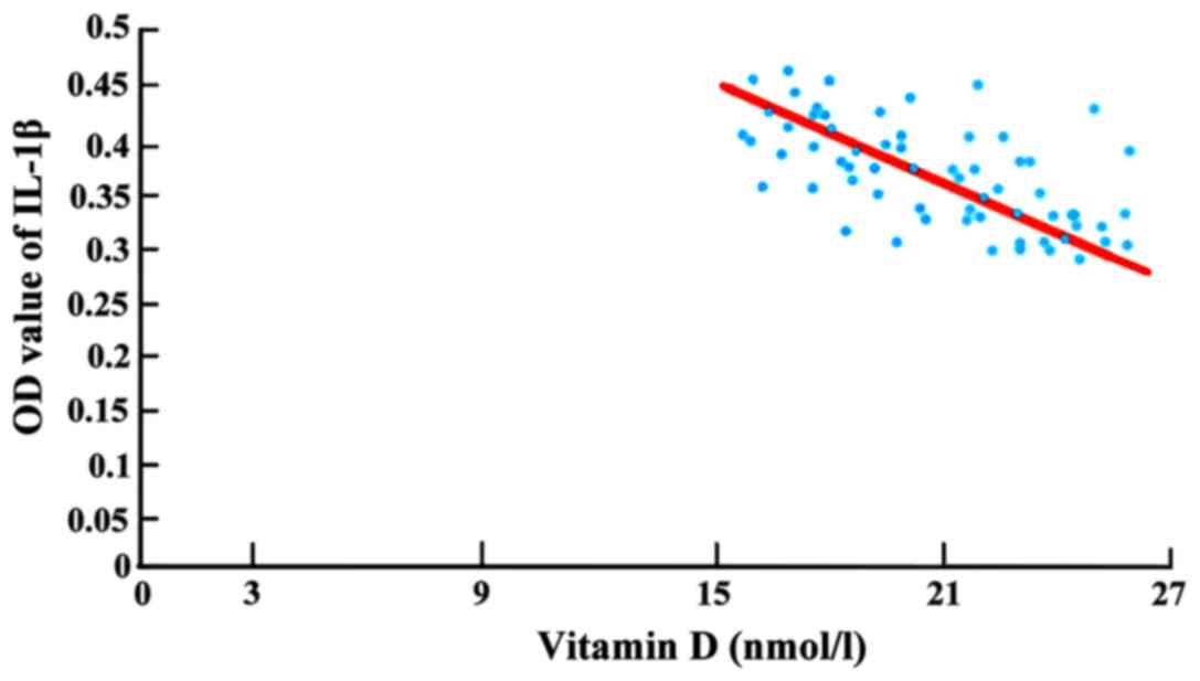
Comparison of IL-1β value in fibrous
rings
The content of IL-1β of inflammatory factor in
fibrous ring of control group was significantly higher than that of
the experimental group, P<0.05 (Fig.
1). Furthermore, vitamin D of control group had a strong
correlation with the content of IL-1β (r= −0.742), with a negative
correlation (Fig. 2).
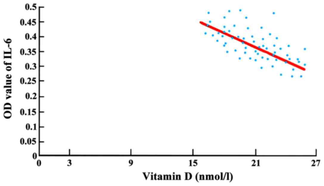
Comparison of IL-6 value in fibrous
rings
The content of IL-6 of inflammatory factor in
fibrous ring of control group was significantly higher than that of
the experimental group, and the difference had statistical
significance, P<0.05 (Fig. 3). In
addition, vitamin D of control group had a strong correlation with
the content of IL-6 (r= −0.715), with a negative correlation
(Fig. 4).
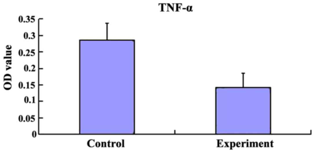
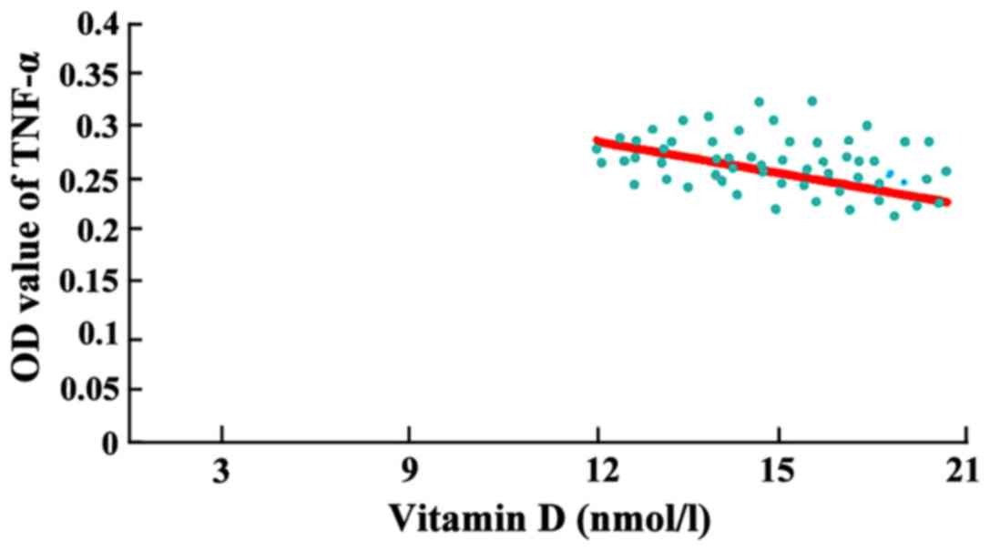
Comparison of TNF-α value in fibrous
rings
The content of tumor necrosis factor TNF-α in
fibrous ring of control group was significantly higher than that of
experimental group, and the difference had statistical
significance, P<0.05 (Fig. 5).
Moreover, vitamin D of the control group had a strong correlation
with the content of TNF-α (r= −0.803), with a negative correlation
(Fig. 6).
Discussion
At present, due to appearance of tuberculosis
drug-resistance bacteria, the prevention and cure of tuberculosis
is facing a serious situation (6).
Therefore, exploring new treatment methods based on the existing
medicines has a significant meaning. Recent research has revealed
that vitamin D plays an important regulatory role in immune system
of patients with tuberculosis. This in turn could improve the
antibacterial ability of Mycobacterium tuberculosis. Thus,
vitamin D could be taken as an adjuvant therapy for the patients
with tuberculosis (7,8).
1, 25-(OH)2-D3, one of active
ingredients of vitamin D, has a verified hormone effect in human
body. This could adjust the immune function of organism to keep
human immunity balance (9). T
lymphocyte subset and monocytes are the target cells of
1,25-(OH)2-D3 immunoregulation, to enhance
the immunity of organism by proliferation promotion (10). The results of the study showed that
the expression of T lymphocyte subsets in patients with spinal
tuberculosis (lacked vitamin D) were lower than those of patients
(vitamin D content was normal). The level of T lymphocyte subsets
in vivo could represent the immunity strength of cells in
human body, as a crucial laboratory index reflecting cell function.
When a person is infected with Mycobacterium tuberculosis,
infection could inhibit the growth of immune cells and weaken
normal immune cell function (11).
The ratio of CD4+ and CD8+ in
T lymphocyte subsets reflects the immunocompetence of the organism.
A high ratio indicates a good immunity, and decreasing ratio
suggests a damaged immune-competence. It is also related to the
severity of tuberculosis. The lower the ratio, the more serious the
tuberculosis (12,13). The present study showed that the
ratio between CD4+/CD8+ of the patients with
spinal tuberculosis (lack vitamin D) were lower than that of
patients with spinal tuberculosis (vitamin D content was normal).
The immunity of human body is regulatory and disequilibrium might
result in an abnormal immunity (14).
Research of specific pathogenesis of spinal
tuberculosis is not completely definite yet. It has been considered
that the occurrence of spinal tuberculosis is related to the
function of cytokines and abnormal immunity in vivo. A study
found that IL-6 in spinal tuberculosis is one of the cytokines that
led to osteoporosis (15). During
the pathologic process, IL-6 could cause decreasing proteoglycan,
so as to influence the compound of fibrocytes, which is one of the
reasons that led to spine inflammatory lesion progress. In
addition, IL-6 also influences the function of immune cells,
leading to pathologic change of intervertebral disc among patients.
A recent study showed that the contents of IL-6 in patients with
spinal tuberculosis were higher than those of normal people
(16). In the present study, the
contents of IL-6 of patients with spinal tuberculosis (lacked
vitamin D) were significantly higher than those of patients (normal
vitamin D content). The expression of IL-6 had a negative
correlation with Vitamin D. Thus, supplement of vitamin D might be
helpful to decrease the generation of IL-6 in the inflammatory
process and protect the intervertebral disc.
Tumor necrosis factor TNF-α has the ability to
stimulate the production of cytokines Th1 and improve immunity of
organism during the development of spinal tuberculosis (17). However, overexpression of TNF-α in
vivo would lead to an immune injury process producing an
undesirable effect on organism (18). In this study, TNF-α of intervertebral
disc tuberculosis lesion collected from the patients with spinal
tuberculosis was detected. It was observed that the contents of
patients with vitamin D deficiency were higher than those of normal
vitamin D group. In the same way, there was a negative correlation
between them. Thus, keeping a proper vitamin D content in
vivo is beneficial in prevention of overexpression of TNF-α in
patients.
IL-1β is a proinflammatory cytokine responsible for
a strong proinflammatory function against spinal tuberculosis
inflammation. During the process of stimulating organism to
compound other cytokines, IL-1β also could activate monocytes and
macrophages at the same time. This in turn leads to inflammatory
infiltration of lesion in patients with spinal tuberculosis
(19). Thus, IL-1β is an important
factor causing inflammation and one of the reasons leading to
abnormal expression of inflammatory cells in patients with spinal
tuberculosis (20). The present
study also found that the expression of IL-1β in vitamin D
deficiency group was higher than that of normal vitamin D group as
for the intervertebral disc lesions of patients with spinal
tuberculosis, and this had a negative correlation with vitamin D
content. Thus, it is suggested that vitamin D might be helpful to
reduce the expression of IL-1β in intervertebral disc lesion of
patients with spinal tuberculosis.
Collectively, the expression of T lymphocyte subsets
in patients with spinal tuberculosis and vitamin D deficiency
significantly reduced, and the immune function decreased as well.
The expressions of IL-1β, IL-6 and TNF-α in lesion were
significantly higher than those of patients with normal vitamin D
content. In addition, the lower the content of vitamin D is, the
more active the expression of inflammatory factors are, which is
not conducive to the recovery of tuberculosis lesions. However, the
number of study samples is not large enough, which may require
further improvement in the following studies.
Acknowledgements
Not applicable.
Funding
No funding was received.
Availability of data and materials
The datasets used and/or analyzed during the current
study are available from the corresponding author on reasonable
request.
Authors' contributions
SHZ drafted the manuscript. XW and SHZ treated the
patients. MYF and HLL analyzed vitamin D levels. FB and TH helped
with T lymphocyte subsets determination and hematoxylin and eosin
staining. HYF contributed to immumohistochemical staining. All
authors read and approved the final version of the manuscript.
Ethics approval and consent to
participate
The study was approved by the Ethics Committee of
Wuhan City Third Hospital (Hubei, China). Written informed consents
were signed by the patients and/or guardians.
Consent for publication
Not applicable.
Competing interests
The authors declare that they have no competing
interests.
References
|
1
|
Garg RK and Somvanshi DS: Spinal
tuberculosis: A review. J Spinal Cord Med. 34:440–454. 2011.
View Article : Google Scholar : PubMed/NCBI
|
|
2
|
Zheng C, Li P and Kan W: Video-assisted
thoracoscopic anterior surgery combined posterior instrumentation
for children with spinal tuberculosis. Eur J Pediatr Surg.
24:83–87. 2014. View Article : Google Scholar : PubMed/NCBI
|
|
3
|
Tuli SM: General principles of
osteoarticular tuberculosis. Clin Orthop Relat Res. 398:11–19.
2002. View Article : Google Scholar
|
|
4
|
Hewison M: Vitamin D and immune function:
An overview. Proc Nutr Soc. 71:50–61. 2012. View Article : Google Scholar : PubMed/NCBI
|
|
5
|
Arya SC and Agarwal N: Vitamin D
deficiency in adult tuberculosis patients. Int J Tuberc Lung Dis.
15:1133–1134. 2011. View Article : Google Scholar : PubMed/NCBI
|
|
6
|
Mathema B, Kurepina NE, Bifani PJ and
Kreiswirth BN: Molecular epidemiology of tuberculosis: Current
insights. Clin Microbiol Rev. 19:658–685. 2006. View Article : Google Scholar : PubMed/NCBI
|
|
7
|
Martineau AR, Timms PM, Bothamley GH,
Hanifa Y, Islam K, Claxton AP, Packe GE, Moore-Gillon JC,
Darmalingam M, Davidson RN, et al: High-dose vitamin D(3) during
intensive-phase antimicrobial treatment of pulmonary tuberculosis:
A double-blind randomised controlled trial. Lancet. 377:242–250.
2011. View Article : Google Scholar : PubMed/NCBI
|
|
8
|
Kota SK, Jammula S, Kota SK, Tripathy PR,
Panda S and Modi KD: Effect of vitamin D supplementation in type 2
diabetes patients with pulmonary tuberculosis. Diabetes Metab
Syndr. 5:85–89. 2011. View Article : Google Scholar : PubMed/NCBI
|
|
9
|
Hughes DA and Norton R: Vitamin D and
respiratory health. Clin Exp Immunol. 158:20–25. 2009. View Article : Google Scholar : PubMed/NCBI
|
|
10
|
Khoo AL, Chai LY, Koenen HJ, Oosting M,
Steinmeyer A, Zuegel U, Joosten I, Netea MG and van der Ven AJ:
Vitamin D(3) down-regulates proinflammatory cytokine response to
Mycobacterium tuberculosis through pattern recognition receptors
while inducing protective cathelicidin production. Cytokine.
55:294–300. 2011. View Article : Google Scholar : PubMed/NCBI
|
|
11
|
Warmelink I, van Altena R, ten Hacken N,
van der Werf T and van der Veer E: Nutritional status and vitamin
D3 during antimicrobial treatment. Lancet. 377:1407–1408, author
reply 1408. 2011. View Article : Google Scholar : PubMed/NCBI
|
|
12
|
Bikle DD: Vitamin D regulation of immune
function. Vitam Horm. 86:1–21. 2011. View Article : Google Scholar : PubMed/NCBI
|
|
13
|
Luong K and Nguyen LT: Impact of vitamin D
in the treatment of tuberculosis. Am J Med Sci. 341:493–498. 2011.
View Article : Google Scholar : PubMed/NCBI
|
|
14
|
Taylor CE and Camargo CA Jr: Impact of
micronutrients on respiratory infections. Nutr Rev. 69:259–269.
2011. View Article : Google Scholar : PubMed/NCBI
|
|
15
|
Raja A: Immunology of tuberculosis. Indian
J Med Res. 120:213–232. 2004.PubMed/NCBI
|
|
16
|
Takahasi H, Suguro T, Okazima Y, Motegi M,
Okada Y and Kakiuchi T: Inflammatory cytokines in the herniated
dise of the lumber. Spine (Phila Pa 1976). 21:218–224. 1996.
View Article : Google Scholar : PubMed/NCBI
|
|
17
|
Weitzmann MN, Cenci S, Rifas L, Brown C
and Pacifici R: Interleukin-7 stimulates osteoclast formation by
up-regulating the T-cell production of soluble osteoclastogenic
cytokines. Blood. 96:1873–1878. 2000.PubMed/NCBI
|
|
18
|
Zganiacz A, Santosuosso M, Wang J, Yang T,
Chen L, Anzulovic M, Alexander S, Gicquel B, Wan Y, Bramson J, et
al: TNF-alpha is a critical negative regulator of type 1 immune
activation during intracellular bacterial infection. J Clin Inves.
113:401–413. 2004. View
Article : Google Scholar
|
|
19
|
Akyol S, Eraslan BS, Etyemez H, Tanriverdi
T and Hanci M: Catabolic cytokine expressions in patients with
degenerative disc disease. Turk Neurosurg. 20:492–499.
2010.PubMed/NCBI
|
|
20
|
Bai X, Wilson SE, Chmura K, Feldman NE and
Chan ED: Morphometric analysis of Th(1) and Th(2) cytokine
expression in human pulmonary tuberculosis. Tuberculosis (Edinb).
84:375–385. 2004. View Article : Google Scholar : PubMed/NCBI
|