Introduction
Sevoflurane is a kind of commonly-used inhalational
general anesthetics. A study has pointed out (1) that sevoflurane can lead to the decline
in cognitive function in the recovery period, which may be related
to the surgical trauma, anesthesia depth, body inflammation, stress
and immune dysfunction. The elderly patients are often complicated
with low metabolic function, poor operation and anesthesia
tolerance, and many perioperative complications, so the
postoperative cognitive dysfunction (POCD) occurs more easily after
operation (2). Hippocampus is an
important brain area involved in learning and memory ability, and
the pathological changes, such as β-amyloid protein deposit and Tau
protein phosphorylation, caused by hippocampal neurons are the
important mechanisms of Alzheimer's disease (3); therefore, it is assumed that POCD
caused by sevoflurane anesthesia may also be related to
abnormalities in hippocampal neuronal structure and function. Cell
autophagy is an important way for eukaryotes to degrade the
misfolded proteins and damaged organelles in the body via
lysosomes, and maintain the homeostasis, which is also known as
type II programmed death (4). The
decreased autophagy is associated with tumorigenesis (4), while the enhanced autophagy is related
to the occurrence of neurodegenerative disease (5). Autophagosomes and specific proteins
light-chain 3-I (LC3-I), LC3-II and Beclin-1 can reflect the
autophagic activity of the body. Dexmedetomidine is a
highly-selective α adrenergic receptor agonist, widely used as an
anesthesia adjuvant drug in clinic, which is characterized by rapid
onset of analgesia and sedation, small side effects, little
inhibition on respiration and circulation and high safety (6,7). This
study aimed to analyze the effects of dexmedetomidine on autophagy
of hippocampal neurons and cognitive dysfunction in aged rats under
sevoflurane anesthesia.
Materials and methods
Animals
Sixty healthy Sprague-Dawley (SD) rats aged 4–6
months of either sex, weighing 150–200 g were purchased from
Laboratory Animal Center of Jilin University (certificate no.: SCXK
(Ji) 2008–0004). They ate and drank freely at room temperature of
22±2°C and relative humidity of 55±2% to adapt to the environment
for 1 week, followed by subsequent experiments. The study was
approved by the Animal Ethics Committee of Nanchang University
(Nanchang, China).
Research methods
The rats were randomly divided into control group
(n=10), sevoflurane group (n=25) and dexmedetomidine + sevoflurane
group (compound group, n=25). The control group was inhaled with
60% O2 for 6 h, the sevoflurane group was inhaled with
3.4–3.6% sevoflurane for 6 h, and the compound group was treated
with intraperitoneal injection of 4 µg/kg dexmedetomidine 1 h
before the inhalation of sevoflurane. Five rats were taken from
each group before sevoflurane anesthesia (T1), immediately after
anesthesia (T2), 12 h after anesthesia (T3) and 24 h after
anesthesia (T4). The expression levels of microtubule-associated
protein 1 LC3-I, LC3-II and Beclin-1 were detected via western
blotting, and the LC3-II/LC3-I value was calculated. Moreover,
Morris water maze test was performed for the five rats in each
group 5 weeks after anesthesia to detect the cognitive function;
the escape latency, times across platform and swimming time in
target quadrant were recorded.
Materials
The rats were placed in a plastic airtight
transparent container, then the container was placed in the ZD-600
thermostatic water bath to maintain the temperature of 37°C, and
the bottom of the container was covered with calcium lime to absorb
the CO2 generated; two side walls of the container were
the air inlet and exhaust port; the air inlet was connected to the
Vapor 2000 anesthetic gas volatilization pot (Dreager, Lübeck,
Germany) for oxygen or sevoflurane delivery (Jiangsu Hengrui
Medicine Co., Ltd., Jiangsu, China), while the exhaust port was
connected to the Vamos multifunctional anesthetic gas monitor
(Dreager) for monitoring the sevoflurane and CO2
concentrations; the abdomen of rats was connected to the probe of
oxygen saturation monitor (Philips Medical Systems B.V., Eindhoven,
The Netherlands).
Western blotting
After anesthesia using chloral hydrate via
intraperitoneal injection at a dose of 300–350mg/kg, the rats were
sacrificed and the brain was removed, the hippocampus tissues were
separated and placed in liquid nitrogen at −80°C. After hippocampus
tissue homogenate, the centrifugation was performed at 4°C at
10,500 × g for 15 min; the supernatant was taken, and the protein
concentration was detected using the bicinchoninic acid (BCA)
protein quantification kit (Jiangsu Nanjing Beyotime Technology
Co., Ltd., Jiangsu, China). The loading buffer was added, followed
by 12% sodium dodecyl sulfate polyacrylamide gel electrophoresis
(SDS-PAGE); the protein was transferred onto the polyvinylidene
fluoride (PVDF) membrane. After sealing with blocking buffer at
37°C for 1 h, rabbit polyclonal LC3-I (dilution: 1:2,000; cat. no.
ab48394), rabbit polyclonal LC3-II (dilution: 1:2,000; cat. no.
ab192890), rabbit polyclonal Beclin-1 (dilution: 1:2,000; cat. no.
ab207612) and rabbit polyclonal GAPDH (dilution: 1:500, cat. no.
ab37168), purchased from Abcam (Cambridge, MA, USA), were added at
4°C overnight. After washing, the goat anti-mouse polyclonal
secondary goat anti-mouse (HRP) IgG antibody (dilution: 1:2,000;
cat. no. ab6789; Abcam, Cambridge, MA, USA) was incubated at 37°C
for 4 h, followed by washing via phosphate buffered saline (PBS)
and ECL development. The results were scanned and saved, and the
semi-quantitative analysis was performed using the Lab Works4.5 gel
imaging software (Invitrogen; Thermo Fisher Scientific, Inc.).
Morris water maze test
Morris water maze test included the place navigation
and space exploration. In place navigation test, the platform was
placed in the third quadrant, and the rats stood on the platform
for 30 sec; the water was poured into the pool wall from different
quadrants, and the time of finding and climbing on platform within
60 sec (escape latency) was recorded. After finding the platform,
the rats stood for 30 sec and rested. If the rats failed to find
the platform within 60 sec, they were led onto the platform to stay
for 15 sec, and the escape latency was recorded as 60 sec. The test
was repeated 4 times per day for 5 days, and the average was taken.
In space exploratory test, the platform was removed, and the rat
was put into the water optionally from the same entry point; the
swimming trajectory within 60 sec was shot using a camera, the
times across platform and the swimming time in original quadrant
(target quadrant) were recorded.
Statistical analysis
Statistical analysis was performed using Statistical
Product and Service Solutions (SPSS) 20.0 software (IBM Corp.,
Armonk, NY, USA). Measurement data are presented as mean ± standard
deviation; one-way analysis of variance (ANOVA) was used for the
comparison among groups, and least-significant difference (LSD)
t-test was used for the pairwise comparison. The repeated measures
analysis of variance (ANOVA) was used for the data comparisons at
different time points. P<0.05 was considered to indicate a
statistically significant difference.
Results
Expression levels of
autophagy-associated proteins at different time points
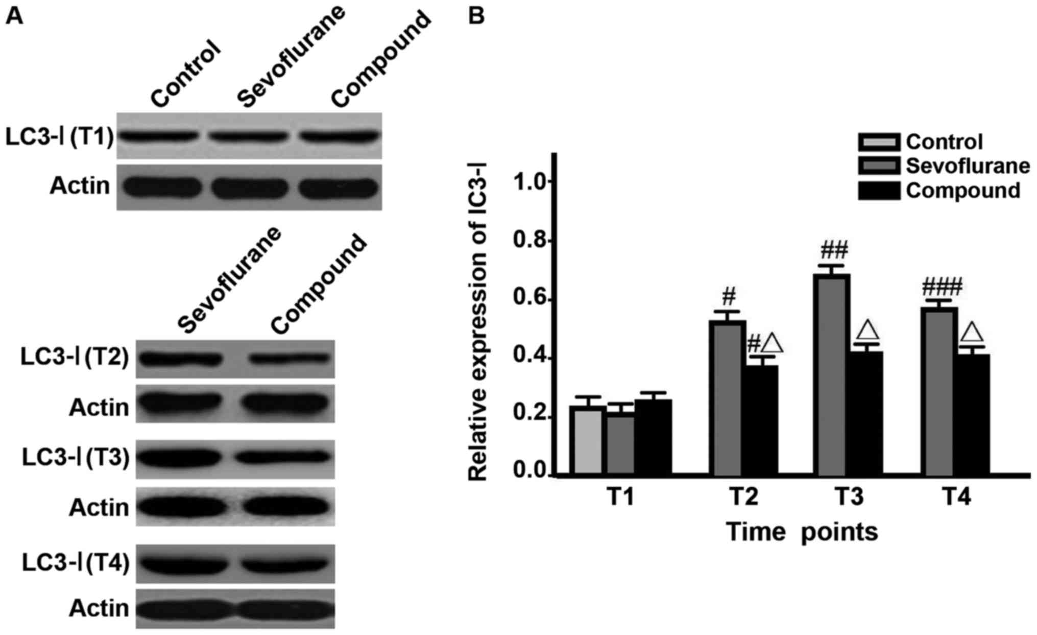
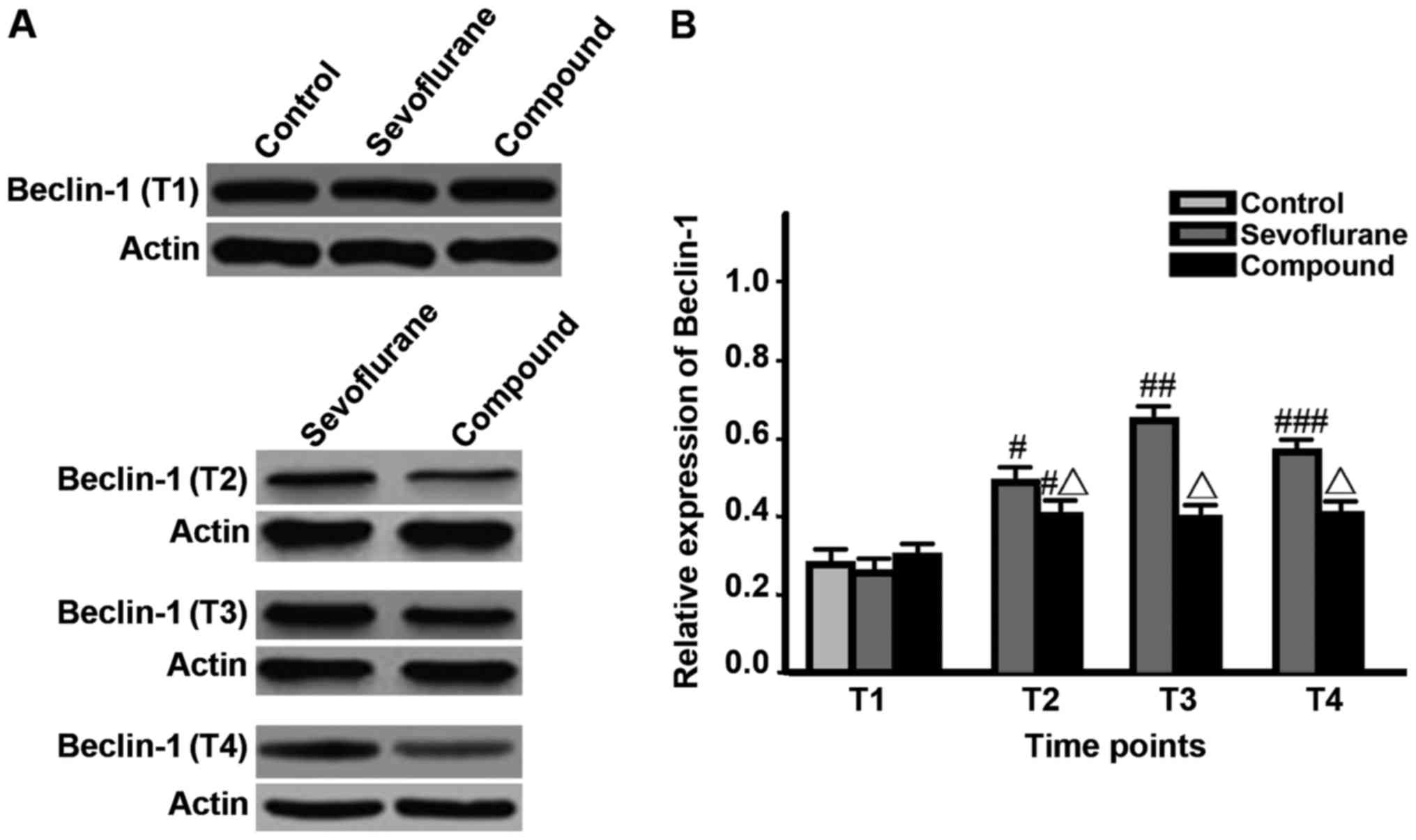
The levels of LC3-I, LC3-II and Beclin-1 in
hippocampal neurons and LC3-II/LC3-I value in sevoflurane group at
T2 were significantly higher than those at T1, and the value was
the highest at T3, but were decreased at T4 (P<0.05); the levels
of proteins and LC3-II/LC3-I value in compound group at T2 were
significantly higher than those at T1 and reached the peak, and
remained unchanged at T3-T4; besides, the levels of proteins and
LC3-II/LC3-I values in compound group at T2-T4 were significantly
lower than those in sevoflurane group (P<0.05) (Figs. 1–3,
Table I).
 | Table I.Comparison of LC3-II/LC3-I values at
different time points. |
Table I.
Comparison of LC3-II/LC3-I values at
different time points.
| Groups | T1 | T2 | T3 | T4 |
|---|
| Control | 1.05±0.26 | – | – | – |
| Sevoflurane | 1.03±0.23 |
0.89±0.13a |
0.92±0.15b |
0.77±0.14c |
| Compound | 1.07±0.28 |
0.67±0.14a,d |
0.68±0.16d |
0.66±0.12d |
Escape latency, times across platform
and swimming time in target quadrant of rats
In sevoflurane group, the escape latency was
prolonged, the times across platform were reduced and the swimming
time in target quadrant was shortened compared with those in
control group (P<0.05); these parameters were significantly
improved in compound group compared with those in sevoflurane group
and control group (P<0.05) (Table
II).
| Table II.Escape latency, times across platform
and swimming time in target quadrant of rats. |
Table II.
Escape latency, times across platform
and swimming time in target quadrant of rats.
| Groups | Escape latency
(s) | Times across platform
(time) | quadrant (s) |
|---|
| Control | 26.7±4.5 | 2.3±0.3 | 14.6±2.3 |
| Sevoflurane |
33.5±4.9a |
1.6±0.3a |
10.4±2.1a |
| Compound |
28.2±4.6b |
2.1±0.4b |
13.2±2.4b |
Discussion
The median effective dose of dexmedetomidine
analgesic on rats is 1 µg/kg (8).
Dexmedetomidine improves the occurrence of cognitive dysfunction
after sevoflurane anesthesia, which may be related to the clearance
of damaged mitochondria, delayed apoptosis, inhibition of
cytochrome c release, reduction of Caspase family
activation, and reduction of apoptosis (9).
In this study, sevoflurane was used on anesthetized
aged SD rats, and the effects of dexmedetomidine on hippocampal
neurons autophagy and cognitive impairment in old rats were
studied.
Morris water maze test is a common method to
evaluate the spatial learning and memory ability of rodents
(10). The results are reliable and
repeatable. The study concluded that compared with rats in the
control group, the rats in sevoflurane group had longer escape
latency; they crossed the platform fewer times, and swam for a
shorter period of time in the target quadrant. The condition of
rats in the composite group was significantly better compared with
that in sevoflurane group. Therefore, we believe that
dexmedetomidine can improve the cognitive dysfunction of
sevoflurane anesthesia in aged rats.
Liu et al (11) showed that the sevoflurane
anesthesia-induced POCD in aged rats is related to the β-amyloid
deposit and Tau protein phosphorylation of hippocampal neurons.
Satomoto et al (12) pointed
out that the sevoflurane anesthesia can cause behavioral changes
and cognitive abnormalities in young rats. Autophagy is a defense
behavior of the body against the environmental stimulus, and the
autophagosomes and specific proteins LC3-I, LC3-II and Beclin-1 can
reflect the autophagic activity of the body (13). LC3-I can bind with the
phosphatidylethanolamin on the surface of autophagy membrane after
the ubiquitin-like modification, forming LC3-II (13), both of which are two protein products
in the autophagy process, and the LC3-II/LC3-I value is better in
reflecting the stability of autophagy. Beclin-1 is a specific gene
involved in autophagy regulation in mammals.
The downregulation of Beclin-1 expression indicates
the decline in autophagy, which is related to the anti-apoptotic
protein Bcl-2 (14). Beclin-1
contains the BH3 structural domain, which binds Beclin-1 and Bcl-2
to inhibit Beclin-1-dependent autophagy-activated pathway through
the interaction with Bcl-2 surface BH3. However, the combination of
Beclin-1 and Bcl-2 does not affect the fact that Bcl-2 binds to the
pro-apoptotic protein Bax, playing an anti-apoptotic effect,
through the structural domain (15).
The results of this study showed that the levels of LC3-I, LC3-II
and Beclin-1 in hippocampal neurons and LC3-II/LC3-I value in
sevoflurane group at T2 were significantly higher than those at T1,
and they reached the peak at T3, but decreased at T4; the level of
proteins and LC3-II/LC3-I values in compound group at T2-T4 were
obviously lower than those in sevoflurane group. Therefore, it is
believed that sevoflurane anesthesia can up-regulate the autophagic
activity of hippocampal neurons, and the anesthesia combined with
dexmedetomidine can reduce the autophagic activity.
This study showed that the mechanism of sevoflurane
anesthesia leading to the cognitive decline of rats was related to
the increased autophagy of hippocampal neurons, providing an
important reference basis for the clinical diagnosis and prevention
of POCD. At the same time, the dexmedetomidine-assisted anesthesia
can improve the cognitive function, providing an important idea for
the application of dexmedetomidine.
Acknowledgements
Not applicable.
Funding
No funding was received.
Availability of data and materials
The datasets used and/or analyzed during the present
study are available from the corresponding author on reasonable
request.
Authors' contributions
CY conceived and designed the study, and drafted the
manuscript. CY, ZF and XL collected, analyzed and interpreted the
experimental data. ZF revised the manuscript for important
intellectual content. All authors read and approved the final
manuscript.
Ethics approval and consent to
participate
The study was approved by the Animal Ethics
Committee of Nanchang University (Nanchang, China).
Consent for publication
Not applicable.
Competing interests
The authors declare that they have no competing
interests.
References
|
1
|
Tao G, Zhang J, Zhang L, Dong Y, Yu B,
Crosby G, Culley DJ, Zhang Y and Xie Z: Sevoflurane induces tau
phosphorylation and glycogen synthase kinase 3β activation in young
mice. Anesthesiology. 121:510–527. 2014. View Article : Google Scholar : PubMed/NCBI
|
|
2
|
Rundshagen I: Postoperative cognitive
dysfunction. Dtsch Arztebl Int. 111:119–125. 2014.PubMed/NCBI
|
|
3
|
Nixon RA: Autophagy, amyloidogenesis and
Alzheimer disease. J Cell Sci. 120:4081–4091. 2007. View Article : Google Scholar : PubMed/NCBI
|
|
4
|
Galluzzi L, Pietrocola F, Bravo-San Pedro
JM, Amaravadi RK, Baehrecke EH, Cecconi F, Codogno P, Debnath J,
Gewirtz DA, Karantza V, et al: Autophagy in malignant
transformation and cancer progression. EMBO J. 34:856–880. 2015.
View Article : Google Scholar : PubMed/NCBI
|
|
5
|
Cai Y, Arikkath J, Yang L, Guo ML,
Periyasamy P and Buch S: Interplay of endoplasmic reticulum stress
and autophagy in neurodegenerative disorders. Autophagy.
12:225–244. 2016. View Article : Google Scholar : PubMed/NCBI
|
|
6
|
Whittington RA, Virág L, Gratuze M, Petry
FR, Noël A, Poitras I, Truchetti G, Marcouiller F, Papon MA, El
Khoury N, et al: Dexmedetomidine increases tau phosphorylation
under normothermic conditions in vivo and in vitro. Neurobiol
Aging. 36:2414–2428. 2015. View Article : Google Scholar : PubMed/NCBI
|
|
7
|
Ni J, Wei J, Yao Y, Jiang X, Luo L and Luo
D: Effect of dexmedetomidine on preventing postoperative agitation
in children: A meta-analysis. PLoS One. 10:e01284502015. View Article : Google Scholar : PubMed/NCBI
|
|
8
|
Sanders RD, Giombini M, Ma D, Ohashi Y,
Hossain M, Fujinaga M and Maze M: Dexmedetomidine exerts
dose-dependent age-independent antinociception but age-dependent
hypnosis in Fischer rats. Anesth Analg. 100:1295–1302. 2005.
View Article : Google Scholar : PubMed/NCBI
|
|
9
|
Zhu M, Wang H, Zhu A, Niu K and Wang G:
Meta-analysis of dexmedetomidine on emergence agitation and
recovery profiles in children after sevoflurane anesthesia:
Different administration and different dosage. PLoS One.
10:e01237282015. View Article : Google Scholar : PubMed/NCBI
|
|
10
|
Zhang C, Hu J, Liu X and Yan J: Effects of
intravenous dexmedetomidine on emergence agitation in children
under sevoflurane anesthesia: A meta-analysis of randomized
controlled trials. PLoS One. 9:e997182014. View Article : Google Scholar : PubMed/NCBI
|
|
11
|
Liu W, Xu J, Wang H, Xu C, Ji C, Wang Y,
Feng C, Zhang X, Xu Z, Wu A, et al: Isoflurane-induced spatial
memory impairment by a mechanism independent of amyloid-beta levels
and tau protein phosphorylation changes in aged rats. Neurol Res.
34:3–10. 2012. View Article : Google Scholar : PubMed/NCBI
|
|
12
|
Satomoto M, Satoh Y, Terui K, Miyao H,
Takishima K, Ito M and Imaki J: Neonatal exposure to sevoflurane
induces abnormal social behaviors and deficits in fear conditioning
in mice. Anesthesiology. 110:628–637. 2009. View Article : Google Scholar : PubMed/NCBI
|
|
13
|
Zhou YF, Wang QX, Zhou HY and Chen G:
Autophagy activation prevents sevoflurane-induced neurotoxicity in
H4 human neuroglioma cells. Acta Pharmacol Sin. 37:580–588. 2016.
View Article : Google Scholar : PubMed/NCBI
|
|
14
|
Liu K, Shi Y, Guo X, Wang S, Ouyang Y, Hao
M, Liu D, Qiao L, Li N, Zheng J, et al: CHOP mediates ASPP2-induced
autophagic apoptosis in hepatoma cells by releasing Beclin-1 from
Bcl-2 and inducing nuclear translocation of Bcl-2. Cell Death Dis.
5:e13232014. View Article : Google Scholar : PubMed/NCBI
|
|
15
|
Trisciuoglio D, de Luca T, Desideri M,
Passeri D, Gabellini C, Scarpino S, Liang C, Orlandi A and Del
Bufalo D: Removal of the BH4 domain from Bcl-2 protein triggers an
autophagic process that impairs tumor growth. Neoplasia.
15:315–327. 2013. View Article : Google Scholar : PubMed/NCBI
|