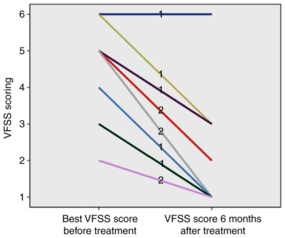
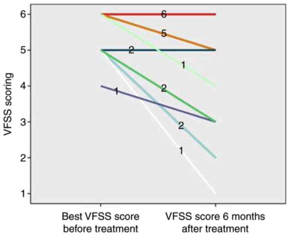
|
1
|
Schneider E, Zimmermann H, Oberwahrenbrock
T, Kaufhold F, Kadas EM, Petzold A, Bilger F, Borisow N, Jarius S,
Wildemann B, et al: Optical coherence tomography reveals distinct
patterns of retinal damage in neuromyelitis optica and multiple
sclerosis. Plos One. 8:e661512013. View Article : Google Scholar : PubMed/NCBI
|
|
2
|
Wan H, He H, Zhang F, Sha Y and Tian G:
Diffusion-weighted imaging helps differentiate multiple sclerosis
and neuromyelitis optica-related acute optic neuritis. J Magn Reson
Imaging. 45:1780–1785. 2017. View Article : Google Scholar : PubMed/NCBI
|
|
3
|
Tan CT, Mao Z, Qiu W, Hu X, Wingerchuk DM
and Weinshenker BG: International consensus diagnostic criteria for
neuromyelitis optica spectrum disorders. Neurology. 86:491–492.
2016. View Article : Google Scholar : PubMed/NCBI
|
|
4
|
Kitley J, Waters P, Woodhall M, Leite MI,
Murchison A, George J, Kuker W, Chandratre S, Vincent A and Palace
J: Neuromyelitis optica spectrum disorders with aquaporin-4 and
myelin-oligodendrocyte glycoprotein antibodies: A comparative
study. Jama Neurol. 71:276–283. 2014. View Article : Google Scholar : PubMed/NCBI
|
|
5
|
Giacoppo S, Soundara RT, Galuppo M,
Pollastro F, Grassi G, Bramanti P and Mazzon E: Purified
Cannabidiol, the main non-psychotropic component of Cannabis
sativa, alone, counteracts neuronal apoptosis in experimental
multiple sclerosis. Eur Rev Med Pharmacol Sci. 19:4906–4919.
2015.PubMed/NCBI
|
|
6
|
Saadoun S, Waters P, Owens GP, Bennett JL,
Vincent A and Papadopoulos MC: Neuromyelitis optica MOG-IgG causes
reversible lesions in mouse brain. Acta Neuropathol Commun.
2:352014. View Article : Google Scholar : PubMed/NCBI
|
|
7
|
Matsuda R, Kezuka T, Umazume A, Okunuki Y,
Goto H and Tanaka K: Clinical profile of Anti-Myelin
oligodendrocyte glycoprotein antibody seropositive cases of optic
neuritis. Neuroophthalmology. 39:213–219. 2015. View Article : Google Scholar : PubMed/NCBI
|
|
8
|
Hoftberger R, Sepulveda M, Armangue T,
Blanco Y, Rostasy K, Calvo AC, Olascoaga J, Ramio-Torrenta L,
Reindl M, Benito-Leon J, et al: Antibodies to MOG and AQP4 in
adults with neuromyelitis optica and suspected limited forms of the
disease. Mult Scler. 21:866–874. 2015. View Article : Google Scholar : PubMed/NCBI
|
|
9
|
Kim SM, Woodhall MR, Kim JS, Kim SJ, Park
KS, Vincent A, Lee KW and Waters P: Antibodies to MOG in adults
with inflammatory demyelinating disease of the CNS. Neurol
Neuroimmunol Neuroinflamm. 2:e1632015. View Article : Google Scholar : PubMed/NCBI
|
|
10
|
Nakajima H, Motomura M, Tanaka K, Fujikawa
A, Nakata R, Maeda Y, Shima T, Mukaino A, Yoshimura S, Miyazaki T,
et al: Antibodies to myelin oligodendrocyte glycoprotein in
idiopathic optic neuritis. BMJ Open. 5:e77662015. View Article : Google Scholar
|
|
11
|
Sato DK, Callegaro D, Lana-Peixoto MA,
Waters PJ, de Haidar JF, Takahashi T, Nakashima I,
Apostolos-Pereira SL, Talim N, Simm RF, et al: Distinction between
MOG antibody-positive and AQP4 antibody-positive NMO spectrum
disorders. Neurology. 82:474–481. 2014. View Article : Google Scholar : PubMed/NCBI
|
|
12
|
Ramanathan S, Prelog K, Barnes EH, Tantsis
EM, Reddel SW, Henderson AP, Vucic S, Gorman MP, Benson LA, Alper
G, et al: Radiological differentiation of optic neuritis with
myelin oligodendrocyte glycoprotein antibodies, aquaporin-4
antibodies, and multiple sclerosis. Mult Scler. 22:470–482. 2016.
View Article : Google Scholar : PubMed/NCBI
|
|
13
|
Akaishi T, Nakashima I, Takeshita T,
Kaneko K, Mugikura S, Sato DK, Takahashi T, Nakazawa T, Aoki M and
Fujihara K: Different etiologies and prognoses of optic neuritis in
demyelinating diseases. J Neuroimmunol. 299:152–157. 2016.
View Article : Google Scholar : PubMed/NCBI
|
|
14
|
Siritho S, Sato DK, Kaneko K, Fujihara K
and Prayoonwiwat N: The clinical spectrum associated with myelin
oligodendrocyte glycoprotein antibodies (anti-MOG-Ab) in thai
patients. Mult Scler. 22:964–968. 2016. View Article : Google Scholar : PubMed/NCBI
|
|
15
|
Martinez-Hernandez E, Sepulveda M, Rostasy
K, Hoftberger R, Graus F, Harvey RJ, Saiz A and Dalmau J:
Antibodies to aquaporin 4, myelin-oligodendrocyte glycoprotein, and
the glycine receptor α1 subunit in patients with isolated optic
neuritis. JAMA Neurol. 72:187–193. 2015. View Article : Google Scholar : PubMed/NCBI
|