Introduction
Doxorubicin is an anthracycline-based topoisomerase
II inhibitor and DNA-modifying agent that has exhibited remarkable
anti-tumor effects against multiple cancer types, including
ovarian, lung and endometrial cancers (1). Doxorubicin is known to act by
intercalating into DNA and interfering with nucleic acid synthesis,
which halts the cell cycle and inhibits DNA transcription (2). However, the clinical utility of
doxorubicin as an anti-tumor drug is hampered by cardiotoxicity and
drug resistance (3).
Doxorubicin-mediated bone loss in cancer patients is produced by an
interaction between oxidative stress and the induction of
transforming growth factor-β (TGF-β) (4). Doxorubicin treatment has also been
indicated to increase the levels of circulating TGF-β in murine
pre-clinical models, and TGF-β promoted osteolytic bone damage,
leading to increased osteoclast-mediated resorption and suppression
of osteoblast differentiation (4).
In numerous previous studies, liposomes have been
used to deliver anti-cancer drugs, including doxorubicin, to local
tumors (5). Liposomal doxorubicin
along with conventional doxorubicin has potential risks of causing
adverse events, including myelosuppression, cardiotoxicity,
alopecia, nausea and vomiting (6).
However, liposomal doxorubicin formulations are associated with
lower reported odds ratios with regard to myelosuppression,
cardiotoxicity and alopecia than conventional doxorubicin on its
own (6). The effects of doxorubicin,
particularly doxorubicin liposomes, on stem cells have remained to
be fully elucidated (7). The aim of
the present study was to evaluate the effects of anionic, cationic
and neutral liposomes loaded with doxorubicin on the viability and
osteogenic differentiation potential of human gingiva-derived stem
cells in two-dimensional culture.
Materials and methods
Preparation of doxorubicin-loaded
liposomes
The thin-lipid-film hydration method was used to
prepare liposomes from the mixture.
1,2-dipalmitoyl-sn-glycero-3-phosphocholine (DPPC, Echelon
Biosciences, Salt Lake City, UT, USA) and cholesterol
(Sigma-Aldrich; Merck KGaA, Darmstadt, Germany) at a weight ratio
of 10:1 was used for the neutral liposome. For the cationic
liposome, DPPC, 1,2-dipalmitoyl-3-trimethylammonium-propane
(chloride salt, 16:0 TAP; Avanti Polar Lipids; Birmingham, AL, USA)
and cholesterol were used at a weight ratio of 5:5:1 and for the
anionic liposome, DPPC, 1,2-dipalmitoyl-sn-glycero-3-phosphoserine
plus sodium salt (Echelon Biosciences) and cholesterol were used at
a weight ratio of 5:5:1.
The lipids were dissolved in dichloromethane and the
solvent was removed via evaporation under reduced pressure at 55°C.
Subsequently, a thin film of lipids was dispersed in distilled
water (lipid concentration of 2.2 mg/ml) with doxorubicin
hydrochloride (LC Laboratories, Woburn, MA, USA) by sonication.
Dialysis was then performed in order to remove the unloaded
doxorubicin using distilled water for 1 h. After disrupting the
liposomes completely with a detergent solution (1% Triton X-100;
Samchun, Pyeongtaek-Si, South Korea), the amount of doxorubicin in
the liposomes was evaluated based on the fluorescence of
doxorubicin (490/570 nm).
Isolation of human gingiva-derived
stem cells
The Institutional Review Board of Seoul St. Mary's
Hospital (College of Medicine, Catholic University of Korea, Seoul,
Republic of Korea) approved the present study (nos. KC17SNSI0606
and KC11SISI0348), and informed consent was obtained from the
participants. Healthy patients visiting the Department of
Periodontics of Seoul St. Mary's Hospital (College of Medicine, The
Catholic University of Korea, Seoul, Republic of Korea) provided
the gingiva tissue for the study. The epithelium of the gingiva was
removed and the tissue was fragmented into 1–2 mm sections. The
tissues were digested with a medium containing dispase (1 mg/ml;
Sigma-Aldrich; Merck KGaA) and collagenase IV (2 mg/ml;
Sigma-Aldrich; Merck KGaA) (8).
Unattached cells were removed from the culture dish. The cells were
then cultured in an incubator with 5% CO2 and 95%
O2 at 37°C, and the medium was changed every two to
three days. For identification, ~1.5×10⁵ cells were
incubated with specific fluorescein isothiocyanate-conjugated mouse
monoclonal antibodies to human CD44 (11-0441-81; 1:200; BD
Biosciences, Franklin Lakes, NJ, USA), CD73 (11-0739-42; 1:40; BD
Biosciences), CD90 (11-0909-42; 1:40; BD Biosciences), CD14
(11-0149-42; 1:40; BD Biosciences) and CD34 (11-0349-42; 1:40; BD
Biosciences) at room temperature for 30 min. Flow cytometric
analysis was performed using a flow cytometer (FACS Canto II; BD
Biosciences,) and FACSDiva software (v8.0.1; BD Biosciences).
The 12 groups of the present study were as follows:
i) Unloaded control group (D0); ii) doxorubicin, 1 µg/ml (D1); iii)
doxorubicin, 10 µg/ml (D10); iv) unloaded anionic group (A0); v)
anionic group loaded with 1 µg/ml doxorubicin (A1); vi) anionic
group loaded with doxorubicin at 10 µg/ml (A10); vii) unloaded
cationic group (C0); viii) cationic group loaded with doxorubicin
at 1 µg/ml (C1); ix) cationic group loaded with doxorubicin at 10
µg/ml (C10); x) unloaded neutral group (N0); xi) neutral group
loaded with doxorubicin at 1 µg/ml (N1); and xii) neutral group
loaded with doxorubicin at 10 µg/ml (N10) (7,9).
Evaluation of cell morphology and
determination of cell viability
The cells (within 10 passages) were seeded in
96-well plates at a density of 2.0×10³ cells/well, and
the cells were cultured in osteogenic medium. The osteogenic medium
consisted of α-minimal essential medium (Gibco; Thermo Fisher
Scientific, Inc., Waltham, MA, USA) containing 15% fetal bovine
serum (Gibco; Thermo Fisher Scientific, Inc.), 100 U/ml penicillin,
100 µg/ml streptomycin (Sigma-Aldrich; Merck KGaA), 200 mM
L-glutamine (Sigma-Aldrich; Merck KGaA), 10 mM ascorbic acid
2-phosphate (Sigma-Aldrich; Merck KGaA), 38 µg/ml dexamethasone and
2 mg/ml glycerophosphate disodium salt hydrate. The morphology of
the cells was observed under an inverted microscope (Leica DM IRM,
Leica Microsystems, Wetzlar, Germany) on days 1, 3 and 7.
On days 1, 3 and 7, a cell viability analysis of the
stem cell spheroids cultured in osteogenic medium was performed.
Cell Counting Kit-8 (CCK-8) stain (Dojindo, Kumamoto, Japan) was
added to the cultures and the stem cells were incubated for 1 h at
37°C. Viable cells were identified by CCK-8 staining, which relies
on the ability of mitochondrial dehydrogenases to oxidize the dye
into a colored formazan product. A microplate reader (BioTek,
Winooski, VT, USA) was used to measure the spectrophotometric
absorbance of the samples at 450 nm.
Alkaline phosphatase activity
assay
The stem cells were seeded on 24-well plates at a
density of 2.0×10⁴ cells/well and the cells were
cultured in the osteogenic medium. On days 1, 5 and 7, alkaline
phosphatase activity assays were performed using a commercially
available kit (K412-500; BioVision, Inc., Milpitas, CA, USA). The
cells were re-suspended in an assay buffer, sonicated and
centrifuged to remove any insoluble material. The supernatant was
mixed with p-nitrophenyl phosphate substrate and incubated at 25°C
for 60 min. The optical density of the resultant p-nitrophenol was
determined spectrophotometrically at 405 nm.
Alizarin Red S staining
Alizarin Red S staining was performed on days 7 and
14. In brief, the cells were washed twice with PBS, fixed with 4%
paraformaldehyde at room temperature and rinsed twice with
deionized water. The cultures were then stained with Alizarin Red S
for 30 min at room temperature. To remove non-specifically bound
dye, the cultures were washed three times with deionized water. The
morphology was evaluated using an inverted microscope (Leica DM
IRM; Leica Microsystems).
Statistical analysis
The results are expressed as the mean ± standard
deviation of the experiments. Normality of the data was assessed
with a Shapiro-Wilk test, and one-way analysis of variance followed
by Tukey's post-hoc test was performed to determine the differences
between the groups using SPSS 12 for Windows (SPSS Inc., Chicago,
IL, USA). P<0.05 was considered to indicate a statistically
significant difference.
Results
Effects of doxorubicin-loaded
liposomes on cell morphology and viability
The expression of the CD44, CD73, CD90, CD14 and
CD34 surface markers was assessed (Fig.
1). The percentages of CD44+, CD73+,
CD90+, CD14+ and CD34+ cells were
100, 99.5, 99.9, 0.1 and 1.8%, respectively. Therefore, the cells
were positive for stem cell surface markers. The control group D0
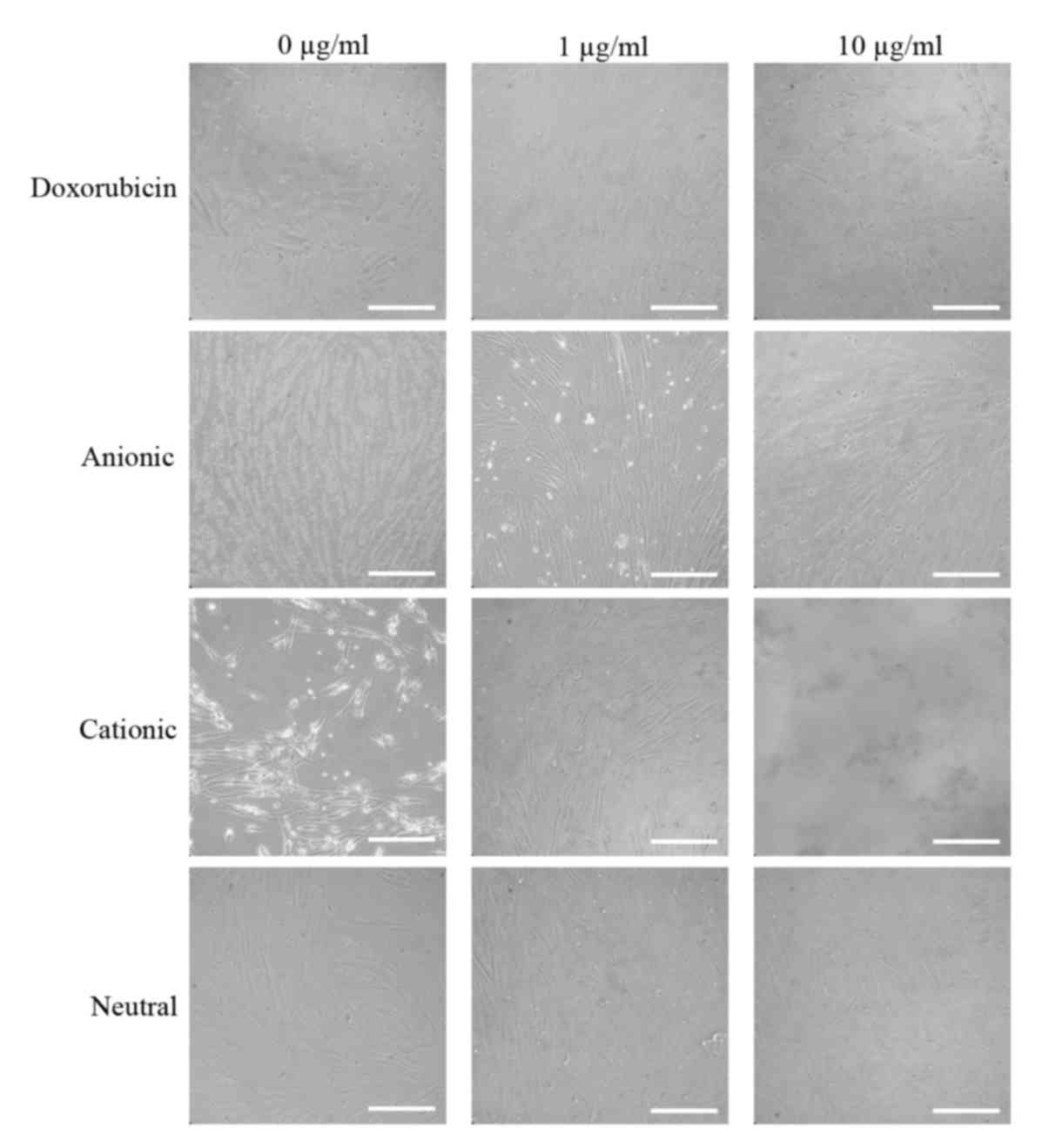
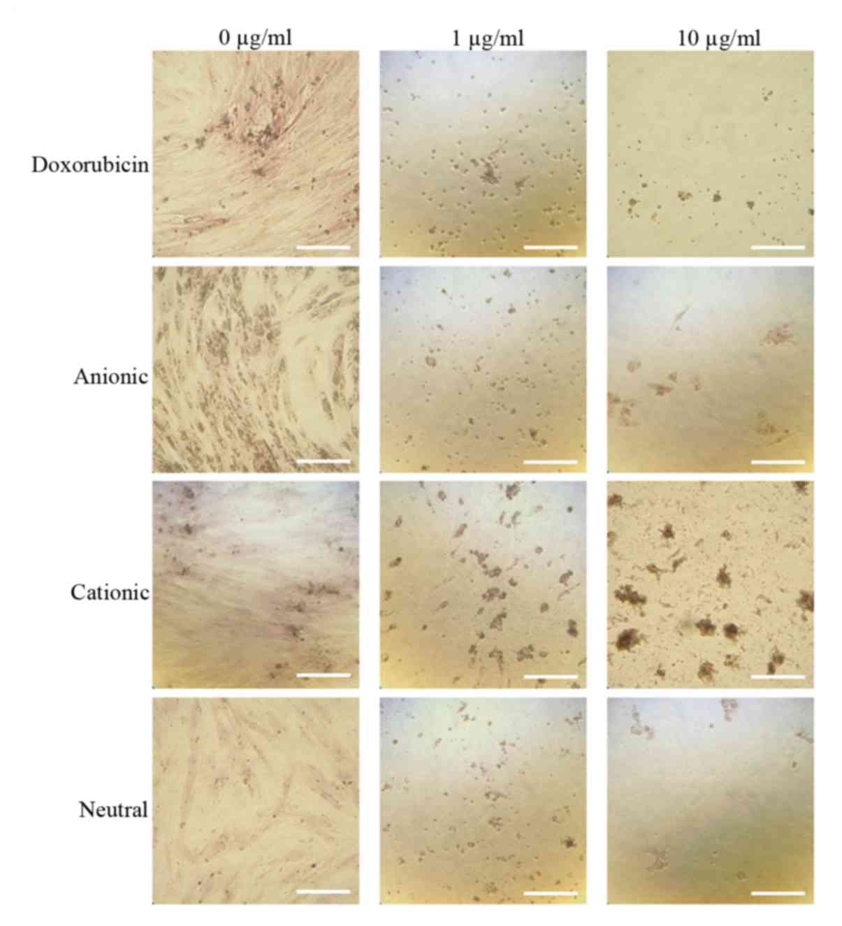
exhibited a normal fibroblast morphology on day 1 (Fig. 2). No significant morphological
changes of stem cells cultured in the osteogenic media were
observed after the addition of anionic, cationic or neutral
doxorubicin (A0, C0 or N0). No noticeable changes were observed
with the addition of doxorubicin at day 1 (D1 or D10). Similar
trends were observed in the anionic and cationic liposomes loaded
with doxorubicin at day 1. However, in the cells treated with the
cationic liposome, noticeable changes in morphology at the highest
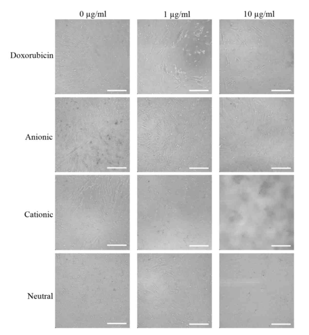
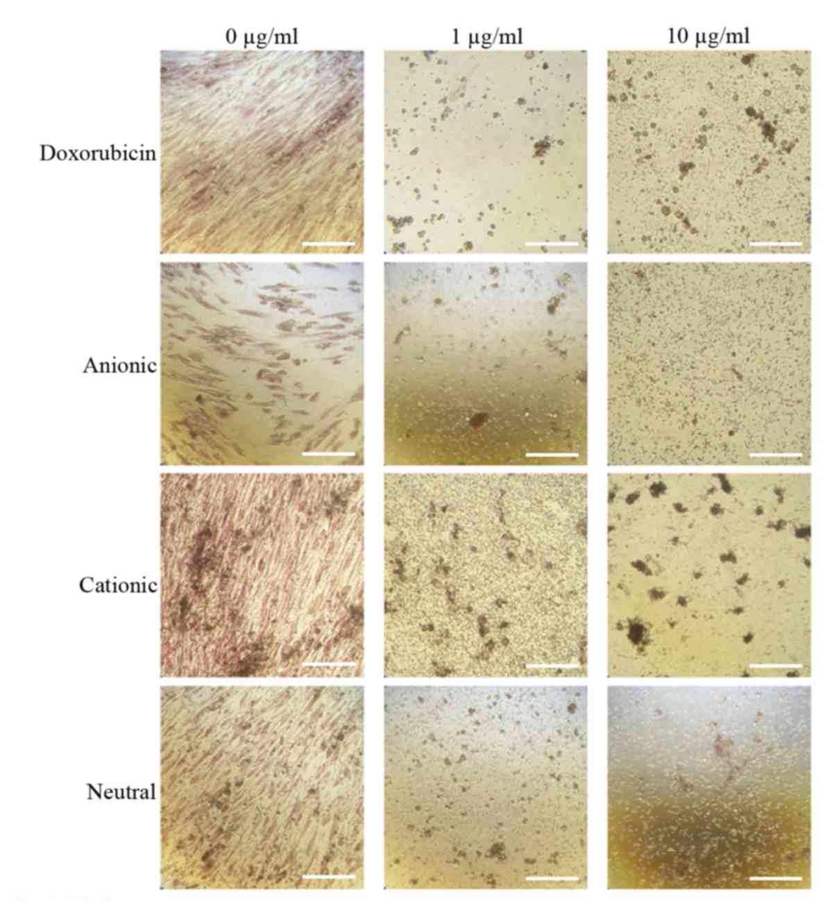
concentration (C10) were noted at day 1. In general, the
morphologies of the stem cells at days 3 were similar to those at
day 1 (Fig. 3). In the cationic
liposome groups, the low concentration of doxorubicin (C1) produced
cells with rounder and more amorphous shapes. More pronounced
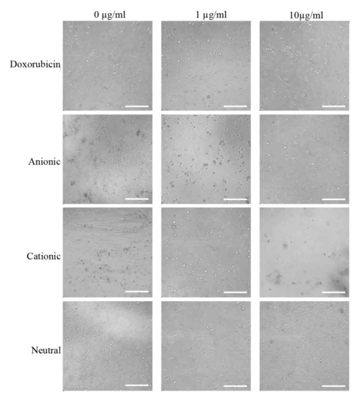
changes in the morphology of the stem cells were seen in the
cationic doxorubicin-loaded groups (C1 and C10) at day 7 (Fig. 4).
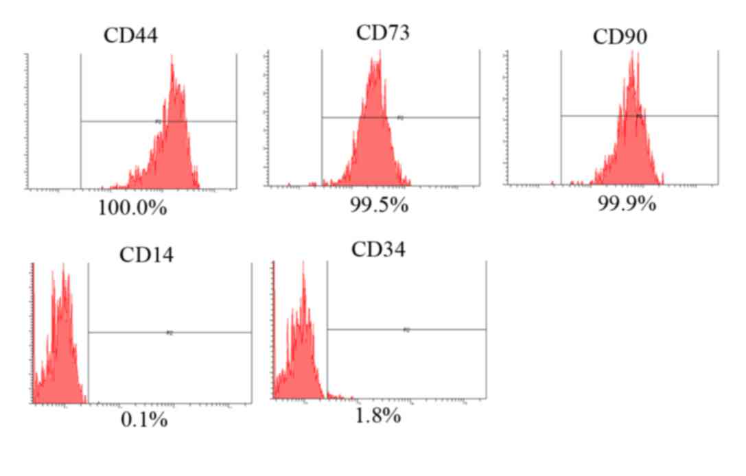 | Figure 1.Evaluation of stem cell surface marker
expression using CD44, CD73, CD90, CD14 and CD34. The percentages
of CD44+, CD73+, CD90+,
CD14+ and CD34+ cells were 100, 99.5, 99.9,
0.1 and 1.8%, respectively. |
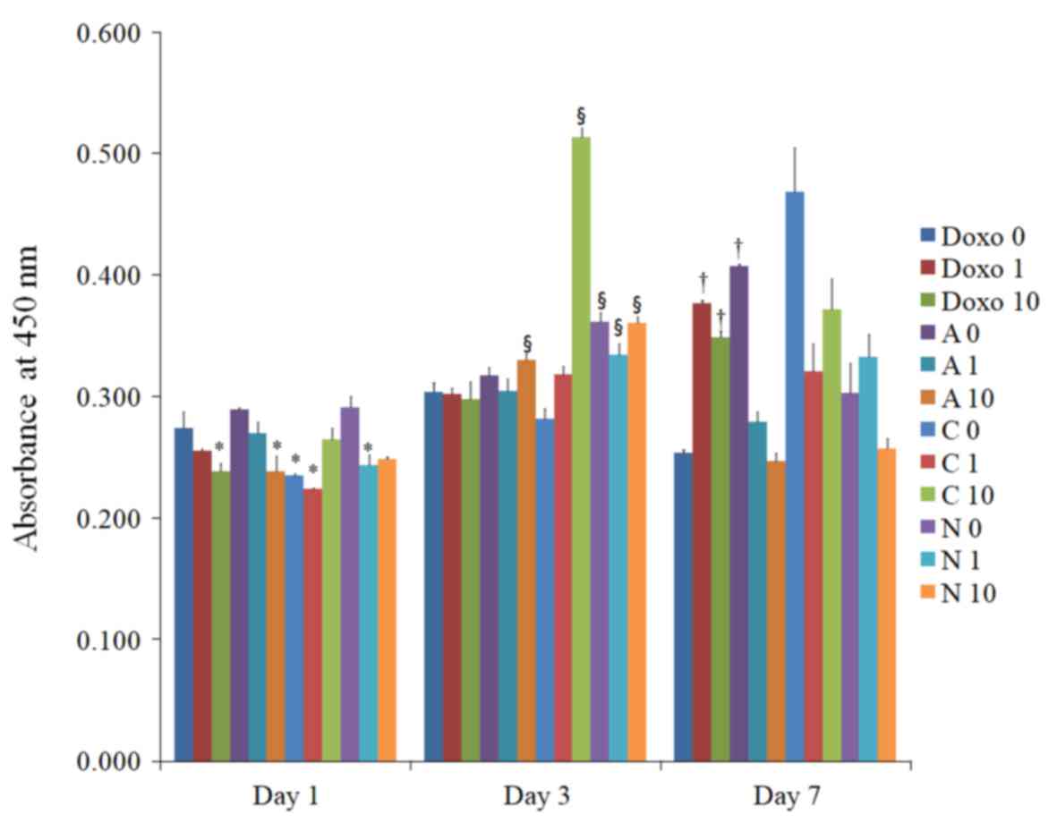
The results of the CCK-8 assay performed on days 1,
3, and 7 are presented in Fig. 5.
Application of doxorubicin at a high concentration on day 1
produced significant differences in cell viability when compared
with that in the D0 group at day 1.
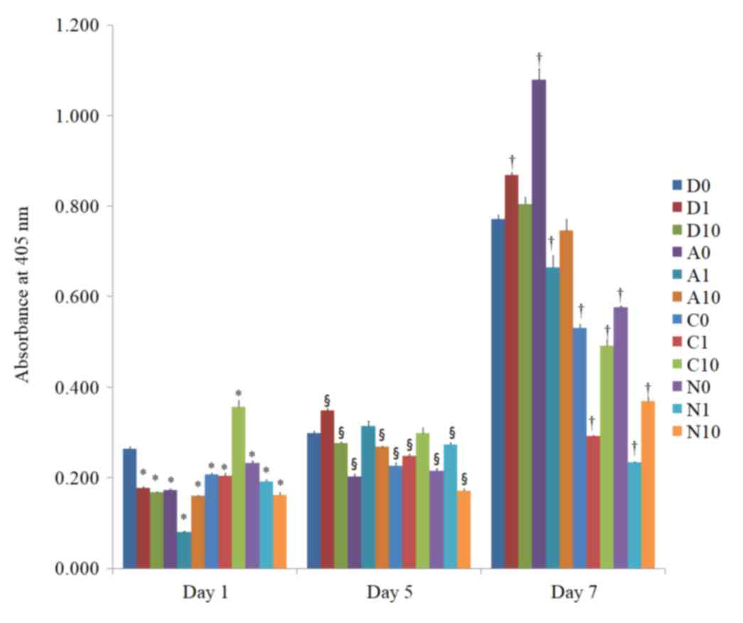
Alkaline phosphatase activity
The alkaline phosphatase activity on days 1, 5 and 7
in the cells treated with doxorubicin is presented in Fig. 6. The alkaline phosphatase activity
increased with longer incubation times. Noticeable decreases in
alkaline phosphatase activity were observed in the D1, D10, A0, A1,
A10, C0, C1, N0, N1 and N10 groups at day 1.
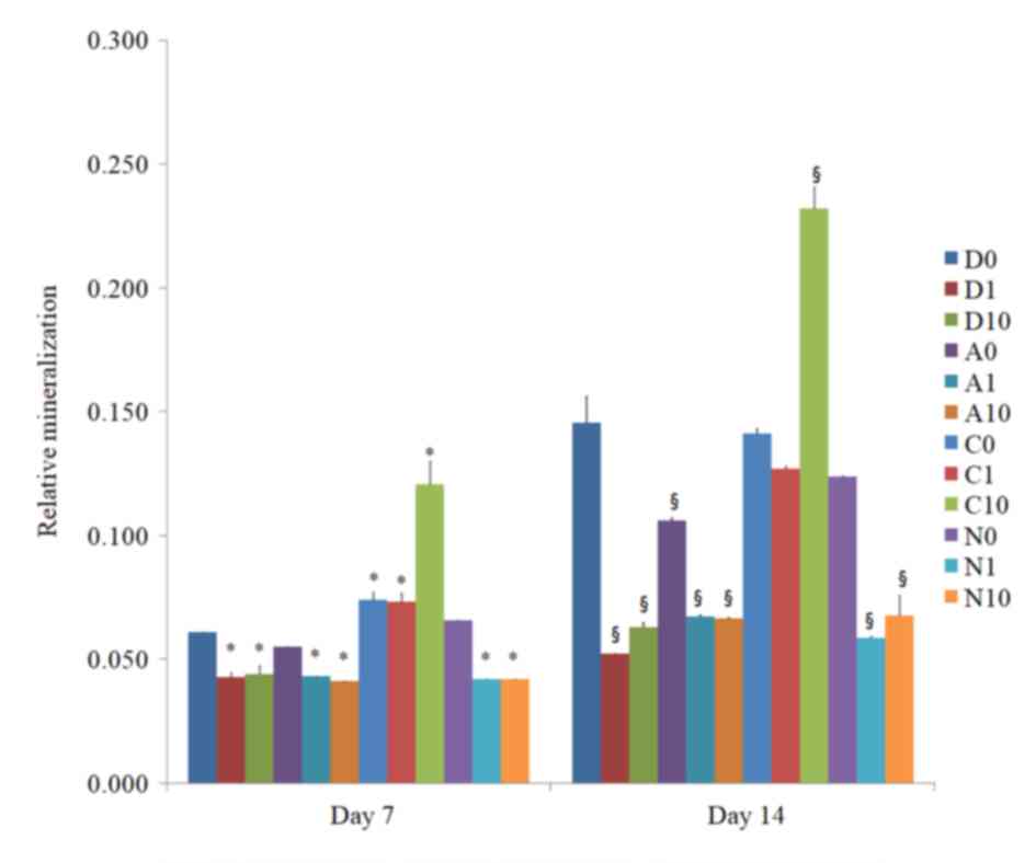
Alizarin Red S staining
The amount of mineralized extracellular deposits was
evaluated in the control groups (D0, A0, C0 and N0) using Alizarin
Red S staining on days 7 and 14 (Figs.
7 and 8). Mineralized
extracellular deposits were noted to in each group and intensity
differed among the groups. An increase in mineralized deposits was
noted on day 14 as compared to day 7. The application of
doxorubicin reduced the Alizarin Red S staining in the
doxorubicin-only group and the different liposome groups. The
morphology of the cationic doxorubicin-loaded groups was affected
the most among the anionic, cationic and neutral doxorubicin-loaded
liposome groups. The relative values at day 7 for D0, D1, D10, A0,
A1, A10, C0, C1, C10, N0, N1 and N10 are presented in Fig. 9. Significant decreases in Alizarin
Red S staining were observed in the D1, D10, A0, A1, A10, N1 and
N10 groups at day 14. Notable morphological changes in the number
of cells were observed in C1 and C10 groups at day 14.
Discussion
In the present study, the effects of liposomes of
different ionic types loaded with doxorubicin on the viability and
osteogenic differentiation potential of gingiva-derived stem cells
in two-dimensional culture were evaluated. The results indicated
that application of doxorubicin with or without liposomes reduced
the cellular viability and osteogenic differentiation. The
doxorubicin-loaded cationic liposomes induced the strongest
reduction in the cell viability and osteogenic differentiation in
the cultured stem cells.
Liposomes are nanoparticle lipid vesicles that are
composed of continuous bilayers of phospholipids surrounding an
aqueous phase; they have been investigated as drug-delivery systems
for improved targeted delivery of therapeutic agents (10). Doxorubicin-loaded liposomes have been
demonstrated to enhance the oral bioavailability of drugs by
modulation of their physicochemical characteristics (11). In a previous study,
doxorubicin-loaded polyethylene glycosylated liposomes were
accumulated and retained in the targeted site, and it was reported
that such liposome complexes increased the therapeutic efficacy of
drugs, thus providing a promising therapeutic approach (10). Ion-pairing technology using a
doxorubicin-cholesteryl hemisuccinate ion-pair complex based on the
conventional thin-film dispersion method produced high drug loading
and a high entrapment efficiency (12). Co-liposomes prepared with gemini
along with a natural zwitterionic lipid, phospholipid and
cholesterol produced H-responsive co-liposomes, which were able to
transport doxorubicin efficiently across doxorubicin-resistant
cancer cells (13).
In the present study, liposomes of different ionic
types were loaded with doxorubicin. The doxorubicin-loaded cationic
liposomes had the greatest effects on cultured stem cells and their
osteogenic differentiation. The differences in the surface charge
may have affected the uptake of doxorubicin by the stem cells
(14). Doxorubicin-induced toxicity
negatively impacts the clinical utility and outcomes (15). The composition of lipids in liposomes
may determine the drug encapsulation efficacy and release kinetics
of doxorubicin, which may impact the clinical toxicity (16). Liposomal co-delivered oleanolic acid
has been reported to attenuate doxorubicin-induced multi-organ
toxicity (15). Another previous
study suggested that miR-1, −21 and −145 may be involved in the
toxicity induced by doxorubicin, which may be considered as targets
for reducing its toxicity (17).
In the present study, the effects of liposomes
loaded of different ionic types loaded with doxorubicin on the
cellular viability and osteogenic differentiation potential of stem
cells in two-dimensional culture were evaluated. The results
indicated that application of doxorubicin reduced the cellular
viability and osteogenic differentiation with or without liposomes.
The doxorubicin-loaded cationic liposomes induced the strongest
reduction in the cell viability and osteogenic differentiation in
the cultured stem cells.
Acknowledgements
Not applicable.
Funding
This study was partly supported by the Research Fund
of Seoul St. Mary's Hospital, The Catholic University of Korea
(Seoul, Korea) in 2017. This work was further supported by the
Basic Science Research Program of the Ministry of Education (grant
no. 2016R1C1B3013951) through the National Research Foundation of
Korea and the financial support of the Catholic Medical Center
Research Foundation in the program year of 2016. This study was
partly supported by the Basic Science Research Program through the
National Research Foundation of Korea (NRF) funded by the Ministry
of Science, Information and Communication Technology & Future
Planning (grant no. NRF-2017R1A1A1A05001307).
Availability of data and materials
All data generated or analyzed during the present
study are included in the published article.
Authors' contributions
HL, JS, GY, HK and JP designed the study, performed
the experiments, were responsible for data collection and analysis,
and participated in drafting the manuscript. All the authors read
and approved the final version of the manuscript.
Ethical approval and consent to
participate
The Institutional Review Board of Seoul St. Mary's
Hospital, College of Medicine, Catholic University of Korea (Seoul,
Korea) approved this study (nos. KC17SNSI0606 and KC11SISI0348).
Informed consent was obtained from all of the participants.
Patient consent for publication
Not applicable.
Competing interests
The authors declare that they have no competing
interests.
References
|
1
|
Ruttala HB, Ramasamy T, Gupta B, Choi HG,
Yong CS and Kim JO: Multiple polysaccharide-drug complex-loaded
liposomes: A unique strategy in drug loading and cancer targeting.
Carbohydr Polym. 173:57–66. 2017. View Article : Google Scholar : PubMed/NCBI
|
|
2
|
Bagnyukova TV, Serebriiskii IG, Zhou Y,
Hopper-Borge EA, Golemis EA and Astsaturov I: Chemotherapy and
signaling: How can targeted therapies supercharge cytotoxic agents?
Cancer Biol Ther. 10:839–853. 2010. View Article : Google Scholar : PubMed/NCBI
|
|
3
|
Zhang Y, Zhai M, Chen Z, Han X, Yu F, Li
Z, Xie X, Han C, Yu L, Yang Y and Mei X: Dual-modified liposome
codelivery of doxorubicin and vincristine improve targeting and
therapeutic efficacy of glioma. Drug Deliv. 24:1045–1055. 2017.
View Article : Google Scholar : PubMed/NCBI
|
|
4
|
Rana T, Chakrabarti A, Freeman M and
Biswas S: Doxorubicin-mediated bone loss in breast cancer bone
metastases is driven by an interplay between oxidative stress and
induction of TGFbeta. PLoS One. 8:e780432013. View Article : Google Scholar : PubMed/NCBI
|
|
5
|
Affram K, Udofot O, Singh M, Krishnan S,
Reams R, Rosenberg J and Agyare E: Smart thermosensitive liposomes
for effective solid tumor therapy and in vivo imaging. PLoS One.
12:e01851162017. View Article : Google Scholar : PubMed/NCBI
|
|
6
|
Fukuda A, Tahara K, Hane Y, Matsui T,
Sasaoka S, Hatahira H, Motooka Y, Hasegawa S, Naganuma M, Abe J, et
al: Comparison of the adverse event profiles of conventional and
liposomal formulations of doxorubicin using the FDA adverse event
reporting system. PLoS One. 12:e01856542017. View Article : Google Scholar : PubMed/NCBI
|
|
7
|
Lee H, Son J, Na CB, Yi G, Koo H and Park
JB: The effects of doxorubicin-loaded liposomes on viability, stem
cell surface marker expression and secretion of vascular
endothelial growth factor of three-dimensional stem cell spheroids.
Exp Ther Med. 15:4950–4960. 2018.PubMed/NCBI
|
|
8
|
Jin SH, Lee JE, Yun JH, Kim I, Ko Y and
Park JB: Isolation and characterization of human mesenchymal stem
cells from gingival connective tissue. J Periodontal Res.
50:461–467. 2015. View Article : Google Scholar : PubMed/NCBI
|
|
9
|
Contrera JF, Matthews EJ, Kruhlak NL and
Benz RD: Estimating the safe starting dose in phase I clinical
trials and no observed effect level based on QSAR modeling of the
human maximum recommended daily dose. Regul Toxicol Pharmacol.
40:185–206. 2004. View Article : Google Scholar : PubMed/NCBI
|
|
10
|
Niu H, Xu M, Li S, Chen J, Luo J, Zhao X,
Gao C and Li X: High-performance liquid chromatography (HPLC)
quantification of liposome-delivered doxorubicin in arthritic
joints of collagen-induced arthritis rats. Med Sci Monit Basic Res.
23:150–158. 2017. View Article : Google Scholar : PubMed/NCBI
|
|
11
|
Daeihamed M, Haeri A, Ostad SN, Akhlaghi
MF and Dadashzadeh S: Doxorubicin-loaded liposomes: Enhancing the
oral bioavailability by modulation of physicochemical
characteristics. Nanomedicine (Lond). 12:1187–1202. 2017.
View Article : Google Scholar : PubMed/NCBI
|
|
12
|
Xu H, Zhang L, Li L, Liu Y, Chao Y, Liu X,
Jin Z, Chen Y, Tang X, He H, et al: Membrane-loaded doxorubicin
liposomes based on ion-pairing technology with high drug loading
and pH-responsive property. AAPS PharmSciTech. 18:2120–2130. 2017.
View Article : Google Scholar : PubMed/NCBI
|
|
13
|
Moitra P, Kumar K, Sarkar S, Kondaiah P,
Duan W and Bhattacharya S: New pH-responsive gemini lipid derived
co-liposomes for efficacious doxorubicin delivery to drug resistant
cancer cells. Chem Commun (Camb). 53:8184–8187. 2017. View Article : Google Scholar : PubMed/NCBI
|
|
14
|
Lee S, Lee SY, Park S, Ryu JH, Na JH, Koo
H, Lee KE, Jeon H, Kwon IC, Kim K and Jeong SY: In vivo NIRF
imaging of tumor targetability of nanosized liposomes in
tumor-bearing mice. Macromol Biosci. 12:849–856. 2012. View Article : Google Scholar : PubMed/NCBI
|
|
15
|
Sarfraz M, Afzal A, Raza SM, Bashir S,
Madni A, Khan MW, Ma X and Xiang G: Liposomal co-delivered
oleanolic acid attenuates doxorubicin-induced multi-organ toxicity
in hepatocellular carcinoma. Oncotarget. 8:47136–47153. 2017.
View Article : Google Scholar : PubMed/NCBI
|
|
16
|
Sreekanth V, Medatwal N, Pal S, Kumar S,
Sengupta S and Bajaj A: Molecular self-assembly of bile
acid-phospholipids controls the delivery of doxorubicin and mice
survivability. Mol Pharm. 14:2649–2659. 2017. View Article : Google Scholar : PubMed/NCBI
|
|
17
|
Razavi-Azarkhiavi K, Jaafari MR, Abnous K,
Razavi BM, Jafarian AH, Hassani FV, Shirani K and Karimi G: The
cardiotoxic mechanism of doxorubicin (DOX) and pegylated liposomal
DOX in mice bearing C-26 colon carcinoma: A study focused on
microRNA role for toxicity assessment of new formulations. Pharm
Res. 34:1849–1856. 2017. View Article : Google Scholar : PubMed/NCBI
|