Introduction
Among gynecological diseases, endometrial cancer
(EC) is the most common one and a threat to the health of women
worldwide; it usually occurs in perimenopausal women around the age
of 50 years (1,2). Over the years, the incidence rate of EC
has displayed a rising trend worldwide; however, the 5-year
survival rate has gradually decreased (3). Since most patients with EC are at stage
I at the time-point of diagnosis, pre-operative assessment provides
benefits regarding the therapeutic strategy selection for EC.
Diagnostic methods for EC usually include
ultrasonography, hysteroscopy and curettage. However, the rates of
misdiagnosis and missed diagnosis are remain high (4). Common diagnostic methods, including
curettage scraping (5), hysteroscopy
(6), magnetic resonance imaging
(7), positron emission
tomography/computed tomography (8)
or traditional transvaginal ultrasound (9), all have their advantages, as well as a
certain insufficiency. Recently, contrast-enhanced ultrasonography
(CEUS), which has been applied in the discrimination of benign from
malignant adnexal masses (10,11), has
attracted the attention of researchers and clinicians due to its
potential in the diagnosis of EC. During the past 10 years, CEUS,
which uses a microbubble contrast agent, e.g., SonoVue or Levovist,
has significantly improved the diagnostic accuracy of
ultrasonography in examining gynecological diseases, including
ovarian tumors (12). Application of
CEUS to determine myometrial invasion and the cancer stage in EC
has also been reported in recent years; however, relevant studies
remain inadequate.
To the best of our knowledge, to date, no previous
study has summarized the accuracy of CEUS in the diagnosis of EC.
In the present study, a meta-analysis was therefore performed to
determine the accuracy of CEUS in diagnosing EC.
Materials and methods
Protocol
The present meta-analysis followed the Preferred
Reporting Items for Systematic Reviews and Meta-Analyses (PRISMA)
recommendations (13).
Study selection criteria
Prior to searching the literature, certain selection
criteria were set for the studies to set standards for determining
the diagnostic value of CEUS in EC. The selection criteria were as
follows: i) The accuracy (sensitivity and specificity) of CEUS in
the diagnosis of EC was evaluated; ii) a gold standard was adopted
to treat and confirm EC, including surgery, histopathology and
appropriate follow-up and iii) the data allowed for construction of
a 2×2 table for true-positives, false-positives, true-negatives and
false-negatives.
Literature search
Articles published until 31 January 2017 were
retrieved from PubMed, EMBASE, Elsevier, Springer and Google
scholar. The search terms were a combination of the following key
words: ‘Ultrasonic angiography’, ‘endometrial carcinoma’, ‘contrast
enhanced ultrasonography’, ‘sensitivity’, ‘specificity’, ‘positive
predictive value’ and ‘negative predictive value’. A manual search
was also performed through searching the reference lists of
relevant articles to expand the number of included studies. In the
case of insufficient data provided in a study, the authors were
contacted. Only studies in English and Chinese language, which met
the selection criteria, were included.
Data collection and extraction
Two independent observers reviewed abstracts to
examine whether they qualified for inclusion according to the
pre-defined criteria. Selected papers were then retrieved,
evaluated for their eligibility, and relevant data were extracted
by the two observers independently. A third observer was consulted
when disagreements occurred. The following items were extracted
according to a fixed protocol: Author, year of publication,
country, study type, original study population, ultrasound modality
and contrast agents. Diagnostic 2×2 contingency tables were
constructed from the selected papers.
Statistical analysis
Pooled estimates of sensitivity, specificity,
likelihood ratios [positive likelihood ratios,
+LR=sensitivity/(1-specificity); negative likelihood ratios,
-LR=specificity/(1-sensitivity)] and diagnostic odds ratios (DORs)
were calculated in the present meta-analysis of the accuracy of
CEUS in diagnosing EC. The fixed-effects model and the
random-effects model were used and heterogeneity result was also
recorded. The summary receiver operating characteristics (sROC)
curve was also constructed. Confidence intervals were calculated
using the F distribution method (14). Forrest plots were drawn to illustrate
the distribution of the data-points in each study in association
with the summary pooled estimate. The heterogeneity of the
sensitivities and specificities were tested by applying the
likelihood ratio test. Heterogeneity of the pooled studies was
tested by using sROC curves as previously described (14). sROC curves were used to calculate the
area under the curve (AUC). Publication bias was assessed using
Deek's funnel plot asymmetry test, through P>0.05 suggesting no
significant publication bias and with Review Manager 5.3 (Cochrane
Collaboration, London, UK). All statistical analyses were performed
using STATA 12.0 (STATA, College Station, TX, USA) except for the
assessment of publication bias with Review Manager 5.3.
Results
Study collection and
characteristics
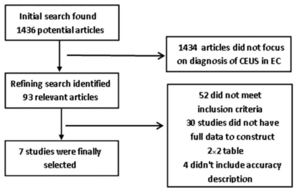
The initial search identified 1,436 references, of
which 93 relevant studies were selected and reviewed. After
reviewing their abstracts, 52 studies were excluded. And after
evaluation of the full-text articles, another 30 references were
excluded. Among the 11 remaining studies, 4 did not meet the
inclusion criteria due to insufficient sensitivity, specificity,
accuracy or correlation values. Thus, 7 studies with a total sample
size of n=275 were finally included in the present meta-analysis
and their data were extracted (15–21). All
selected studies are in accordance with the inclusion criteria. A
flow chart depicting the selection process is presented in Fig. 1.
As indicated in Table
I, the 7 included studies were published between 2008 and 2015.
Among the studies, three were prospective studies and five were
retrospective. The number of cases varied between 28 and 68. All
studies used certain types of CEUS, including regular CEUS, color
Doppler CEUS and combination of 2-dimensional (2D) and 3D CEUS. The
microbubble contrast agent used in all studies was SonoVue. In all
of the studies, CE was pathologically confirmed in addition to the
diagnosis by CEUS.
 | Table I.Summary of studies included in the
present meta-analysis. |
Table I.
Summary of studies included in the
present meta-analysis.
| Author, year | Country | Study type | Mean age (years) | Cases (n) | Modality | Contrast agent | (Refs.) |
|---|
| Wang, 2012 | China | Retro | 56 | 37 | Color Doppler
CEUS | SonoVue | (15) |
| Zhou et al,
2015 | China | Prosp | 50 | 68 | 2D and 3D CEUS | SonoVue | (16) |
| Song et al,
2009 | China | Prosp | None | 35 | CEUS | SonoVue | (17) |
| Ding et al,
2013 | China | Retro | 55 | 40 | Color Doppler
CEUS | SonoVue | (18) |
| Liu et al,
2011 | China | Retro | 56 | 37 | Color Doppler
CEUS | SonoVue | (19) |
| Sun et al,
2008 | China | Retro | 55 | 30 | Color Doppler
CEUS | SonoVue | (20) |
| Zhang et al,
2011 | China | Retro | None | 28 | Color Doppler
CEUS | SonoVue | (21) |
Accuracy of CEUS in the diagnosis of
EC
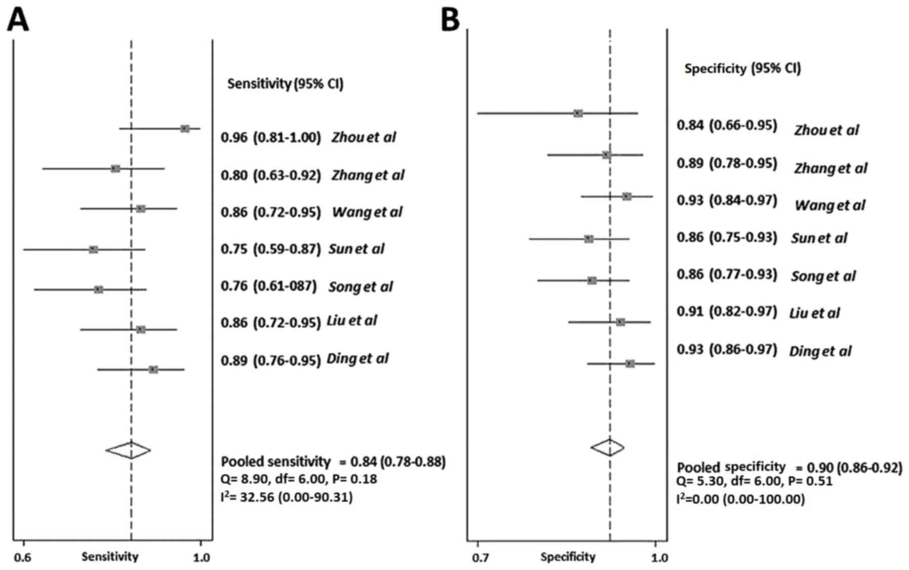
For the analysis CEUS accuracy in the diagnosis of
EC, heterogeneity results demonstrated that the data was
homogeneous with P=0.461. Thus, a fixed model in STATA was
utilized. Parameters for the analysis of CEUS accuracy in the
diagnosis of EC are shown in Table
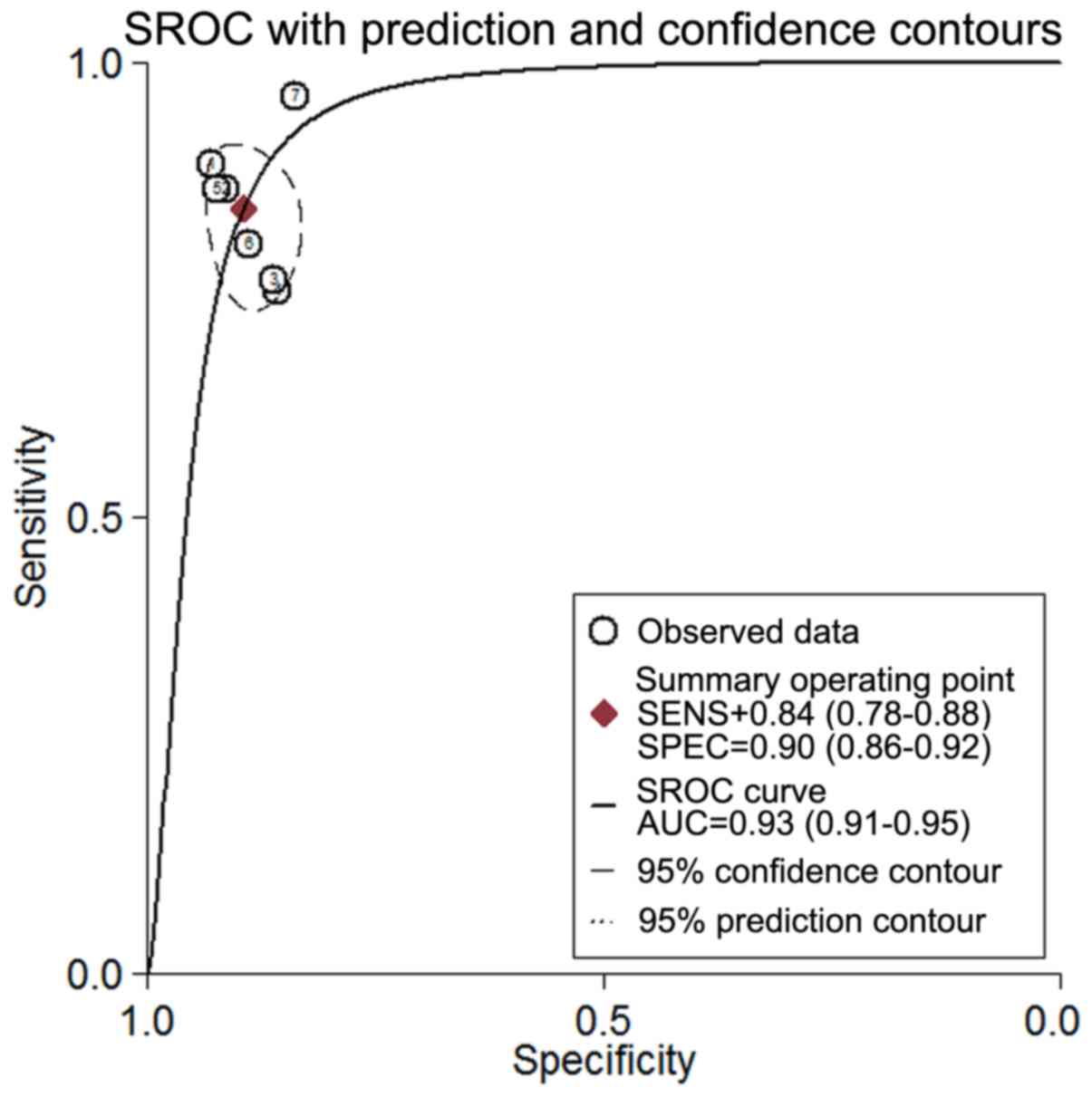
II. As presented in Fig. 2A and
B, the pooled sensitivity of CEUS in the diagnosis of EC was
84% [95% confidence interval (CI), 0.78–0.88], while the pooled
specificity was 90% (95% CI, 0.86–0.92). The positive likelihood
ratio (+LR) of CEUS was 8.0 (95% CI, 5.9–10.8) and the negative
likelihood ratio (-LR) was 0.18 (95% CI, 0.13–0.25). The DOR was 44
(95% CI, 26–77). The AUC under the sROC curve was 0.93 (Fig. 3). Moderate heterogeneity was observed
for the sensitivity, specificity and DOR with I2 values
of 32.56, 34.68 and 41.2%, respectively.
| Table II.Parameters for the analysis of CEUS
accuracy in the diagnosis of EC. |
Table II.
Parameters for the analysis of CEUS
accuracy in the diagnosis of EC.
| Parameter | Estimate | 95% CI |
|---|
| Sensitivity | 0.84 | (0.78–0.88) |
| Specificity | 0.90 | (0.86–0.92) |
| Positive likelihood
ratio | 8.0 | (5.9–10.8) |
| Negative likelihood
ratio | 0.18 | (0.13–0.25) |
| Diagnostic odds
ratio | 44 | (26–77) |
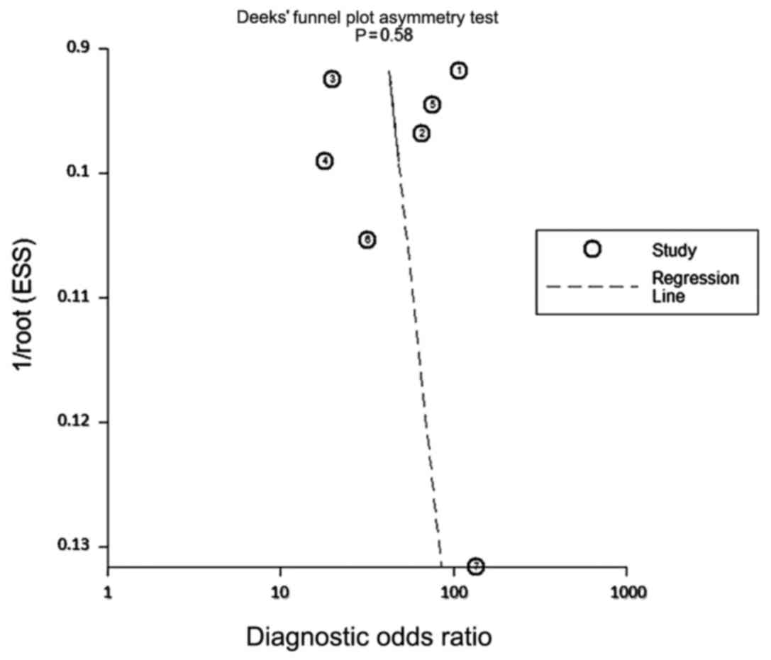
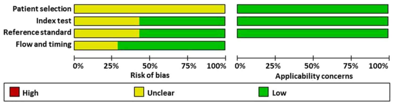
Assessment of bias
The publication bias was examined using Deeks'
funnel plot asymmetry test (Fig. 4).
No significant publication bias was observed for the DOR of CEUS in
the diagnosis of EC (P=0.58). The result of the risk of bias
assessment performed with Review Manager 5.3 is presented in
Fig. 5. The results demonstrated
that no study exhibited a high risk of bias in patient selection,
index test, reference standard, trial process or timing of the
studies.
Discussion
In recent years, CEUS has been proved to be
efficient in the diagnosis of numerous diseases, e.g., liver cancer
(22). In more recent years, the
diagnostic value of CEUS in EC has gained the attention of
researchers and clinicians. In China, an increasing number of
clinicians choose to use CEUS to diagnose uterine lesions,
including uterine fibroid (23).
Sconfienza et al (24)
studied the efficacy of CEUS in the diagnosis of uterine fibroids
and demonstrated that CEUS was effective in determining the degree
of vascular occlusion at the end of superselective uterine fibroid
embolization procedures. Based on a study by Seitz and Strobel
(25), CEUS was approved for
diagnostic liver imaging in adults and children in the USA.
However, compared with the application of CEUS in other fields, its
use in the diagnosis of EC still requires further research.
In the present study, a meta-analysis was performed
to summarize the accuracy of CEUS in diagnosing EC. Two reviewers
were involved in the study to analyze the literature independently
to reduce the bias of researchers. The meta-analysis also followed
the PRISMA recommendations (13),
which suggests that only studies containing at least four of the
Quality Assessment of Diagnostic Accuracy Studies criteria
(26) are included and in addition,
all patients were confirmed by pathological analysis.
The present meta-analysis study provided a pooled
sensitivity and specificity of 84% (95% CI, 0.78–0.88) and 90% (95%
CI, 0.86–0.92), respectively, with a DOR of 44 (95% CI, 26–77) and
an AUC of 0.93. Moderate heterogeneity was observed for the
sensitivity, specificity and DOR. The present results suggest that
CEUS has a high accuracy in the diagnosis of EC. Furthermore, the
present meta-analysis indicated a high +LR of 8.0 (95% CI,
5.9–10.8) and a low -LR of 0.18 (95% CI, 0.13–0.25), suggesting
that the diagnostic test performs well in correctly identifying the
true disease state, which should also be confirmed by
histopathological analysis. The publication bias was examined using
bot Deeks' funnel plot asymmetry test and Review Manager 5.3, and
no significant publication bias was observed.
Of note, the present study had several limitations.
First, due to the limited number of cases and studies included,
further evidences are still required. Furthermore, all studies that
met the inclusion criteria for the meta-analysis were from China.
This may be due to the application of CEUS having been widely
adopted for diagnosing EC in China, but are not that common in
other countries. Finally, the studies were limited by language, as
the literature search only included those in English and
Chinese.
In conclusion, the present meta-analysis indicates
that CEUS is valuable in the diagnosis of EC. Additional clinical
data and studies are still required to confirm the present results
and to further develop the diagnostic application of CEUS in
EC.
Acknowledgements
Not applicable.
Funding
No funding was received.
Availability of data and materials
The datasets used and/or analyzed during the current
study are available from the corresponding author on reasonable
request.
Authors' contributions
JG conducted the analysis and wrote the manuscript;
and JT collected and performed a preliminary analysis of
references, revised the manuscript, and approved it for
submission.
Ethics approval and consent to
participate
Not applicable.
Patient consent for publication
Not applicable.
Competing interests
The authors declare that they have no competing
interests.
References
|
1
|
Dal Maso L, Augustin LS, Karalis A,
Talamini R, Franceschi S, Trichopoulos D, Mantzoros CS and La
Vecchia C: Circulating adiponectin and endometrial cancer risk. J
Clin Endocrinol Metab. 89:1160–1163. 2014. View Article : Google Scholar
|
|
2
|
Wright JD, Barrena Medel NI, Sehouli J,
Fujiwara K and Herzog TJ: Contemporary management of endometrial
cancer. Lancet. 379:1352–1360. 2012. View Article : Google Scholar : PubMed/NCBI
|
|
3
|
Siegel RL, Miller KD and Jemal A: Cancer
statistics, 2015. CA Cancer J Clin. 65:5–29. 2015. View Article : Google Scholar : PubMed/NCBI
|
|
4
|
de Boer SM, Nout RA, Jürgenliemk-Schulz
IM, Jobsen JJ, Lutgens LC, van der Steen-Banasik EM, Mens JW, Slot
A, Stenfert Kroese MC, Oerlemans S, et al: Long-term impact of
endometrial cancer diagnosis and treatment on health-related
quality of life and cancer survivorship: Results from the
randomized PORTEC-2 trial. Int J Radiat Oncol Biol Phys.
93:797–809. 2015. View Article : Google Scholar : PubMed/NCBI
|
|
5
|
Kurosawa H, Ito K, Nikura H, Takano T,
Nagase S, Utsunomiya H, Otsuki T, Toyoshima M, Nagai T, Tanaka S,
et al: Hysteroscopic inspection and total curettage are
insufficient for discriminating endometrial cancer from atypical
endometrial hyperplasia. Tohoku J Exp Med. 228:365–70. 2012.
View Article : Google Scholar : PubMed/NCBI
|
|
6
|
Crispi CP, Vanin CM, Dibi RP, Kato SK and
Pesssini SA: Postmenopausal bleeding: Findings and accuracy of
hysteroscopy and histopathologic in the diagnosis of endometrial
cancer. J Minimal Invasive Gynecol. 18:S832011. View Article : Google Scholar
|
|
7
|
Bakir B, Sanli S, Bakir VL, Ayas S, Yildiz
SO, Iyibozkurt AC, Kartal MG and Yavuz E: Role of diffusion
weighted MRI in the differential diagnosis of endometrial cancer,
polyp, hyperplasia, and physiological thickening. Clin Imaging.
41:86–94. 2017. View Article : Google Scholar : PubMed/NCBI
|
|
8
|
Bollineni VR, Ytrehauge S,
Bollinenibalabay O, Salvesen HB and Haldorsen IS: High diagnostic
value of FDG-PET/CT in endometrial cancer: Systematic review and
meta-analysis of the literature. J Nucl Med. 57:879–885. 2016.
View Article : Google Scholar : PubMed/NCBI
|
|
9
|
Jacobs I, Gentry-Maharaj A, Burnell M,
Manchanda R, Singh N, Sharma A, Ryan A, Seif MW, Amso NN, Turner G,
et al: Sensitivity of transvaginal ultrasound screening for
endometrial cancer in postmenopausal women: A case-control study
within the UKCTOCS cohort. Lancet Oncol. 12:38–48. 2011. View Article : Google Scholar : PubMed/NCBI
|
|
10
|
Palmieri VO, Santovito D, Marano G,
Minerva F, Ricci L, D'Alitto F, Angelelli G and Palasciano G:
Contrast-enhanced ultrasound in the diagnosis of hepatocellular
carcinoma. Radiol Med. 120:627–633. 2015. View Article : Google Scholar : PubMed/NCBI
|
|
11
|
Dietrich CF, Averkiou MA, Correas JM,
Lassau N, Leen E and Piscaglia F: An EFSUMB introduction into
dynamic contrast-enhanced ultrasound (DCE-US) for quantification of
tumour perfusion. Ultraschall Med. 33:344–351. 2012. View Article : Google Scholar : PubMed/NCBI
|
|
12
|
Wang J, Lv F, Fei X, Cui Q, Wang L, Gao X,
Yuan Z, Lin Q, Lv Y and Liu A: Study on the characteristics of
contrast-enhanced ultrasound and its utility in assessing the
microvessel density in ovarian tumors or tumor-like lesions. Int J
Biol Sci. 7:600–606. 2011. View Article : Google Scholar : PubMed/NCBI
|
|
13
|
Moher D, Liberati A, Tetzlaff J, Altman DG
and PRISMA Group: Preferred reporting items for systematic reviews
and meta-analyses: The PRISMA statement. Ann Intern Med.
151:264–269. 2009. View Article : Google Scholar : PubMed/NCBI
|
|
14
|
Puli SR, Kalva N, Bechtold ML,
Pamulaparthy SR, Cashman MD, Estes NC, Pearl RH, Volmar FH, Dillon
S, Shekleton MF and Forcione D: Diagnostic accuracy of endoscopic
ultrasound in pancreatic neuroendocrine tumors: A systematic review
and meta-analysis. World J Gastroenterol. 19:3678–3684. 2013.
View Article : Google Scholar : PubMed/NCBI
|
|
15
|
Wang AZ, Liu CY, Xie Q and Wu XP:
Contrast-enhanced ultrasound and magnetic resonance imaging
diagnosis value and differential diagnosis for myometrial invasion
of stage I endometrial carcinoma. Shaanxi Yi Xue Za Zhi. 41:80–83.
2012.(In Chinese).
|
|
16
|
Zhou HL, Xiang H, Duan L, Shahai G, Liu H,
Li XH and Mou RX: Application of combined two-dimensional and
three-dimensional transvaginal contrast enhanced ultrasound in the
diagnosis of endometrial carcinoma. Biomed Res Int.
2015:2927432015. View Article : Google Scholar : PubMed/NCBI
|
|
17
|
Song Y, Yang J, Liu Z and Shen K:
Preoperative evaluation of endometrial carcinoma by
contrast-enhanced ultrasonography. BJOG. 116:298–299. 2009.
View Article : Google Scholar
|
|
18
|
Ding Y, Guo Y, Guan L, Wang H, Zhang D and
Wang Y: Application value of contrast-enhanced ultrasound for stage
of endometrial carcinoma. Chongqing Med J. 42:2103–2106. 2013.
|
|
19
|
Liu CY, Wang XF, Xie Q, Wan BB, Sheng X,
Wang M and Zhao LH: Diagnostic value of contrast-enhanced
ultrasound for myometrial invasion of stage I endometrial
carcinoma. Chinese J Med Image Technol. 27:1443–1446. 2011.
|
|
20
|
Sun ZJ, Yang JX, Shen K, et al:
Contrast-enhanced ultrasound in the evaluation of myometrial
invasion in endometrial carcinoma. J Reproduct Med. 2008.
|
|
21
|
Zhang XZ, Zhao HY, Peng M and Wei WB:
Application of contrast-enhanced ultrasound in the myometrial
invasion of endometrial carcinoma. J Bengbu Med Col. 36((3)):
285–287. 2011.
|
|
22
|
Lencioni R, Piscaglia F and Bolondi L:
Contrast-enhanced ultrasound in the diagnosis of hepatocellular
carcinoma. J Hepatol. 48:848–857. 2008. View Article : Google Scholar : PubMed/NCBI
|
|
23
|
Zhou XD, Ren XL, Zhang J, He GB, Zheng MJ,
Tian X, Li L, Zhu T, Zhang M, Wang L and Luo W: Therapeutic
response assessment of high intensity focused ultrasound therapy
for uterine fibroid: Utility of contrast-enhanced ultrasonography.
Eur J Radiol. 62:289–294. 2007. View Article : Google Scholar : PubMed/NCBI
|
|
24
|
Sconfienza LM, Lacelli F, Gazzo P,
Gandolfo N, Gravano M and Serafini G: Is contrast-enhanced
ultrasound (CEUS) effective in the assessment of outcomes and in
the follow up of uterine fibroids after superselective uterine
fibroids embolization (SUFE) when compared to dynamic magnetic
resonance (MR)? Ultraschall in Der Medizin. 29((S1))2008.
|
|
25
|
Seitz K and Strobel D: A Milestone:
Approval of CEUS for diagnostic liver imaging in adults and
children in the USA. Ultraschall Med. 37:229–232. 2016. View Article : Google Scholar : PubMed/NCBI
|
|
26
|
Whiting P, Rutjes AW, Reitsma JB, Bossuyt
PM and Kleijnen J: The development of QUADAS: A tool for the
quality assessment of studies of diagnostic accuracy included in
systematic reviews. BMC Med Res Methodol. 3:252003. View Article : Google Scholar : PubMed/NCBI
|