Introduction
Parkinson's disease (PD) is a degenerative disease
of the central nervous system that primarily affects the motor
system. Early symptoms of PD include slow-motion shaking, limb
stiffness, walking difficulties, and often accompanied by mental
problems such as depression, anxiety and sleep disorders (1). PD occurs in middle-aged and elderly
people, and its incidence is positively correlated with age, and is
higher in men than in women (2). In
the past 20 years, according to statistics, the incidence of PD in
China has increased by nearly 20 times (3). This chronic, long-term progressive
disease can only be alleviated by the present medical treatment.
Late stage of PD often manifests as dementia and loss of the
ability to live independently, which brings great mental impact and
economic pressure to patients and their families (2). Approximately 60% of PD patients
progress to severe disability after 5 years of onset, and no less
than 30% of patients eventually die of PD complications, among
which acne and sepsis are two most common types (4). Etiology of PD has not yet been
elucidated, and it may be related to age, oxidative stress, free
radicals and calcium ions and other factors. Under the combined
action of genetic and environmental factors, oxidative stress,
calcium overload, apoptosis and other mechanisms cause nigral
striatum containing melanin neurons (mainly DA neurons) to undergo
degeneration and apoptosis (5). The
diagnosis of this disease is mainly based on clinical symptoms. It
can be diagnosed only after neurological examinations and other
diseases are excluded (6).
Phosphatidylinositol 3-kinase (PI3K) is a
macromolecular protein with serine/threonine kinase activity and
plays an important role in cell signal transduction (7). PI3K is activated when the cells are
stimulated. Activation of PI3K promotes the conversion of PIP2 into
PIP3, so as to promote recruitment of protein kinase B (Akt). After
phosphorylation by protein kinase K (PDK), Akt directly or
indirectly activates rapamycin target protein (mTOR) and mTOR is
phosphorylated into p-mTOR, thereby exerting a series of biological
effects such as inhibition of apoptosis and promotion of
proliferation (8). PI3K/Akt/mTOR
signaling pathway plays a protective role for nerve energy cells by
participating in oxidative stress and negatively regulating
apoptosis (9,10). c-Jun amino-terminal kinase 3 (JNK3)
is a protein that regulates programmed cell death and is mainly
present in neurons in substantia nigra pars compacta. c-Jun's
apoptosis-inducing function and its special anatomic location make
it closely related to various neurodegenerative diseases such as
Alzheimer's disease and PD (11,12).
In the present study, effect of PI3K/Akt/mTOR
pathway on the expression of JNK3 in PD mice was studied in order
to further explain the pathogenesis of PD and provide a new target
for the treatment of PD.
Materials and methods
Test animals
A total of 200 male Sprague-Dawley rats with a body
mass of 254.7±33.9 g were purchased from Shanghai SLAC Laboratory
Animal Co., Ltd. (Shanghai, China) [production license SCXK
(Shanghai) 2012-0002]. Rats were fed with basal diet (Shanghai SLAC
Laboratory Animal Co., Ltd.) at a temperature of 21–25°C and a
humidity of approximately 60%. Rats were allowed to access water
freely. The study was approved by the Ethics Committee of The First
Affiliated Hospital of Henan University (Kaifeng, China).
Main reagents
Rotenone (Nanjing Xinfan Biotechnology Co., Ltd.,
Shanghai, China), sunflower oil (Shanghai Lisheng Chemicals Co.,
Ltd., Shanghai, China), LY294002 (Cell Signaling Technology, Inc.,
Danvers, MA, USA), rapamycin (Dalian Meilun Biotechnology Co.,
Ltd., Dalian, China), rabbit anti-rat JNK3/p-mTOR/β-actin
monoclonal antibody and goat anti-rabbit IgG secondary antibody
(cat. nos. E-AB-60615, E-AB-63517, E-AB-30419 and E-AB-1003,
respectively; Wuhan Elabscience Biotechnology Co., Ltd., Wuhan,
China), TRIzol (Genenode, Beijing, China), RIPA lysate (Shanghai
Huzhen Biological Technology Co., Ltd., Shanghai, China), PMSF
(Nanjing Shengxing Biotechnology Co., Ltd., Nanjing, China), BCA
protein kit (Shanghai Beiyi Bioequip Information Co., Ltd.,
Shanghai, China), reverse transcription kit and fluorescence
quantitative PCR kit (both from Guangzhou Huijun Biotechnology Co.,
Ltd., Guangzhou, China). Primers were synthesized by Sangon Biotech
Co., Ltd. (Shanghai, China).
Rat PD model construction and
grouping
After two weeks of adaptive feeding, PD rat model
was prepared by continuous subcutaneous injection of low dose
rotenone. Rotenone was dissolved in sunflower oil to prepare an
emulsion of 1.5 mg/ml. Rats were weighed every morning at 9
o'clock, and emulsion was used at a dose of 1.5 mg/kg. Once a day,
every 7 days counts as a cycle. Injection was discontinued for a
day after each cycle and a total of 3 cycles were performed. After
the completion of screening, model animals were grouped. Criteria
for successful modeling (13):
Occasional paralysis of limbs, inconvenience of walking, inability
to eat, gradual loss of ability to resist arrest, yellowing of hair
and bow back. Two rats died after 3 cycles, 18 failed to meet PD
modeling criteria, and finally 180 rats were modeled successfully.
The 180 rats were randomly divided into 4 groups including A, B, C
and D, with 45 in each group. Group A was control group and was
given physiological saline. Group B was LY294002 group, LY294002 as
PI3K inhibitor was used at a concentration of 10 mmol/l, and the
lateral ventricle was injected at a dose of 10 µl/kg according to
the weight of each rat. Group C was rapamycin (a specific inhibitor
of mTOR protein) group and rapamycin (0.32 ng in 4 µl) was injected
into the lateral ventricle. Group D was LY294002 combined with
rapamycin group, and the dose and injection method were the same as
above.
Detection of JNK3 expression by
RT-qPCR
TRIzol extraction of total RNA from rat substantia
nigra tissue (100 mg) was performed according to the instructions
of the kit. A total of 20 µl reaction system was prepared and cDNA
was synthesized according to the following conditions: 37°C for 15
min and 85°C for 5 sec. A total of 2 µl of reverse transcription
product was used for the PCR reaction according to the instructions
of TaqMan qPCR kit (Thermo fisher, Waltham, MA, USA) and GAPDH was
used as endogenous control. Reaction conditions were: 95°C for 60
sec, followed by 40 cycles of 95°C for 10 sec and 60°C for 30 sec.
This experiment was performed in triplicate manner and data were
processed using 2−ΔCq method (14). Primer sequences are listed in
Table I.
 | Table I.Primer sequences. |
Table I.
Primer sequences.
| Items | Forward | Reverse |
|---|
| GAPDH |
5′-TGGTCTACATGTTCCAGTATGACT-3′ |
5′-CCATTTGATGTTAGCGGGATCTC-3′ |
| JNK3 |
5′-CCACGCAGATCAAACAGGA-3′ |
5′-CCACGCAGATCAAACAGGA-3′ |
Western blot analysis
Protein lysate was prepared using 10 ml of RIPA and
100 µl of PMS protease inhibitor to extract total protein form 100
mg rat substantia nigra tissue. BCA method was used to measure
protein concentration. A total of 50 µl of protein was mixed with
200 µl of 5X loading buffer and subjected to SDS-PAGE
electrophoresis. After that, gel transfer was performed at 4
mA/cm2 for 1 h. Blocking with 5% BSA was performed for 1
h, followed by incubation with JNK3 monoclonal antibody (1:1,000),
p-mTOR monoclonal antibody (1:1,000) and β-actin monoclonal
antibody (1: 5,000) for 16 h at 4°C. After washing with PBS 3
times, membrans were washed 3 times with PBS, followed by
incubation with goat anti-IgG secondary antibody (1:5,000) at room
temperature for 1 h. ECL luminescence reagent was added to develop
signals and Quantity One software (Bio Rad, Hercules, CA, USA) was
used to scan membranes. Relative expression level of the
JNK3/p-mTOR protein (grayscale) of JNK3/p-mTOR grayscale of
β-actin.
Statistical analysis
SPSS 19.0 statistical package (IBM Corp., Armonk,
NY, USA) was used for all analyses. Results were expressed as mean
± standard deviation (SD), and were analyzed using non-parametric
K-S test according to data distribution characteristics.
Relationship between JNK3 and p-mTOR protein expression was
analyzed using Pearson's correlation, and the significance level
was α=0.05.
Results
Detection of JNK3 mRNA expression by
RT-qPCR
Expression level of JNK3 mRNA in experimental group
B, C and D was 0.79±0.03, 0.50±0.01 and 0.17±0.01, respectively,
which was significantly lower than that in control group A
(1.00±0.05; P<0.01). Expression of JNK3 mRNA in group C and D
was significantly lower than that in group B (P<0.01), and
expression level of JNK3 mRNA in group D was significantly lower
than that in group C (P<0.01) (Fig.
1).
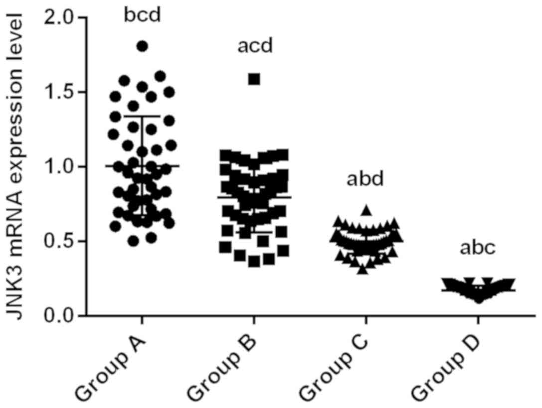 | Figure 1.Detection of JNK3 mRNA expression by
RT-qPCR. Normalized expression level of JNK3 mRNA in experimental
group B, C and D was 0.79±0.03, 0.50±0.01 and 0.17±0.01,
respectively, which was significantly lower than that in control
group A (1.00±0.05; P<0.01). Expression of JNK3 mRNA in group C
and D was significantly lower than that in group B (P<0.01), and
expression level of JNK3 mRNA in group D was significantly lower
than that in group C (P<0.01). aP<0.01, compared
with group A. bP<0.01, compared with group B.
cP<0.01, compared with group C.
dP<0.01, compared with group D. JNK3, c-Jun
amino-terminal kinase 3. |
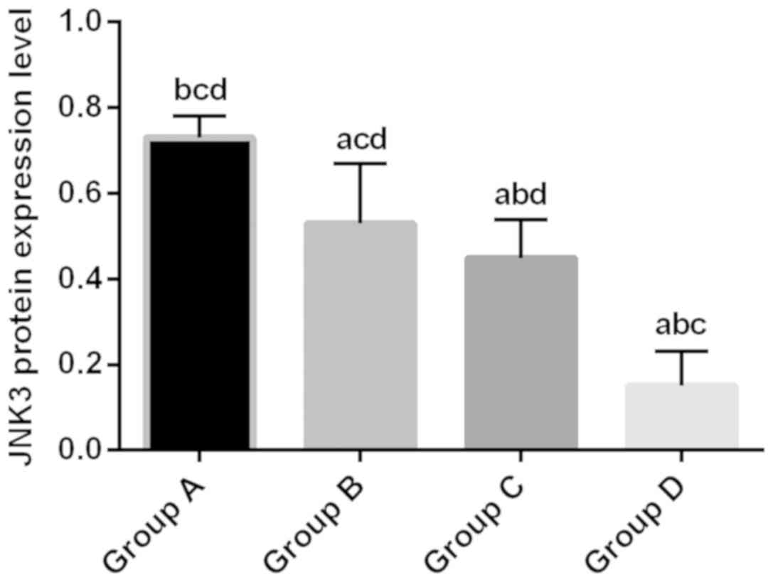
Western blot analysis of JNK3
protein
Relative expression level of JNK3 protein in
experimental groups B, C and D was 0.53±0.14, 0.45±0.09 and
0.15±0.08, respectively, which was significantly lower than that in
control group (0.73±0.05; P<0.05). Relative expression level of
JNK3 protein in group C and D was significantly lower than that in
group B (P<0.01). Relative expression level of JNK3 protein in
group D was significantly lower than that in group C (P<0.01)
(Fig. 2).
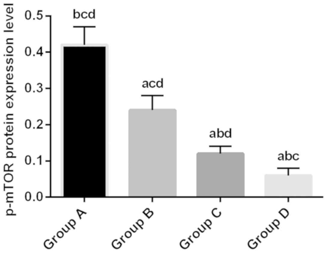
Western blot analysis of p-mTOR
protein expression
Relative expression level of p-mTOR protein in the
experimental group B, C and D was 0.24±0.04, 0.12±0.02 and
0.06±0.02, respectively, which was significantly lower than that in
control group A (0.42±0.05; P<0.05). Relative expression level
of p-mTOR protein in group C and D was significantly lower than
that in group B (P<0.01). Relative expression level of JNK3
protein in group D was significantly lower than that in group C
(P<0.01) (Fig. 3).
Correlation analysis of JNK3 and
p-mTOR protein expression
Pearson's correlation analysis showed that
expression of JNK3 mRNA was positively correlated with JNK3 protein
expression and Pearson's correlation coefficient was 0.98
(P<0.01). There was also a positive correlation between the
expression of JNK3 mRNA and the expression of p-mTOR protein and
Pearson's correlation coefficient was 0.95 (P<0.01). Besides,
expression of JNK3 protein was positively correlated with the
expression of p-mTOR protein, and the Pearson's correlation
coefficient was 0.93 (P<0.01) (Table
II).
| Table II.Correlation analysis of JNK3 and
p-mTOR protein expression. |
Table II.
Correlation analysis of JNK3 and
p-mTOR protein expression.
| Pearson's correlation
coefficient | JNK3 mRNA | JNK3 protein | p-mTOR protein |
|---|
| JNK3 mRNA | 1 | / | / |
| JNK3 protein | 0.98 | 1 | / |
| p-mTOR protein | 0.95 | 0.93 | 1 |
Discussion
PD is the most common disabling disease in
middle-aged and elderly people. Its main pathological feature is
degeneration and necrosis of DA neurons in the nigrostriatal
(15). PI3K/AKT/mTOR signaling
pathway plays an important regulatory role in many life activities
such as cell metabolism, cell growth, biosynthesis, as well as
development of cancer, degenerative diseases, and diabetes
(16,17). JNK3 is one of the major MAPK family
members in the brain. It is reported that calcium overload and
oxygen free radicals can activate JNK3, thereby inducing the
expression of apoptotic proteins such as p53 and FasL and inducing
the expression of caspases, so as to accelerate the process of cell
apoptosis (3). Therefore, this study
aimed to investigate the effect of PI3K/Akt/mTOR signaling pathway
on the expression of JNK3 in PD mice, so as to further explain the
pathogenesis of PD and provide a new target for the treatment of
PD.
LY294002 is a specific inhibitor of PI3K, and
rapamycin is a specific inhibitor of mTOR. Both of them can inhibit
PI3K/Akt/mTOR signaling pathway. In this study, the relative
expression levels of JNK3 mRNA and JNK3 protein were decreased when
LY294002 and rapamycin were administered, and the combination of
the two inhibitors resulted in the lowest expression level,
indicating that both PI3K inhibitor and rapamycin inhibitor can
inhibit the expression of JNK3, and the inhibitory effect is
enhanced when they are used together. At the same time, the
relative expression level of p-mTOR protein was also decreased
after inhibiting PI3K and/or mTOR, indicating that mTOR indeed
participates in the PI3K/Akt/mTOR signaling pathway. Heras-Sandoval
et al (18) also found that
PI3K/AKT pathway can regulate mTOR activity in Alzheimer's disease
and PD, and this signaling pathway can protect neuronal cells by
regulating autophagy. This regulatory effect has also been reported
by Singh et al (19).
Correlation analysis showed that JNK3 mRNA expression was
positively correlated with JNK3 protein expression, which was in
line with the central law. There was a positive correlation between
the expression of JNK3 mRNA and the expression of p-mTOR protein.
The positive correlation between the expression of JNK3 protein and
expression of p-mTOR protein indicates that the inhibition of
PI3K/Akt/mTOR signaling pathway can downregulate the expression of
JNK3. Expression of JNK3 decreased with inhibition degree of
PI3K-Akt-mTOR signaling pathway. Expression level of JNK3 mRNA in
group C was always lower than that in group B, indicating that the
inhibitory effect of LY294002 was better than that of rapamycin.
This may be because mTOR is a downstream effector gene of PI3K.
Although mTOR was inhibited in group C but PI3K was not inhibited,
PI3K may exert its inhibitory action through other pathways.
Therefore, JNK3, PI3K, Akt, and mTOR are all expected to become
therapeutic targets for PD.
One of the advantages of this study is that the PD
rat model prepared by the rotenone method is similar to the natural
progression of human PD. Rotenone can poison cells by inhibiting
nerve cell respiration and disrupting the chain of oxidative
phosphorylation (20). Betarbet
et al (21) was the first to
successfully establish rotenone-induced PD model. This model has
been studied and shows a high degree of consistency with humans in
behavioral changes and pathological features. In contrast to mice,
we chose rats for model construction. Due to its larger brain
volume, it is more conducive to the nigrostriatal localization and
the extraction of substantia nigra tissue. Regarding drug
administration, continuous injection of low doses not only reduces
the death of rats, but also mimics the chronic progression of
PD.
Our study also has some limitations. Pathogenesis of
PD is still unclear and it is hard to mimic the damage of other
brain regions outside the substantia nigra (22). Although JNK3 mRNA expression was
positively correlated with JNK3 protein expression, JNK3 mRNA
expression was positively correlated with p-mTOR protein expression
and JNK3 protein expression was positively correlated with p-mTOR
protein expression, Pearson's coefficient alone cannot explain the
causal relationship between them. Although numerous studies
(23,24) have demonstrated that mTOR is a
downstream target of Akt, the PI3K/Akt/mTOR signaling pathway is
one of the most widely studied signaling pathways involved in cell
growth and apoptosis. Raha et al (25) also found that activation of MAPK
family can inhibit PI3K/Akt/mTOR signaling pathway by inducing
autophagy, and JNK3 is also a member of MAPK family, so JNK3 may
also act on mTOR. However, whether this interaction is direct or
indirect is unknown. In this study, inhibition of PI3K/Akt/mTOR
signaling pathway indeed downregulated JNK3 expression.
In conclusion, inhibition of the PI3K/Akt/mTOR
signaling pathway leads to a decrease in expression level of JNK3.
Expression level of JNK3 decreases with increase in degree of
inhibition of PI3K-Akt-mTOR signaling pathway, thereby protecting
dopaminergic neurons and improving PD.
Acknowledgements
Not applicable.
Funding
This study was supported by the project of ‘Effects
of ketamine on the pathological process and cognitive function of
synuclein in Parkinson's disease (project approval no.
81600940).
Availability of data and materials
The datasets used and/or analyzed during the present
study are available from the corresponding author on reasonable
request.
Authors' contributions
YC conceived and designed this study. XZ and YW were
responsible for rat PD model construction and grouping. YC and JS
performed PCR and western blot analysis. All authors read and
approved the final manuscript.
Ethics approval and consent to
participate
The study was approved by the Ethics Committee of
The First Affiliated Hospital of Henan University (Kaifeng,
China).
Patient consent for publication
Not applicable.
Competing interests
The authors declare that they have no competing
interests.
References
|
1
|
Khwanraj K, Madlah S, Grataitong K and
Dharmasaroja P: Comparative mRNA expression of eEF1A isoforms and a
PI3K/Akt/mTOR pathway in a cellular model of Parkinson's disease.
Parkinsons Dis. 2016:87160162016.PubMed/NCBI
|
|
2
|
Chong ZZ, Shang YC, Wang S and Maiese K: A
critical kinase cascade in neurological disorders: PI 3-K, Akt, and
mTOR. Future Neurol. 7:733–748. 2012. View Article : Google Scholar : PubMed/NCBI
|
|
3
|
Pei B, Yang M, Qi X, Shen X, Chen X and
Zhang F: Quercetin ameliorates ischemia/reperfusion-induced
cognitive deficits by inhibiting ASK1/JNK3/caspase-3 by enhancing
the Akt signaling pathway. Biochem Biophys Res Commun. 478:199–205.
2016. View Article : Google Scholar : PubMed/NCBI
|
|
4
|
Chan CS, Guzman JN, Ilijic E, Mercer JN,
Rick C, Tkatch T, Meredith GE and Surmeier DJ: ‘Rejuvenation’
protects neurons in mouse models of Parkinson's disease. Nature.
447:1081–1086. 2007. View Article : Google Scholar : PubMed/NCBI
|
|
5
|
Thevathasan W, Coyne TJ, Hyam JA, Kerr G,
Jenkinson N, Aziz TZ and Silburn PA: Pedunculopontine nucleus
stimulation improves gait freezing in Parkinson disease.
Neurosurgery. 69:1248–1253; discussion 1254. 2011. View Article : Google Scholar : PubMed/NCBI
|
|
6
|
Dewey DC, Miocinovic S, Bernstein I,
Khemani P, Dewey RB III, Querry R, Chitnis S and Dewey RB Jr:
Automated gait and balance parameters diagnose and correlate with
severity in Parkinson disease. J Neurol Sci. 345:131–138. 2014.
View Article : Google Scholar : PubMed/NCBI
|
|
7
|
Zhang L, Wang H, Xu J, Zhu J and Ding K:
Inhibition of cathepsin S induces autophagy and apoptosis in human
glioblastoma cell lines through ROS-mediated PI3K/AKT/mTOR/p70S6K
and JNK signaling pathways. Toxicol Lett. 228:248–259. 2014.
View Article : Google Scholar : PubMed/NCBI
|
|
8
|
Heavey S, O'Byrne KJ and Gately K:
Strategies for co-targeting the PI3K/AKT/mTOR pathway in NSCLC.
Cancer Treat Rev. 40:445–456. 2014. View Article : Google Scholar : PubMed/NCBI
|
|
9
|
Manfredi GI, Dicitore A, Gaudenzi G,
Caraglia M, Persani L and Vitale G: PI3K/Akt/mTOR signaling in
medullary thyroid cancer: A promising molecular target for cancer
therapy. Endocrine. 48:363–370. 2015. View Article : Google Scholar : PubMed/NCBI
|
|
10
|
Asati V, Mahapatra DK and Bharti SK:
PI3K/Akt/mTOR and Ras/Raf/MEK/ERK signaling pathways inhibitors as
anticancer agents: Structural and pharmacological perspectives. Eur
J Med Chem. 109:314–341. 2016. View Article : Google Scholar : PubMed/NCBI
|
|
11
|
Yeh YH, Wang SW, Yeh YC, Hsiao HF and Li
TK: Rhapontigenin inhibits TGF-β-mediated epithelial mesenchymal
transition via the PI3K/AKT/mTOR pathway and is not associated with
HIF-1α degradation. Oncol Rep. 35:2887–2895. 2016. View Article : Google Scholar : PubMed/NCBI
|
|
12
|
Wen XR, Fu YY, Liu HZ, Wu J, Shao XP,
Zhang XB, Tang M, Shi Y, Ma K, Zhang F, et al: Neuroprotection of
sevoflurane against ischemia/reperfusion-induced brain injury
through inhibiting JNK3/caspase-3 by enhancing Akt signaling
pathway. Mol Neurobiol. 53:1661–1671. 2016. View Article : Google Scholar : PubMed/NCBI
|
|
13
|
von Wrangel C, Schwabe K, John N, Krauss
JK and Alam M: The rotenone-induced rat model of Parkinson's
disease: Behavioral and electrophysiological findings. Behav Brain
Res. 279:52–61. 2015. View Article : Google Scholar : PubMed/NCBI
|
|
14
|
Livak KJ and Schmittgen TD: Analysis of
relative gene expression data using real time quantitative PCR and
the 2 (Delta Delta C(T)) method. Methods. 25:402–408. 2001.
View Article : Google Scholar : PubMed/NCBI
|
|
15
|
Granato M, Rizzello C, Gilardini Montani
MS, Cuomo L, Vitillo M, Santarelli R, Gonnella R, D'Orazi G,
Faggioni A and Cirone M: Quercetin induces apoptosis and autophagy
in primary effusion lymphoma cells by inhibiting PI3K/AKT/mTOR and
STAT3 signaling pathways. J Nutr Biochem. 41:124–136. 2017.
View Article : Google Scholar : PubMed/NCBI
|
|
16
|
Hou X, Zhao M, Wang T and Zhang G:
Upregulation of estrogen receptor mediates migration, invasion and
proliferation of endometrial carcinoma cells by regulating the
PI3K/AKT/mTOR pathway. Oncol Rep. 31:1175–1182. 2014. View Article : Google Scholar : PubMed/NCBI
|
|
17
|
Mabuchi S, Kuroda H, Takahashi R and
Sasano T: The PI3K/AKT/mTOR pathway as a therapeutic target in
ovarian cancer. Gynecol Oncol. 137:173–179. 2015. View Article : Google Scholar : PubMed/NCBI
|
|
18
|
Heras-Sandoval D, Pérez-Rojas JM,
Hernández-Damián J and Pedraza-Chaverri J: The role of
PI3K/AKT/mTOR pathway in the modulation of autophagy and the
clearance of protein aggregates in neurodegeneration. Cell Signal.
26:2694–2701. 2014. View Article : Google Scholar : PubMed/NCBI
|
|
19
|
Singh AK, Kashyap MP, Tripathi VK, Singh
S, Garg G and Rizvi SI: Neuroprotection through rapamycin-induced
activation of autophagy and PI3K/Akt1/mTOR/CREB signaling against
amyloid-β-induced oxidative stress, synaptic/neurotransmission
dysfunction, and neurodegeneration in adult rats. Mol Neurobiol.
54:5815–5828. 2017. View Article : Google Scholar : PubMed/NCBI
|
|
20
|
Ablat N, Lv D, Ren R, Xiaokaiti Y, Ma X,
Zhao X, Sun Y, Lei H, Xu J, Ma Y, et al: Neuroprotective effects of
a standardized flavonoid extract from safflower against a
rotenone-induced rat model of Parkinson's disease. Molecules.
21:11072016. View Article : Google Scholar
|
|
21
|
Betarbet R, Sherer TB, MacKenzie G,
Garcia-Osuna M, Panov AV and Greenamyre JT: Chronic systemic
pesticide exposure reproduces features of Parkinson's disease. Nat
Neurosci. 3:1301–1306. 2000. View
Article : Google Scholar : PubMed/NCBI
|
|
22
|
Xiong ZK, Lang J, Xu G, Li HY, Zhang Y,
Wang L, Su Y and Sun AJ: Excessive levels of nitric oxide in rat
model of Parkinson's disease induced by rotenone. Exp Ther Med.
9:553–558. 2015. View Article : Google Scholar : PubMed/NCBI
|
|
23
|
Zhang Y, Kwok-Shing Ng P, Kucherlapati M,
Chen F, Liu Y, Tsang YH, de Velasco G, Jeong KJ, Akbani R,
Hadjipanayis A, et al: A pan-cancer proteogenomic atlas of
PI3K/AKT/mTOR pathway alterations. Cancer Cell. 31:820–832.e3.
2017. View Article : Google Scholar : PubMed/NCBI
|
|
24
|
Baek SH, Ko JH, Lee JH, Kim C, Lee H, Nam
D, Lee J, Lee SG, Yang WM, Um JY, et al: Ginkgolic acid inhibits
invasion and migration and TGF-β-induced EMT of lung cancer cells
through PI3K/Akt/mTOR inactivation. J Cell Physiol. 232:346–354.
2017. View Article : Google Scholar : PubMed/NCBI
|
|
25
|
Raha S, Yumnam S, Hong GE, Lee HJ,
Saralamma VV, Park HS, Heo JD, Lee SJ, Kim EH, Kim JA, et al:
Naringin induces autophagy-mediated growth inhibition by
downregulating the PI3K/Akt/mTOR cascade via activation of MAPK
pathways in AGS cancer cells. Int J Oncol. 47:1061–1069. 2015.
View Article : Google Scholar : PubMed/NCBI
|